patrick c. cullinan, do, nbpns, fccm, facoep, facoiperioperative management of hemodynamics...
TRANSCRIPT

Patrick C. Cullinan, DO, NBPNS, FCCM, FACOEP, FACOI Associate Clinical Professor, UIWSOM, San Antonio, Texas
Adjunct Assistant Professor, University of Texas Health Science Center, Department
of Emergency Medicine, San Antonio, Texas
Methodist, Santa Rosa and Baptist Health Systems, San Antonio, Texas

NONE

Indications for ultrasonography
Traditional evaluation of hemodynamic instability Preload
Cardiac Right Ventricle
Left Ventricle
TTE Hemodynamic Assessment Preload
Cardiac Right Ventricle
Left Ventricle
Feasibility of training

Hemodynamic instability
Infective endocarditis
Aortic dissection
Unexplained hypoxemia
Intracardiac thrombus
Perioperative management of hemodynamics
Procedural guidance
Evaluation for PTX or pleural effusion
Evaluation of ICP
Evaluation of trauma patient
Airway mgmt
Beaulieu and Marik. Chest 2005;128:881
Crit Care Med May 2007 Suppl.

Table 1. Core emergency ultrasound applications.
Trauma
Intrauterine Pregnancy
AAA
Cardiac
Biliary
Urinary Tract
DVT
Soft-tissue/musculoskeletal
Thoracic
Ocular
Procedural Guidance
Emergency Ultrasound Guidelines, ACEP Policy Statement 2008

Table 2. Other emergency ultrasound
applications (adjunct or emerging).
Advanced Echo
Transesophageal Echo
Bowel (including intussusception, appendicitis,
pyloric stenosis, diverticulitis, SBO obstruction
Adnexal Pathology
Testicular
Transcranial Doppler
Contrast Studies
Emergency Ultrasound Guidelines, ACEP Policy Statement 2008

Preload
Crystalloid or Colloid
Cardiac function
Right Ventricle – Milrinone, Niseritide, Nitric Oxide
Left Ventricle – Dobutamine, Isoprel
Afterload
Norepinephrine, Dopamine, Epinephrine, Vasopressin

CVP
PAOP
RVEDVI

Central Venous Catheter – IJ, SC
Pulmonary Artery Catheter
Esophageal Doppler
Arterial Wave Form Analysis

“PE is inaccurate and
often cannot be trusted
in the critically ill”Kirkpatrick AW, Sustic A, Blaivas
M. Crit Care Med 2007;35:S123
“CVP assessment was
inaccurate and highly
variable”
Cook DJ. Am J Med Sci 1990
http://www.hmc.org.qa/hmc/heartviews/H-V-v4%20N3/images/Fig%201%20pg%20126a.JPG

Controversy
Highly invasive
Associated morbidity and risks
Diagnostic utility
Correlation of pressure and volume indices
Physician competence
http://www.dannyburk.com/images/old-faithful.jpgConnors AF et al. JAMA 1996;276:889
Shure D. NEJM 2006;354:2273
Kumar A et al. Crit Care Med 2004;32:691
Iberti et al. JAMA 1990;264:2928

Randomized trial of ARDS management
Hemodynamic protocol guided by
Blood pressure
Urinary output
Physical exam
PLUS
PAC or CVP data

Multicenter
Inclusion
Ventilation
PaO2:FIO2 <300
Bilateral infiltrates
No left ventricular failure
Measured values
PAOP, CI
CVP
Non-measure values
Lactate, SvO2, mixed venous

1001 randomized
501 PAC vs 480 CVC
Results – NO DIFFERENCE
Death in first 60 days
Ventilator free days
ICU free days
Shock resolution
Organ function – lung, kidney

Kumar A et al. Crit Care Med 2004;32:691

Clinical exam and
static cardiac filling
pressures are
correct about 50%
of the time
Michard F and Teboul JL. Chest 2002;121:2000
Feissel et al. Int Care Med 2004;30:1834
http://www1.istockphoto.com/file_thumbview_approve/2512836/2/istockphoto_2512836_coin_flip.jpg


Feissel et al. Int Care Med 2004;30:1834
Septic, ventilated patients
TTE, Subxiphoidal long axis view
Volume responsive = >15% increase in CO
Respiratory change in IVC diameter >12% had a 93% PPV and 92% NPV


Marcelino P et al. Rev Port Pneumol. 2006 Nov-Dec;12(6):637-58
16 bed medical/surgical ICU
560 pt w/ 477 IVC evaluations
APACHE II – 24 SAPS II – 56
ICU LOS - 12 days +/- 19 days
RESULTS
IVC index < 25% and CVP > 13
IVC index> 51 % and CVP < 7
IVC >20 mm and CVP > 13


Flow = Cross sectional area (CSA) x Average velocity
Average velocity not usually measured directly
VTI = velocity-time integral
Area under the velocity curve for a single beat
Represents ‘stroke distance’
SV = VTI * CSA


2 2. 7 8 5C S A r D
S V C S A V T I
C O S V H R
COCI
BSA

Accurate measurement of CSA
Weakest link in the calculation
VTI very good for assessing change in cardiac output with therapy, by following changes in VTI, since CSA is largely invariant in an individual
Measures forward flow only
Regurgitant fraction not considered
May over-estimate systemic cardiac output
Echocardiographic window in mechanically ventilated patients may be poor


Pulmonary artery (RV) systolic pressure
2
m a x4 T VP A S P V R A P


Marcelino P et al. Acta Med Port. 2006 Sep-Oct;19(5):363-71.
Prospective study in Medical/Surgical ICU
Inclusion:
SBP < 90 or MAP < 60 and nonresponsive to fluid challenge over 30 minutes
Goals:
Exclude significant cardiac dysfunction (Tamponade)
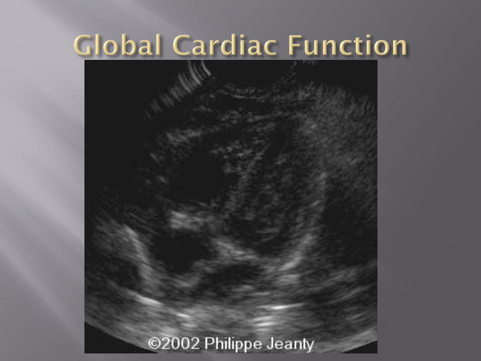
Evaluate global cardiac function (EF, CI)
Evaluate IVC (preload)

198/208 enrolled (4.5% unable to be examined)
APACHE II – 30 SAPS II – 69
Mortality 51%
Mechanical Ventilation 82%
Diseases
Cardiac 44% with severe 14% (28/87)
AS
Endocarditis
Dilated cardiomyopathy
Tamponade
Results
CI and IVC Index correlated with mortality
Significant rate of unexpected cardiac abnormalities

Manasia et al. J Card Vasc Anes. 2005;19:155
10 one hour training sessions
Limited TTE
LV fxn, RWMA, pericard. effusion
Correctly interpreted 84%
94% completed studies
Study time 10.5 +/- 4.2 min
Sonoheart elite brochure, Sonosite
Limited TTE changed mgmt in 37%
of patients


Established credentialing process and literature to support its use ACEP Policy Statement: Emergency Ultrasound Guidelines 2008.
Documented success with current methods of training McCarter et al. Ann Surg 2000;231:689.
15-20 scans = 90% sen., 99% spec. and 99% accurate
50 scans = 96% sen. and 100% accuracy

ACEP Policy Statement: Emergency Ultrasound Guidelines 2001

ACC/AHA Clinical Competence Statement on Echocardiography. JACC 2003
Total = 150-200

LV function?
RV function?
Pericardial effusion present? Tamponade?
Volume status? Dynamic IVC assessment
Beaulieu Y. Crit Care Med 2007;35:S144


Physical exam is sensitive and specific for evaluating shock in the critically ill patient?
1) True
2) False

The FACTT Trial demonstrated that a PA catheter when compared to a CVP monitor improved
1) Mortality
2) Ventilator free days
3) Shock resolution
4) None of the above

Which of the below is most correct.
When predicting volume responsiveness during hypotension
1) Physical exam is superior to static cardiac pressure measurements
2) Static cardiac pressure measurements are superior to physical exam
3) Both static cardiac pressure measurements and physical exam are sensitive and specific for volume resuscitation
4) Neither static cardiac pressure measurements or physical exam are sensitive or specific for volume resuscitation

Which is the most correct
1) RV:LV ratio > 1 reflects the need for more volume
2) IVC index > 50% is consistent with a CVP > 13
3) A flattened septum noted on a parasternal short axis demonstrates a RV pressure overload state
4) VTI is a poor measurement to follow cardiac response to volume resuscitation