patterns of immunotherapy-induced pneumonitis in patients
TRANSCRIPT

Picard et al. J Med Case Reports (2021) 15:332 https://doi.org/10.1186/s13256-021-02926-y
CASE REPORT
Patterns of immunotherapy-induced pneumonitis in patients with non-small-cell lung cancer: a case seriesSarah Picard1,2*† , Desiree Goh1,2†, Ashley Tan2,3, Nisha Sikotra3, Eli Gabbay4,5 and Tim Clay2,6,7
Abstract
Background: Immunotherapy has become an efficacious option in the management of solid organ malignancies. Immune-related adverse events including pneumonitis are well described and may be particularly of concern in patients receiving immunotherapy for non-small-cell lung cancer.
Case presentations: In this paper, we describe three cases of immunotherapy-induced pneumonitis occurring in the management of lung malignancy. Our cases include a 54-year-old Caucasian woman with squamous cell lung cancer who was successfully rechallenged with immunotherapy after prior significant pneumonitis, a 65-year-old Caucasian man with metastatic squamous cell lung cancer who developed pneumonitis after multiple cycles of uneventful immunotherapy, and a 73-year-old Caucasian man with squamous cell lung cancer who developed early-onset pneumonitis with rebound on steroid taper.
Conclusions: This case series has provided further insight into the presentation and risk factors for pneumonitis in patients with non-small-cell lung cancer. Each of the cases of immunotherapy-induced pneumonitis illustrates the different potential patterns that may arise when immunotherapy-induced pneumonitis develops. This case series provides key learning points that may assist physicians managing non-small-cell lung cancer with immunotherapy.
Keywords: Pneumonitis, Immunotherapy, Malignancy, NSCLC, irAE
© The Author(s) 2021. Open Access This article is licensed under a Creative Commons Attribution 4.0 International License, which permits use, sharing, adaptation, distribution and reproduction in any medium or format, as long as you give appropriate credit to the original author(s) and the source, provide a link to the Creative Commons licence, and indicate if changes were made. The images or other third party material in this article are included in the article’s Creative Commons licence, unless indicated otherwise in a credit line to the material. If material is not included in the article’s Creative Commons licence and your intended use is not permitted by statutory regulation or exceeds the permitted use, you will need to obtain permission directly from the copyright holder. To view a copy of this licence, visit http:// creat iveco mmons. org/ licen ses/ by/4. 0/. The Creative Commons Public Domain Dedication waiver (http:// creat iveco mmons. org/ publi cdoma in/ zero/1. 0/) applies to the data made available in this article, unless otherwise stated in a credit line to the data.
BackgroundImmunotherapy is increasingly prescribed for solid organ and hematological malignancies, with striking improve-ments in progression-free and overall survival in some cancers [1]. Immunotherapeutic drugs may cause life-threatening immune-related adverse events (irAEs) including pneumonitis, complicating their use in lung cancer. Patients with advanced non-small-cell lung can-cer (NSCLC) have a higher incidence of pneumonitis from immunotherapy (4.7% incidence) compared with the overall incidence of pneumonitis in patients with
other solid organ malignancies receiving immunotherapy (2.92% incidence) [2]. In this paper, we describe three cases of immunotherapy-induced pneumonitis occurring in the management of lung malignancy.
Immune checkpoint inhibitors in routine clinical practice today target programmed death 1 (PD-1), pro-grammed death ligand 1 (PD-L1), and cytotoxic T-lym-phocyte-associated protein 4 (CTLA-4). These pathways are expressed in normal tissue as a mechanism to control the immune system, and they can become dysregulated in the tumor microenvironment [3]. Inhibition of these proteins with monoclonal antibodies causes upregula-tion of the immune system [3, 4], allowing it to target and destroy cancer cells [2, 5]. However, T cells may also attack noncancerous cells, resulting in irAEs [5, 6] including pneumonitis.
Open Access
*Correspondence: [email protected]†Sarah Picard and Desiree Goh contributed equally as co-first authors1 Sir Charles Gairdner Hospital, Hospital Ave, Nedlands, WA 6009, AustraliaFull list of author information is available at the end of the article
Page 2 of 9Picard et al. J Med Case Reports (2021) 15:332
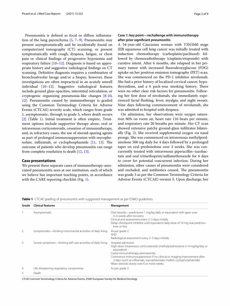
Pneumonitis is defined as focal or diffuse inflamma-tion of the lung parenchyma [5, 7–9]. Pneumonitis may present asymptomatically and be incidentally found on computerized tomography (CT) scanning, or present symptomatically with cough, dyspnea, fatigue, or chest pain or clinical findings of progressive hypoxemia and respiratory failure [10–12]. Diagnosis is based on appro-priate history and suggestive radiological findings on CT scanning. Definitive diagnosis requires a combination of bronchoalveolar lavage and/or a biopsy; however, these investigations are often impractical in an acutely unwell individual [10–12]. Suggestive radiological features include ground glass opacities, interstitial reticulation, or cryptogenic organizing pneumonia-like changes [8-10, 12]. Pneumonitis caused by immunotherapy is graded using the Common Terminology Criteria for Adverse Events (CTCAE) severity scale, which ranges from grade 1, asymptomatic, through to grade 5, where death occurs [2] (Table 1). Initial treatment is often empiric. Treat-ment options include supportive therapy alone, oral or intravenous corticosteroids, cessation of immunotherapy, and, in refractory cases, the use of steroid-sparing agents as part of prolonged immunosuppression with mycophe-nolate, infliximab, or cyclophosphamide [11, 13]. The outcome of patients who develop pneumonitis can range from complete resolution to death [12, 13].
Case presentationsWe present three separate cases of immunotherapy-asso-ciated pneumonitis seen at our institution, each of which we believe has important teaching points, in accordance with the CARE reporting checklist (Fig. 1).
Case 1: key point—rechallenge with immunotherapy after prior significant pneumonitisA 54-year-old Caucasian woman with T3N3M0 stage IIIB squamous cell lung cancer was initially treated with induction chemotherapy (carboplatin/paclitaxel) fol-lowed by chemoradiotherapy (cisplatin/etoposide) with curative intent. After 6 months, she relapsed in her pri-mary tumor with increased fluorodeoxyglucose (FDG) uptake on her positron emission tomography (PET) scan. She was commenced on the PD-1 inhibitor nivolumab. She had a prior history of localized cervical cancer, hypo-thyroidism, and a 6 pack-year smoking history. There were no other clear risk factors for pneumonitis. Follow-ing her first dose of nivolumab, she immediately expe-rienced facial flushing, fever, myalgia, and night sweats. Nine days following commencement of nivolumab, she was admitted to hospital with dyspnea.
On admission, her observations were oxygen satura-tion 80% on room air, heart rate 116 beats per minute, and respiratory rate 20 breaths per minute. Her CT scan showed extensive patchy ground-glass infiltrates bilater-ally (Fig. 2). She received supplemental oxygen via nasal prongs. She was commenced on intravenous methylpred-nisolone 500 mg daily for 4 days followed by a prolonged taper on oral prednisolone over 3 weeks. She was con-currently treated with intravenous piperacillin–tazobac-tam and oral trimethoprim/sulfamethoxazole for 6 days to cover for potential concurrent infection. During her admission, other causes of pneumonitis were considered and excluded, and antibiotics ceased. The pneumonitis was grade 3 as per the Common Terminology Criteria for Adverse Events (CTCAE) version 5. Upon discharge, her
Table 1 CTCAE grading of pneumonitis with suggested management as per ESMO guidelines
CTCAE Common Terminology Criteria for Adverse Events, ESMO European Society for Medical Oncology
Grade Clinical features Management
1 Asymptomatic Oral steroids—prednisone 1 mg/kg daily or equivalent with taper over 4–6 weeks after recovery
Clinical and assessment every 2–3 days initiallyDelay checkpoint inhibitor until equivalent daily dose of 10 mg oral predniso-
lone or less
2 Symptomatic—limiting instrumental activities of daily living As per grade 2ANDRadiological assessment every 2–3 days initially
3 Severe symptoms—limiting self-care activities of daily living Hospital admissionHigh-dose intravenous corticosteroids (methylprednisolone 2–4 mg/kg/day or
equivalent)Cease immunotherapy permanentlyCommence immunosuppression if no clinical or imaging improvement after
2 days (such as infliximab, mycophenolate mofetil, cyclophosphamide)Wean steroids slowly over 6 or more weeks
4 Life-threatening respiratory compromise As per grade 3
5 Death

Page 3 of 9Picard et al. J Med Case Reports (2021) 15:332
cardiorespiratory parameters were within normal range, and she had returned to her premorbid function.
Two months after treatment, the FDG-PET scan showed an initial partial metabolic response, with incomplete resolution of the changes due to pneumoni-tis (Fig. 3). Over time, the pneumonitis settled, but there
was cancer progression on FDG-PET after 5 months off nivolumab. On discussion between the patient and her treating team, nivolumab was reinstituted and treatment continued for a period of 18 months with a best response of stable disease. No further episodes of pneumonitis were observed. The patient subsequently died from cancer-associated venous thromboembolism unrelated to her prior immunotherapy.
Diagnosis Date
Case 3 Immunotherapy Commenced
Case 3 Admission #1
Case 3 Admission #2
Case 3 Death
Case 2 Immunotherapy Commenced
Case 1 Immunotherapy Commenced
Case 1 Admission #1
Case 2 Admission #1
Case 2 Death
Case 1 Death
0 1 2 3 4 5 6 7 8 9 10 11 12 13 14 15 16 17 18 19 20 21 22 23 24 25 26 27 28
Time (Months)
Immunotherapy Induced Pneumonis Key Event Timeline
Fig. 1 Timeline of key events
Fig. 2 Case 1—chest computed tomography (CT) revealing signs of pneumonitis
Fig. 3 Case 1—CT chest 2 months after commencement of pneumonitis treatment with incomplete resolution of pneumonitis changes
Page 4 of 9Picard et al. J Med Case Reports (2021) 15:332
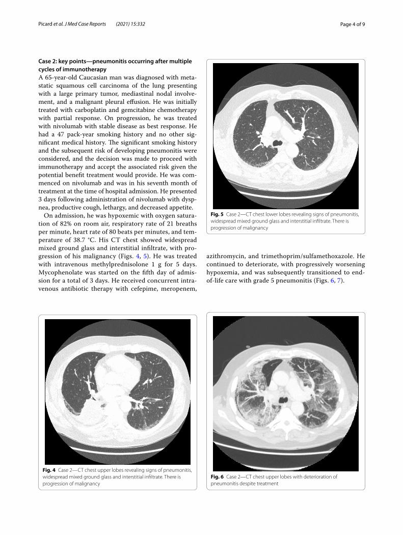
Case 2: key points—pneumonitis occurring after multiple cycles of immunotherapyA 65-year-old Caucasian man was diagnosed with meta-static squamous cell carcinoma of the lung presenting with a large primary tumor, mediastinal nodal involve-ment, and a malignant pleural effusion. He was initially treated with carboplatin and gemcitabine chemotherapy with partial response. On progression, he was treated with nivolumab with stable disease as best response. He had a 47 pack-year smoking history and no other sig-nificant medical history. The significant smoking history and the subsequent risk of developing pneumonitis were considered, and the decision was made to proceed with immunotherapy and accept the associated risk given the potential benefit treatment would provide. He was com-menced on nivolumab and was in his seventh month of treatment at the time of hospital admission. He presented 3 days following administration of nivolumab with dysp-nea, productive cough, lethargy, and decreased appetite.
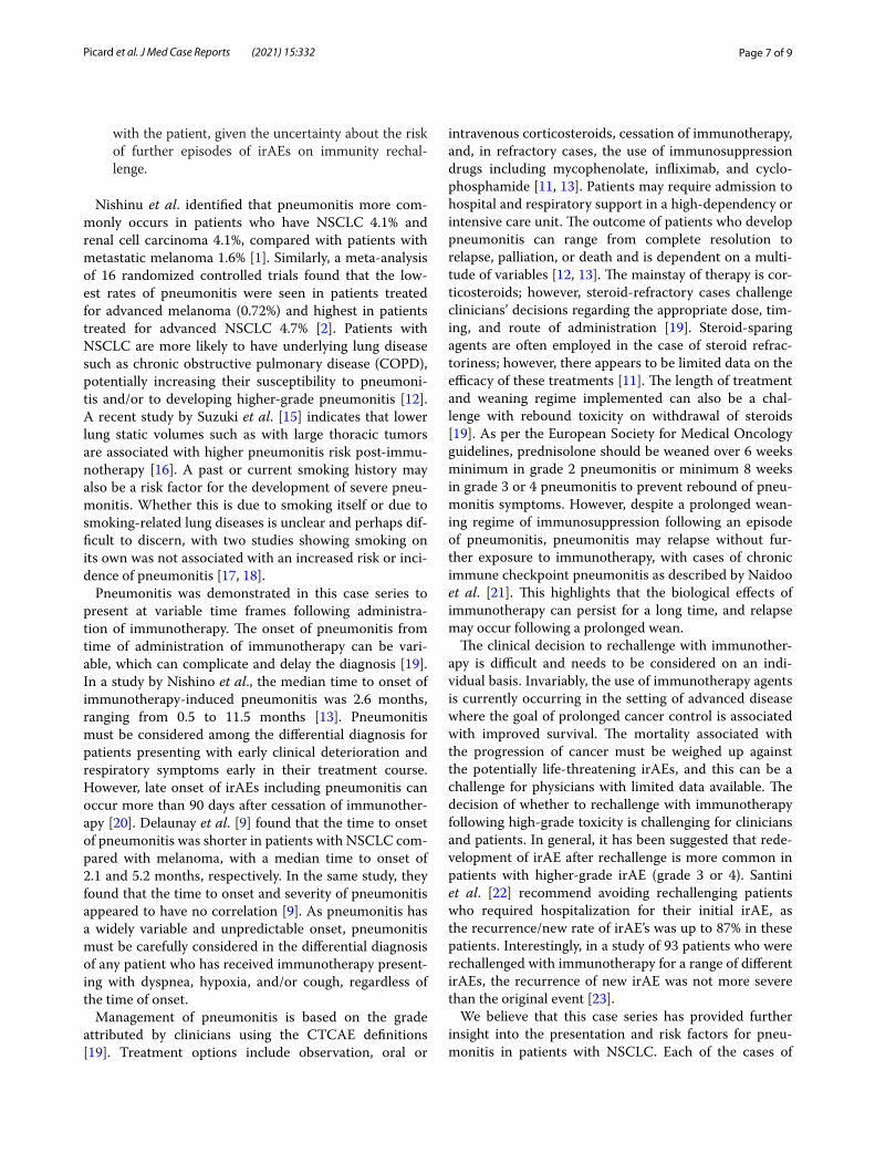
On admission, he was hypoxemic with oxygen satura-tion of 82% on room air, respiratory rate of 21 breaths per minute, heart rate of 80 beats per minutes, and tem-perature of 38.7 °C. His CT chest showed widespread mixed ground glass and interstitial infiltrate, with pro-gression of his malignancy (Figs. 4, 5). He was treated with intravenous methylprednisolone 1 g for 5 days. Mycophenolate was started on the fifth day of admis-sion for a total of 3 days. He received concurrent intra-venous antibiotic therapy with cefepime, meropenem,
azithromycin, and trimethoprim/sulfamethoxazole. He continued to deteriorate, with progressively worsening hypoxemia, and was subsequently transitioned to end-of-life care with grade 5 pneumonitis (Figs. 6, 7).
Fig. 4 Case 2—CT chest upper lobes revealing signs of pneumonitis, widespread mixed ground glass and interstitial infiltrate. There is progression of malignancy
Fig. 5 Case 2—CT chest lower lobes revealing signs of pneumonitis, widespread mixed ground glass and interstitial infiltrate. There is progression of malignancy
Fig. 6 Case 2—CT chest upper lobes with deterioration of pneumonitis despite treatment

Page 5 of 9Picard et al. J Med Case Reports (2021) 15:332
Case 3: key points—early‑onset pneumonitis with rebound on steroid taperA 73-year-old Caucasian man with was diagnosed with T3N1M0 squamous cell carcinoma of the lung. He received treatment with chemoradiotherapy (weekly carboplatin/paclitaxel) with curative intent. An early FDG-PET scan showed partial response, and consolida-tion immunotherapy durvalumab, a PD-L1 inhibitor, was commenced. He had a background of emphysema, cerebellar hematoma, cerebrovascular disease, coronary artery disease, hyperlipidemia, and hypothyroidism. He was an ex-smoker with a 75 pack-year history. The signif-icant smoking history, emphysema, and recent radiother-apy were considered as risk factors for the development of pneumonitis. After consideration, treatment pro-ceeded, given the clear survival benefits demonstrated in the PACIFIC clinical trial [14]. Six days following the first dose of durvalumab, he developed dyspnea and a produc-tive cough.
The patient was admitted to hospital. His initial obser-vations showed oxygen saturation was 96% on room air with a respiratory rate of 20 breaths per minute, temper-ature of 37 °C, and heart rate of 78 breaths per minute. His CT scan of the chest showed diffuse peribronchial ground-glass changes and a decrease in size of the lung cancer (Figs. 8, 9). He was admitted for 6 days and com-menced on oral prednisolone 100 mg with a weaning regime and concurrent doxycycline for grade 3 pneumo-nitis. The patient improved clinically, his observations were within normal cardiorespiratory parameters, and he was discharged on a prolonged steroid taper. The patient was reviewed by his respiratory physician and medical
oncologist in clinic 3 weeks after discharge from hospi-tal. Immunotherapy was not recommenced. A repeat CT scan showed marked resolution of the ground-glass changes, and he had returned to his premorbid function (Fig. 10).
One week after clinic review, the patient was readmit-ted to hospital with increasing dyspnea and hemoptysis. He had been on corticosteroids for a period of 4 weeks and 4 days at this time. He was hypoxemic at 92% oxygen saturation on 4 L of oxygen per minute via nasal prongs,
Fig. 7 Case 2—CT chest lower lobes with deterioration of pneumonitis despite treatment
Fig. 8 Case 3—CT chest before immunotherapy commenced
Fig. 9 Case 3—CT chest after immunotherapy given revealing signs of pneumonitis, diffuse peribronchial ground-glass changes. There is decrease in size of lung cancer

Page 6 of 9Picard et al. J Med Case Reports (2021) 15:332
had a respiratory rate of 20 breaths per minute and heart rate of 95 beats per minute, and was afebrile. A repeat CT scan of the chest showed recurrence of diffuse ground-glass infiltrates, in keeping with acute pneumonitis, and no change in the size of the lung cancer (Fig. 11).
On readmission, oral corticosteroids were reesca-lated to prednisolone 100 mg. The patient rapidly dete-riorated, with worsening hypoxemia requiring admission
to Intensive Care Unit (ICU). Immunosuppression was increased to pulse methylprednisolone 1 g daily with subsequent administration of intravenous cyclophos-phamide. Broad antimicrobial therapy was administered to cover for concurrent infection (including ceftriaxone, piperacillin–tazobactam, meropenem, trimethoprim/sul-famethoxazole, and azithromycin over the period of his admission). Respiratory support included supplemental oxygen via high-flow nasal prongs and periodic nonin-vasive ventilation. Intubation and mechanical ventilation were deemed inappropriate in view of the guarded prog-nosis and in discussion with the patient and his family. Despite maximal therapy, the patient’s condition deterio-rated, and he died from grade 5 pneumonitis 18 days fol-lowing admission.
Discussion and conclusionCheckpoint inhibitor immunotherapy now enjoys wide-spread use in cancer care. Severe adverse events with sin-gle-agent PD-1 or PD-L1 inhibition are uncommon but well described—they can be unpredictable with regard to timing and severity and can result in significant morbid-ity and, occasionally, mortality. With any presentation, irAEs must be considered in the differential diagnosis and acted on promptly. Pneumonitis remains a toxicity of particular concern for clinicians treating NSCLC with greatest incidence of immunotherapy-induced pneumo-nitis [2]. In the current context, Coronavirus 19 infection also remains an important differential, and in jurisdic-tions with community transmission, it is particularly important to exclude this diagnosis urgently.
The first case highlights that immunotherapy rechal-lenge is possible but must occur after consideration of the therapeutic options and a discussion on risks and benefits. It is important for clinicians to highlight the uncertainty about the risk of further episodes of irAEs on immunotherapy rechallenge.
Our case series demonstrates:
1. Patients with underlying lung pathology or smok-ing history are susceptible to developing high-grade pneumonitis.
2. Immunotherapy-induced pneumonitis can present at any time after commencement of immunotherapy.
3. The severity of immunotherapy-induced pneumoni-tis does not appear to be correlated with the time-frame of onset.
4. Immunotherapy-induced pneumonitis can flare or recur during the period of steroid taper.
5. Following careful consideration, immunotherapy rechallenge can occur for some patients with prior immunotherapy-induced pneumonitis. A careful dis-cussion of the potential risks and benefits is required
Fig. 10 Case 3—CT chest after treatment of pneumonitis with initial recovery
Fig. 11 Case 3—CT chest after relapse of pneumonitis. There is no change in size of lung cancer
Page 7 of 9Picard et al. J Med Case Reports (2021) 15:332
with the patient, given the uncertainty about the risk of further episodes of irAEs on immunity rechal-lenge.
Nishinu et al. identified that pneumonitis more com-monly occurs in patients who have NSCLC 4.1% and renal cell carcinoma 4.1%, compared with patients with metastatic melanoma 1.6% [1]. Similarly, a meta-analysis of 16 randomized controlled trials found that the low-est rates of pneumonitis were seen in patients treated for advanced melanoma (0.72%) and highest in patients treated for advanced NSCLC 4.7% [2]. Patients with NSCLC are more likely to have underlying lung disease such as chronic obstructive pulmonary disease (COPD), potentially increasing their susceptibility to pneumoni-tis and/or to developing higher-grade pneumonitis [12]. A recent study by Suzuki et al. [15] indicates that lower lung static volumes such as with large thoracic tumors are associated with higher pneumonitis risk post-immu-notherapy [16]. A past or current smoking history may also be a risk factor for the development of severe pneu-monitis. Whether this is due to smoking itself or due to smoking-related lung diseases is unclear and perhaps dif-ficult to discern, with two studies showing smoking on its own was not associated with an increased risk or inci-dence of pneumonitis [17, 18].
Pneumonitis was demonstrated in this case series to present at variable time frames following administra-tion of immunotherapy. The onset of pneumonitis from time of administration of immunotherapy can be vari-able, which can complicate and delay the diagnosis [19]. In a study by Nishino et al., the median time to onset of immunotherapy-induced pneumonitis was 2.6 months, ranging from 0.5 to 11.5 months [13]. Pneumonitis must be considered among the differential diagnosis for patients presenting with early clinical deterioration and respiratory symptoms early in their treatment course. However, late onset of irAEs including pneumonitis can occur more than 90 days after cessation of immunother-apy [20]. Delaunay et al. [9] found that the time to onset of pneumonitis was shorter in patients with NSCLC com-pared with melanoma, with a median time to onset of 2.1 and 5.2 months, respectively. In the same study, they found that the time to onset and severity of pneumonitis appeared to have no correlation [9]. As pneumonitis has a widely variable and unpredictable onset, pneumonitis must be carefully considered in the differential diagnosis of any patient who has received immunotherapy present-ing with dyspnea, hypoxia, and/or cough, regardless of the time of onset.
Management of pneumonitis is based on the grade attributed by clinicians using the CTCAE definitions [19]. Treatment options include observation, oral or
intravenous corticosteroids, cessation of immunotherapy, and, in refractory cases, the use of immunosuppression drugs including mycophenolate, infliximab, and cyclo-phosphamide [11, 13]. Patients may require admission to hospital and respiratory support in a high-dependency or intensive care unit. The outcome of patients who develop pneumonitis can range from complete resolution to relapse, palliation, or death and is dependent on a multi-tude of variables [12, 13]. The mainstay of therapy is cor-ticosteroids; however, steroid-refractory cases challenge clinicians’ decisions regarding the appropriate dose, tim-ing, and route of administration [19]. Steroid-sparing agents are often employed in the case of steroid refrac-toriness; however, there appears to be limited data on the efficacy of these treatments [11]. The length of treatment and weaning regime implemented can also be a chal-lenge with rebound toxicity on withdrawal of steroids [19]. As per the European Society for Medical Oncology guidelines, prednisolone should be weaned over 6 weeks minimum in grade 2 pneumonitis or minimum 8 weeks in grade 3 or 4 pneumonitis to prevent rebound of pneu-monitis symptoms. However, despite a prolonged wean-ing regime of immunosuppression following an episode of pneumonitis, pneumonitis may relapse without fur-ther exposure to immunotherapy, with cases of chronic immune checkpoint pneumonitis as described by Naidoo et al. [21]. This highlights that the biological effects of immunotherapy can persist for a long time, and relapse may occur following a prolonged wean.
The clinical decision to rechallenge with immunother-apy is difficult and needs to be considered on an indi-vidual basis. Invariably, the use of immunotherapy agents is currently occurring in the setting of advanced disease where the goal of prolonged cancer control is associated with improved survival. The mortality associated with the progression of cancer must be weighed up against the potentially life-threatening irAEs, and this can be a challenge for physicians with limited data available. The decision of whether to rechallenge with immunotherapy following high-grade toxicity is challenging for clinicians and patients. In general, it has been suggested that rede-velopment of irAE after rechallenge is more common in patients with higher-grade irAE (grade 3 or 4). Santini et al. [22] recommend avoiding rechallenging patients who required hospitalization for their initial irAE, as the recurrence/new rate of irAE’s was up to 87% in these patients. Interestingly, in a study of 93 patients who were rechallenged with immunotherapy for a range of different irAEs, the recurrence of new irAE was not more severe than the original event [23].
We believe that this case series has provided further insight into the presentation and risk factors for pneu-monitis in patients with NSCLC. Each of the cases of

Page 8 of 9Picard et al. J Med Case Reports (2021) 15:332
immunotherapy-induced pneumonitis illustrates the different potential patterns that may arise when immu-notherapy-induced pneumonitis develops. Given the var-iable onset and severity of pneumonitis, we recommend that immunotherapy-induced pneumonitis must always be considered as a differential, regardless of the time of onset. Special consideration should be given to patients with underlying lung disease or a significant smoking his-tory, given the higher risk of mortality. The decision to rechallenge patients, while difficult, is possible. Early ini-tiation and perhaps prolonged immunosuppressive ther-apy may be required to optimize patient outcomes.
AbbreviationsNSCLC: Non-small-cell lung cancer; irAE: Immune-related adverse events; FDG-PET: Fluorodeoxyglucose positron emission tomography; CT: Computerized tomography; PD-1: Programmed death 1; PD-L1: Programmed death ligand 2; CTLA-4: Cytotoxic T-lymphocyte-associated protein 4; ESMO: European Society for Medical Oncology.
AcknowledgementsThis case series was supported by the Bendat Respiratory Research and Devel-opment Fund and in-kind funding.
Authors’ contributionsSP, DG, NS, and AT gathered data that contributed to the manuscript. SP and DG analyzed the data and wrote the manuscript, including ongoing editing and revisions. EG and TC provided technical expertise, supervised, edited, and proofread drafts of the paper. All authors reviewed the final manuscript.
FundingThis case series was supported by the Bendat Respiratory Research and Devel-opment Fund and in-kind funding.
Availability of data and materialsData sharing is not applicable to this article as no datasets were generated or analyzed during the current study.
Declarations
Ethics approval and consent to participateThe authors are accountable for all aspects of the work in ensuring that ques-tions related to the accuracy or integrity of any part of the work are appropri-ately investigated and resolved. All procedures performed in studies involving human participants were in accordance with the ethical standards of the institutional and/or national research committee(s) and with the Helsinki Dec-laration (as revised in 2013). Ethics approval was obtained from the St John of God Health Care Human Research Ethics Committee (ref 1536), who approved the study as satisfying the ethical requirements under the National Health and Medical Council’s National Statement on Ethical Conduct in Human Research (NHMRC, 2007).
Consent for publicationWritten informed consent was obtained from the deceased patients’ family members for the publication of this case report and for any images. A copy of the written consent is available for review by the Editor-in-Chief of this journal. The manuscript was prepared in accordance with the CARE reporting checklist.
Competing interestsThere are no conflicts of interest to disclose. All authors have completed the ICMJE uniform disclosure form.
Author details1 Sir Charles Gairdner Hospital, Hospital Ave, Nedlands, WA 6009, Australia. 2 St John of God Hospital, 12 Salvado Rd, Subiaco, WA 6008, Australia. 3 Fiona Stanley Hospital, 11 Robin Warren D, Murdoch, WA 6150, Australia. 4 The Uni-versity of Notre Dame, Fremantle, Australia. 5 St John of God, Subiaco, Australia. 6 Adjunct Associate Professor of Medicine, Edith Cowan University, Joondalup, WA, Australia. 7 Bendat Family Respiratory Research and Development Fund, Subiaco, Australia.
Received: 24 February 2021 Accepted: 24 May 2021
References 1. Nishino M, Giobbie-Hurder A, Hatabu H, Ramaiya NH, Hodi FS. Incidence
of programmed cell death 1 inhibitor-related pneumonitis in patients with advanced cancer: a systematic review and meta-analysis. JAMA Oncol. 2016;2(12):1607–16.
2. Wu J, Hong D, Zhang X, Lu X, Miao J. PD-1 inhibitors increase the inci-dence and risk of pneumonitis in cancer patients in a dose-independent manner: a meta-analysis. Sci Rep. 2017;7:44173.
3. Nishino M, Sholl LM, Hodi FS, Hatabu H, Ramaiya NH. Anti-PD-1-related pneumonitis during cancer immunotherapy. N Engl J Med. 2015;373(3):288–90.
4. Sato K, Akamatsu H, Murakami E, Sasaki S, Kanai K, Hayata A, et al. Correla-tion between immune-related adverse events and efficacy in non-small cell lung cancer treated with nivolumab. Lung Cancer. 2018;115:71–4.
5. Luo W, Wang Z, Tian P, Li W. Safety and tolerability of PD-1/PD-L1 inhibitors in the treatment of non-small cell lung cancer: a meta-analysis of randomized controlled trials. J Cancer Res Clin Oncol. 2018;144(10):1851–9.
6. Khunger M, Rakshit S, Pasupuleti V, Hernandez AV, Mazzone P, Stevenson J, et al. Incidence of pneumonitis with use of programmed death 1 and programmed death-ligand 1 inhibitors in non-small cell lung cancer: a systematic review and meta-analysis of trials. Chest. 2017;152(2):271–81.
7. Kato T, Masuda N, Nakanishi Y, Takahashi M, Hida T, Sakai H, et al. Nivolumab-induced interstitial lung disease analysis of two phase II stud-ies patients with recurrent or advanced non-small-cell lung cancer. Lung Cancer. 2017;104:111–8.
8. Naidoo J, Wang X, Woo KM, Iyriboz T, Halpenny D, Cunningham J, et al. Pneumonitis in patients treated with anti-programmed death-1/pro-grammed death ligand 1 therapy. J Clin Oncol. 2017;35(7):709–17.
9. Delaunay M, Cadranel J, Lusque A, Meyer N, Gounant V, Moro-Sibilot D, et al. Immune-checkpoint inhibitors associated with interstitial lung disease in cancer patients. Eur Respir J. 2017;50(2):1700050.
10. Helber HA, Hada AL, Pio RB, Moraes PHZ, Gomes DBD. Immunother-apy-induced pneumonitis: cases report. Einstein (Sao Paulo Brazil). 2018;16(2):eRC4030.
11. Castanon E. Anti-PD1-induced pneumonitis: capturing the hidden enemy. Clin Cancer Res. 2016;22(24):5956–8.
12. Rickard F, Hyams C, Low AT. Pneumonitis: a serious adverse effect of PD-L1 inhibitors including pembrolizumab. BMJ Case Rep. 2018;2018.
13. Nishino M, Ramaiya NH, Awad MM, Sholl LM, Maattala JA, Taibi M, et al. PD-1 inhibitor-related pneumonitis in advanced cancer patients: radiographic patterns and clinical course. Clin Cancer Res. 2016;22(24):6051–60.
14. Antonia SJ, Villegas A, Daniel D, Vicente D, Murakami S, Hui R, et al. Dur-valumab after chemoradiotherapy in stage III non-small-cell lung cancer. N Engl J Med. 2017;377(20):1919–29.
15. Suzuki Y, Karayama M, Uto T, Fujii M, Matsui T, Asada K, et al. Assessment of immune-related interstitial lung disease in patients with NSCLC treated with immune checkpoint inhibitors: a multicenter prospective study. J Thorac Oncol. 2020;15(8):1317–27.
16. Pérol M. Multidisciplinary approach of immune checkpoint inhibitor-related pneumonitis: a key to address knowledge and management gaps. J Thorac Oncol. 2020;15(8):1261–4.
17. Cui P, Liu Z, Wang G, Ma J, Qian Y, Zhang F, et al. Risk factors for pneu-monitis in patients treated with anti-programmed death-1 therapy: a case-control study. Cancer Med. 2018;7(8):4115–20.

Page 9 of 9Picard et al. J Med Case Reports (2021) 15:332
• fast, convenient online submission
•
thorough peer review by experienced researchers in your field
• rapid publication on acceptance
• support for research data, including large and complex data types
•
gold Open Access which fosters wider collaboration and increased citations
maximum visibility for your research: over 100M website views per year •
At BMC, research is always in progress.
Learn more biomedcentral.com/submissions
Ready to submit your researchReady to submit your research ? Choose BMC and benefit from: ? Choose BMC and benefit from:
18. Ahn MJ, Gandhi L, Hamid O, Hellmann MD, Garon EB, Ramalingam SS, et al. 459P Risk of pneumonitis in patients with advanced NSCLC treated with pembrolizumab in KEYNOTE-001. Ann Oncol. 2015;26:ix125.
19. Sears CR, Peikert T, Possick JD, Naidoo J, Nishino M, Patel SP, et al. Knowl-edge gaps and research priorities in immune checkpoint inhibitor-related pneumonitis. An official American Thoracic Society research statement. Am J Respir Crit Care Med. 2019;200(6):e31–43.
20. Couey MA, Bell RB, Patel AA, Romba MC, Crittenden MR, Curti BD, et al. Delayed immune-related events (DIRE) after discontinuation of immuno-therapy: diagnostic hazard of autoimmunity at a distance. J Immunother Cancer. 2019;7(1):165.
21. Naidoo J, Cottrell TR, Lipson EJ, Forde PM, Illei PB, Yarmus LB, et al. Chronic immune checkpoint inhibitor pneumonitis. J Immunother Cancer. 2020;8(1):e000840.
22. Santini FC, Rizvi H, Plodkowski AJ, Ni A, Lacouture ME, Gambarin-Gelwan M, et al. Safety and efficacy of re-treating with immunotherapy after immune-related adverse events in patients with NSCLC. Cancer Immunol Res. 2018;6(9):1093–9.
23. Simonaggio A, Michot JM, Voisin AL, Le Pavec J, Collins M, Lallart A, et al. Evaluation of readministration of immune checkpoint inhibitors after immune-related adverse events in patients with cancer. JAMA Oncol. 2019;5(9):1310–7.
Publisher’s NoteSpringer Nature remains neutral with regard to jurisdictional claims in pub-lished maps and institutional affiliations.