pdf (3 mb)
TRANSCRIPT

Cardiovascular, Pulmonary and Renal Pathology
Obligatory Role for Interleukin-13 in ObstructiveLesion Development in Airway Allografts
Vibha N. Lama,* Hiroaki Harada,† Linda N. Badri,*Andrew Flint,‡ Cory M. Hogaboam,‡
Andrew McKenzie,§ Fernando J. Martinez,*Galen B. Toews,* Bethany B. Moore,* andDavid J. Pinsky†
From the Division of Pulmonary and Critical Care Medicine,*
Division of Cardiovascular Medicine,† and the Department of
Pathology,‡ University of Michigan Health System, Ann Arbor,
Michigan; and the Laboratory of Molecular Biology,§ Medical
Research Council, Cambridge, United Kingdom
The pathogenesis of bronchiolitis obliterans (BO), acommon and devastating obliterative disorder ofsmall airways following lung transplantation, re-mains poorly understood. Lesions are characterizedin their early stages by lymphocyte influx that evolvesinto dense fibrotic infiltrates. Airway specimenstaken from patients with histological BO revealed in-filtrating myofibroblasts, which strongly expressedthe signaling chain of the high affinity interleukin-13(IL-13) receptor IL-13R�1. Because IL-13 has proin-flammatory and profibrotic actions, a contributoryrole for IL-13 in BO development was examined usingmurine models of orthotopic and heterotopic tra-cheal transplantation. Compared with airwayisografts, allografts exhibited a significant increase inrelative IL-13 mRNA and protein levels. Allogeneictracheas transplanted into IL-13-deficient mice wereprotected from BO in both transplant models. Flowcytometric analysis of orthotopic transplant tissuedigests revealed markedly fewer infiltrating mononu-clear phagocytes and CD3� T lymphocytes in IL-13-deficient recipients. Furthermore, protection fromluminal obliteration, collagen deposition, and myofi-broblast infiltration was observed in heterotopicairways transplanted into the IL-13�/� recipients.Transforming growth factor-�1 expression was sig-nificantly decreased in tracheal allografts into IL-13�/� recipients, compared to wild-type counter-parts. These human and murine data implicate IL-13as a critical effector cytokine driving cellular recruit-ment and subsequent fibrosis in clinical and ex-
perimental BO. (Am J Pathol 2006, 169:47–60; DOI:
10.2353/ajpath.2006.050975)
Bronchiolitis obliterans syndrome (BOS) is the major fac-tor limiting quality of life as well as long-term survival afterlung transplantation. BO is a histological diagnosis, char-acterized by fibrosis and obliteration of the small air-ways.1 BOS, the clinical correlate of BO, is diagnosed byirreversible deterioration of pulmonary function in the ab-sence of other causes.2 BOS is seen in up to 60% of lungtransplant recipients by 5 years after transplantation.There are currently no meaningful therapeutic interven-tions, providing a compelling rationale to understand fullyBOS pathogenesis as this may lead to development ofnew therapeutic approaches.
Data from human as well as animal studies suggestassociations between inflammatory cells/mediators andBOS. Lymphocytic bronchitis is considered a risk factorfor development of BOS in humans,3 and lymphocyticinfiltration precedes development of luminal obliterationin animal models of tracheal transplantation.4,5 Similarly,several markers of inflammation (eg, neutrophilia,6 IL-8,7
and monocyte chemoattractant protein-1 (MCP-1/CCL2)8,9) are predictive of development of BO in hu-mans. However, the failure of immunosuppressivetherapy and anti-inflammatory agents to demonstrate ef-ficacy in preventing or treating this disease has focusedattention on BOS as a fibroproliferative response to air-way epithelial injury. Evidence for this is provided bystudies suggesting a role for transforming growth factor-�(TGF-�), a major profibrotic factor, in the pathogenesis ofBOS.7,10,11
Supported by grants from the National Institutes of Health (K23 HL077719to V.N.L.; R01 HL55397 to D.J.P.; R01HL69448 to D.J.P.; HL071586 toB.B.M.; and P50HL56402 to G.B.T., C.M.H., F.J.M., and B.B.M.) and theAmerican Lung Association (RG-1059-N to V.N.L.).
V.N.L. and H.H. contributed equally to this work.
Accepted for publication April 3, 2006.
Address reprint requests to Vibha N. Lama, M.D., M.S., Division ofPulmonary and Critical Care Medicine, University of Michigan HealthSystem, 1150 W. Medical Center Drive, Ann Arbor, MI 48109-0644. E-mail: [email protected].
American Journal of Pathology, Vol. 169, No. 1, July 2006
Copyright © American Society for Investigative Pathology
DOI: 10.2353/ajpath.2006.050975
47
Interleukin-13 (IL-13), a Th2 cytokine produced by mul-tiple cell types, including T cells and natural killer cells,has been implicated in both inflammatory cell recruitmentand remodeling in the lung.12,13 IL-13 exerts a strongprofibrotic effect by promoting fibroblast proliferation, ex-tracellular matrix deposition, adhesion molecule expres-sion, profibrotic cytokine secretion, and contraction ofcollagen gels14–20 and has been associated with bothairway12,21 and alveolar fibrosis.22 Neutralization of IL-13can ameliorate Schistosoma mansoni-induced hepatic fi-brosis23 as well as bleomycin-induced alveolar fibrosis.24
To investigate the role of IL-13 in the development ofBO, human specimens with BO were evaluated for ex-pression of the IL-13 receptor, and the causal role forIL-13 in BO was studied in two well-established murinetracheal transplant models. These experiments used astrategy wherein airways were implanted into IL-13 gene-deleted recipients, and the effect on the inflammatoryand fibrotic components of BO were studied. The ratio-nale for these experiments is that IL-13 has been impli-cated in inflammation, airway remodeling, and tissue fi-brosis, conditions that are replicated in the growingobliterative lesion of BO.
Materials and Methods
Human Lung Biopsy Specimens
Fifteen transbronchial biopsy (TBBx) samples with apathological diagnosis of BO, defined by the standard-ized criteria of the International Society of Heart and LungTransplantation (ISHLT),25 were identified under an Insti-tutional Review Board-approved protocol. Patients fromwhom these biopsies were obtained included 13 femalesand two males with a median age of 48 � 11 years(range, 19–63 years) at the time of transplantation. Pri-mary indications for transplantation in these cases in-cluded emphysema with or without �-1 antitrypsin defi-ciency (n � 12), primary pulmonary hypertension (n � 2),and interstitial lung diseases (n � 1). Although all patientsmet the criteria of BOS as defined by the ISHLT guide-lines,2 they were at different stages of BOS at the time ofbiopsy (BOS 0 (n � 1), BOS 0p (n � 1), BOS 1 (n � 5),BOS 2 (n � 3), and BOS 3 (n � 5)). Further characteris-tics of the cases analyzed are shown in Table 1. Controlsincluded 14 TBBx specimens demonstrating terminalbronchioles but no evidence of BO. These biopsies wereobtained from patients who had no evidence of clinicalBOS and were matched to BO cases by time after trans-plantation. Time-matched control for one biopsy casecould not be obtained. TBBx specimens used in the
present study were processed and paraffin-embeddedaccording to standard clinical laboratory procedures.
Immunohistochemistry for Human Samples
Contiguous sections of paraffin wax-embedded TBBxspecimens were analyzed immunohistochemically for lo-calization of �-smooth muscle actin (�-SMA) and IL-13receptor �1 (IL-13R�1). Immunohistochemical stainingwith human IL-13R�1 antibody (R&D Systems, Minneap-olis, MN) was performed as previously described.26
Staining for �-SMA (Sigma-Aldrich, St. Louis, MO) wasdone according to standard clinical laboratoryprocedures.
Mice
BALB/c and C57Bl/6 mice were purchased from JacksonLaboratories (Bar Harbor, ME). The generation of IL-13�/� mice has been previously described.27 This geno-type, backcrossed onto the BALB/c background for eightgenerations, was bred at the University of Michigan. Allmice were used at 7 to 15 weeks of age (25 to 30 g). Allexperiments were performed according to protocols ap-proved by the University Committee on the Use and Careof Animals at the University of Michigan.
Tracheal Transplant Models
Two well-established tracheal transplant models wereused to study the effect of IL-13 on the pathogenesis ofallograft rejection. The first model was the previouslydescribed double lumen airway (orthotopic) transplantmodel for studying chronic airway rejection.28,29 Briefly,after anesthesia, donor mice were exsanguinated, andwhole trachea was harvested by transecting below thecricoid cartilage distal to the carinal bifurcation understerile conditions. Recipient mice were similarly anesthe-tized, and the whole trachea was exposed. Distal (theseventh intercartilaginous space) and proximal (immedi-ately subjacent to the cricoid cartilage area) orifices po-sitioned on the recipient trachea were anastomosed withboth ends of the tracheal graft. This air permissive modelmimics lymphocytic bronchitis and allows studies focus-ing on inflammation. Isogeneic tracheal transplants wereperformed using C57Bl/6 mice as both recipients anddonors or BALB/c mice as both recipients and donors.Allogeneic tracheal grafts were performed using donor(C57Bl/6) airway tracheas and transplanting them intoBALB/c or IL-13�/� recipient mice. Mixed lymphocytereactions confirmed that splenocyte responses toC57Bl/6 stimulators were similar between BALB/c andIL-13�/� mice. Experiments were also performed usingreverse donor/recipient transplantation protocols, usingBALB/c or IL-13�/� mice as donors and C57Bl/6 mice asrecipients. All surgeries were performed using a Leitz-Wild surgical microscope (Urban Engineering, Burbank,CA) under �16 magnification.
A heterotopic model, characterized by complete epi-thelial loss and fibroproliferative plug formation by 4
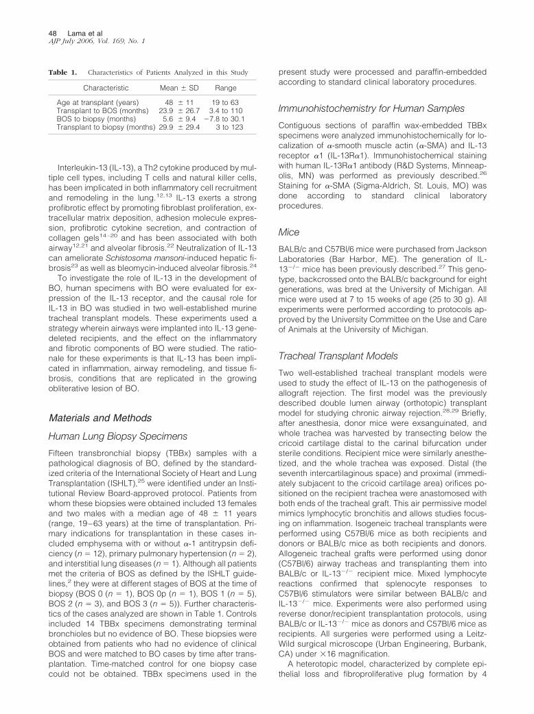
Table 1. Characteristics of Patients Analyzed in this Study
Characteristic Mean � SD Range
Age at transplant (years) 48 � 11 19 to 63Transplant to BOS (months) 23.9 � 26.7 3.4 to 110BOS to biopsy (months) 5.6 � 9.4 �7.8 to 30.1Transplant to biopsy (months) 29.9 � 29.4 3 to 123
48 Lama et alAJP July 2006, Vol. 169, No. 1
weeks after transplant as previously described,4,11,30
was also used. Donor tracheas were harvested as de-scribed above. The trachea was inserted in a subcuta-neous pouch created after a 3-mm incision and bluntdissection in the back of the neck. The skin pocket wasclosed with a size 5.0 nylon suture.
Histopathological Evaluation of TrachealTransplants
Grafts were harvested at the indicated time points andembedded en bloc in Tissue Freezing Medium (TriangleBiomedical Sciences, Durham, NC) in Disposable BaseMolds (Richard-Allan Scientific, Kalamazoo, MI) in liquidnitrogen and stored at �80°C until the time of analysis orwere formalin-fixed and paraffin-embedded. For the or-thotopic model histochemical staining was performed forelastin (Accustain; Sigma-Aldrich) to determine graft lu-minal occlusion on 5-�m-thick sections. Immunostainingwas also performed using serial adjacent sections fromeach group, with primary antibodies directed against apan-T-cell marker (hamster anti-mouse CD3; BD Pharm-ingen, San Diego, CA) and a macrophage marker (anti-mouse F4/80; BD Pharmingen). Morphometric measure-ments of cross-sectional areas were performed by blindlytracing both epithelial and subepithelial areas using acomputer-assisted image analysis system (AxoCamHR;Carl Zeiss Microimaging, Thornwood, NY). All sampleswere analyzed at 1 week after transplantation, becausethat was the time of maximal lumen narrowing and cellu-lar infiltration in allografts.
For the heterotopic model, transplants were harvestedfor evaluation at 7, 14, 21, and 28 days after implantation(n � 4–6 at each time point). Contiguous sections, 6 �mthick, obtained from the center of each tracheal segmentwere stained with hematoxylin and eosin and elastictrichrome. Four histological features (airway lining epithe-lial loss, leukocyte infiltration, luminal obliteration, andextracellular matrix deposition) were assessed semi-quantitatively, in a blinded fashion, using a modified scor-ing system based on those previously described by au-thors in this field.5,31 The scoring used was as follows: 1)airway lining epithelial loss (0, no change; 1�, �25%circumference loss; 2�, 25 to 50% circumference loss;3�, 50 to 75% circumference loss; 4�, �75% circumfer-ence loss); 2) leukocyte infiltration (0, no change; 1�,sparse perivascular infiltrates involving some vessels;2�, perivascular infiltrates involving most vessels withextension into adjacent connective tissue; 3�, diffuse butmild to moderate panmural infiltration; 4�, diffusemarked panmural infiltration with striking subepithelialand intraepithelial component); 3) luminal obliteration (0,no change; 1�, �25% obliteration; 2�, 25 to 50% oblit-eration; 3�, 50 to 75% obliteration; 4�, �75% oblitera-tion); and 4) extracellular matrix deposition (0, nochange; 1�, �25% deposition; 2�, 25 to 50% deposi-tion; 3�, 50 to 75% deposition; 4�, �75% deposition).
mRNA Isolation and PCR Analysis
Total RNA was isolated from mouse tracheas using theRNeasy mini kit (Qiagen, Inc., Valencia, CA) as per themanufacturer’s instructions. The TaqMan real-time poly-merase chain reaction (PCR) primers for IL-13, TGF-�1,MCP-1, and �-actin were purchased from Applied Bio-systems (Foster City, CA). Reverse transcriptase (RT)-PCR and real-time PCR were performed using the one-step QuantiTech Probe RT-PCR kit (Qiagen) as per themanufacturer’s instructions. Results were normalized to�-actin amplified from the same RNA mix in each sample.
Protein Analysis by ELISA
Harvested grafts were homogenized in 500 �l of CellLyticTM mammalian tissue lysis reagent (Sigma-Aldrich)containing protease inhibitors (Complete Protease tab-lets; Roche, Manheim, Germany). Cellular debris wasremoved by centrifugation at 10,000 rpm for 10 minutes.Aliquots of tracheal homogenate were then assayed byspecific ELISA for cytokines. Measurements of MCP-1,regulated on activation normal T cell expressed and se-creted (RANTES), and IL-13 were performed on trachealhomogenates using ELISA kits from R&D Systems (Min-neapolis, MN).
Plasminogen Activator Inhibitor-1/Luciferase(PAIL) Assay for TGF-�1
Tracheal homogenates were prepared as describedabove, and 250 �l of the tracheal homogenate was acid-treated, then neutralized to activate latent TGF-�1 withinthe sample. These tracheal homogenates were then an-alyzed in a bioassay for TGF-�1 activity (PAIL cell assay)as described previously.22
Sircol Assay for Collagen Measurements
Tracheal collagen levels were determined by harvestingheterotopic tracheal transplants at day 28. Animals wereeuthanized, and tracheas were harvested and snap-fro-zen in liquid nitrogen. Before analysis, two tracheas werehomogenized in 500 �l of normal saline and spun at 2000rpm for 10 minutes. Aliquots of homogenate (100 �l) werethen assayed for total tracheal collagen levels and com-pared with a standard curve prepared from rat tail colla-gen using the Sircol collagen dye binding assay (Accu-rate Chemical and Scientific Corp., Westbury, NY)according to the manufacturer’s instructions.
FACS Analysis
Infiltrating inflammatory cells were isolated from trans-planted airway graft tissue by using the Medimachine(DakoCytomation, Glostrup, Denmark) to isolate cells asper the manufacturer’s protocol. Immunophenotyping ofgraft-infiltrating leukocytes was accomplished by flow cy-tometric analysis. Isolated cells were stained for multipa-
Interleukin-13 in Bronchiolitis Obliterans 49AJP July 2006, Vol. 169, No. 1
rameter fluorescence-activated cell sorting (FACS) anal-ysis using phycoerythrin-labeled anti-mouse CD3, andfluorescein isothiocyanate-labeled anti-mouse macro-phage (F4/80; Caltag Laboratories, Burlingame, CA).Cells were prepared in staining buffer consisting of Ca2�-and Mg2�-free Dulbecco’s phosphate-buffered saline(Mediatech, Herndon, VA) with 0.05% sodium azide and1% fetal bovine serum. To reduce nonspecific binding ofFcII/III receptors, 5-minute incubation at 4°C with purifiedanti-2.4G2 (BD Biosciences, San Diego, CA) was per-formed. After incubation for 30 minutes at 4°C in the darkwith the indicated fluorescent antibodies (1:100), cellswere washed twice in staining buffer, and populations oflymphocytes and macrophages were then identified us-ing a FACSCalibur system (BD Biosciences). Cell detec-tor voltages (gating) were set using unstained lympho-cytes isolated from the spleen. Absolute numbers ofspecific cell populations were derived from percentagesof cells determined by FACS analysis, multiplied by thetotal number of cells isolated from grafts (determined bymanual cell counts performed using a hemocytometer).
Neutralization of IL-13
Anti-murine IL-13 polyclonal antibodies were raised byimmunizing New Zealand White rabbits with recombinantmurine IL-13 (R&D Systems) as described before32 andwere a generous gift of Dr. Nick Luckacs (University ofMichigan). IL-13 immunoneutralization studies were per-formed by daily intraperitoneal injection of anti-murineIL-13 (0.2 ml) in the wild-type recipients of orthotopictracheal transplantation from days 1 to 6 after surgery.
Statistics
Student’s t-tests were used to determine P values whencomparing two groups. When comparing three or moregroups, analysis of variance was performed with aposthoc Bonferroni test to determine which groupsshowed significant differences. A value of P � 0.05 wasconsidered significant.
Results
Detection of Myofibroblasts in HumanBO Lesions
Myofibroblasts are recognized as key effector cells intissue fibrosis because of their enhanced ability to syn-thesize interstitial collagen. The composition of the fi-brotic lesions in human BO has not been fully character-ized. Tissue sections from 15 biopsy samples withpathologist-confirmed diagnosis of BO were stained witha monoclonal antibody to �-SMA, a marker of myofibro-blasts. The characteristics of the cases analyzed areshown in Table 1. Bronchial tissue was identified by thepresence of characteristic bronchial epithelium and orpresence of smooth muscle bundles. Mesenchymal cellsexpressing �-SMA were identified in all 15 samples of
BO. Myofibroblasts were identified predominantly in thelamina propria (Figure 1A) and varied in number from afew cells to marked infiltration. These cells were not seenin the lung airway tissue of biopsies in which BO was notpresent (Figure 1B, inset). In cases of BO marked byintraluminal obstruction, myofibroblasts were identified inthe polypoid obstructing lesions (Figure 1C). This findingsupports the involvement of myofibroblast accumulationin the fibroproliferative lesion of BOS.
Expression of IL-13R�1 in Myofibroblasts in BO
We next determined whether the myofibroblasts seen inlesions of BO demonstrate expression of IL-13R�1, thesignaling chain of the high affinity IL-13 receptor. Immu-nohistochemical analysis indicated that IL-13R�1 wasexpressed abundantly in myofibroblasts seen in terminalbronchi in transbronchial biopsy specimens demonstrat-ing BO. Representative staining to identify myofibroblastswith �-SMA is shown in Figure 1D. IL-13R�1 and controlstaining is shown in a serial section (Figure 1, E and F). Inaddition IL-13R�1 expression was seen on epithelial cellsand smooth muscle bundles.
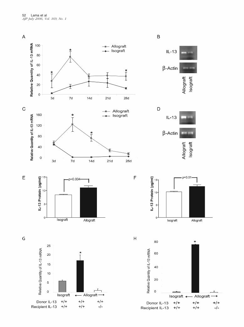
IL-13 mRNA and Protein Expression in MurineTracheal Allograft Models
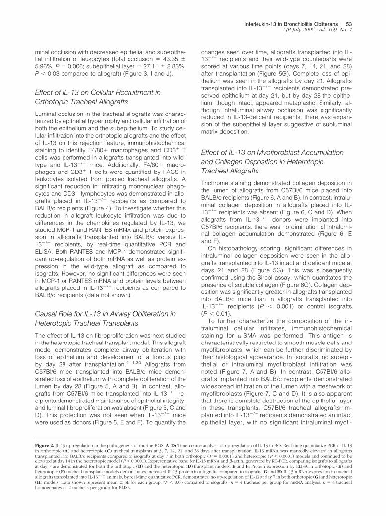
To determine whether IL-13 expression is altered in thesetting of BO, IL-13 mRNA was measured at various timepoints after orthotopic (Figure 2, A and B) and heterotopic(Figure 2, C and D) tracheal transplantation. IL-13 expres-sion peaked at day 7 after transplantation in both models(Figure 2, A and C). Furthermore, protein expression ofIL-13 by specific ELISA at day 7 confirmed significantlyincreased levels of IL-13 in allografts compared to isografts(Figure 2, E and F). To differentiate the contribution of donorgraft versus infiltrating recipient cells to the up-regulation ofIL-13 seen in allografts, we measured IL-13 mRNA expres-sion in allografts implanted into IL-13�/� recipients. Allo-grafts placed into IL-13�/� recipients showed no IL-13mRNA elevation in either model (Figure 2, G and H). Thesedata suggest that infiltrating recipient cells comprise thedominant source of the elevated IL-13 mRNA expressed intracheal allografts. To determine whether expression ofanother important Th2 cytokine, IL-4, was modulated inIL-13�/� recipients, we measured graft expression of IL-4 atday 7, the time of maximal IL-13 up-regulation. No statisti-cally significant difference was noted in levels of IL-4 mRNAbetween transplanted tracheal allografts placed into IL-13�/� and wild-type recipients in both orthotopic and het-erotopic models (P � 0.46 and 0.35, respectively).
Role for IL-13 in Orthotopic TrachealTransplants
We next sought to determine the influence of IL-13 on theluminal narrowing of orthotopic tracheal allografts (Figure3). Tracheal isografts (BALB/c into BALB/c) maintainedtheir normal architecture and demonstrated no significant
50 Lama et alAJP July 2006, Vol. 169, No. 1

inflammatory infiltrates or luminal encroachment (Figure3, A and B). Allografted tracheal transplants (C57Bl/6 intoBALB/c) demonstrated partial concentric graft luminalocclusion (55.43 � 6.07% compared to 25.30 � 5.32% inisografts, P � �0.001; Figure 3, C and D) secondary tocharacteristic thickening of the epithelial and subepithe-lial layer.28,29 To study the role of IL-13, C57Bl/6 tracheaswere transplanted into allogeneic IL-13�/� recipients.These experiments showed a significant decrease in lu-minal occlusion in allografts transplanted into IL-13�/�
recipients when compared to allografts placed into IL-13-sufficient, BALB/c mice with diminution in both the epi-thelial and subepithelial components (total occlusion �
37.37 � 4.30%, P � 0.004; subepithelial layer � 24.28 �6.07%, P � 0.001 compared to allograft) (Figure 3, E andF). In contrast to these data, no protection was seen whenIL-13�/� donor tracheas were implanted into allogeneic,C57Bl/6 recipients (48.49 � 3.71%; Figure 3, G and H).These data demonstrate that recipient, but not donor,IL-13 gene-deleted mice are protected from airway lumi-nal obliteration following allograft implantation. To defin-itively implicate IL-13 in the rejection process, IL-13 im-munoneutralization studies were performed by injectinganti-murine IL-13 in the allograft recipient (C57Bl/6 intoBALB/c). Immunoneutralization of IL-13 between days 1and 6 of transplantation significantly decreased graft lu-
Figure 1. Immunohistochemistry staining for �-SMA and IL-13R�1 in TBBx specimens demonstrating evidence of BO. A: �-SMA-positive (stained brown)mesenchymal cells (myofibroblasts) are demonstrated in the submucosa of bronchi in a patient with BOS. B: TBBx specimen from a patient without BOS showingnormal bronchial epithelium with underlying smooth muscle bundle and absence of myofibroblast infiltration in the lamina propria and the lumen. C: �-SMAstaining (brown) of a TBBx specimen demonstrates bronchi cut tangentially as evidenced by smooth muscle bundles (arrows). The lumen is filled withmesenchymal cells that stain positively for �-SMA, demonstrating the presence of myofibroblasts. D–F: Contiguous sections from C (marked by the rectangle)are shown at higher magnification stained with �-SMA (D), IL-13R�1 (stained red) (E), and the negative control for IL-13R�1 (F). Original magnifications: �100(A and C); �200 (B); �400 (D–F). These findings are representative of all 15 patients examined.
Interleukin-13 in Bronchiolitis Obliterans 51AJP July 2006, Vol. 169, No. 1
52 Lama et alAJP July 2006, Vol. 169, No. 1
minal occlusion with decreased epithelial and subepithe-lial infiltration of leukocytes (total occlusion � 43.35 �5.96%, P � 0.006; subepithelial layer � 27.11 � 2.83%,P � 0.03 compared to allograft) (Figure 3, I and J).
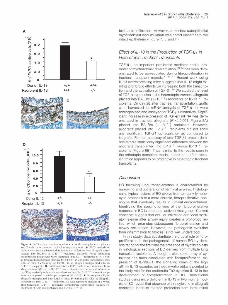
Effect of IL-13 on Cellular Recruitment inOrthotopic Tracheal Allografts
Luminal occlusion in the tracheal allografts was charac-terized by epithelial hypertrophy and cellular infiltration ofboth the epithelium and the subepithelium. To study cel-lular infiltration into the orthotopic allografts and the effectof IL-13 on this rejection feature, immunohistochemicalstaining to identify F4/80� macrophages and CD3� Tcells was performed in allografts transplanted into wild-type and IL-13�/� mice. Additionally, F4/80� macro-phages and CD3� T cells were quantified by FACS inleukocytes isolated from pooled tracheal allografts. Asignificant reduction in infiltrating mononuclear phago-cytes and CD3� lymphocytes was demonstrated in allo-grafts placed in IL-13�/� recipients as compared toBALB/c recipients (Figure 4). To investigate whether thisreduction in allograft leukocyte infiltration was due todifferences in the chemokines regulated by IL-13, westudied MCP-1 and RANTES mRNA and protein expres-sion in allografts transplanted into BALB/c versus IL-13�/� recipients, by real-time quantitative PCR andELISA. Both RANTES and MCP-1 demonstrated signifi-cant up-regulation of both mRNA as well as protein ex-pression in the wild-type allograft as compared toisografts. However, no significant differences were seenin MCP-1 or RANTES mRNA and protein levels betweenallografts placed in IL-13�/� recipients as compared toBALB/c recipients (data not shown).
Causal Role for IL-13 in Airway Obliteration inHeterotopic Tracheal Transplants
The effect of IL-13 on fibroproliferation was next studiedin the heterotopic tracheal transplant model. This allograftmodel demonstrates complete airway obliteration withloss of epithelium and development of a fibrous plugby day 28 after transplantation.4,11,30 Allografts fromC57Bl/6 mice transplanted into BALB/c mice demon-strated loss of epithelium with complete obliteration of thelumen by day 28 (Figure 5, A and B). In contrast, allo-grafts from C57Bl/6 mice transplanted into IL-13�/� re-cipients demonstrated maintenance of epithelial integrity,and luminal fibroproliferation was absent (Figure 5, C andD). This protection was not seen when IL-13�/� micewere used as donors (Figure 5, E and F). To quantify the
changes seen over time, allografts transplanted into IL-13�/� recipients and their wild-type counterparts werescored at various time points (days 7, 14, 21, and 28)after transplantation (Figure 5G). Complete loss of epi-thelium was seen in the allografts by day 21. Allograftstransplanted into IL-13�/� recipients demonstrated pre-served epithelium at day 21, but by day 28 the epithe-lium, though intact, appeared metaplastic. Similarly, al-though intraluminal airway occlusion was significantlyreduced in IL-13-deficient recipients, there was expan-sion of the subepithelial layer suggestive of subluminalmatrix deposition.
Effect of IL-13 on Myofibroblast Accumulationand Collagen Deposition in HeterotopicTracheal Allografts
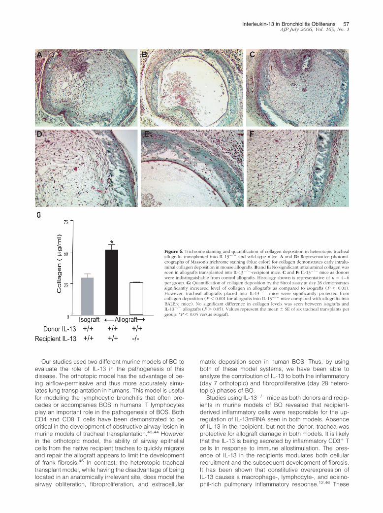
Trichrome staining demonstrated collagen deposition inthe lumen of allografts from C57Bl/6 mice placed intoBALB/c recipients (Figure 6, A and B). In contrast, intralu-minal collagen deposition in allografts placed into IL-13�/� recipients was absent (Figure 6, C and D). Whenallografts from IL-13�/� donors were implanted intoC57Bl/6 recipients, there was no diminution of intralumi-nal collagen accumulation demonstrated (Figure 6, Eand F).
On histopathology scoring, significant differences inintraluminal collagen deposition were seen in the allo-grafts transplanted into IL-13 intact and deficient mice atdays 21 and 28 (Figure 5G). This was subsequentlyconfirmed using the Sircol assay, which quantitates thepresence of soluble collagen (Figure 6G). Collagen dep-osition was significantly greater in allografts transplantedinto BALB/c mice than in allografts transplanted intoIL-13�/� recipients (P � 0.001) or control isografts(P � 0.01).
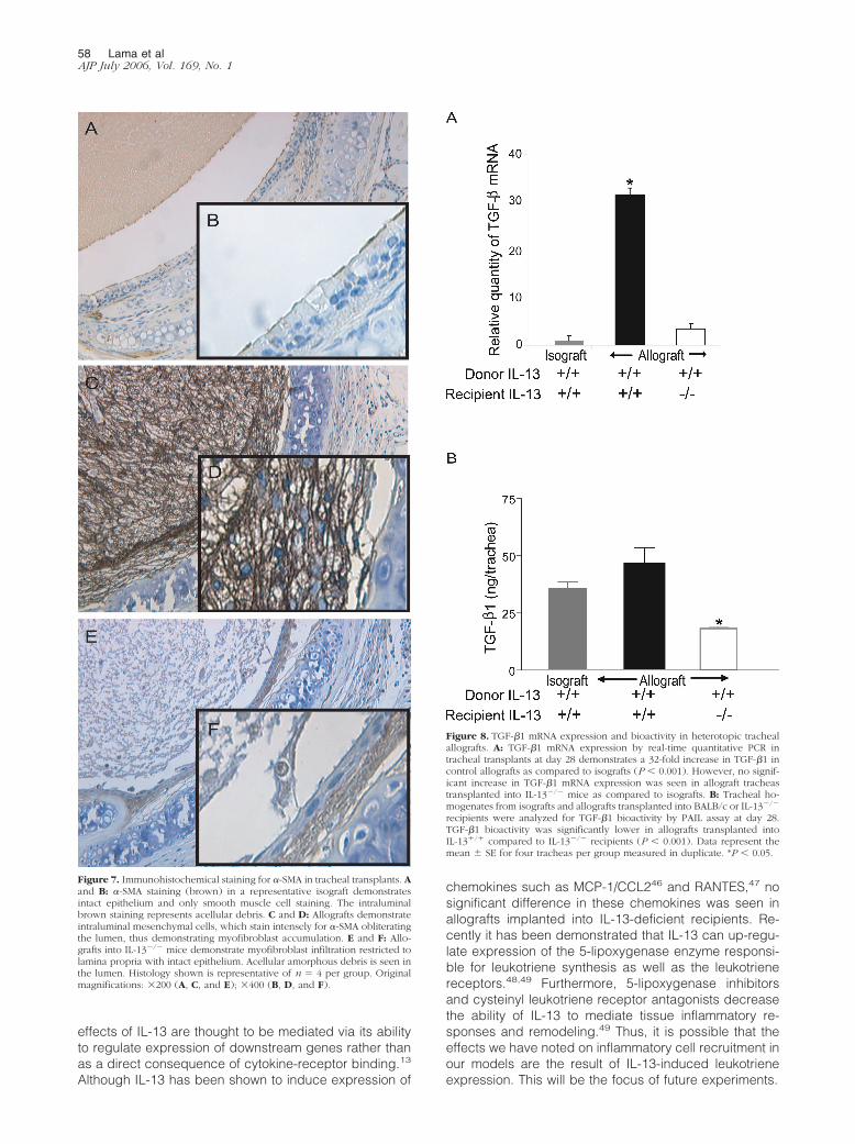
To further characterize the composition of the in-traluminal cellular infiltrates, immunohistochemicalstaining for �-SMA was performed. This antigen ischaracteristically restricted to smooth muscle cells andmyofibroblasts, which can be further discriminated bytheir histological appearance. In isografts, no subepi-thelial or intraluminal myofibroblast infiltration wasnoted (Figure 7, A and B). In contrast, C57Bl/6 allo-grafts implanted into BALB/c recipients demonstratedwidespread infiltration of the lumen with a meshwork ofmyofibroblasts (Figure 7, C and D). It is also apparentthat there is complete destruction of the epithelial layerin these transplants. C57Bl/6 tracheal allografts im-planted into IL-13�/� recipients demonstrated an intactepithelial layer, with no significant intraluminal myofi-
Figure 2. IL-13 up-regulation in the pathogenesis of murine BOS. A–D: Time-course analysis of up-regulation of IL-13 in BO. Real-time quantitative PCR of IL-13in orthotopic (A) and heterotopic (C) tracheal transplants at 3, 7, 14, 21, and 28 days after transplantation. IL-13 mRNA was markedly elevated in allograftstransplanted into BALB/c recipients compared to isografts at day 7 in both orthotopic (P � 0.0001) and heterotopic (P � 0.0001) models and continued to beelevated at day 14 in the heterotopic model (P � 0.0001). Representative band for IL-13 mRNA and �-actin, generated by RT-PCR, comparing isografts to allograftsat day 7 are demonstrated for both the orthotopic (B) and the heterotopic (D) transplant models. E and F: Protein expression by ELISA in orthotopic (E) andheterotopic (F) tracheal transplant models demonstrates increased IL-13 protein in allografts compared to isografts. G and H: IL-13 mRNA expression in trachealallografts transplanted into IL-13�/� animals, by real-time quantitative PCR, demonstrated no up-regulation of IL-13 at day 7 in both orthotopic (G) and heterotopic(H) models. Data shown represent mean � SE for each group. *P � 0.05 compared to isografts. n � 4 tracheas per group for mRNA analysis. n � 4 trachealhomogenates of 2 tracheas per group for ELISA.
Interleukin-13 in Bronchiolitis Obliterans 53AJP July 2006, Vol. 169, No. 1
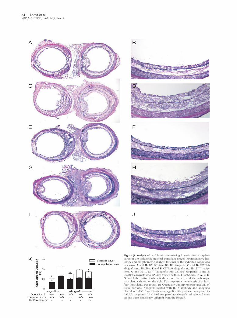
Figure 3. Analysis of graft luminal narrowing 1 week after transplan-tation in the orthotopic tracheal transplant model. Representative his-tology and morphometric analysis for each of the indicated conditionsis shown. A and B: BALB/c into BALB/c isografts. C and D: C57Bl/6allografts into BALB/c. E and F: C57Bl/6 allografts into IL-13�/� recip-ients. G and H: IL-13�/� allografts into C57Bl/6 recipients. I and J:C57Bl/6 allografts into BALB/c treated with IL-13 antibody. In A, C, E,G, and I the native trachea is shown on the left, and the orthotopictransplant is shown on the right. Data represent the analysis of at leastfour transplants per group. K: Quantitative morphometric analysis oftissue sections. Allografts treated with IL-13 antibody and allograftsplaced in IL-13�/� recipients were significantly protected compared toBALB/c recipients. *P � 0.05 compared to allografts. All allograft con-ditions were statistically different from the isograft.
54 Lama et alAJP July 2006, Vol. 169, No. 1
broblasts infiltration. However, a modest subepithelialmyofibroblast accumulation was noted underneath theintact epithelium (Figure 7, E and F).
Effect of IL-13 in the Production of TGF-�1 inHeterotopic Tracheal Transplants
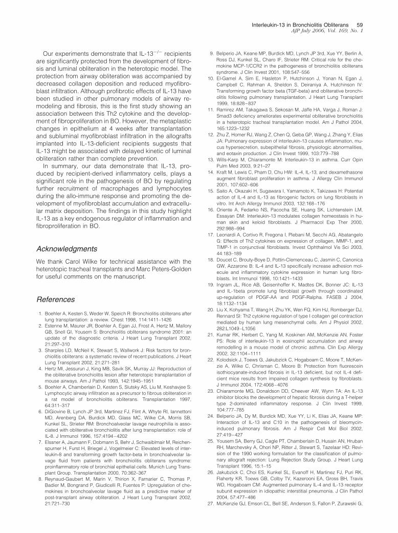
TGF-�1, an important profibrotic mediator and a pro-moter of myofibroblast differentiation,33,34 has been dem-onstrated to be up-regulated during fibroproliferation intracheal transplant models.11,35–37 Recent work usingIL-13-overexpressing mice suggests that IL-13 might ex-ert its profibrotic effects via increasing both the transcrip-tion and the activation of TGF-�1.38 We studied the levelof TGF-� expression in the heterotopic tracheal allograftsplaced into BALB/c (IL-13�/�) recipients or IL-13�/� re-cipients. On day 28 after tracheal transplantation, graftswere harvested for mRNA analysis of TGF-�1 or werehomogenized and assayed for TGF-�1 bioactivity. Signif-icant increase in expression of TGF-�1 mRNA was dem-onstrated in tracheal allografts (P � 0.001, Figure 8A)placed into BALB/c (IL-13�/�) recipients. However,allografts placed into IL-13�/� recipients did not showany significant TGF-�1 up-regulation as compared toisografts. Further, bioassay of total TGF-�1 protein dem-onstrated a statistically significant difference between theallografts transplanted into IL-13�/� versus IL-13�/� re-cipients (Figure 8B). Thus, similar to the results seen inthe orthotopic transplant model, a lack of IL-13 in recip-ient mice appears to be protective in heterotopic trachealtransplants.
Discussion
BO following lung transplantation is characterized bynarrowing and obliteration of terminal airways. Histologi-cally, typical lesions of BO evolve from an early lympho-cytic bronchitis to a more chronic, fibroproliferative phe-notype that eventually results in luminal encroachment.Identifying the specific drivers of the fibroproliferativeresponse in BO is an area of active investigation. Currentconcepts suggest that cellular infiltration and local medi-ator release after airway injury creates a profibrotic mi-lieu, which promotes subsequent fibroproliferation andairway obliteration. However, the pathogenic evolutionfrom inflammation to fibrosis is not well understood.
In this study, data substantiate the crucial role of fibro-proliferation in the pathogenesis of human BO by dem-onstrating for the first time the presence of myofibroblastsin histological sections of BO derived from multiple lungtransplant recipients. Although a pleiotropic array of cy-tokines has been associated with fibroproliferation, ex-pression of IL-13R�1, the signaling chain of the highaffinity IL-13 receptor, on these myofibroblasts pointed tothe likely role for the profibrotic Th2 cytokine IL-13 in thedevelopment of fibroproliferation in BO. Translationalstudies using mice deficient in IL-13 in two murine mod-els of BO reveal that absence of this cytokine in allograftrecipients leads to marked protection from intraluminal
Figure 4. FACS analysis and immunohistochemical staining for macrophagesand T cells in orthotopic tracheal transplant model. A: FACS analysis ofF4/80� cells (macrophages) identified in cell isolations from allografts trans-planted into BALB/c or IL-13�/� recipients. Markedly fewer infiltratingmononuclear phagocytes were identified in IL-13�/� recipients (P � 0.05).B: Immunohistochemical staining for F4/80� in allografts transplanted intoBALB/c mice. C: Staining for F4/80� in an allograft transplanted into anIL-13�/� recipients. D: FACS analysis for CD3� cells in cell isolations fromallografts into BALB/c or IL-13�/� mice. Significantly decreased infiltrationby CD3-positive lymphocytes was demonstrated in IL-13�/� allograft recip-ients as compared to wild-type recipients (P � 0.05). E: Staining for CD3 inallografts transplanted into BALB/c mice. F: Staining for CD3 in allograftstransplanted into IL-13�/� recipients. All data represent analysis at 1 weekafter transplant. IL-13�/� recipients demonstrate significantly reduced re-cruitment of both macrophages and T cells (n � 4).
Interleukin-13 in Bronchiolitis Obliterans 55AJP July 2006, Vol. 169, No. 1

obliteration, mononuclear cell infiltration, and fibrosis. Incontrast, deficiency of IL-13 in donor tissue was not as-sociated with a reduction in either fibroproliferation or BO.These data demonstrate that IL-13 secreted by infiltratinginflammatory cells contributes to the development of fi-brosis in BO.
Even though BO has been recognized as a fibroticdisorder of the small airways for a number of years, this isthe first study to demonstrate that myofibroblasts arepresent in human histological sections demonstrating ev-idence of BO. The presence of myofibroblasts helps ex-plain the excessive deposition of extracellular matrixseen in these patients, as myofibroblasts are known to be
potent producers of collagen and other extracellular ma-trix proteins.39,40 In fact, the data shown here demon-strate that in the murine models there is a strong corre-lation between the presence of myofibroblasts and theaccumulation of collagen. In humans, myofibroblastshave previously been associated with airway remodelingin asthma41 as well as alveolar fibrosis in idiopathic pul-monary fibrosis.39,40 Furthermore, early myofibroblast in-filtration predicts chronic renal allograft dysfunction inrenal transplant recipients.42 Additional studies to deter-mine whether the degree of myofibroblast infiltration inthe bronchial tissue correlates with clinical course of thedisease would be useful in making clinical predictions.
Figure 5. Histopathology (hematoxylin and eo-sin) of luminal fibrosis 28 days after transplanta-tion in the heterotopic tracheal transplant model.A and B: Representative photomicrographs ofheterotopic trachea allografts (C57Bl/6 allograftsinto BALB/c-recipient mice) demonstrate epithe-lial injury, complete airway obliteration, and ma-trix deposition. C and D: Allografts into IL-13�/
�-recipient mice show intact epithelium and nointraluminal cellular infiltration or airway obliter-ation. The deposit in the center represents acel-lular debris as this is not an air-permissive model.E and F: IL-13�/� tracheas used as donors intoC57Bl/6 mice do not demonstrate any protectionfrom airway obliteration. G: Quantitative analysisof histopathological sections of tracheal allograftstransplanted into IL-13�/� and IL-13�/� recipi-ents. Histology shown is representative of n �4–6 per group. Original magnifications: �200 (A,C, and E); �400 (B, D, and F). *P � 0.05.
56 Lama et alAJP July 2006, Vol. 169, No. 1
Our studies used two different murine models of BO toevaluate the role of IL-13 in the pathogenesis of thisdisease. The orthotopic model has the advantage of be-ing airflow-permissive and thus more accurately simu-lates lung transplantation in humans. This model is usefulfor modeling the lymphocytic bronchitis that often pre-cedes or accompanies BOS in humans. T lymphocytesplay an important role in the pathogenesis of BOS. BothCD4 and CD8 T cells have been demonstrated to becritical in the development of obstructive airway lesion inmurine models of tracheal transplantation.43,44 Howeverin the orthotopic model, the ability of airway epithelialcells from the native recipient trachea to quickly migrateand repair the allograft appears to limit the developmentof frank fibrosis.45 In contrast, the heterotopic trachealtransplant model, while having the disadvantage of beinglocated in an anatomically irrelevant site, does model theairway obliteration, fibroproliferation, and extracellular
matrix deposition seen in human BOS. Thus, by usingboth of these model systems, we have been able toanalyze the contribution of IL-13 to both the inflammatory(day 7 orthotopic) and fibroproliferative (day 28 hetero-topic) phases of BO.
Studies using IL-13�/� mice as both donors and recip-ients in murine models of BO revealed that recipient-derived inflammatory cells were responsible for the up-regulation of IL-13mRNA seen in both models. Absenceof IL-13 in the recipient, but not the donor, trachea wasprotective for allograft damage in both models. It is likelythat the IL-13 is being secreted by inflammatory CD3� Tcells in response to immune allostimulation. The pres-ence of IL-13 in the recipients modulates both cellularrecruitment and the subsequent development of fibrosis.It has been shown that constitutive overexpression ofIL-13 causes a macrophage-, lymphocyte-, and eosino-phil-rich pulmonary inflammatory response.12,46 These
Figure 6. Trichrome staining and quantification of collagen deposition in heterotopic trachealallografts transplanted into IL-13�/� and wild-type mice. A and D: Representative photomi-crographs of Masson’s trichrome staining (blue color) for collagen demonstrates early intralu-minal collagen deposition in mouse allografts. B and E: No significant intraluminal collagen wasseen in allografts transplanted into IL-13�/�-recipient mice. C and F: IL-13�/� mice as donorswere indistinguishable from control allografts. Histology shown is representative of n � 4–6per group. G: Quantification of collagen deposition by the Sircol assay at day 28 demonstratessignificantly increased level of collagen in allografts as compared to isografts (P � 0.01).However, tracheal allografts placed into IL-13�/� mice were significantly protected fromcollagen deposition (P � 0.001 for allografts into IL-13�/� mice compared with allografts intoBALB/c mice). No significant difference in collagen levels was seen between isografts andIL-13�/� allografts (P � 0.05). Values represent the mean � SE of six tracheal transplants pergroup. *P � 0.05 versus isograft.
Interleukin-13 in Bronchiolitis Obliterans 57AJP July 2006, Vol. 169, No. 1
effects of IL-13 are thought to be mediated via its abilityto regulate expression of downstream genes rather thanas a direct consequence of cytokine-receptor binding.13
Although IL-13 has been shown to induce expression of
chemokines such as MCP-1/CCL246 and RANTES,47 nosignificant difference in these chemokines was seen inallografts implanted into IL-13-deficient recipients. Re-cently it has been demonstrated that IL-13 can up-regu-late expression of the 5-lipoxygenase enzyme responsi-ble for leukotriene synthesis as well as the leukotrienereceptors.48,49 Furthermore, 5-lipoxygenase inhibitorsand cysteinyl leukotriene receptor antagonists decreasethe ability of IL-13 to mediate tissue inflammatory re-sponses and remodeling.49 Thus, it is possible that theeffects we have noted on inflammatory cell recruitment inour models are the result of IL-13-induced leukotrieneexpression. This will be the focus of future experiments.
Figure 7. Immunohistochemical staining for �-SMA in tracheal transplants. Aand B: �-SMA staining (brown) in a representative isograft demonstratesintact epithelium and only smooth muscle cell staining. The intraluminalbrown staining represents acellular debris. C and D: Allografts demonstrateintraluminal mesenchymal cells, which stain intensely for �-SMA obliteratingthe lumen, thus demonstrating myofibroblast accumulation. E and F: Allo-grafts into IL-13�/� mice demonstrate myofibroblast infiltration restricted tolamina propria with intact epithelium. Acellular amorphous debris is seen inthe lumen. Histology shown is representative of n � 4 per group. Originalmagnifications: �200 (A, C, and E); �400 (B, D, and F).
Figure 8. TGF-�1 mRNA expression and bioactivity in heterotopic trachealallografts. A: TGF-�1 mRNA expression by real-time quantitative PCR intracheal transplants at day 28 demonstrates a 32-fold increase in TGF-�1 incontrol allografts as compared to isografts (P � 0.001). However, no signif-icant increase in TGF-�1 mRNA expression was seen in allograft tracheastransplanted into IL-13�/� mice as compared to isografts. B: Tracheal ho-mogenates from isografts and allografts transplanted into BALB/c or IL-13�/�
recipients were analyzed for TGF-�1 bioactivity by PAIL assay at day 28.TGF-�1 bioactivity was significantly lower in allografts transplanted intoIL-13�/� compared to IL-13�/� recipients (P � 0.001). Data represent themean � SE for four tracheas per group measured in duplicate. *P � 0.05.
58 Lama et alAJP July 2006, Vol. 169, No. 1
Our experiments demonstrate that IL-13�/� recipientsare significantly protected from the development of fibro-sis and luminal obliteration in the heterotopic model. Theprotection from airway obliteration was accompanied bydecreased collagen deposition and reduced myofibro-blast infiltration. Although profibrotic effects of IL-13 havebeen studied in other pulmonary models of airway re-modeling and fibrosis, this is the first study showing anassociation between this Th2 cytokine and the develop-ment of fibroproliferation in BO. However, the metaplasticchanges in epithelium at 4 weeks after transplantationand subluminal myofibroblast infiltration in the allograftsimplanted into IL-13-deficient recipients suggests thatIL-13 might be associated with delayed kinetic of luminalobliteration rather than complete prevention.
In summary, our data demonstrate that IL-13, pro-duced by recipient-derived inflammatory cells, plays asignificant role in the pathogenesis of BO by regulatingfurther recruitment of macrophages and lymphocytesduring the allo-immune response and promoting the de-velopment of myofibroblast accumulation and extracellu-lar matrix deposition. The findings in this study highlightIL-13 as a key endogenous regulator of inflammation andfibroproliferation in BO.
Acknowledgments
We thank Carol Wilke for technical assistance with theheterotopic tracheal transplants and Marc Peters-Goldenfor useful comments on the manuscript.
References
1. Boehler A, Kesten S, Weder W, Speich R: Bronchiolitis obliterans afterlung transplantation: a review. Chest 1998, 114:1411–1426
2. Estenne M, Maurer JR, Boehler A, Egan JJ, Frost A, Hertz M, MalloryGB, Snell GI, Yousem S: Bronchiolitis obliterans syndrome 2001: anupdate of the diagnostic criteria. J Heart Lung Transplant 2002,21:297–310
3. Sharples LD, McNeil K, Stewart S, Wallwork J: Risk factors for bron-chiolitis obliterans: a systematic review of recent publications. J HeartLung Transplant 2002, 21:271–281
4. Hertz MI, Jessurun J, King MB, Savik SK, Murray JJ: Reproduction ofthe obliterative bronchiolitis lesion after heterotopic transplantation ofmouse airways. Am J Pathol 1993, 142:1945–1951
5. Boehler A, Chamberlain D, Kesten S, Slutsky AS, Liu M, Keshavjee S:Lymphocytic airway infiltration as a precursor to fibrous obliteration ina rat model of bronchiolitis obliterans. Transplantation 1997,64:311–317
6. DiGiovine B, Lynch JP 3rd, Martinez FJ, Flint A, Whyte RI, IannettoniMD, Arenberg DA, Burdick MD, Glass MC, Wilke CA, Morris SB,Kunkel SL, Strieter RM: Bronchoalveolar lavage neutrophilia is asso-ciated with obliterative bronchiolitis after lung transplantation: role ofIL-8. J Immunol 1996, 157:4194–4202
7. Elssner A, Jaumann F, Dobmann S, Behr J, Schwaiblmair M, Reichen-spurner H, Furst H, Briegel J, Vogelmeier C: Elevated levels of inter-leukin-8 and transforming growth factor-beta in bronchoalveolar la-vage fluid from patients with bronchiolitis obliterans syndrome:proinflammatory role of bronchial epithelial cells. Munich Lung Trans-plant Group. Transplantation 2000, 70:362–367
8. Reynaud-Gaubert M, Marin V, Thirion X, Farnarier C, Thomas P,Badier M, Bongrand P, Giudicelli R, Fuentes P: Upregulation of che-mokines in bronchoalveolar lavage fluid as a predictive marker ofpost-transplant airway obliteration. J Heart Lung Transplant 2002,21:721–730
9. Belperio JA, Keane MP, Burdick MD, Lynch JP 3rd, Xue YY, Berlin A,Ross DJ, Kunkel SL, Charo IF, Strieter RM: Critical role for the che-mokine MCP-1/CCR2 in the pathogenesis of bronchiolitis obliteranssyndrome. J Clin Invest 2001, 108:547–556
10. El-Gamel A, Sim E, Hasleton P, Hutchinson J, Yonan N, Egan J,Campbell C, Rahman A, Sheldon S, Deiraniya A, Hutchinson IV:Transforming growth factor beta (TGF-beta) and obliterative bronchi-olitis following pulmonary transplantation. J Heart Lung Transplant1999, 18:828–837
11. Ramirez AM, Takagawa S, Sekosan M, Jaffe HA, Varga J, Roman J:Smad3 deficiency ameliorates experimental obliterative bronchiolitisin a heterotopic tracheal transplantation model. Am J Pathol 2004,165:1223–1232
12. Zhu Z, Homer RJ, Wang Z, Chen Q, Geba GP, Wang J, Zhang Y, EliasJA: Pulmonary expression of interleukin-13 causes inflammation, mu-cus hypersecretion, subepithelial fibrosis, physiologic abnormalities,and eotaxin production. J Clin Invest 1999, 103:779–788
13. Wills-Karp M, Chiaramonte M: Interleukin-13 in asthma. Curr OpinPulm Med 2003, 9:21–27
14. Kraft M, Lewis C, Pham D, Chu HW: IL-4, IL-13, and dexamethasoneaugment fibroblast proliferation in asthma. J Allergy Clin Immunol2001, 107:602–606
15. Saito A, Okazaki H, Sugawara I, Yamamoto K, Takizawa H: Potentialaction of IL-4 and IL-13 as fibrogenic factors on lung fibroblasts invitro. Int Arch Allergy Immunol 2003, 132:168–176
16. Oriente A, Fedarko NS, Pacocha SE, Huang SK, Lichtenstein LM,Essayan DM: Interleukin-13 modulates collagen homeostasis in hu-man skin and keloid fibroblasts. J Pharmacol Exp Ther 2000,292:988–994
17. Leonardi A, Cortivo R, Fregona I, Plebani M, Secchi AG, AbatangeloG: Effects of Th2 cytokines on expression of collagen, MMP-1, andTIMP-1 in conjunctival fibroblasts. Invest Ophthalmol Vis Sci 2003,44:183–189
18. Doucet C, Brouty-Boye D, Pottin-Clemenceau C, Jasmin C, CanonicaGW, Azzarone B: IL-4 and IL-13 specifically increase adhesion mol-ecule and inflammatory cytokine expression in human lung fibro-blasts. Int Immunol 1998, 10:1421–1433
19. Ingram JL, Rice AB, Geisenhoffer K, Madtes DK, Bonner JC: IL-13and IL-1beta promote lung fibroblast growth through coordinatedup-regulation of PDGF-AA and PDGF-Ralpha. FASEB J 2004,18:1132–1134
20. Liu X, Kohyama T, Wang H, Zhu YK, Wen FQ, Kim HJ, Romberger DJ,Rennard SI: Th2 cytokine regulation of type I collagen gel contractionmediated by human lung mesenchymal cells. Am J Physiol 2002,282:L1049–L1056
21. Kumar RK, Herbert C, Yang M, Koskinen AM, McKenzie AN, FosterPS: Role of interleukin-13 in eosinophil accumulation and airwayremodelling in a mouse model of chronic asthma. Clin Exp Allergy2002, 32:1104–1111
22. Kolodsick J, Toews G, Jakubzick C, Hogaboam C, Moore T, McKen-zie A, Wilke C, Chrisman C, Moore B: Protection from fluoresceinisothiocyanate-induced fibrosis in IL-13 deficient, but not IL-4 defi-cient mice results from impaired collagen synthesis by fibroblasts.J Immunol 2004, 172:4068–4076
23. Chiaramonte MG, Donaldson DD, Cheever AW, Wynn TA: An IL-13inhibitor blocks the development of hepatic fibrosis during a T-helpertype 2-dominated inflammatory response. J Clin Invest 1999,104:777–785
24. Belperio JA, Dy M, Burdick MD, Xue YY, Li K, Elias JA, Keane MP:Interaction of IL-13 and C10 in the pathogenesis of bleomycin-induced pulmonary fibrosis. Am J Respir Cell Mol Biol 2002,27:419–427
25. Yousem SA, Berry GJ, Cagle PT, Chamberlain D, Husain AN, HrubanRH, Marchevsky A, Ohori NP, Ritter J, Stewart S, Tazelaar HD: Revi-sion of the 1990 working formulation for the classification of pulmo-nary allograft rejection: Lung Rejection Study Group. J Heart LungTransplant 1996, 15:1–15
26. Jakubzick C, Choi ES, Kunkel SL, Evanoff H, Martinez FJ, Puri RK,Flaherty KR, Toews GB, Colby TV, Kazerooni EA, Gross BH, TravisWD, Hogaboam CM: Augmented pulmonary IL-4 and IL-13 receptorsubunit expression in idiopathic interstitial pneumonia. J Clin Pathol2004, 57:477–486
27. McKenzie GJ, Emson CL, Bell SE, Anderson S, Fallon P, Zurawski G,
Interleukin-13 in Bronchiolitis Obliterans 59AJP July 2006, Vol. 169, No. 1

Murray R, Grencis R, McKenzie AN: Impaired development of Th2cells in IL-13-deficient mice. Immunity 1998, 9:423–432
28. Minamoto K, Pinsky DJ: Recipient iNOS but not eNOS deficiencyreduces luminal narrowing in tracheal allografts. J Exp Med 2002,196:1321–1333
29. Minamoto K, Harada H, Lama VN, Fedarau MA, Pinsky DJ: Recipro-cal regulation of airway rejection by the inducible gas-forming en-zymes heme oxygenase and nitric oxide synthase. J Exp Med 2005,202:283–294
30. Medoff BD, Seung E, Wain JC, Means TK, Campanella GS, Islam SA,Thomas SY, Ginns LC, Grabie N, Lichtman AH, Tager AM, Luster AD:BLT1-mediated T cell trafficking is critical for rejection and obliterativebronchiolitis after lung transplantation. J Exp Med 2005, 202:97–110
31. Belperio JA, Keane MP, Burdick MD, Lynch JP 3rd, Xue YY, Li K, RossDJ, Strieter RM: Critical role for CXCR3 chemokine biology in thepathogenesis of bronchiolitis obliterans syndrome. J Immunol 2002,169:1037–1049
32. Matsukawa A, Hogaboam CM, Lukacs NW, Lincoln PM, Evanoff HL,Strieter RM, Kunkel SL: Expression and contribution of endogenousIL-13 in an experimental model of sepsis. J Immunol 2000,164:2738–2744
33. Sime PJ, Xing Z, Graham FL, Csaky KG, Gauldie J: Adenovector-mediated gene transfer of active transforming growth factor-beta1induces prolonged severe fibrosis in rat lung. J Clin Invest 1997,100:768–776
34. Thannickal VJ, Lee DY, White ES, Cui Z, Larios JM, Chacon R,Horowitz JC, Day RM, Thomas PE: Myofibroblast differentiation bytransforming growth factor-beta1 is dependent on cell adhesion andintegrin signaling via focal adhesion kinase. J Biol Chem 2003,278:12384–12389
35. Aris RM, Walsh S, Chalermskulrat W, Hathwar V, Neuringer IP: Growthfactor upregulation during obliterative bronchiolitis in the mousemodel. Am J Respir Crit Care Med 2002, 166:417–422
36. Liu M, Suga M, Maclean AA, St George JA, Souza DW, Keshavjee S:Soluble transforming growth factor-beta type III receptor gene trans-fection inhibits fibrous airway obliteration in a rat model of Bronchi-olitis obliterans. Am J Respir Crit Care Med 2002, 165:419–423
37. Zhou H, Latham CW, Zander DS, Margolin SB, Visner GA: Pirfenidoneinhibits obliterative airway disease in mouse tracheal allografts.J Heart Lung Transplant 2005, 24:1577–1585
38. Lee CG, Homer RJ, Zhu Z, Lanone S, Wang X, Koteliansky V, Shipley
JM, Gotwals P, Noble P, Chen Q, Senior RM, Elias JA: Interleukin-13induces tissue fibrosis by selectively stimulating and activating trans-forming growth factor beta(1). J Exp Med 2001, 194:809–821
39. Phan SH: The myofibroblast in pulmonary fibrosis. Chest 2002,122:286S–289S
40. Gabbiani G: The myofibroblast: a key cell for wound healing andfibrocontractive diseases. Prog Clin Biol Res 1981, 54:183–194
41. Brewster CE, Howarth PH, Djukanovic R, Wilson J, Holgate ST, RocheWR: Myofibroblasts and subepithelial fibrosis in bronchial asthma.Am J Respir Cell Mol Biol 1990, 3:507–511
42. Badid C, Desmouliere A, Babici D, Hadj-Aissa A, McGregor B, Le-francois N, Touraine JL, Laville M: Interstitial expression of alpha-SMA: an early marker of chronic renal allograft dysfunction. NephrolDial Transplant 2002, 17:1993–1998
43. Higuchi T, Jaramillo A, Kaleem Z, Patterson GA, Mohanakumar T:Different kinetics of obliterative airway disease development in hete-rotopic murine tracheal allografts induced by CD4� and CD8� Tcells. Transplantation 2002, 74:646–651
44. Higuchi T, Maruyama T, Jaramillo A, Mohanakumar T: Induction ofobliterative airway disease in murine tracheal allografts by CD8�CTLs recognizing a single minor histocompatibility antigen. J Immu-nol 2005, 174:1871–1878
45. Fernandez FG, Jaramillo A, Chen C, Liu DZ, Tung T, Patterson GA,Mohanakumar T: Airway epithelium is the primary target of allograftrejection in murine obliterative airway disease. Am J Transplant 2004,4:319–325
46. Zhu Z, Ma B, Zheng T, Homer RJ, Lee CG, Charo IF, Noble P, EliasJA: IL-13-induced chemokine responses in the lung: role of CCR2 inthe pathogenesis of IL-13-induced inflammation and remodeling.J Immunol 2002, 168:2953–2962
47. Lee PJ, Zhang X, Shan P, Ma B, Lee CG, Homer RJ, Zhu Z, Rincon M,Mossman BT, Elias JA: ERK1/2 mitogen-activated protein kinaseselectively mediates IL-13-induced lung inflammation and remodel-ing in vivo. J Clin Invest 2006, 116:163–173
48. Thivierge M, Stankova J, Rola-Pleszczynski M: IL-13 and IL-4 up-regulate cysteinyl leukotriene 1 receptor expression in human mono-cytes and macrophages. J Immunol 2001, 167:2855–2860
49. Vargaftig BB, Singer M: Leukotrienes mediate murine bronchopulmo-nary hyperreactivity, inflammation, and part of mucosal metaplasiaand tissue injury induced by recombinant murine interleukin-13. Am JRespir Cell Mol Biol 2003, 28:410–419
60 Lama et alAJP July 2006, Vol. 169, No. 1