pdf - annals of surgical innovation and research
TRANSCRIPT

BioMed Central
Annals of Surgical Innovation and Research
ss
Open AcceResearch articleA new surgical ventricular restoration technique to reset residual myocardium's fiber orientation: the "KISS" procedureMarco CirilloAddress: Cardiovascular Department, Heart Surgery Unit, Poliambulanza Foundation Hospital, Brescia, Italy
Email: Marco Cirillo - [email protected]
AbstractBackground: The history of surgical reconstruction of the left ventricle after an anteriormyocardial infarction shows an evolution of techniques which tend to a more and more physiologicrestoration of ventricular shape and volume, with increasing attention to the orientation ofmyocardial fibers.
Methods: We set a new surgical procedure for endoventricular patch reconstruction techniquewith the aim to rebuild a physiologic shape and volume of the left ventricle caring about realignmentof myocardial fibers orientation. Peculiarities of this reconstruction are the shape of the patch(reduction of minor axis compared with currently used oval-shaped patch) and the asymmetricalway of suturing it inside the ventricle.
Results: We present a detailed description of operative steps of this procedure, and we add somerelevant surgical hints to clarify its peculiarities. Most of the patients operated on with thistechnique showed the original renewal of apical rotation and left ventricular torsion as specificindex of the restoration of physiologic fiber orientation: we report an exemplary case of at-sightrecovery of apical rotation in the operating room.
Conclusion: This technique can represent a reproducible new way to realign myocardial fibers ina near-normal setting, improving the physiological restoration of ischemically injured left ventricle.It could be also the basis to reconsider surgical treatment for heart failure.
BackgroundSurgery for left ventricular anterior reconstruction in thesetting of ischemic cardiomyopathy has a long history. Ithas evolved over the years from linear closure of largeaneurysms to present geometric reconstruction by meansof endoventricular patch [1-4]. Surgical and clinical out-come have extensively been revised in many retrospectivestudies, highlighting a variability in the results of this sur-gical treatment [5-10]. The internal border zone betweennormal and infarcted tissue was usually encircled by the
Fontan purse-string, a running circular suture with theaim to approach residual walls and reduce the tissue gapto be closed by the patch. The original circular patch usedin the reconstruction of the residual tissue gap has beenreplaced by an oval-shaped patch [11,12], able to assure abetter ventricular shape and volume, which have beenpointed out as two of the major determinants of steadylong-term results [13-16]. Thanks to the improving out-comes of this approach, a new chapter of left ventricularvolume reduction has opened [17-19], supported by new
Published: 23 June 2009
Annals of Surgical Innovation and Research 2009, 3:6 doi:10.1186/1750-1164-3-6
Received: 2 January 2009Accepted: 23 June 2009
This article is available from: http://www.asir-journal.com/content/3/1/6
© 2009 Cirillo; licensee BioMed Central Ltd. This is an Open Access article distributed under the terms of the Creative Commons Attribution License (http://creativecommons.org/licenses/by/2.0), which permits unrestricted use, distribution, and reproduction in any medium, provided the original work is properly cited.
Page 1 of 9(page number not for citation purposes)

Annals of Surgical Innovation and Research 2009, 3:6 http://www.asir-journal.com/content/3/1/6
and accurate studies on left ventricular geometry andfunction [20], with particular interest in myocardial fiberdisposition [21,22].
MethodsWe here describe a detailed surgical plan of a novel tech-nique aiming at realigning residual myocardial fibers in aphysiologic setting. It is a logical consequence of the stateof the art of this surgical treatment and derives from stud-ies about the pathological changes that intervene in themyocardium after an acute infarction. The technique hasbeen improved since 2002, until its standard applicationin 21 patients (2005–2008). As an indirect evidence ofrestored fiber disposition, we observed in several patients,immediately after the surgical correction in the operatingroom and during the follow-up, the renewal of apicalrotation and, consequently, of left ventricular torsion, apeculiar movement of the normal heart that optimizesejection efficiency and energy expenditures: it was lost dueto the dilation of the apex and to the loss of contractilemyocardium and its renewal, although postulated, wasnever demonstrated before in any technique of ventricularreconstruction [23,24].
Failure to obtain its renewal during a so long clinical his-tory of endoventricular reconstruction testifies that it doesnot exclusively depend on revascularization and reshap-ing itself, but on specific solutions addressing fiber rear-rangement.
Surgical procedureWe called this surgical procedure "KISS", acrostic for"Keep fIbers orientation with Strip patch reShaping": thename underlines its peculiar action to let normal walls"kiss" together, restoring anatomic fibers' contiguity andorientation. Peculiarities of this reconstruction are:
1. the shape of the patch (reduction of minor axiscompared with currently used oval-shaped patch, tai-lored major axis and arrow-shaped ends);
2. the absence of Fontan purse-string;
3. the asymmetrical way of suturing it inside the ven-tricle.
Preliminary phases are common to the standard operativemanagement: median sternotomy, mild hypothermic(33°C) cardiopulmonary bypass, aortic cross clamping,intermittent cold blood cardioplegia and warm reper-fusate. Left ventricle is opened, as usually, through ananterolateral incision two cm aside the left anteriordescending artery.
Endoventricular reconstruction surgery was performed bymeans of a very narrow, strip-shaped patch, with arrow-shaped ends, whose minor axis is less than one cm wideand major axis is proportional to the width of infarctedarea. The patch used was made either by glutaraldehyde-treated autologous pericardium or by knitted Dacron(Hemashield Platinum Finesse, 0.8 × 7.6 cm, Boston Sci-entific, Natick, MA, USA).
Operative steps1. Once opened the left ventricle, boundaries betweennormal and fibrotic tissue, if present, are identified; incase of akinetic, non-fibrotic area, thinning of the area andinformation derived from echocardiographical and/ormagnetic resonance studies are used to identify the limitsfrom normal myocardium.
2. The Fontan purse-string is not used, to avoid a circulardisposition of the border zone between normal andfibrotic muscle that alters the spatial geometry of myocar-dial walls and forces myocardial fibers to a non-physio-logic orientation; in this way, we let the suture to redirectfibers, without any pre-conditioned deformation.
3. The limit of the fibrosis near the apex is then identified:this is the distal end of the patch and will form the newapex; it corresponds to the angle between the end ofnecrosis and the septum and is defined as "Apex stitch"(Figure 1).
Apex StitchFigure 1Apex Stitch. Angle between the end of necrosis and the septum: it corresponds to the lower end of the patch and will be the new apex (apex stitch).
Page 2 of 9(page number not for citation purposes)

Annals of Surgical Innovation and Research 2009, 3:6 http://www.asir-journal.com/content/3/1/6
4. The limit of fibrotic tissue in the interventricular sep-tum (near the aortic valve) is identified: this is the upperend of the patch and is defined as "High stitch" (Figure 2).
5. The distance between these two points (the high andthe apex stitches) along the interventricular septum is thelength (major axis) of the patch that is then tailoredaccording to this measure with arrow-shaped ends (Figure3).
6. The upper end of the patch is secured to the High stitchby a 4.0 polypropylene monofilament (Prolene, Ethicon),tied at its half length with few knots (Figure 2).
7. All the septal rim of the patch is then sutured along thebasal border of the fibrotic interventricular septum, fromthe high to the apex stitch, with a continuous runningsuture using the 4.0 polypropylene monofilament: in thissuture there is almost a patch-tissue equivalence, that isthe length of the patch and the length of the myocardialwall are quite equal (Figure 4).
8. The lateral rim of the patch is then secured to the lateralborder zone between fibrotic and normal tissue of theventricular wall with the other half of 4.0 monofilament:this line is widened by the dilation and the curvature ofthe left ventricle, so the length of the patch is smaller thanthe length of the ventricular wall (patch-tissue mismatch)(Figure 5A, B): surgeon must fit the length of lateral wallto the length of the patch, shrinking in this way the ven-tricular wall to a shorter line, that is, forcing the orienta-tion of residual myocardium (and myofibers' lamina)
High StitchFigure 2High Stitch. Upper limit of fibrotic tissue in the interven-tricular septum: it is the upper end of the patch (high stitch).
Patch measure and tailoringFigure 3Patch measure and tailoring. The distance between these two points (the high and the apex stitches) is the length (major axis) of the patch that is then tailored according to this measure with an arrow-shaped end.
Septal SutureFigure 4Septal Suture. All the inferior (septal) rim of the patch is sutured along the basal border of the fibrotic interventricular septum, from the high to the apex stitch (patch-tissue equiva-lence).
Page 3 of 9(page number not for citation purposes)

Annals of Surgical Innovation and Research 2009, 3:6 http://www.asir-journal.com/content/3/1/6
towards a tighter disposition, compensating the dilationand spacing due to akinetic/dyskinetic portion of the wall.
9. The two sutures end at the Apex stitch and are tiedtogether (Figure 6).
10. Ventriculotomy was closed by overlapping the freeedges in a vest-over-pants fashion, in order to occlude theexcluded chamber, with or without a felt strip to reinforcethe suture (Figure 7).
Relevant surgical hints• The first running suture ("septal") has quite the samelength as the patch: this part of the ventricular wall usuallybecomes thinner rather than dilated, due to the anatomyof the septum, embedded between the two ventricles, witha dual layers structure vascularized by two coronary sys-tems; in some cases is possible to compensate a greaterdilation of this part with the suture, stretching the residualmyocardium to a shorter patch, but in most of the casesthere is a patch-tissue equivalence.
• Position of the new apex is often determined by the endof the necrotic tissue; otherwise, its relocation should beguided by normal geometry; normal values of diastolicleft ventricular length are 80 ± 9 mm [25], so this shouldbe the maximum value of the new chamber, adding patchlength to the length of the normal upper part of interven-tricular septum (from aortic valve to akinetic/fibrotic sep-tum); new apex should also be near the level of rightventricular apex, as is in the normal heart.
• Given that usually we can reach the upper septum at 2–3 cm from the aortic valve, it follows that the length of thepatch should be 5–6 cm in most cases; the end of rightventricle is another reference point to set the new apex.
• The second suture ("lateral") forces the lateral wall to fitto the patch, stretching myocardial fibers towards a
Lateral SutureFigure 5Lateral Suture. The lateral border zone between fibrotic and normal tissue of the ventricular wall is secured to the superior (lateral) rim of the patch (patch-tissue mismatch). Panels A and B depict the stretching of the suture that approaches and fits the lateral wall to the patch. The arrow in panel B shows how the suture redirects fiber orientation, approaching a farer point of the myocardial wall to a nearer point on the patch.
Final appearanceFigure 6Final appearance. The two sutures end at the Apex stitch and are tied together.
Ventriculotomy closureFigure 7Ventriculotomy closure. Ventriculotomy was closed by overlapping the free edges; note the "goffered" lateral wall, due to length compensation obtained by the suture.
Page 4 of 9(page number not for citation purposes)

Annals of Surgical Innovation and Research 2009, 3:6 http://www.asir-journal.com/content/3/1/6
shorter line, thus realigning them in a narrower setting,representing the key characteristic of this technique. Forthis reason the lateral sutured myocardial border appears"goffered" at the end of the procedure, to compensate thediscrepancy between patch and myocardial suture rims(Figure 7).
• Converting the setting to a geometrical figure, as previ-ously described [26], we have a circular segment in whichthe chord is the basal line of the fibrotic interventricularseptum and the arc is the semicircular line of lateral bor-der zone between fibrotic and normal tissue of the ven-tricular wall; the surgical suture approaches the arc to thechord, reducing the arc's length of about one half.
• The gross realignment of residual myocardial fibers atthe border zone in order to have a recovery in theiroblique spatial orientation can only be obtained by a nar-row (small minor axis) patch, which forces the realign-ment, while a wider (great minor axis) patch tends tomaintain fibers direction in the setting of a dilated leftventricle, eliminating the length compensation inducedby this technique.
• The use of a patch is of primary importance for fibers'realignment: despite this technique could seem like a lin-ear suture, only the use of the patch makes to grade thestretching of the two sutures possible, differentiating thefitting of the two rims (septal and lateral) to the relativeborder of the patch; otherwise, it would be impossible toadjust different fibers' orientation along inferior septum(less dilated = fibers less divergent) and lateral wall (moredilated = fibers more divergent).
• The key features of this surgical technique are the smallshort axis of the patch and the asymmetrical suture at thetwo sides of the patch: the first one approaches residualmyocardium as close as possible; the second differentiatesthe reconstruction of the dilated border zones to re-estab-lish a physiologic contiguity of myocardial lamina.
ResultsOverall ResultsTwenty-one patients were operated on with this tech-nique. Mean age was 60.8 ± 12.5 years. Eleven of themwere male. Eight patients had a mitral regurgitation ≥grade 3 and were contemporarily treated with mitral ringanuloplasty. All patients received complete coronaryrevascularization. Patch dimensions were 1.0 ± 0.3 × 5.9 ±1.0 (short × long axis, cm). No hospital deaths werereported. At a mean follow-up time of 1.95 ± 0.96 years,all patients were alive and in functional NYHA class = 1.3± 0.5. Two-dimensional speckle tracking imaging, a relia-ble ecocardiographical tool for rotation movements of theheart, was used to assess left ventricular torsion in pres-
ence of a good acoustic window (16 out of 21 patients).Echocardiographical data are reported in Table 1.
Exemplary caseA 52-years-old male patient, ZD, had the acute episode ofmyocardial infarction on August 2001, treated withthrombolysis. On September 2001 he performed a coro-nary angiography showing critical stenoses of left anteriordescending (LAD) and right coronary arteries. He thenunderwent off-pump surgical revascularization by meansof three coronary grafts (diagonal, anterior descendingand right coronary arteries). The left ventricular functionwas already compromised (EF = 30%). Postoperativecourse was uneventful. Since 2004 he suffered from sev-eral episodes of heart failure. On July 2004, coronary re-angiography showed a mild stenosis of the graft on LAD(50%) and left ventricular dysfunction (EF = 28%) withanterolateral aneurysm. Grade 4 mitral regurgitation wasevident. Ventriculography showed a completely upset LVgeometry with consequent loss of ventricular apex andtorsion movement (Additional file 1). On May 2005, thepatient was reoperated (remodeling time from acute inf-arction to surgery = 49.7 months). Endoventricularreshaping was performed as described. Patch dimensionswere 1 × 6 cm. A 30-mm rigid ring mitral anuloplasty wasassociated. Left anterior descending artery was grafted bya saphenous vein. Once weaned from extracorporeal cir-culation, left ventricle showed the renewal of apical rota-tion and, as a consequence, of left ventricular torsion, asevident in Additional file 2. Echocardiographical data arereported in Table 2. Patient was discharged to a rehabili-tation hospital eight days after the operation and is nowin first functional NYHA class. A 3D full volume echocar-diography was performed at late follow-up (19 months),
Table 1: Overall series data
Preop Follow-up data(1.95 ± 0.96 years)
EDV 222.8 ± 66.7 136.2 ± 39.3
EDVI 122.0 ± 34.4 73.1 ± 35.2
ESV 159.3 ± 50.7 88.0 ± 31.0
ESVI 87.5 ± 27.9 35.2 ± 16.1
EF 27.9 ± 6.9 36.4 ± 8.5
LV TORSION 2.5 ± 4.6 7.7 ± 3.4
EDV = end-diastolic volume (ml); EDVI = end-diastolic volume index (ml/m2); ESV = end-systolic volume (ml); ESVI = end-systolic volume index (ml/m2); EF = ejection fraction. LV torsion is reported in degrees.
Page 5 of 9(page number not for citation purposes)

Annals of Surgical Innovation and Research 2009, 3:6 http://www.asir-journal.com/content/3/1/6
showing a still present apical rotation at several apicalslices (Additional file 3).
Due to a poor echocardiographical window, this patientwas not studied by 2D speckle tracking echocardiography.Some of patients studied by this method were reported assingle clinical cases [27,28], demonstrating the renewaland the persistence during time of apical rotation and LVtorsion.
DiscussionLeft ventricular structure and alterations induced by myocardial infarctionThe left ventricle is characterized by a unique myocardialtissue structure [29,30]. The necrosis induced by myocar-dial infarction alters the myocardial continuum, forcing
the whole ventricle to work at a lower efficiency level.Necrotic area spaces out the residual normal tissue andnegative remodeling following the acute episode [31]completely alters ventricular geometry and mechanic.Nevertheless, it is demonstrated from in vivo studies thatmyofibers orientation in the thickness of normal myocar-dium is not changed after myocardial infarction [32] andthat transmural courses of fiber orientation angles nearinfarct zones were similar to those of normal myocardium[33]. This means that infarct site demarcates at the end ofnecrotic process, with an extension related to the tributaryarea of the occluded coronary vessel and to the wavefrontphenomenon of ischemic cell death [34]: definitiveboundaries are characterized by a still normal myocar-dium interlaced to the necrotic region. This is shown alsoin gross histological samples [35]: a normally structuredmyocardium ends without graduation where starts thefibrotic infarcted tissue. It is also evident that alterationsoccurring in myocardium adjacent to an infarction con-sists with myocytes elongation and myofiber rearrange-ment (slippage), as well as changes in orientation of thelaminar structure of the ventricular wall [36].
The key point to understand how myocardial structure isaltered by coronary vessel occlusion is the relative posi-tion of epicardial coronary vessel and fiber orientation.
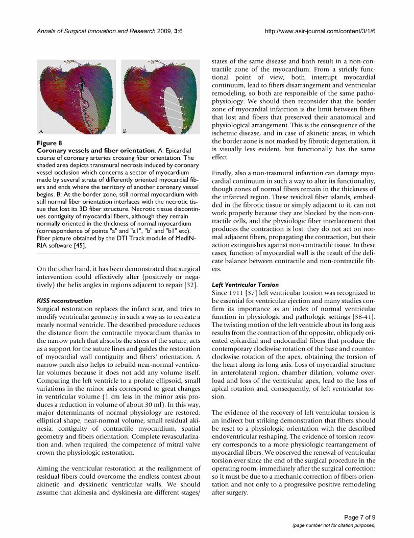
Coronary arteries have an epicardial course not to besqueezed during systole, while fiber orientation is chang-ing throughout myocardium thickness, from epicardiumto endocardium, following a continuous angular gradientwhich is crossed more or less perpendicularly by vesselsdirection (Figure 8A). The transmural necrosis induced bycoronary vessel occlusion concerns a sector of myocar-dium made by several strata of differently oriented myo-cardial fibers and ends where the territory of anothercoronary vessel begins (Figure 8A, shaded area). At theborder zone, still normal myocardium with still normalfiber orientation interlaces with the necrotic tissue thatlost its 3D fiber structure. Necrotic tissue discontinuescontinuity and contiguity of myocardial fibers, althoughthey remain normally oriented in the thickness of normalmyocardium (Figure 8B, correspondence of points "a"and "a1", "b" and "b1" etc).
The infarcted area alters regional and then global functionof the left ventricle, expands and dilates the ventricularchamber and dislocates adjacent, still anatomically nor-mal, non-infarcted myocardium. Consequently, fibers inthe normal myocardium are displaced by the necrotic,thin, akinetic/dyskinetic portion of ventricular wall and(although normally oriented inside the residual wall)their orientation and spatial disposition is completelyupset.
Table 2: Exemplary case data
Preop Postop7 days
Postop19 months
EDD 75 65 68
ESD 59 54 51
APICALDIAMETER
70 36 35
LLD 82 79 78
LLS 82 76 76
EDV 256 160 136
EDVI 135 84 69
ESV 150 120 95
ESVI 79 62 48
EF 28 35 38
WMSI 2.88 1,88 1,56
MR 4 0 0
NYHA 4 1 1
EDD = end-diastolic diameter (mm); ESD = end-systolic diameter (mm); APICAL DIAMETER = internal diastolic diameter two centimeters above the ventricular apex (mm); LLD = diastolic longitudinal length (mm); LLS = systolic longitudinal length (mm); EDV = end-diastolic volume (ml); EDVI = end-diastolic volume index (ml/m2); ESV = end-systolic volume (ml); ESVI = end-systolic volume index (ml/m2); EF = ejection fraction; WMSI = wall motion score index; MR = mitral regurgitation; NYHA = New York Health Association class.
Page 6 of 9(page number not for citation purposes)
Annals of Surgical Innovation and Research 2009, 3:6 http://www.asir-journal.com/content/3/1/6
On the other hand, it has been demonstrated that surgicalintervention could effectively alter (positively or nega-tively) the helix angles in regions adjacent to repair [32].
KISS reconstructionSurgical restoration replaces the infarct scar, and tries tomodify ventricular geometry in such a way as to recreate anearly normal ventricle. The described procedure reducesthe distance from the contractile myocardium thanks tothe narrow patch that absorbs the stress of the suture, actsas a support for the suture lines and guides the restorationof myocardial wall contiguity and fibers' orientation. Anarrow patch also helps to rebuild near-normal ventricu-lar volumes because it does not add any volume itself.Comparing the left ventricle to a prolate ellipsoid, smallvariations in the minor axis correspond to great changesin ventricular volume (1 cm less in the minor axis pro-duces a reduction in volume of about 30 ml). In this way,major determinants of normal physiology are restored:elliptical shape, near-normal volume, small residual aki-nesia, contiguity of contractile myocardium, spatialgeometry and fibers orientation. Complete revasculariza-tion and, when required, the competence of mitral valvecrown the physiologic restoration.
Aiming the ventricular restoration at the realignment ofresidual fibers could overcome the endless contest aboutakinetic and dyskinetic ventricular walls. We shouldassume that akinesia and dyskinesia are different stages/
states of the same disease and both result in a non-con-tractile zone of the myocardium. From a strictly func-tional point of view, both interrupt myocardialcontinuum, lead to fibers disarrangement and ventricularremodeling, so both are responsible of the same patho-physiology. We should then reconsider that the borderzone of myocardial infarction is the limit between fibersthat lost and fibers that preserved their anatomical andphysiological arrangement. This is the consequence of theischemic disease, and in case of akinetic areas, in whichthe border zone is not marked by fibrotic degeneration, itis visually less evident, but functionally has the sameeffect.
Finally, also a non-trasmural infarction can damage myo-cardial continuum in such a way to alter its functionality,though zones of normal fibers remain in the thickness ofthe infarcted region. These residual fiber islands, embed-ded in the fibrotic tissue or simply adjacent to it, can notwork properly because they are blocked by the non-con-tractile cells, and the physiologic fiber interlacement thatproduces the contraction is lost: they do not act on nor-mal adjacent fibers, propagating the contraction, but theiraction extinguishes against non-contractile tissue. In thesecases, function of myocardial wall is the result of the deli-cate balance between contractile and non-contractile fib-ers.
Left Ventricular TorsionSince 1911 [37] left ventricular torsion was recognized tobe essential for ventricular ejection and many studies con-firm its importance as an index of normal ventricularfunction in physiologic and pathologic settings [38-41].The twisting motion of the left ventricle about its long axisresults from the contraction of the opposite, obliquely ori-ented epicardial and endocardial fibers that produce thecontemporary clockwise rotation of the base and counter-clockwise rotation of the apex, obtaining the torsion ofthe heart along its long axis. Loss of myocardial structurein anterolateral region, chamber dilation, volume over-load and loss of the ventricular apex, lead to the loss ofapical rotation and, consequently, of left ventricular tor-sion.
The evidence of the recovery of left ventricular torsion isan indirect but striking demonstration that fibers shouldbe reset to a physiologic orientation with the describedendoventricular reshaping. The evidence of torsion recov-ery corresponds to a more physiologic rearrangement ofmyocardial fibers. We observed the renewal of ventriculartorsion ever since the end of the surgical procedure in theoperating room, immediately after the surgical correction:so it must be due to a mechanic correction of fibers orien-tation and not only to a progressive positive remodelingafter surgery.
Coronary vessels and fiber orientationFigure 8Coronary vessels and fiber orientation. A: Epicardial course of coronary arteries crossing fiber orientation. The shaded area depicts transmural necrosis induced by coronary vessel occlusion which concerns a sector of myocardium made by several strata of differently oriented myocardial fib-ers and ends where the territory of another coronary vessel begins. B: At the border zone, still normal myocardium with still normal fiber orientation interlaces with the necrotic tis-sue that lost its 3D fiber structure. Necrotic tissue discontin-ues contiguity of myocardial fibers, although they remain normally oriented in the thickness of normal myocardium (correspondence of points "a" and "a1", "b" and "b1" etc). Fiber picture obtained by the DTI Track module of MedIN-RIA software [45].
Page 7 of 9(page number not for citation purposes)

Annals of Surgical Innovation and Research 2009, 3:6 http://www.asir-journal.com/content/3/1/6
Current Limitations and Future DevelopmentsAt present, we have only indirect, "gross" evidence of fiberrealignment, but we can not correct fiber disposition inorder to obtain a "histological" realignment. Fastimprovement in medical imaging techniques able to visu-alize fiber orientation (diffusion tensor magnetic reso-nance, limited until now by heart movement) will be veryuseful in this topic and will probably lead to a preopera-tive assessment full of key data for the surgeon. Someadvanced technology laboratories [42,43] are alreadystudying the use of integrated imaging techniques foraccurate planning of surgical procedures (for example,tumor removal in the brain with 3D visualization of cere-bral fibers, tissue and structures). It would be reasonableto foresee a 3D surgical planning of ventricular reshapingwith reference points for fiber disposition that will guidethe surgeon during the reconstruction. A so called "fiber-based" therapy could open unexpected improvements inthe surgical treatment of the failing heart. For example,the Batista operation and the surgical reshaping in the set-ting of dilated cardiomyopathy had not the expected dif-fusion, perhaps because volume reduction did not takecare of fiber alignment at the suture site, but caused itselfa fiber disarrangement, suturing together non-contiguousparts of ventricular walls with different fiber disposition.A fiber-based physiological view could also help us in theevaluation and/or optimization of medical therapies,mainly in case of new drugs or of non-responder patients.
Finally, basic science implications of fiber-based left ven-tricular function could let us use an old knowledge thatwas forgotten until now, due to the imaging limitationsand to the complex 3D anatomy of myocardium, but thatcould represent the basis for a new approach to the failingheart.
ConclusionThe knowledge of anatomical structure of left ventricle isvery old [44] but it has never been applied in clinical prac-tice. The described endoventricular reconstruction tendsto grossly realign fibers in a more physiologic manner,approaching them together and re-establishing theiroblique disposition and orientation, compensating in thisway the spacing and the anomalies induced by myocar-dial infarction. The evidence of the renewal of left ven-tricular torsion could open the way to implement theevaluation of the results of this surgical procedure. Varia-bility in outcome could be overcome with a multifactorialapproach to restoration taking care of all aspects of nor-mal physiology of ventricular function, in which fibers-based procedures will have a key role. Future develop-ment in medical imaging will probably confirm and sup-port the main role of fibers' disposition in the surgicaltreatment of failing hearts.
Competing interestsThe author declares that they have no competing interests.
Authors' contributionsMC set the theoretical bases and conceived the surgicalprocedure, performing all of the operations. Thedescribed surgical technique and its peculiar result (theoriginal, never described before renewal of left ventricularapical rotation and torsion) are to be considered his ownintellectual property.
Additional material
References1. Likoff W, Bailey CP: Excision of myocardial aneurysm, report of
a successful case. J Am Med Assoc 1955, 158:915.2. Reddy SB, Cooley DA, Duncan JM, Norman JC: Left ventricular
aneurysm: twenty year surgical experience with 1572patients at the Texas Heart Institute. Cardiovasc Dis 1981,8(2):165-186.
3. Jatene AD: Left ventricular aneurysmectomy. Resection orreconstruction. J Thorac Cardiovasc Surg 1985, 89:321-331.
4. Dor V, Saab M, Coste P, Kornaszewska M, Montiglio F: Left ven-tricular aneurysm: a new surgical approach. Thorac CardiovascSurg 1989, 37:11-19.
5. Lundblad R, Abdelnoor M, Svennevig JL: Surgery for left ventricu-lar aneurysm: early and late survival after simple linearrepair and endoventricular patch plasty. J Thorac CardiovascSurg 2004, 128:449-56.
6. Di Donato M, Sabatier M, Dor V, Genuini GF, Toso A, Maioli M,Stanley AWH, Athanasuleas C, Buckberg G: Effects of the Dor pro-
Additional file 1Exemplary Patient: preoperative left ventriculography. Preoperative left ventriculography of the exemplary patient with large dyskinesia: normal geometry and torsion movement are completely lost.Click here for file[http://www.biomedcentral.com/content/supplementary/1750-1164-3-6-S1.mpg]
Additional file 2Exemplary Patient: renewal of left ventricular torsion at the end of operation. Head view of the heart at the end of reconstruction surgery of the same exemplary patient, normal and slow motion of the same sequence. Cardiopulmonary bypass is off. Ventilation is temporarily off. The counterclockwise torsion movement towards the right ventricle is evi-dent, marked by the displacement of the saphenous graft on left anterior descending artery.Click here for file[http://www.biomedcentral.com/content/supplementary/1750-1164-3-6-S2.mpg]
Additional file 3Exemplary Patient: late full volume echocardiography. Simultaneous display of nine short axis views generated from an apical full volume acquisition with 4D mode (Vivid 7, GE Medical Systems, Norway) of the exemplary patient 19 months after the operation. Apical rotation is evi-dent towards the right ventricle.Click here for file[http://www.biomedcentral.com/content/supplementary/1750-1164-3-6-S3.mpg]
Page 8 of 9(page number not for citation purposes)

Annals of Surgical Innovation and Research 2009, 3:6 http://www.asir-journal.com/content/3/1/6
cedure on left ventricular dimension and shape and geomet-ric correlates of mitral regurgitation one year after surgery.J Thorac Cardiovasc Surg 2001, 121:91-6.
7. Yotsumoto G, Sakata R, Ueno T, Iguro Y, Kinjo T, Kobayashi A, Mat-sumoto K, Tei C, Otsuji Y, Tanaka Y: Late development of mitralregurgitation after left ventricular reconstruction surgery.Ann Thorac Cardiovasc Surg 2005, 11:159-63.
8. Athanasuleas CL, Stanley WH Jr, Buckberg GD, Dor V, Di Donato M,Blackstone EH, the RESTORE Group: Surgical anterior ventricu-lar endocardial restoration (SAVER) in the dilated remod-eled ventricle after anterior myocardial infarction. J Am CollCardiol 2001, 37:1199-1209.
9. Kono T, Sabbah HN, Stein PD, Brymer JF, Khaja F: Left ventricularshape as a determinant of functional mitral regurgitation inpatients with severe heart failure secondary to either coro-nary artery disease or idiopathic dilated cardiomyopathy.Am J Cardiol 1991, 68:355-9.
10. Raman J, Dixit A, Bolotin G, Jeevanandam V: Failure modes of leftventricular reconstruction or the Dor procedure: a multi-institutional perspective. Eur J Cardiothorac Surg 2006,30:347-352.
11. Bolooki H, DeMarchena E, Mallon S, Katariya K, Barron M, BolookiM, Thurer RJ, Novak S, Duncan RC: Factors affecting late surgicalremodeling of left ventricular aneurysms. J Thorac CardiovascSurg 2003, 126:374-85.
12. Isomura T, Horii T, Suma H, Buckberg GD, the RESTORE Group:Septal anterior ventricular exclusion operation (Pacopexy)for ischemic dilated cardiomyopathy: treat form not disease.Eur J Cardiothorac Surg 2006, 295:S245-S250.
13. Solomon SD, Skali H, Anavekar NS, Bourgoun M, Barvik S, Ghali JK,Warnica JW, Khrakovskaya M, Arnold JMO, Schwartz Y, Velazquez EJ,Califf RM, McMurray JV, Pfeffer MA: Changes in ventricular sizeand function in patients treated with valsartan, captopril, orboth after myocardial infarction. Circulation 2005,111:3411-3419.
14. Cirillo M, Amaducci A, Brunelli F, Dalla Tomba M, Parrella P, Tasca G,Troise G, Quaini E: Determinants of postinfarction remodelingaffect outcome and left ventricular geometry after surgicaltreatment of ischemic cardiomyopathy. J Thorac Cardiovasc Surg2004, 127:1648-56.
15. Jiang L, Huang Y, Hunyor S, dos Remedios CG: Cardiomyocyteapoptosis is associated with increased wall stress in chronicfailing left ventricle. Eur Heart J 2003, 24(8):742-51.
16. White HD, Norris RM, Brown MA, Brandt PW, Whitlock RM, WildCJ: Left ventricular endsystolic volume as the major determi-nant of survival after recovery from myocardial infarction.Circulation 1987, 76:44-51.
17. Batista RJV, Verde J, Neri P, Bocchino L, Takeshita N, Bhayana JN,Bergsland J, Graham S, Houck JP, Salerno TA: Partial left ventri-culectomy to treat end-stage heart disease. Ann Thorac Surg1997, 64:634-8.
18. Suma H: Left ventriculoplasty for nonischemic dilated cardio-myopathy. Semin Thorac Cardiovasc Surg 2001, 13(4):514-21.
19. Menicanti L, Di Donato M, Frigiola A, Buckberg G, Santambrogio C,Ranucci M, Santo D: Ischemic mitral regurgitation: intraven-tricular papillary muscle imbrication without mitral ringduring left ventricular restoration. J Thorac Cardiovasc Surg 2002,123:1041-50.
20. Spotnitz HM: Macro design, structure, and mechanics of theleft ventricle. J Thorac Cardiovasc Surg 2000, 119:1053-77.
21. Torrent Guasp F, Ballester M, Buckberg GD, Carreras F, Flotats A,Carrió I, Ferreira A, Samuels LE, Narula J: Spatial orientation ofthe ventricular muscle band: physiologic contribution andsurgical implication. J Thorac Cardiovasc Surg 2001, 122(2):389-92.
22. Torrent Guasp F, Caralps Riera JM, Ballester Rodés M: Cuatro pro-puestas para la remodelaciòn ventricular en el tratamientode la miocardiopatia dilatada. Rev Esp Cardiol 1997, 50:682-88.
23. Garot J, Pascal O, Diebold B, Derumeaux G, Gerber BL, Dubois-Randé JL, Lima JAC, Guéret P: Alterations of systolic left ven-tricular twist after acute myocardial infarction. Am J PhysiolHeart Circ Physiol 2002, 282(1):H357-H362.
24. Setser RM, Kasper JM, Lieber ML, Starling RC, McCarthy PM, WhiteRD: Persistent abnormal left ventricular systolic torsion indilated cardiomyopathy after partial left ventriculectomy. JThorac Cardiovasc Surg 2003, 126:48-55.
25. Di Donato M, Dabic P, Castelvecchio S, Santambrogio C, Brankovic J,Collarini L, Joussef T, Frigiola A, Buckberg G, Menicanti L, theRESTORE Group: Left ventricular geometry in normal andpost-anterior myocardial infarction patients: sphericityindex and 'new' conicity index comparisons. Eur J CardiothoracSurg 2006, 29:S225-S230.
26. Cirillo M, Arpesella G: Rewind the heart: A novel technique toreset heart fibers' orientation in surgery for ischemic cardi-omyopathy. Med Hypotheses. 2008, 70(4):848-854.
27. Cirillo M, Villa E, Campana M, Troise G: Renewal of left ventricu-lar torsion after modified surgical anterior ventricular resto-ration. J Cardiovasc Med (Hagerstown) 2008, 9(11):1142-1146.
28. Cirillo M, Villa E, Troise G: Improvement of left ventricularfunction after modified surgical ventricular restoration:good, better, best. Heart Surg Forum 2008, 11(5):E266-E269.
29. Hawthorne EW: Dynamic geometry of the left ventricle. Am JCardiol 1966, 18:566-573.
30. Kennedy JW, Baxley WA, Figley MM, Dodge HT, Blackmon JR: Thenormal left ventricle in man. Circulation 1966, 34(2):272-278.
31. Chareonthaitawee P, Christian TF, Hirose K, Gibbons RJ, RumbergerJ: Relation of initial infarct size to extent of left ventricularremodeling in the year after acute myocardial infarction. JAm Coll Cardiol 1995, 25:567-73.
32. Walker JC, Guccione JM, Jiang Y, Zhang P, Wallace AW, Hsu EW,Ratcliffe MB: Helical myofiber orientation after myocardial inf-arction and left ventricular surgical restoration in sheep. JThorac Cardiovasc Surg 2005, 129:382-90.
33. Chen J, Song S, Liu W, McLean M, Allen JS, Tan J, Wickline SA, Yu X:Remodeling of cardiac fiber structure after infarction in ratsquantified with diffusion tensor MRI. Am J Physiol Heart Circ Phys-iol 2003, 285:H946-H954.
34. Reimer KA, Lowe JEMD, Margaret M, Rasmussen MD, Robert B, Jen-nings MD: The Wavefront Phenomenon of Ischemic CellDeath. Circulation 1977, 56(5):786-794.
35. Lutgens E, Daemen MJAP, de Muinck ED, Debets J, Leenders P, SmitsJFM: Chronic myocardial infarction in the mouse: cardiacstructural and functional changes. Cardiovascular Research 1999,41:586-593.
36. Zimmerman SD, Criscione J, Covell JW: Remodeling in myocar-dium adjacent to an infarction in the pig left ventricle. Am JPhysiol Heart Circ Physiol 2004, 287:H2697-H2704.
37. Mall FP: On the muscular architecture of the ventricles of thehuman heart. Am J Anat 1911, 11:211-278.
38. Taber LA, Yang M, Podszus WW: Mechanics of left ventriculartorsion. J Biomech 1996, 29(6):645-52.
39. Notomi Y, Setser RM, Shiota T, Martin-Miklovic MG, Weaver JA,Popovic ZB, Yamada H, Greenberg NL, White RD, Thomas JD:Assessment of left ventricular torsional deformation by Dop-pler tissue imaging. Validation study with tagged magneticresonance imaging. Circulation 2005, 111:1141-47.
40. Notomi Y, Srinath G, Shiota T, Martin-Miklovic MG, Beachler L, How-ell K, Oryszak SJ, Deserranno DG, Freed AD, Greenberg NL, You-noszai A, Thomas JD: Maturational and Adaptive Modulation ofLeft Ventricular Torsional Biomechanics. Circulation 2006,113:2534-2541.
41. Kanzaki H, Nakatani S, Yamada N, Urayama S, Miyatake K, KitakazeM: Impaired Systolic torsion in dilated cardiomyopathy:Reversal of apical rotation at mid-systole characterized withmagnetic resonance tagging method. Basic Res Cardiol. 2006,101(6):465-470.
42. National Alliance for Medical Image Computing: Project housed atSurgical Planning Laboratory (SPL). [http://na-mic.org].Department of Radiology, Brigham and Women's Hospital, Boston,MA, USA
43. Center for Imaging Science: Medical Imaging and Computa-tional Anatomy. [http://www.cis.jhu.edu/]. John Hopkins Univer-sity, Baltimore, MD, USA
44. Harvey W: Exercitatio Anatomica de Motu Cordis et San-guinis in Animalibus. Frankfurt 1628.
45. Peyrat JM, Sermesant M, Pennec X, Delingette H, Xu C, McVeigh ER,Ayache N: A Computational Framework for the StatisticalAnalysis of Cardiac Diffusion Tensors: Application to a SmallDatabase of Canine Hearts. IEEE Transactions on Medical Imaging2007, 26(111500-1514 [http://www-sop.inria.fr/asclepios/].
Page 9 of 9(page number not for citation purposes)