nicu resident manual chapter 5 clinical...
TRANSCRIPT

CLINICAL CHAPTER
I. Newborn Resuscitation and Intubation Guidelines II. First Hour of Life Protocol
III. Surfactant Therapy IV. Gestational Age Assessment V. Group B Streptococcal Infections
VI. Blood Product Transfusion Guidelines VII. ROP Surveillance
VIII. Intraventricular Hemorrhage IX. 1-2-3 MN Newborn Hearing Screening Program X. MN State Newborn Screening
XI. Osteopenia of Prematurity XII. Cleft Lip or Palate
XIII. Organ Donation XIV. End of Life Care XV. Immunizations
XVI. Fluid and Electrolyte stabilization: Hypoglycemia Hyperkalemia
XVII. References
I. NEWBORN RESUSCITATION AND INTUBATION GUIDELINES
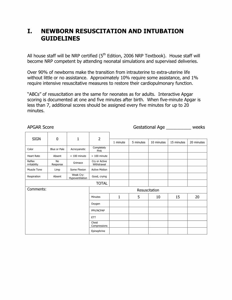
All house staff will be NRP certified (5th Edition, 2006 NRP Textbook). House staff will become NRP competent by attending neonatal simulations and supervised deliveries. Over 90% of newborns make the transition from intrauterine to extra-uterine life without little or no assistance. Approximately 10% require some assistance, and 1% require intensive resuscitative measures to restore their cardiopulmonary function. “ABCs” of resuscitation are the same for neonates as for adults. Interactive Apgar scoring is documented at one and five minutes after birth. When five-minute Apgar is less than 7, additional scores should be assigned every five minutes for up to 20 minutes. APGAR Score Gestational Age __________ weeks
SIGN 0 1 2
1 minute 5 minutes 10 minutes 15 minutes 20 minutes
Color Blue or Pale Acrocyanotic Completely Pink
Heart Rate Absent < 100 minute > 100 minute Reflex irritability
No Response Grimace Cry or Active
Withdrawal
Muscle Tone Limp Some Flexion Active Motion
Respiration Absent Weak Cry: Hypoventilation Good, crying
TOTAL
Resuscitation
Minutes 1 5 10 15 20
Oxygen
PPV/NCPAP
ETT Chest Compressions
Comments:
Epinephrine

Complete documentation of the events taking place during resuscitation must also include a narrative description of all interventions performed. Birth
Routine Care • Provide
warmth • Clear airway • Dry • Assess color
• Term gestation? • Clear amniotic fluid? • Breathing or crying? • Good muscle tone?
• Provide warmth • Position; clear airway*
(as necessary) • Dry, stimulate,
reposition
Observational Care
• Give supplemental oxygen
• Provide positive-pressure ventilation*
• Provide positive-pressure ventilation*
• Administer chest compressions*
Post-resuscitation Care
• Administer epinephrine
Absent HR for > 10 minutes
Persistent bradycardia or cyanosis or failure to ventilate
* Endotracheal intubation
may be considered at several steps.
• Evaluate respirations, heart rate, and color
Recheck effectiveness of • Ventilation • Chest compressions • Endotracheal
intubation • Epinephrine delivery
Consider possibility of • Hypovolemia
• Consider: • Airway malformations • Lung problems, such
as - Pneum
othorax - Diaphr
agmatic hernia • Congenital heart
disease Consider discontinuing resuscitation
No
Yes
Apneic or HR < 100
HR < 60 HR > 60
HR < 60
Persistently cyanotic
Pink Cyanotic
30 sec
30 sec
30 sec
Appr
oxim
ate
Tim
e
Birth
Routine Care • Provide warmth • Clear airway • Dry • Assess color
• Term gestation? • Clear amniotic fluid? • Breathing or crying? • Good muscle tone?
• Provide warmth • Position; clear airway*
(as necessary) • Dry, stimulate, reposition
Observational Care
• Give supplemental oxygen
• Provide positive-pressure ventilation*
• Provide positive-pressure ventilation* • Administer chest compressions*
Post-resuscitation Care
• Administer epinephrine
Absent HR for > 10 minutes
Persistent bradycardia or cyanosis or failure to ventilate
* Endotracheal intubation may be considered at several steps.
• Evaluate respirations, heart rate, and color
Recheck effectiveness of • Ventilation • Chest compressions • Endotracheal intubation • Epinephrine delivery
Consider possibility of • Hypovolemia
• Consider: • Airway malformations • Lung problems, such as - Pneumothorax - Diaphragmatic hernia • Congenital heart disease
No
Yes
Apneic or HR < 100
HR < 60 HR > 60
HR < 60
Breathing HR < 100 and pink
Effective ventilation HR > 100 and pink
Persistently cyanotic
Pink Cyanotic
30 sec
30 sec
30 sec
Appr
oxim
ate
Tim
e
INTUBATION GUIDELINES FOR NEONATES Pediatric residents, by the end of their residency training, will be proficient at neonatal airway management, including intubations, bag/mask ventilation, Neopuff ventilation, and oral airway placement. Intubation attempts should not compromise patient stability. Patients should maintain a heart rate greater than or equal to 100 beats/minute, and oxygen saturation greater than or equal to 90%. The most experienced provider available should perform the intubations on unstable neonates or neonates at risk for adverse outcomes. These cases include:
- extremely preterm neonates (less than or equal to 26 weeks’ gestation); - unstable neonates (unable to establish oxygenation with positive pressure
ventilation); - and neonates requiring CPR;
and certain congenital anomalies: - congenital diaphragmatic hernia; - known airway obstruction; - micrognathia; - cleft palate; - hydrops.
Pre-medication with atropine, morphine, and a short-acting muscle relaxant should be used for all non-emergent intubations. This has been shown to decrease the time and number of attempts needed to successfully intubate and reduce the incidence of severe desaturations.
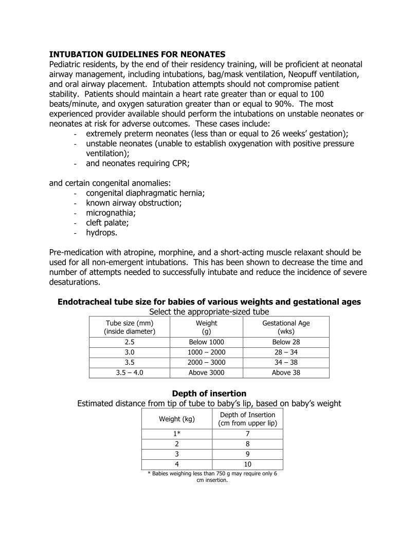
Endotracheal tube size for babies of various weights and gestational ages
Select the appropriate-sized tube Tube size (mm) (inside diameter)
Weight (g)
Gestational Age (wks)
2.5 Below 1000 Below 28 3.0 1000 – 2000 28 – 34 3.5 2000 – 3000 34 – 38
3.5 – 4.0 Above 3000 Above 38
Depth of insertion
Estimated distance from tip of tube to baby’s lip, based on baby’s weight
Weight (kg) Depth of Insertion (cm from upper lip)
1* 7 2 8 3 9 4 10
* Babies weighing less than 750 g may require only 6 cm insertion.
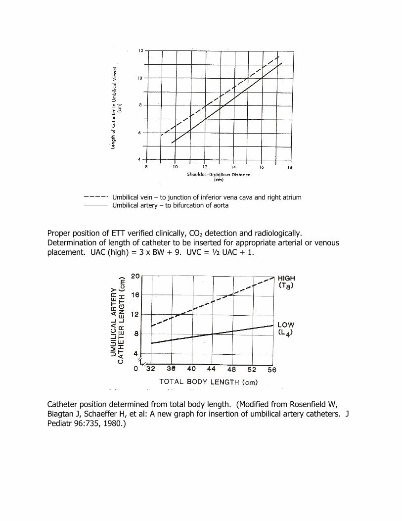
Umbilical vein – to junction of inferior vena cava and right atrium Umbilical artery – to bifurcation of aorta
Proper position of ETT verified clinically, CO2 detection and radiologically. Determination of length of catheter to be inserted for appropriate arterial or venous placement. UAC (high) = 3 x BW + 9. UVC = ½ UAC + 1.
Catheter position determined from total body length. (Modified from Rosenfield W, Biagtan J, Schaeffer H, et al: A new graph for insertion of umbilical artery catheters. J Pediatr 96:735, 1980.)

II. FIRST HOUR OF LIFE PROTOCOL Criteria for Inserting Central Lines:
If infant is <1kg and is intubated, place UAC/UVC. If infant is < 1kg and is stable on CPAP or NC, place UAC and PIV. If infant is > 1kg and is intubated, place UAC if frequent lab or BP monitoring is
anticipated. Consider UVC if needed for medications or long term TPN. If infant is > 1kg and is stable on CPAP or NC place PIV only If attempts at placing umbilical lines are unsuccessful after 20-30 minutes,
procedure should be aborted and PIV placed. However, every effort should be made to procure blood for initial lab evaluation at this time.
For stable infants who need central IV access try to place a PICC within 3 days of life and remove UVC by day of life 3 if possible.
If infant is unstable, requiring pressors or PGE umbilical venous catheter may be left in place longer than 3 days
Consider double lumen UVC only if infant is >1kg and if multiple access is needed (PPHN, cardiac dz, etc)
Upon Admission to NICU:
The infant’s initial glucose should be checked within 30 minutes of birth (not admission). It may be drawn from UVC if UVC is being inserted (NNP to insert low UVC, draw labs, wait for glucose results and then advance UVC. If glucose if low, a D10W bolus can be administered safely through a low UVC before UVC is advanced).
Follow up glucoses should be checked at 1 hr and 1 ½ hrs of life with all high-risk infants.
Admission nurse should draw initial labs (if no central lines placed). Use IRMA for initial blood gas, Hct and glucose. If admission nurse is uncomfortable drawing initial labs, she/he should ask for assistance.
A mask/cap must be worn by anyone in the infant’s room during UAC/UVC insertion
Consider pushing first dose of Ampicillin (obtain from Pyxis) Goal is to have infants admitted, stable and antibiotics started within 1
hour of birth
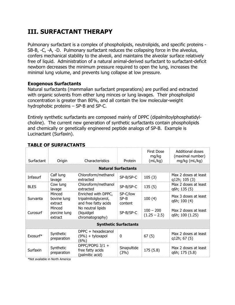
III. SURFACTANT THERAPY Pulmonary surfactant is a complex of phospholipids, neutrolipids, and specific proteins - SB-B, -C, -A, -D. Pulmonary surfactant reduces the collapsing force in the alveolus, confers mechanical stability to the alveoli, and maintains the alveolar surface relatively free of liquid. Administration of a natural animal-derived surfactant to surfactant-deficit newborn decreases the minimum pressure required to open the lung, increases the minimal lung volume, and prevents lung collapse at low pressure. Exogenous Surfactants Natural surfactants (mammalian surfactant preparations) are purified and extracted with organic solvents from either lung minces or lung lavages. Their phospholipid concentration is greater than 80%, and all contain the low molecular-weight hydrophobic proteins – SP-B and SP-C. Entirely synthetic surfactants are composed mainly of DPPC (dipalmitoylphosphatidyl-choline). The current new generation of synthetic surfactants contain phospholipids and chemically or genetically engineered peptide analogs of SP-B. Example is Lucinactant (Surfaxin). TABLE OF SURFACTANTS
Surfactant Origin Characteristics Protein
First Dose mg/kg
(mL/kg)
Additional doses (maximal number)
mg/kg (mL/kg)
Natural Surfactants
Infasurf Calf lung lavage
Chloroform/methanol extracted SP-B/SP-C 105 (3) Max 2 doses at least
q12h; 105 (3)
BLES Cow lung lavage
Chloroform/methanol extracted SP-B/SP-C 135 (5) Max 2 doses at least
q6h; 135 (5)
Survanta Minced bovine lung extract
Enriched with DPPC, tripalmitolglycerol, and free fatty acids
SP-C/low SP-B content
100 (4) Max 3 doses at least q6h; 100 (4)
Curosurf Minced porcine lung extract
No neutral lipids (liquidgel chromatography)
SP-B/SP-C 100 – 200 (1.25 – 2.5)
Max 2 doses at least q6h; 100 (1.25)
Synthetic Surfactants
Exosurf* Synthetic preparation
DPPC + hexadecanol (9%) + tyloxapol (6%)
0 67 (5) Max 2 doses at least q12h; 67 (5)
Surfaxin Synthetic preparation
DPPC/POPG 3/1 + free fatty acids (palmitic acid)
Sinapultide (3%) 175 (5.8) Max 2 doses at least
q6h; 175 (5.8)
*Not available in North America
Studies demonstrated 40% reduction in the odds of neonatal death after surfactant treatment, either natural or synthetic administered as a prophylactic or rescue treatment. Both types of surfactant and both treatment strategies have significantly reduced the risk of pulmonary air leaks by 30-50%. Chronic lung disease has not been significantly reduced because the increase in survival is mainly among extremely premature infants. Prophylactic treatment (treatment within the first 30 minutes of birth), instilling surfactant before the onset of respiratory distress syndrome, has been shown to partially avoid barotrauma and vascular injury resulting from mechanical ventilation. Infants born at, or less than, 26 weeks’ gestation are eligible for prophylactic treatment. Early rescue (before two hours of life) is associated with decreased risk of neonatal mortality and significant reduction in the incidence of pneumothorax. Infants born at greater than 26 weeks’ gestation, and less than 32 weeks, should receive surfactant without delay if they require supplemental oxygen with FiO2 greater than 0.3 on nasal CPAP or mechanical ventilation at 90 minutes of life. Inactivation of surfactant is involved in the pathogenesis of various respiratory disorders, including meconium aspiration syndrome, pneumonia, and sepsis. These disorders may represent potential targets for surfactant therapy. Administration Dosing is usually two divided aliquots and administered via a catheter in the endotracheal tube. Infants are manually ventilated during administration to ensure maximal dispersion. Each half-dose is injected over 1-2 minutes, and infant’s head and torso are rotated 30-45 degrees to the right for the first half-dose, and 30-45 degrees to the left for the second half-dose. Transient oxygen desaturation and mild bradycardia are frequently observed during administration, and may require adjustments of the ventilation and FiO2 or interruption of surfactant administration. Improvement in gas exchange after administration is usually rapid (within a few minutes) and ventilation pressure and volume must be adjusted while monitoring tidal volume and PaCO2. Because oxygenation improves rapidly, continuous monitoring of oxygen saturation during and after administration is mandatory. Approximately one-third of treated infants still require mechanical ventilation with an FiO2 greater than 0.3, six hours after the first done; these infants are eligible for retreatment. Infants less than 26 weeks’ gestation with a diagnosis of respiratory distress syndrome may receive a second dose of surfactant, if they continue intubated on mechanical ventilation, regardless of the inspired oxygen concentration. Infants greater than 26 weeks’ gestation with a diagnosis of respiratory distress syndrome should receive a second dose, if they continue on mechanical ventilation and require greater than 30% inspired oxygen. If infant remains intubated on oxygen concentration between 21-29%, consider a second dose of surfactant. There is no proven benefit to more than two additional doses.
IV. GESTATIONAL AGE ASSESSMENT
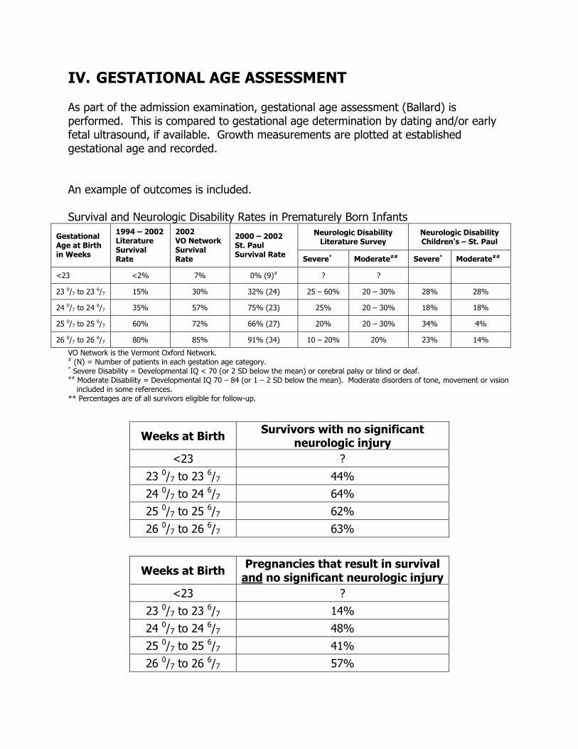
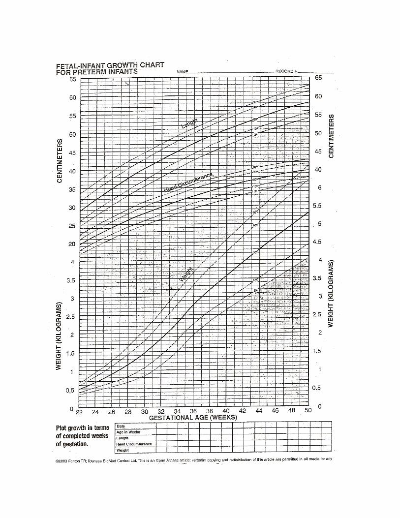
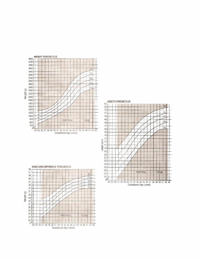
As part of the admission examination, gestational age assessment (Ballard) is performed. This is compared to gestational age determination by dating and/or early fetal ultrasound, if available. Growth measurements are plotted at established gestational age and recorded. An example of outcomes is included. Survival and Neurologic Disability Rates in Prematurely Born Infants
Neurologic Disability Literature Survey
Neurologic Disability Children's – St. Paul
Gestational Age at Birth in Weeks
1994 – 2002 Literature Survival Rate
2002 VO Network Survival Rate
2000 – 2002 St. Paul Survival Rate Severe* Moderate## Severe* Moderate##
<23 <2% 7% 0% (9)# ? ?
23 0/7 to 23 6/7 15% 30% 32% (24) 25 – 60% 20 – 30% 28% 28%
24 0/7 to 24 6/7 35% 57% 75% (23) 25% 20 – 30% 18% 18%
25 0/7 to 25 6/7 60% 72% 66% (27) 20% 20 – 30% 34% 4%
26 0/7 to 26 6/7 80% 85% 91% (34) 10 – 20% 20% 23% 14%
VO Network is the Vermont Oxford Network. # (N) = Number of patients in each gestation age category. * Severe Disability = Developmental IQ < 70 (or 2 SD below the mean) or cerebral palsy or blind or deaf. ## Moderate Disability = Developmental IQ 70 – 84 (or 1 – 2 SD below the mean). Moderate disorders of tone, movement or vision
included in some references. ** Percentages are of all survivors eligible for follow-up.
Weeks at Birth Survivors with no significant neurologic injury
<23 ? 23 0/7 to 23 6/7 44% 24 0/7 to 24 6/7 64% 25 0/7 to 25 6/7 62% 26 0/7 to 26 6/7 63%
Weeks at Birth Pregnancies that result in survival and no significant neurologic injury
<23 ? 23 0/7 to 23 6/7 14% 24 0/7 to 24 6/7 48% 25 0/7 to 25 6/7 41% 26 0/7 to 26 6/7 57%
V. GROUP B STREPTOCOCCAL INFECTIONS Group B streptococcus remains the most common cause of neonatal bacterial infections in the United States in infants >2500 gms. The mainstay for prevention of Group B strep infections in neonates, as suggested by the CDC, is screening pregnant women for Group B strep colonization and providing intrapartum antibiotic prophylaxis (IAP). Approximately 30% of women have asymptomatic Group B strep colonization during pregnancy. Nearly 50% of infants who pass through a colonized birth canal become colonized, and 1-2% of colonized infants developed invasive disease; 75% of cases of Group B strep infections are early onset (0-6 six days). Most infants with early onset disease become ill within the first 24 hours. Transmission is vertical in nearly all cases, and occurs shortly before or during delivery. Septicemia occurs in 25-40%, pneumonia 35-55%, and meningitis in 5-10%. Mortality is 4-6%, and higher in preterm infants. Late onset disease occurs at 3-4 weeks with a range of 7 days-3 months. Term and preterm infants are equally susceptible. Intrapartum antibiotic prophylaxis has not decreased the incidence of late onset disease. Late onset Group B strep disease presents as bacteremia, meningitis, osteomyelitis, septic arthritis, adenitis, and cellulitis. Transmission is mainly horizontal. Universal screening of all pregnant women at 35-37 weeks’ gestational age is a recommended standard of care. Intrapartum prophylaxis is indicated for all Group B strep carriers, except for those where cesarean section delivery is planned in the absence of labor with intact membranes.

IAP INDICATED
• Previous infant with invasive GBS disease
• GBS bacteriuria during current pregnancy
• Positive GBS screening culture during current pregnancy (unless a planned cesarean delivery is performed in the absence of labor or membrane rupture)
• Unknown GBS status AND any of the following: Delivery at < 37 weeks’ gestation Membranes ruptured for > 18 hours Intrapartum fever (temperature > 38.0oC [> 100.4oF])2
IAP NOT INDICATED
• Previous pregnancy with a positive GBS screening culture (unless a culture also was positive during the current pregnancy or previous infant with invasive GBS disease)
• Planned cesarean delivery performed in the absence of labor or membrane rupture (regardless of GBS culture status)
• Negative vaginal and rectal GBS screening culture in late gestation, regardless of intrapartum risk factors
Indications for intrapartum antimicrobial prophylaxis (IAP) to prevent early-onset group B streptococcal (GBS) disease using a universal prenatal culture screening strategy at 35 to 37 weeks’ gestation for all women.
1 Exceptions: women with GBS bacteriuria during the current pregnancy or women with a previous infant with invasive GBS disease.
2 If chorioamnionitis is suspected, broad-spectrum antimicrobial therapy that includes an agent known to be active against GBS should replace GBS IAP.
When appropriate, antibiotics are administered more than four hours prior to delivery; only 1.2% (instead of 47%) of infants are colonized. Appropriate maternal antibiotics are penicillin, ampicillin, cefozolin, and vancomycin. Incidence of early onset Group B strep disease has decreased to 0.34 cases/1,000 live births with maternal screening and intrapartum antibiotic prophylaxis. (This is down from 1-4 cases/1,000 live births before the above screening and IAP). Ninety-five (95) percent of early onset Group B strep infection presents within the first 24 hours after birth. Management of infants whose mothers received IAP remains controversial. The CDC and AAP provide algorithm for management of infants born to mothers who have received IAP. Full diagnostic work-up includes CBC, CRP, blood culture, and lumbar puncture (if sepsis is suspected). Thirty-eight (38) percent of infants with culture-proving meningitis have negative blood cultures. Chest radiograph should be obtained with respiratory symptoms. Ampicillin and gentamicin are started empirically. Length of therapy depends on the infant’s clinical course and laboratory results.
Vaginal and rectal GBS cultures at 35-37 weeks’ gestation for ALL pregnant women1

Empiric management of a neonate whose mother received intrapartum antimicrobial prophylaxis (IAP) for prevention of early-onset group B streptococcal (GBS) disease1 or suspected chorioamnionitis. The algorithm is not an exclusive course of management. Variations that incorporate individual circumstances or institutional preferences may be appropriate.
1 If no maternal IAP for GBS was administered despite an indication being present, data are insufficient on which to recommend a single management strategy. 2 Includes complete blood cell (CBC) count with differential, blood culture, and chest radiograph if
respiratory abnormalities are present. When signs of sepsis are present, a lumbar puncture, if feasible, should be performed.
3 Duration of therapy varies depending on results of blood culture, cerebrospinal fluid findings (if obtained), and the clinical course of the infant. If laboratory results and clinical course do not indicate bacterial infection, duration may be as short as 48 hours.
4 CBC including white blood cell count with differential and blood culture. 5 Applies only to penicillin, ampicillin, or cefazolin and assumes recommended dosing regimens. 6 A healthy-appearing infant who was U>U 38 weeks’ gestation at delivery and whose mother received U>U 4
hours of IAP before delivery may be discharged home after 24 hours if other discharge criteria have been met and a person able to comply fully with instructions for home observation will be present. If any one of these conditions is not met, the infant should be observed in the hospital for at least 48 hours and until criteria for discharge are achieved.
VI. BLOOD PRODUCT TRANSFUSION GUIDELINES The decision to treat anemia should be based on etiology, severity, and condition of the infant. If infant is born with signs of hypovolemia, shock, or anemia, immediate volume expansion is indicated with saline and plasma, followed by blood if indicated. Packed red blood cells are used routinely. If coagulation factors are needed, use packed red blood cells and fresh frozen plasma. Begin with 20 ml/kg over 30-60 minutes. More may be required, but care must be taken to accurately assess the need for further volume support. Uncross-matched O negative red blood cells are available in an emergency. If infant develops severe anemia chronically in utero, but is hemodynamically well compensated without findings of hypotension, tachycardia, or poor perfusion, packed red blood cells should be given without delay. Smaller increments should be given – 10 ml/kg – in separate transfusion until hemoglobin normalizes. If infant is hydropic with severe anemia and variable hemodynamic decompensation, an isovolumetric partial exchange transfusion, using packed red blood cells, should be accomplished without delay. Pretransfusion work-up: Requires newborn work-up during current admission period. Surgical RBC ordering recommendations: Newborn open cavity, 20 ml/kg; PDA ligation, 50 ml/kg.
St. Paul Children’s NICU Revised Transfusion Guidelines 2011
Supporting Data:
*Transfusions are associated with an increased risk for BPD, NEC, and ROP (Valieva, et al)
*Studies have found no association between maintaining a higher Hct at birth and prevention of IVH (Valieva, et al)
*Volume of blood given may lead to volume overload in the lungs leading to higher oxygen requirements (Kasat et al)
*Treatments aimed at maintaining Hct levels above 32% incurred additional cost without demonstrable benefit. Restrictive transfusion policies were not associated with adverse outcome (Ohls, R)
*Neonates managed without recourse to transfusion guidelines are two times more likely to receive a blood transfusion as those managed in compliance with guidelines (Bell, EF)
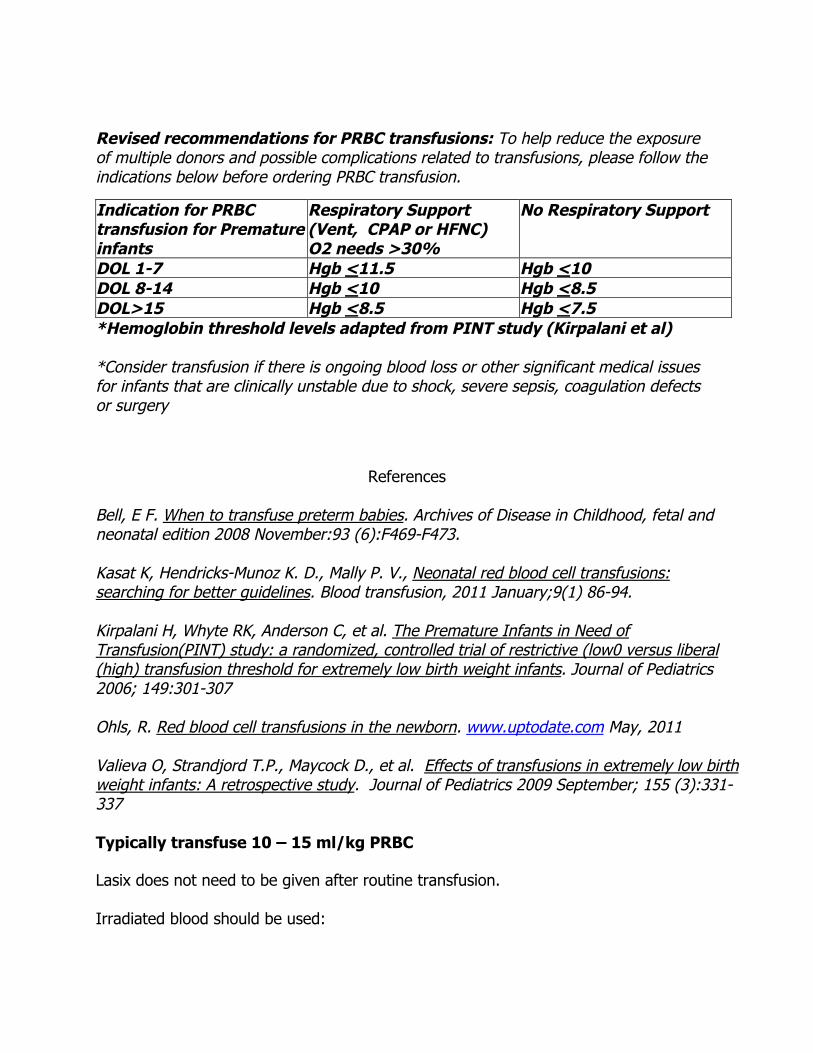
Revised recommendations for PRBC transfusions: To help reduce the exposure of multiple donors and possible complications related to transfusions, please follow the indications below before ordering PRBC transfusion.
Indication for PRBC transfusion for Premature infants
Respiratory Support (Vent, CPAP or HFNC) O2 needs >30%
No Respiratory Support
DOL 1-7 Hgb <11.5 Hgb <10 DOL 8-14 Hgb <10 Hgb <8.5 DOL>15 Hgb <8.5 Hgb <7.5 *Hemoglobin threshold levels adapted from PINT study (Kirpalani et al) *Consider transfusion if there is ongoing blood loss or other significant medical issues for infants that are clinically unstable due to shock, severe sepsis, coagulation defects or surgery
References Bell, E F. When to transfuse preterm babies. Archives of Disease in Childhood, fetal and neonatal edition 2008 November:93 (6):F469-F473. Kasat K, Hendricks-Munoz K. D., Mally P. V., Neonatal red blood cell transfusions: searching for better guidelines. Blood transfusion, 2011 January;9(1) 86-94. Kirpalani H, Whyte RK, Anderson C, et al. The Premature Infants in Need of Transfusion(PINT) study: a randomized, controlled trial of restrictive (low0 versus liberal (high) transfusion threshold for extremely low birth weight infants. Journal of Pediatrics 2006; 149:301-307 Ohls, R. Red blood cell transfusions in the newborn. www.uptodate.com May, 2011 Valieva O, Strandjord T.P., Maycock D., et al. Effects of transfusions in extremely low birth weight infants: A retrospective study. Journal of Pediatrics 2009 September; 155 (3):331-337 Typically transfuse 10 – 15 ml/kg PRBC Lasix does not need to be given after routine transfusion. Irradiated blood should be used:

- When there is a suspicion of immune deficiency (DiGeorge Syndrome, absent thymus, aortic arch abnormality, family history);
- The infant has received intrauterine transfusion and is to receive a double-volume exchange transfusion;
- The blood is from a primary family member (directed donor blood); - Acellular blood components (fresh frozen plasma and cryoprecipitate) do not need
to be irradiated. Guidelines For Administering Blood Products Packed red blood cells: Concentrated red blood cells, have most plasma and platelets removed. CMV safe is either leukocyte-depleted, or CMV negative. Average hematocrit = 55 – 60%. Volume of blood to be available in surgery varies with procedure: rate of transfusion, 5 cc/kg/hour; 2 ml/kg/hour if patient has insipient congestive heart failure. Platelets: Platelets are suspended in a small amount of plasma. Average volume is 50-70 ml/unit. Transfusion guidelines:
- Severe thrombocytopenia (platelet count less than 20,000); - Platelet count less than 50,000:
in patients who require a surgery; in patients with active hemorrhage; risk of eminent bleeding; increased risk for intraventricular hemorrhage (less than 32 weeks post
conceptual age). Higher platelet counts may be used at discretion of neonatologist for extremely low birth-weight premature infants. Dose is 10-20 cc/kg (one unit for every 10 kg body weight) will increase platelet count by 50,000. Full-volume platelets are preferred to volume-reduced platelets because process of volume reduction activates platelets, causing degranulation, making them less effective. Administer no faster than 2-3 cc/minute via syringe pump. If volume overload is a problem, administer total dose over 1-2 hours. Fresh frozen plasma: Fresh frozen plasma is the portion of blood that contains clotting factors and protein. Type AB plasma may be used in an emergency. Indications are:
- Massive hemorrhage; - Multiple clotting deficiencies (liver disease, disseminated intravascular
coagulation). Dose for acute hemorrhage is 15-30 cc/kg. Clotting deficiencies 10-15 cc/kg. May be repeated up to three times each 24 hours as necessary. Monitor for congestive heart
failure, secondary to volume overload. Rate of administration for hemorrhage is dependent on patient’s condition. For clotting deficiencies, administer over 2-3 hours. Cryoprecipitate: Cryoprecipitate is concentrated fibrinogen, 15 ml/unit. Indication is hypofibrinogenemia, disseminated intravascular coagulation. Dosage: body weight less than 2.5 kg, give 0.4 units (6 ml/kg of body weight); 2.5-5 kg, give one unit (15 ml); 5-10 kg, give 1-2 units. Infants who can tolerate or who need extra intravascular volume, administer as much of the unit as tolerated to reduce donor exposure.
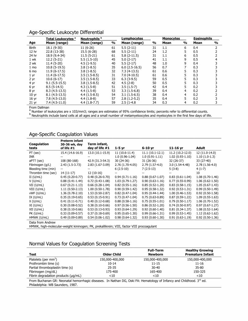
Age-Specific Leukocyte Differential Total Leukocytes a Neutrophils b Lymphocytes Monocytes Eosinophils
Age Mean (range) Mean (range) % Mean (range) % Mean % Mean % Birth 18.1 (9-30) 11 (6-26) 61 5.5 (2-11) 31 1.1 6 0.4 2 12 hr 22.8 (13-38) 15.5 (6-28) 68 5.5 (2-11) 24 1.2 5 0.5 2 24 hr 18.9 (9.4-34) 11.5 (5-21) 61 5.8 (2-11.5) 31 1.1 6 0.5 2 1 wk 12.2 (5-21) 5.5 (1.5-10) 45 5.0 (2-17) 41 1.1 9 0.5 4 2 wk 11.4 (5-20) 4.5 (1-9.5) 40 5.5 (2-17) 48 1.0 9 0.4 3 1 mo 10.8 (5-19.5) 3.8 (1-8.5) 35 6.0 (2.5-16.5) 56 0.7 7 0.3 3 6 mo 11.9 (6-17.5) 3.8 (1-8.5) 32 7.3 (4-13.5) 61 0.6 5 0.3 3 1 yr 11.4 (6-17.5) 3.5 (1.5-8.5) 31 7.0 (4-10.5) 61 0.6 5 0.3 3 2 yr 10.6 (6-17) 3.5 (1.5-8.5) 33 6.3 (3-9.5) 59 0.5 5 0.3 3 4 yr 9.1 (5.5-15.5) 3.8 (1.5-8.5) 42 4.5 (2-8) 50 0.5 5 0.3 3 6 yr 8.5 (5-14.5) 4.3 (1.5-8) 51 3.5 (1.5-7) 42 0.4 5 0.2 3 8 yr 8.3 (4.5-13.5) 4.4 (1.5-8) 53 3.3 (1.5-6.8) 39 0.4 4 0.2 2 10 yr 8.1 (4.5-13.5) 4.4 (1.5-8.5) 54 3.1 (1.5-6.5) 38 0.4 4 0.2 2 16 yr 7.8 (4.5-13.0) 4.4 (1.8-8) 57 2.8 (1.2-5.2) 35 0.4 5 0.2 3 21 yr 7.4 (4.5-11.0) 4.4 (1.8-7.7) 59 2.5 (1-4.8 34 0.3 4 0.2 3 From Dallman a Number of leukocytes are x 103/mm3; ranges are estimates of 95% confidence limits; percents refer to differential counts. b Neutrophils include band cells at all ages and a small number of metamyeloocytes and myelocytes in the first few days of life.
Age-Specific Coagulation Values
Coagulation tests
Preterm infant 30-36 wk, day of life #1
Term infant, day of life #1 1-5 yr 6-10 yr 11-16 yr Adult
PT (sec) 15.4 (14.6-16.9) 13.0 (10.1-15.9) 11 (10.6-11.4) 11.1 (10.1-12.1) 11.2 (10.2-12.0) 12 (11.0-14.0) INR -- -- 1.0 (0.96-1.04) 1.0 (0.91-1.11) 1.02 (0.93-1.10) 1.10 (1.0-1.3) aPTT (sec) 108 (80-168) 42.9 (31.3-54.3) 30 (24-36) 31 (26-36) 32 (26-37) 33 (27-40) Fibrinogen (g/L) 2.43 (1.5-3.73) 2.83 (1.67-3.09) 2.76 (1.70-4.05) 2.79 (1.57-4.0) 3.0 (1.54-4.48) 2.78 (1.56-4.0) Bleeding time (min) -- -- 6 (2.5-10) 7 (2.5-13) 5 (3-8) 4 (1-7) Thrombin time (sec) 14 (11-17) 12 (10-16) -- -- -- 10 II (U/mL) 0.45 (0.20-0.77) 0.48 (0.26-0.70) 0.94 (0.71-1.16) 0.88 (0.67-1.07) 0.83 (0.61-1.04) 1.08 (0.70-1.46) V (U/mL) 0.88 (0.41-1.44) 0.72 (0.43-1.08) 1.03 (0.79-1.27) 0.90 (0.63-1.16) 0.77 (0.55-0.99) 1.06 (0.62-1.50) VII (U/mL) 0.67 (0.21-1.13) 0.66 (0.28-1.04) 0.82 (0.55-1.16) 0.85 (0.52-1.20) 0.83 (0.58-1.15) 1.05 (0.67-1.43) VIII (U/mL) 1.11 (0.50-2.13) 1.00 (0.50-1.78) 0.90 (0.59-1.42) 0.95 (0.58-1.32) 0.92 (0.53-1.31) 0.99 (0.50-1.49) vWF (U/mL) 1.36 (0.78-2.10) 1.53 (0.50-2.87) 0.82 (0.47-1.04) 0.95 (0.44-1.44) 1.00 (0.46-1.53) 0.92 (0.50-1.58) IX (U/mL) 0.35 (0.19-0.65) 0.53 (0.15-0.91) 0.73 (0.47-1.04) 0.75 (0.63-0.89) 0.87 (0.59-1.22) 1.09 (0.55-1.63) X (U/mL) 0.41 (0.11-0.71) 0.40 (0.12-0.68) 0.88 (0.58-1.16) 0.75 (0.55-1.01) 0.79 (0.50-1.17) 1.06 (0.70-1.52) XI (U/mL) 0.30 (0.08-0.52) 0.38 (0.10-0.66) 0.97 (0.56-1.50) 0.86 (0.52-1.20) 0.74 (0.50-0.97) 0.97 (0.67-1.27) XII (U/mL) 0.38 (0.10-0.66) 0.53 (0.13-0.93) 0.93 (0.64-1.29) 0.92 (0.60-1.40) 0.81 (0.34-1.37) 1.08 (0.52-1.64) PK (U/mL) 0.33 (0.09-0.57) 0.37 (0.18-0.69) 0.95 (0.65-1.30) 0.99 (0.66-1.31) 0.99 (0.53-1.45) 1.12 (0.62-1.62) HMWK (U/mL) 0.49 (0.09-0.89) 0.54 (0.06-1.02) 0.98 (0.64-1.32) 0.93 (0.60-1.30) 0.91 (0.63-1.19) 0.92 (0.50-1.36)
Data from Andrew HMWK, high-molecular-weight kininogen; PK, prekallikrein; VIII, factor VIII procoagulant
Normal Values for Coagulation Screening Tests
Test Older Child Full-Term Newborn
Healthy Growing Premature Infant
Platelets (per mm3) 150,000-400,000 150,000-400,000 150,000-400,000 Prothrombin time (s) 10-14 11-15 11-16 Partial thromboplastin time (s) 25-35 30-40 35-80 Fibrinogen (mg/dL) 175-400 165-400 150-325 Fibrin degradation products (µg/mL) <10 <10 <10
From Buchanan GR: Neonatal hemorrhagic diseases. In Nathan DG, Oski FA: Hematology of Infancy and Childhood. 3rd ed. Philadelphia: WB Saunders, 1987.
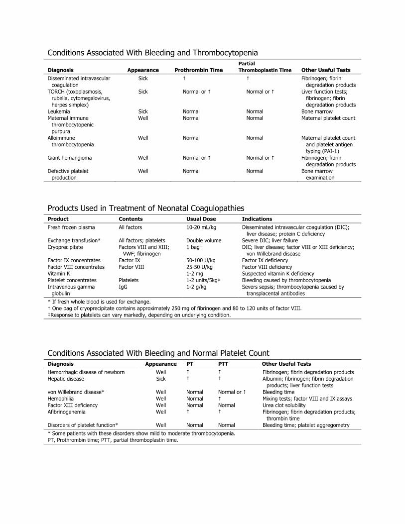
Conditions Associated With Bleeding and Thrombocytopenia
Diagnosis Appearance Prothrombin Time Partial Thromboplastin Time Other Useful Tests
Disseminated intravascular coagulation
Sick Fibrinogen; fibrin degradation products
TORCH (toxoplasmosis, rubella, cytomegalovirus, herpes simplex)
Sick Normal or Normal or Liver function tests; fibrinogen; fibrin degradation products
Leukemia Sick Normal Normal Bone marrow Maternal immune
thrombocytopenic purpura
Well Normal Normal Maternal platelet count
Alloimmune thrombocytopenia
Well Normal Normal Maternal platelet count and platelet antigen typing (PAI-1)
Giant hemangioma Well Normal or Normal or Fibrinogen; fibrin degradation products
Defective platelet production
Well Normal Normal Bone marrow examination
Products Used in Treatment of Neonatal Coagulopathies Product Contents Usual Dose Indications Fresh frozen plasma All factors 10-20 mL/kg Disseminated intravascular coagulation (DIC);
liver disease; protein C deficiency Exchange transfusion* All factors; platelets Double volume Severe DIC; liver failure Cryoprecipitate Factors VIII and XIII;
VWF; fibrinogen 1 bag† DIC; liver disease; factor VIII or XIII deficiency;
von Willebrand disease Factor IX concentrates Factor IX 50-100 U/kg Factor IX deficiency Factor VIII concentrates Factor VIII 25-50 U/kg Factor VIII deficiency Vitamin K 1-2 mg Suspected vitamin K deficiency Platelet concentrates Platelets 1-2 units/5kg‡ Bleeding caused by thrombocytopenia Intravenous gamma
globulin IgG 1-2 g/kg Severs sepsis; thrombocytopenia caused by
transplacental antibodies
* If fresh whole blood is used for exchange. † One bag of cryoprecipitate contains approximately 250 mg of fibrinogen and 80 to 120 units of factor VIII. ‡Response to platelets can vary markedly, depending on underlying condition.
Conditions Associated With Bleeding and Normal Platelet Count Diagnosis Appearance PT PTT Other Useful Tests Hemorrhagic disease of newborn Well Fibrinogen; fibrin degradation products Hepatic disease Sick Albumin; fibrinogen; fibrin degradation
products; liver function tests von Willebrand disease* Well Normal Normal or Bleeding time Hemophilia Well Normal Mixing tests; factor VIII and IX assays Factor XIII deficiency Well Normal Normal Urea clot solubility Afibrinogenemia Well Fibrinogen; fibrin degradation products;
thrombin time Disorders of platelet function* Well Normal Normal Bleeding time; platelet aggregometry
* Some patients with these disorders show mild to moderate thrombocytopenia. PT, Prothrombin time; PTT, partial thromboplastin time.
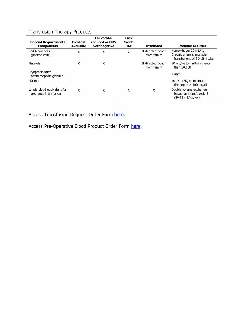
Transfusion Therapy Products
Special Requirements Components
Freshest Available
Leukocyte-reduced or CMV
Seronegative
Lack Sickle HGB Irradiated Volume to Order
Red blood cells (packed cells)
X X X If directed donor from family
Hemorrhage: 20 mL/kg Chronic anemia: multiple
transfusions of 10-15 mL/kg Platelets X X If directed donor
from family 10 mL/kg to mailtain greater
than 50,000 Cryoprecipitated
antihemophilic globulin 1 unit
Plasma 10-15mL/kg to maintain fibrinogen > 100 mg/dL
Whole blood equivalent for exchange transfusion
X X X X Double volume exchange based on infant’s weight (80-85 mL/kg/vol)
Access Transfusion Request Order Form here. Access Pre-Operative Blood Product Order Form here.

VII. RETINOPATHY OF PREMATURITY SURVEILLANCE
Retinopathy of prematurity (ROP) is a vasculo-proliferative disorder occurring almost exclusively in premature infants. Almost all ROP-related vision loss is associated with severe disease. Approximately 420 infants/year experience vision loss from ROP. New vessel growth regresses about 80% of the time with the other 20% progressing to neuro-vascularization, scar formation, hemorrhage, or retinal detachment. Prematurity is the greatest risk factor for ROP. Hyperoxia is a widely accepted risk factor. There is no safe level of arterial PaO2. The American Academy of Pediatrics guidelines recommends oxygen saturation monitoring if supplemental oxygen is required beyond the emergency period for newborns delivered before 36 weeks of gestation. Premature newborns are screened according to the following guidelines. Hypertension, reflex bradycardia, apneic events can be caused by the eye exam, or the mydriatic eye drops. The location and sequential retinal changes provide timely recognition of disease that is amendable to therapy and helps to provide prognosis. Retinopathy of prematurity is classified by location, zones I-III, and severity stage I-V. The extent of disease is measured around the retina in clock hours (1-12) with special designation for the accelerated inflammation with poor prognosis, plus disease. (See diagram of retina) If the ROP reaches (threshold) severity, the risk of poor retinal outcome increases to about 50%. Complications of ROP include varying degrees of visual impairment, including retinal detachment and blindness in the worse cases. Later complications include glaucoma, strabismus, cataracts, and amblyopia. Treatment of ROP is with cryotherapy or laser therapy. Cryotherapy freezes the avascular retina, preventing further abnormal vessel proliferation. Laser therapy, Argon, or iodide laser therapy photocoagulates the avascular periphery of the retina. This is less invasive and less traumatic to the eye than cryotherapy, and requires no anesthetics. Complications include scarring, choroidal hemorrhage, and pain.
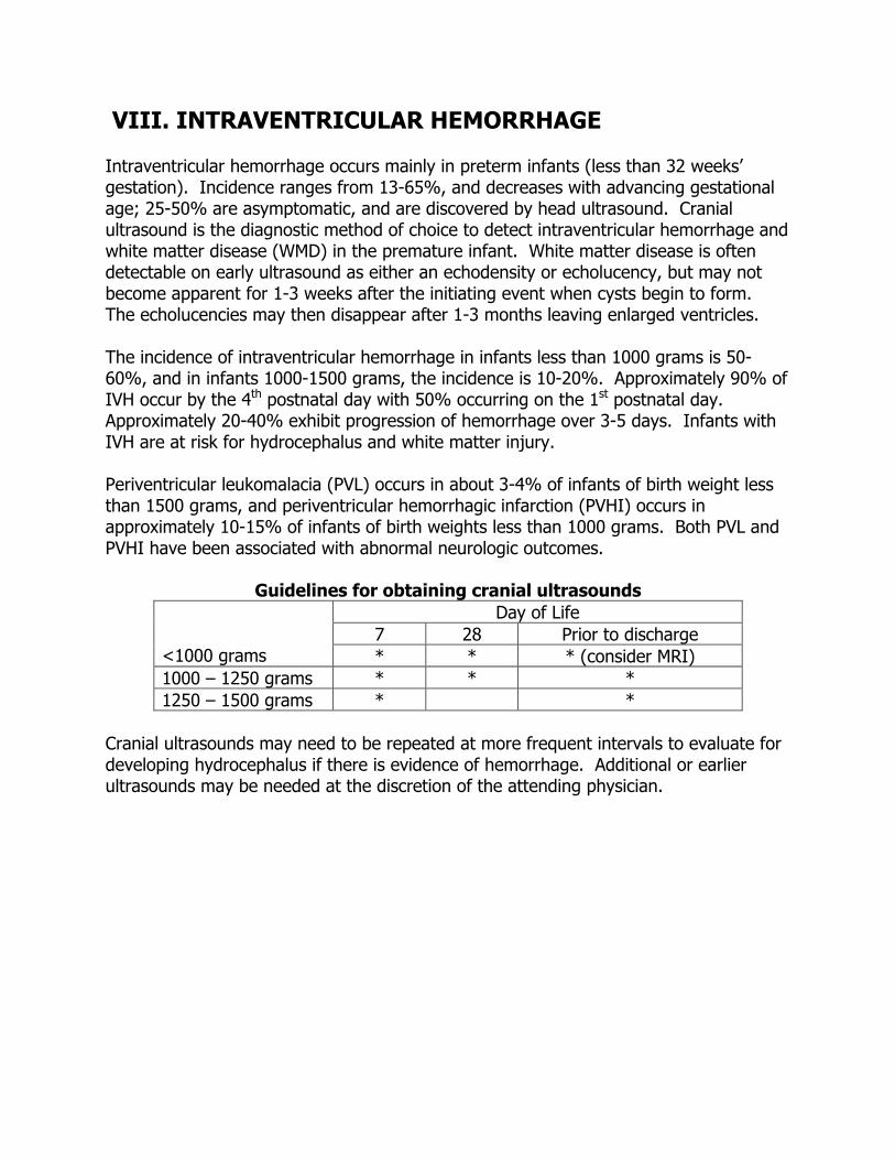
VIII. INTRAVENTRICULAR HEMORRHAGE
Intraventricular hemorrhage occurs mainly in preterm infants (less than 32 weeks’ gestation). Incidence ranges from 13-65%, and decreases with advancing gestational age; 25-50% are asymptomatic, and are discovered by head ultrasound. Cranial ultrasound is the diagnostic method of choice to detect intraventricular hemorrhage and white matter disease (WMD) in the premature infant. White matter disease is often detectable on early ultrasound as either an echodensity or echolucency, but may not become apparent for 1-3 weeks after the initiating event when cysts begin to form. The echolucencies may then disappear after 1-3 months leaving enlarged ventricles. The incidence of intraventricular hemorrhage in infants less than 1000 grams is 50-60%, and in infants 1000-1500 grams, the incidence is 10-20%. Approximately 90% of IVH occur by the 4th postnatal day with 50% occurring on the 1st postnatal day. Approximately 20-40% exhibit progression of hemorrhage over 3-5 days. Infants with IVH are at risk for hydrocephalus and white matter injury. Periventricular leukomalacia (PVL) occurs in about 3-4% of infants of birth weight less than 1500 grams, and periventricular hemorrhagic infarction (PVHI) occurs in approximately 10-15% of infants of birth weights less than 1000 grams. Both PVL and PVHI have been associated with abnormal neurologic outcomes.
Guidelines for obtaining cranial ultrasounds Day of Life
7 28 Prior to discharge <1000 grams * * * (consider MRI) 1000 – 1250 grams * * * 1250 – 1500 grams * *
Cranial ultrasounds may need to be repeated at more frequent intervals to evaluate for developing hydrocephalus if there is evidence of hemorrhage. Additional or earlier ultrasounds may be needed at the discretion of the attending physician.
IX. “1-2-3” MINNESOTA NEWBORN HEARING SCREENING PROGRAM
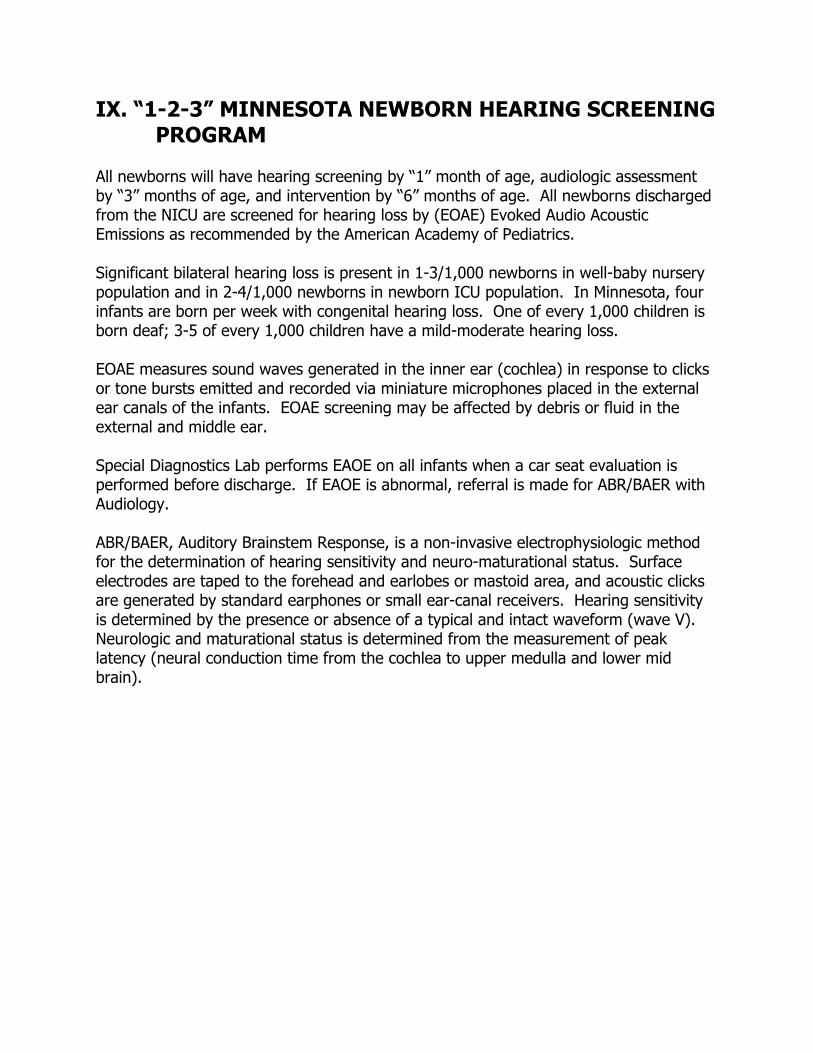
All newborns will have hearing screening by “1” month of age, audiologic assessment by “3” months of age, and intervention by “6” months of age. All newborns discharged from the NICU are screened for hearing loss by (EOAE) Evoked Audio Acoustic Emissions as recommended by the American Academy of Pediatrics. Significant bilateral hearing loss is present in 1-3/1,000 newborns in well-baby nursery population and in 2-4/1,000 newborns in newborn ICU population. In Minnesota, four infants are born per week with congenital hearing loss. One of every 1,000 children is born deaf; 3-5 of every 1,000 children have a mild-moderate hearing loss. EOAE measures sound waves generated in the inner ear (cochlea) in response to clicks or tone bursts emitted and recorded via miniature microphones placed in the external ear canals of the infants. EOAE screening may be affected by debris or fluid in the external and middle ear. Special Diagnostics Lab performs EAOE on all infants when a car seat evaluation is performed before discharge. If EAOE is abnormal, referral is made for ABR/BAER with Audiology. ABR/BAER, Auditory Brainstem Response, is a non-invasive electrophysiologic method for the determination of hearing sensitivity and neuro-maturational status. Surface electrodes are taped to the forehead and earlobes or mastoid area, and acoustic clicks are generated by standard earphones or small ear-canal receivers. Hearing sensitivity is determined by the presence or absence of a typical and intact waveform (wave V). Neurologic and maturational status is determined from the measurement of peak latency (neural conduction time from the cochlea to upper medulla and lower mid brain).
X. MINNESOTA STATE NEWBORN SCREENING
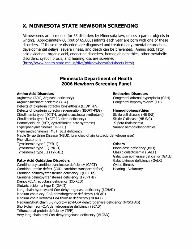
All newborns are screened for 53 disorders by Minnesota law, unless a parent objects in writing. Approximately 60 (out of 65,000) infants each year are born with one of these disorders. If these rare disorders are diagnosed and treated early, mental retardation, developmental delays, severe illness, and death can be prevented. Amino acid, fatty acid oxidation, organic acid, endocrine disorders, hemoglobinopathies, other metabolic disorders, cystic fibrosis, and hearing loss are screened. (http://www.health.state.mn.us/divs/phl/newborn/factsheets.html)
Minnesota Department of Health 2006 Newborn Screening Panel
Amino Acid Disorders Endocrine Disorders Arginemia (ARG, Arginase deficiency) Congenital adrenal hyperplasia (CAH) Argininosuccinate acidemia (ASA) Congenital hypothyroidism (CH) Defects of biopterin cofactor biosynthesis (BIOPT-BS) Defects of biopterin cofactor regeneration (BIOPT-REG) Hemoglobinopathies Citrullinemia type I (CIT-I, argininosuccinate synthetase) Sickle cell disease (HB S/S) Citrullinemia type II (CIT-II, citrin deficiency) Sickle-C disease (HB S/C) Homocystinuria (HCY, cystathionine beta synthase) S-βeta thalassemia Hyperphenylalaninemia (H-PHE) Variant hemoglobinopathies Hypermethioninemia (MET, I/III deficiency) Maple Syrup Urine Disease (MSUD, branched-chain ketoacid dehydrogenase) Phenylketonuria Tyrosinemia type I (TYR-1) Others Tyrosinemia type II (TYR-II) Biotinidase deficiency (BIO) Tyrosinemia type III (TYR-III) Classic galactosemia (GALT) Galactose epimerase deficiency (GALE) Fatty Acid Oxidation Disorders Galactokinase deficiency (GALK) Carnitine acylcarnitine translocase deficiency (CACT) Cystic fibrosis Carnitine uptake defect (CUD, carnitine transport defect) Hearing - Voluntary Carnitine palmitoyltransferase deficiency I (CPT-1a) Carnitine palmitoyltransferase deficiency II (CPT-II) Dienoyl-CoA reductase deficiency (DE-RED) Glutaric acidemia type II (GA-II) Long-chain hydroxyacyl-CoA dehydrogenase deficiency (LCHAD) Medium-chain acyl-CoA dehydrogenase deficiency (MCAD) Medium-chain ketoacyl-CoA thiolase deficiency (MCKAT) Medium/Short chain L-3-hydroxy acyl-CoA dehydrogenase deficiency (M/SCHAD) Short-chain acyl-CoA dehydrogenase deficiency (SCAD) Trifunctional protein deficiency (TFP) Very long-chain acyl-CoA dehydrogenase deficiency (VLCAD)
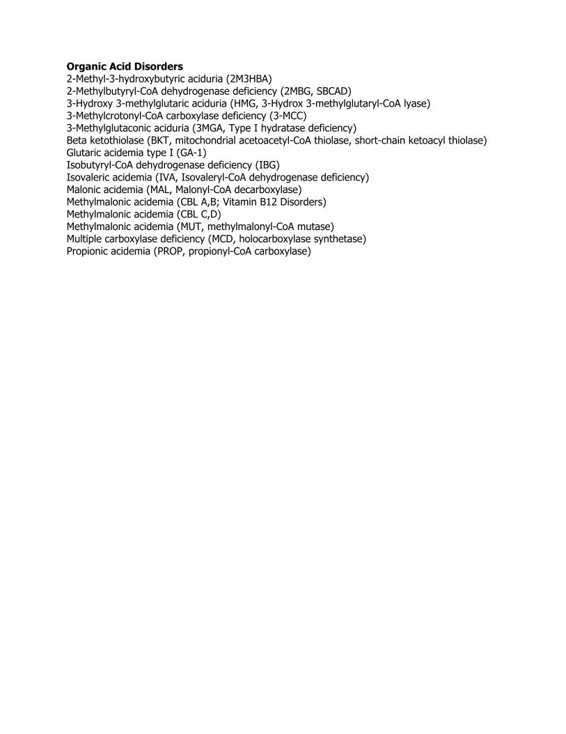
Organic Acid Disorders 2-Methyl-3-hydroxybutyric aciduria (2M3HBA) 2-Methylbutyryl-CoA dehydrogenase deficiency (2MBG, SBCAD) 3-Hydroxy 3-methylglutaric aciduria (HMG, 3-Hydrox 3-methylglutaryl-CoA lyase) 3-Methylcrotonyl-CoA carboxylase deficiency (3-MCC) 3-Methylglutaconic aciduria (3MGA, Type I hydratase deficiency) Beta ketothiolase (BKT, mitochondrial acetoacetyl-CoA thiolase, short-chain ketoacyl thiolase) Glutaric acidemia type I (GA-1) Isobutyryl-CoA dehydrogenase deficiency (IBG) Isovaleric acidemia (IVA, Isovaleryl-CoA dehydrogenase deficiency) Malonic acidemia (MAL, Malonyl-CoA decarboxylase) Methylmalonic acidemia (CBL A,B; Vitamin B12 Disorders) Methylmalonic acidemia (CBL C,D) Methylmalonic acidemia (MUT, methylmalonyl-CoA mutase) Multiple carboxylase deficiency (MCD, holocarboxylase synthetase) Propionic acidemia (PROP, propionyl-CoA carboxylase)
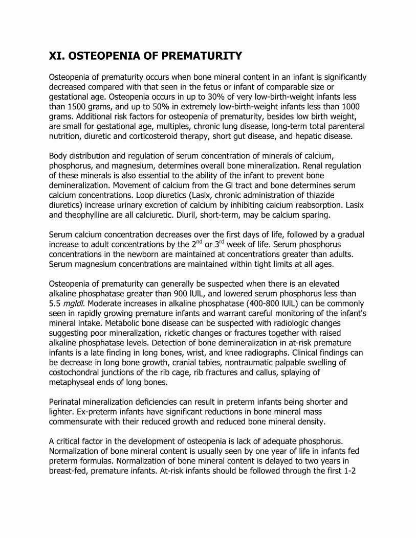
XI. OSTEOPENIA OF PREMATURITY Osteopenia of prematurity occurs when bone mineral content in an infant is significantly decreased compared with that seen in the fetus or infant of comparable size or gestational age. Osteopenia occurs in up to 30% of very low-birth-weight infants less than 1500 grams, and up to 50% in extremely low-birth-weight infants less than 1000 grams. Additional risk factors for osteopenia of prematurity, besides low birth weight, are small for gestational age, multiples, chronic lung disease, long-term total parenteral nutrition, diuretic and corticosteroid therapy, short gut disease, and hepatic disease. Body distribution and regulation of serum concentration of minerals of calcium, phosphorus, and magnesium, determines overall bone mineralization. Renal regulation of these minerals is also essential to the ability of the infant to prevent bone demineralization. Movement of calcium from the Gl tract and bone determines serum calcium concentrations. Loop diuretics (Lasix, chronic administration of thiazide diuretics) increase urinary excretion of calcium by inhibiting calcium reabsorption. Lasix and theophylline are all calciuretic. Diuril, short-term, may be calcium sparing. Serum calcium concentration decreases over the first days of life, followed by a gradual increase to adult concentrations by the 2nd or 3rd week of life. Serum phosphorus concentrations in the newborn are maintained at concentrations greater than adults. Serum magnesium concentrations are maintained within tight limits at all ages. Osteopenia of prematurity can generally be suspected when there is an elevated alkaline phosphatase greater than 900 lUlL, and lowered serum phosphorus less than 5.5 mgldl. Moderate increases in alkaline phosphatase (400-800 lUlL) can be commonly seen in rapidly growing premature infants and warrant careful monitoring of the infant's mineral intake. Metabolic bone disease can be suspected with radiologic changes suggesting poor mineralization, ricketic changes or fractures together with raised alkaline phosphatase levels. Detection of bone demineralization in at-risk premature infants is a late finding in long bones, wrist, and knee radiographs. Clinical findings can be decrease in long bone growth, cranial tabies, nontraumatic palpable swelling of costochondral junctions of the rib cage, rib fractures and callus, splaying of metaphyseal ends of long bones. Perinatal mineralization deficiencies can result in preterm infants being shorter and lighter. Ex-preterm infants have significant reductions in bone mineral mass commensurate with their reduced growth and reduced bone mineral density. A critical factor in the development of osteopenia is lack of adequate phosphorus. Normalization of bone mineral content is usually seen by one year of life in infants fed preterm formulas. Normalization of bone mineral content is delayed to two years in breast-fed, premature infants. At-risk infants should be followed through the first 1-2
years with monitoring growth parameters, possibly alkaline phosphatase, phosphate, and vitamin D levels during and after switching from premature nutrition. The stability of bones is adapted to local muscle forces. This has been shown in very low-birth-weight infants, who had greater gains in body weight, fat-free mass, forearm length, and bone mineral content with an exercise program, consisting of passive resistance and compression of all extremity joints, starting when stable. Fetuses have had resistance training against the uterine muscle. Sick preterm infants tend to be very inactive, naturally or iatrogenically. Prevention of osteopenia of prematurity is the best approach. Osteopenia of prematurity nutritional therapy protocol should be followed from birth. Calcium and phosphorus content and ratio should be optimized in hyperalimentation. Initiate enteral feedings and optimize calcium, phosphorus content by adding human milk fortifier or premature formula. If premature infant is on unfortified breast milk for a prolonged period of time, consider calcium, phosphorus supplementation or feeding some premature formula. Switch from Lasix to an anti-calciuretic diuretic (chlorothiazide) as soon as possible. Limit use of aminophylline and steroids. Maintain vitamin D intake, especially with GI, hepatic, or renal dysfunction. More would be required with cholestasis, direct bilirubin level greater than 2. In cases of severe cholestasis, consider the use of 25-hydroxy vitamin D supplements. Initiate occupational therapy, passive range of motion and compression of joints to enhance bone mineralization and bone mineral content in very low-birth-weight infants when stable.
XII. CLEFT LIP OR PALATE Affected infants will have a number of medical issues and potential complications requiring multidisciplinary evaluation and care. A child with a cleft lip/palate, or other craniofacial abnormalities, has multiple and complex problems, including early feeding and nutritional concerns, middle ear disease, hearing deficiencies, deviations in speech and resonance, dental, facial, and orthodontic abnormalities, and psycho-social adjustment problems. Appropriate consultation should be obtained from Plastic Surgery (Dr. Robert Wood), Genetics, Audiology and/or Pediatric Otolaryngology, and Speech Pathology, with follow-up with Dr. Wood at Gillette Children's Hospital. Cleft lip with or without cleft palate, and cleft palate alone, have a combined incidence of 1-2/1,000. They are the 4th most common birth defect. Cleft lip and palate incidence is 1 in 700 births with cleft palate alone occurring 1 in 2500 births. Native Americans have the higher incidence in the United States. Cleft lip with cleft palate is the most common presentation. The etiology of cleft lip and/or cleft palate is largely unknown. It is believed to be of multifactorial etiology with genetic and environmental factors interacting to shift a complex process of morphogenesis, resulting in clefting. Development of the face and upper lip occurs between the 5th and 9th weeks of pregnancy. Palatal development occurs between the 6th and 11th weeks of pregnancy. Patients with oral clefts exhibit other anomalies 21-37%, such as cardiovascular, musculoskeletal, facial dysmorphia, and genitourinary system disturbances. Numerous syndromes and chromosomal abnormalities have been associated with oral cleft. Cleft lip with cleft palate is the most common presentation. More males are affected than females. More males have complete clefts. The unilateral clefts are most common on the left side. Infants with associated anomalies are more likely to have combined cleft lip/palate, or cleft palate rather than a cleft lip alone. They are also more likely to be small for gestational age.

XIII. ORGAN DONATION Organ and tissue donations provide hope for people with organ failure or tissue disease and injuries. A person who is an organ donor can potentially help up to eight people. A person who is a tissue donor can potentially help up to 50 people. Donations can occur after brain death or cardiac death. Organs that can be donated are heart, lungs, pancreas, intestines, kidneys, and liver. Tissues to be donated are corneas, heart valves, veins/arteries, bone, connective tissue, and skin. Life Source is the not-for-profit organ procurement organization for our four-state region. They are our bridge between donation and transplantation. They identify potential donors, matching donors with recipients, coordinating clinical donation activities, arranging surgical recovery, support donor families, and increasing public awareness about donation. All patient deaths (imminent brain death and cardiac death) must be referred to Life Source. Call Life Source when brain death is imminent, cardiac death or asystole occurs, or family mentions donation. Do not mention donation to the family. A donation coordinator will evaluate your patient to determine donor designation and possible procurement. A Life Source coordinator will collaborate with the healthcare team to develop an appropriate communication plan if the family mentions or has questions about donation. Call Life Source when the patient has expired for possible tissue and eye donation, even if the patient has been previously referred.
XIV. END OF LIFE CARE More infants die in the neonatal period than at any other time in childhood. Pediatric residency provides neonatal end-of-life training and supportive care if needed in these situations. Caring for terminally ill children is a multidisciplinary task. At Children's Hospitals and Clinics of Minnesota - St. Paul, notify Social Work, bereavement team, and chaplaincy. Appropriate end-of-life care is palliative comfort care. Parents will need to understand their infant's condition and the reason for palliative care. Bereavement process for parents starts before the termination of active medical treatment. Encourage parents to touch, care, and obtain items (pictures, blankets, clothes) that they can remember their infant after death. Private rooms provide a quiet private location for end-of-life care. Staff (nursing and medical) presence and involvement is important for parents. Autopsy request should be discussed at an appropriate time before or after death - not at the time of death. Consent for autopsy, or denial, needs to be documented on the hospital Autopsy Consent Form. Follow-up with parents, with or without an autopsy, is important to the parents. It is acceptable to attend funerals of your patients, if you would like. Always consider cultural norms with the death of your patients.
XV. IMMUNIZATIONS All medically stable infants should be given vaccines as recommended for their age. A medically stable infant is defined as one who does not require on-going management for serious infections, metabolic disease, acute renal, cardiovascular, or respiratory tract illness, and who demonstrates a clinical course of sustained recovery and pattern of steady growth. To avoid multiple injections and superimposed local reactions, two-week intervals of recommended vaccines may be reasonable. Consistent weight gain by a preterm infant is important before receipt of the first dose of hepatitis B vaccine, as this is predictive of immune responsiveness. Medically stable, thriving infants, weighing less than 2000 grams, demonstrate predictable, consistent, and sufficient hepatitis B antibody responses, and should receive the first dose of hepatitis B vaccine as early as 30 days of chronologic age, regardless of gestational age or birth weight. Starting the hepatitis B series at one month of age, regardless of the weight of the preterm infant, offers more options for implementing the immunization schedule in the NICU setting, lessens the number of simultaneous injections at two months of age, provides earlier protection to vulnerable preterm infants, more likely to receive multiple blood products and undergo surgical interventions, and decreases the risk of horizontal transmission from occult hepatitis B chronic carriers among family members, hospital visitors, and other caregivers. Medically stable preterm infants, weighing more than 2000 grams, born to hepatitis B, surface antigen negative mothers, may receive the first dose of hepatitis B vaccine at birth, or shortly thereafter. Only monovalent hepatitis B vaccine should be used for infants from birth-six weeks of age. Because all preterm infants are considered at increased risk for complications of influenza, two doses of an inactivated influenza vaccine, given one month a part, should be offered for these infants, beginning at six months of chronologic age before the onset of influenza season. Household contacts should also receive inactivated influenza vaccine. RSV pneumonia is a major cause of serious pediatric respiratory disease from November - April, especially among infants who have chronic lung disease and those born prematurely. Palivizumab (Synagis TM), a recombinant monoclonal antibody against RSV, is effective in decreasing the incidence of RSV pneumonia in high-risk infants. Palivizumab use does not interfere with the provision of routine childhood immunizations to preterm or low-birth-weight infants. Infants at risk for RSV will be given Synagis, 15 mg/kg IM monthly, from November-April. Patients who are to be given this treatment include: gestational age less than 28 weeks, and less than 12 months postnatal age at start of RSV season; gestational age 29-32 weeks and postnatal age less than or equal to six months at start of RSV season; gestational age 33-35 weeks and additional risk factors, who are younger than six months of age at the start of the RSV season; childcare attendants; school age siblings; exposure to environmental air pollutants, congenital abnormality of the airway, or severe
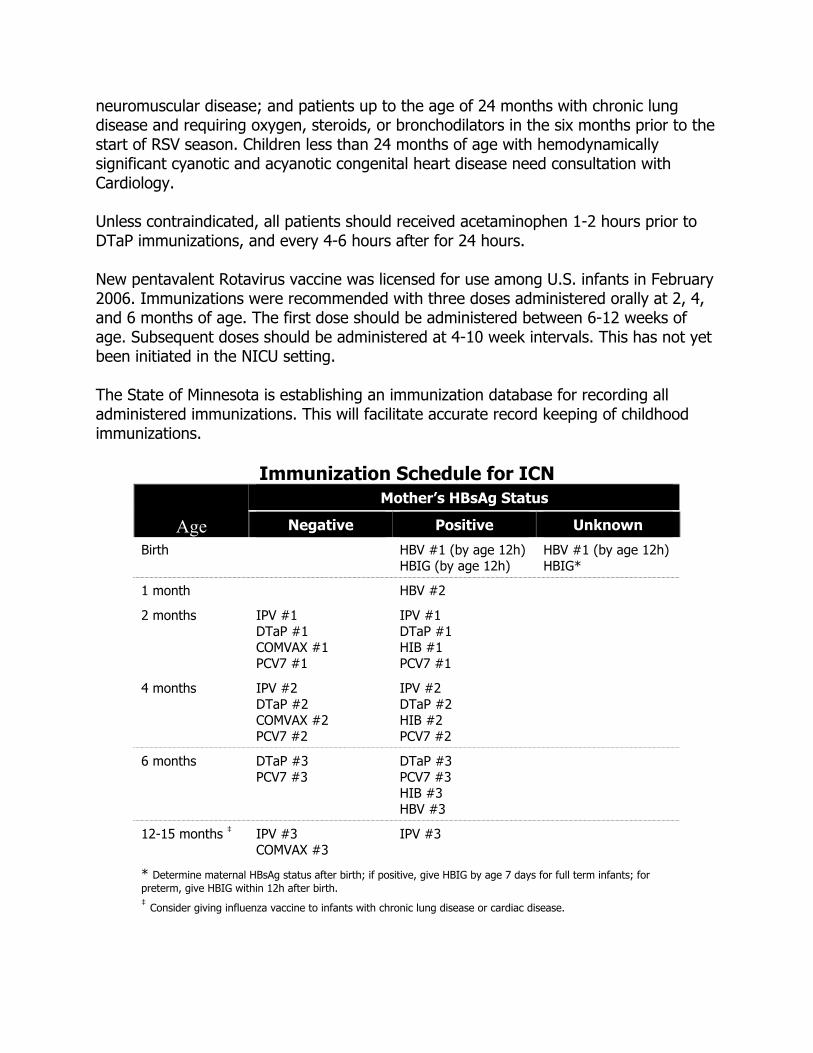
neuromuscular disease; and patients up to the age of 24 months with chronic lung disease and requiring oxygen, steroids, or bronchodilators in the six months prior to the start of RSV season. Children less than 24 months of age with hemodynamically significant cyanotic and acyanotic congenital heart disease need consultation with Cardiology. Unless contraindicated, all patients should received acetaminophen 1-2 hours prior to DTaP immunizations, and every 4-6 hours after for 24 hours. New pentavalent Rotavirus vaccine was licensed for use among U.S. infants in February 2006. Immunizations were recommended with three doses administered orally at 2, 4, and 6 months of age. The first dose should be administered between 6-12 weeks of age. Subsequent doses should be administered at 4-10 week intervals. This has not yet been initiated in the NICU setting. The State of Minnesota is establishing an immunization database for recording all administered immunizations. This will facilitate accurate record keeping of childhood immunizations.
Immunization Schedule for ICN Mother’s HBsAg Status
Age Negative Positive Unknown
Birth HBV #1 (by age 12h) HBIG (by age 12h)
HBV #1 (by age 12h) HBIG*
1 month HBV #2
2 months IPV #1 DTaP #1 COMVAX #1 PCV7 #1
IPV #1 DTaP #1 HIB #1 PCV7 #1
4 months IPV #2 DTaP #2 COMVAX #2 PCV7 #2
IPV #2 DTaP #2 HIB #2 PCV7 #2
6 months DTaP #3 PCV7 #3
DTaP #3 PCV7 #3 HIB #3 HBV #3
12-15 months ‡ IPV #3 COMVAX #3
IPV #3
* Determine maternal HBsAg status after birth; if positive, give HBIG by age 7 days for full term infants; for preterm, give HBIG within 12h after birth. ‡ Consider giving influenza vaccine to infants with chronic lung disease or cardiac disease.

Hepatitis B Immunoprophylaxis Scheme by Infant Birth Weight1 Maternal
Status Infant > 2000g Infant < 2000g
HbsAg positive
Hepatitis B vaccine + HBIG (within 12 h of birth) Continue vaccine series beginning at 1-2 mo of
age according to recommended schedule for infants born to HBsAg-positive mothers (see following table)
Check anti-HBs and HBsAg after completion of vaccine series2
HBsAg-negative infants with anti-HBs levels > 10 mIU/mL are protected and need no further medical management
HBsAg-negative infants with anti-HBs levels < 10 mIU/mL should be reimmunized with 3 doses at 2-mo intervals and retested
Infants who are HBsAg positive should receive appropriate follow-up, including medical evaluation for chronic liver disease
Hepatitis B vaccine + HBIG (within 12h of birth) Continue vaccine series beginning at 1-2 mo of
age according to recommended schedule for infants born to HBsAg-positive mothers (see following table)
Immunize with 4 vaccine doses; do not count birth dose as part of vaccine series
Check anti-HBs and HBsAg after completion of vaccine series2
HBsAg-negative infants with anti-HBs levels > 10 mIU/mL are protected and need no further medical management
HBsAg-negative infants with anti-HBs levels < 10 mIU/mL should be reimmunized with 3 doses at 2-mo intervals and retested
Infants who are HBsAg positive should receive appropriate follow-up, including medical evaluation for chronic liver disease
HBsAg status unknown
Test mother for HBsAg immediately after admission for delivery
Hepatitis B vaccine (by 12h) Administer HBIG (within 7 days) if mother tests
HBsAg positive
Continue vaccine series beginning at 1-2 mo of age according to recommended schedule based on mother’s HBsAg result (see following table)
Test mother for HBsAg immediately after admission for delivery
Hepatitis B vaccine (by 12h) Administer HBIG if mother tests HBsAg positive
or if mother’s HBsAg result is not available within 12h of birth
Continue vaccine series beginning at 1-2 mo of age according to recommended schedule based on mother’s HBsAg result (see following table)
Immunize with 4 vaccine doses; do not count birth dose as part of vaccine series
HBsAg negative
Hepatitis B vaccine at birth3
Continue vaccine series beginning at 1-2 mo of
age (see following table) Follow-up anti-HBs and HBsAg testing not
needed
Hepatitis B vaccine dose 1-30 days of chronologic age if medically stable, or at hospital discharge if before 30 days of chronologic age
Continue vaccine series beginning at 1-2 mo of age (see following table)
Follow-up anti-HBs and HBsAg testing not needed
HBsAg indicates hepatitis B surface antigen; HBIG, hepatitis B Immune Globulin; anti-HBs, antibody to hepatitis B surface antigen. 1 Extremes of gestational age and birth weight no longer are a consideration for timing of hepatitis B vaccine doses. 2 Test at 9 to 18 months of age, generally at the next well-child visit after completion of the primary series. Use testing method that allows determination of a protective concentration of anti-HBs (>10mIU/mL).
3 The first dose may be delayed until after hospital discharge for an infant who weighs > 2000 g and whose mother is HBsAg negative, but only if a physician’s order to withhold the birth dose and a copy of the mother’s original HBsAg-negative laboratory are documented in the infant’s medical record.

Hepatitis B Vaccine Schedules for Infant, by Maternal Hepatitis B Surface Antigen (HBsAg) Status1, 2
Single Antigen Vaccine Single Antigen + Combination
Maternal HBsAg Status Dose Age Dose Age Positive 13
HBIG4 2 35
Birth (<12h) Birth (<12h) 1-2 mo 6 mo
13 HBIG 2 3 45
Birth (<12h) Birth (<12h) 2 mo 4 mo 6 mo (Pediatrix)
or 12-15 mo (Comvax)
Unknown6 13 2 35
Birth (<12h) 1-2 mo 6 mo
13 2 3 45
Birth (<12h) 2 mo 4 mo 6 mo (Pediatrix)
or 12-15 mo (Comvax)
Negative 13,7 2 35
Birth (before discharge) 1-2 mo 6 mo
13,7 2 3 45
Birth (before discharge) 2 mo 4 mo 6 mo (Pediatrix)
or 12-15 mo (Comvax) 1 Centers for Disease Control and Prevention. A comprehensive immunization strategy to eliminate transmission of hepatitis B virus infection in the United States. Recommendations of the Advisory Committee on Immunization Practices (ACIP) part 1: immunization os infants, children, and adolescents. MMWR Recomm Rep. 2005;54(RR-16):1-23 2 See previous table for vaccine schedules for preterm infants weighing < 2000g. 3 Recombivax HB or Engerix-B should be used for the birth dose. Comvax and Pediarix cannot be administered at birth or before 6 weeks of age. 4 Hepatitis B Immune Globulin (0.5 mL) administered intramuscularly in a separate site from vaccine. 5 The final dose in the vaccine series should not be administered intramuscularly in a separate site from vaccine. 6 Mothers should have blood drawn and tested for HBsAg as soon as possible after admission for delivery; if the mother is found to be HBsAg positive, the infant should receive HBIG as soon as possible but no later than 7 days of age. 7 On a case-by-case basis and only in rare circumstances, the first dose may be delayed until after hospital discharge for an infant who weighs > 2000g and whose mother is HBsAg negative, but only if a physician’s order to withhold the birth dose and a copy of the mother’s original; HBsAg-negative laboratory report are documented in the infant’s medical record.

XVI. Fluid and Electrolyte Stabilization
HYPOGLYCEMIA
Glucose falls postnatally and reaches a point no lower than 30 mg/dL in uncompromised term infants between 1-2 hours of age. Levels stabilize by 4-6 hours of age in the range of 45-80 mg/dL. In compromised high-risk infants, blood glucose may fall to subnormal levels, or may not rise appropriately postnatally. Etiology of Hypoglycemia – Infant with prematurity, IUGR, IDM, IGDM, sepsis, chronic intrauterine stress or asphyxia, hypothermia, heart failure, erythroblastosis fetalis, and polycythemia.
• Premature infants, IURG, and infants with chronic intrauterine asphyxia may deplete their glycogen stores needed to maintain glucose homeostasis after birth. IDM, IGDM, and infants with hemolytic disorders may have hyperinsulinemia. IUGR or asphyxiates infants may have deficit catecholamine excretion or exhaustion of catecholamine response, or be unable to use pathways of gluconeogenesis.
Evaluation- Plasma glucose (lab) (higher value) is more accurate than POC glucose. STAT confirmatory plasma glucose test should be sent to the lab if glucose screening reveals low glucose levels. If glucose is low on POC testing, therapy should be initiated and not wait for the confirmatory results from the lab. Intervention- is needed with glucometer reading <20 mg/dL, or <40 mg/dL and NPO,
or preterm or <40 mg/dL after feeding, or <40 mg/dL and symptomatic 1. Symptomatic infants during initial transition period (12-24 hours of life) - glucose
less than 40 mg/dL should be given bolus of 2 cc/kg of D10W, followed by a continuous infusion of 5-8 mg/kg/minute (80-115 ml/kg/day of D10W). Failure to provide continuous infusion may result in rebound recurrence of hypoglycemia.
2. At-risk infants who are stable and asymptomatic – should be offered feeds within one hour after birth and have POC glucose tested 30 minutes after a feed until stable.
3. At-risk infants who are NPO and asymptomatic – start IV glucose rates of 80-100 mg/kg/day of D10W (5.5-7 mg of glucose/kg/minute) and check glucose 30-60 minutes of life. Infants less than 25 weeks’ gestation should be started at a GIR of 4.5-6 mg/kg/minute (D10W at 66-86 mg/kg/day). If glucose is less than 40 mg/dL during the first four hours of life (less than 45 mg/dL at 4-24 hours of

life), give IV glucose of 2 cc/kg of D10W, followed by an increase in GIR by 10-20% or to 8 mg/kg/minutes (whichever is greater).
Total fluid intake, both oral and IV fluids, must be carefully monitored to avoid fluid overload and hyponatremia. If a high enough GIR cannot be delivered using D12.5W within a reasonable fluid goal (120 cc/kg/day), a central venous catheter will need to be placed for concentrated glucose infusion.
Length of glucose screening is risk-dependent- 1. LGA >4 kg – IDM infants should have glucose levels monitored at least until 12 hours of age.
2. <34 weeks’ gestational age, term small for gestational age infants, infants <2.5 kg – should have glucose levels monitored at least until 24 hours of age.
Weaning IV dextrose infusion- When blood glucose levels have been stable (AC glucose >60 mg/dL for 12-24 hours without IV glucose bolus, begin decreasing IV infusion by 1-2 cc/hour every 3-4 hours (decrease GIR by 10-20%).
HYPERKALEMIA
(Central Serum Potassium 6.5 mEq/l or >) is a medical emergency that requires close patient observation, continuous cardiac monitoring, and measurement of serum potassium levels. Treatment is indicated when serum potassium is greater than 7 mEq/l. Etiology of Hyperkalemia in Neonates • Factitious (most common): Hemolized blood from heel stick, thrombocytosis.
Send repeat sample STAT before starting treatment, unless EKG changes indicate hyperkalemia.
• Decreased removal of potassium: Acute renal failure, positive potassium balance in premature infants during the first days of life, adrenal failure in CAH and medications (Captopril).
• Increased load of potassium: Hemoloysis, IVH, hematoma, excess potassium administration.
• Redistribution: Elevated potassium, secondary to metabolic acidosis and sepsis, NEC, and medications (Digoxin).
Evaluation of Hyperkalemia Determination of etiology and management: Electrolytes, BUN, creatinine, platelet
count, blood gases, ionized calcium, total calcium, and magnesium levels.

EKG changes progress with increasing potassium level: peak T-waves, prolonged PR interval, loss of P wave, widening QRS, sign wave QRST, first
degree AV block, ventricular dysrhythmia and, finally, asystole. Treatment of Hyperkalemia Hyperkalemia with dehydration should respond to fluid resuscitation. • Immediately change to an IV solution without potassium. If on gentamicin, hold
dose, pending evaluation of renal status, and gentamicin trough levels. The effects of hyperkalemia can be worsened by hypocalcemia and hypomagnesemia.
Hyperkalemia with Cardiac Changes • With cardiac monitoring, give 100 mg/kg/dose (1 ml/kg/dose) IV of 10% calcium
gluconate or 20 mg/kg/dose (0.2 ml/kg/dose) of 10% calcium chloride over 10 minutes. This will decrease myocardial excitability and prevent cardiac arrhythmias. May repeat calcium dose in 10 minutes, if abnormal cardiac changes persist. Administration of calcium does not lower serum potassium levels.
• If patient is acidotic, give sodium bicarbonate, 1-2 mg/kg IV over 10-20 minutes. Inducing alkalosis will drive potassium into cells. Correct respiratory acidosis first before administering sodium bicarbonate.
• Give insulin to assist driving potassium into the intracellular fluid compartment. If infant is normoglycemic, administer insulin and glucose together as a bolus to prevent hypoglycemia. The ratio should be 1 unit of insulin to 4 gm of glucose given as a bolus. 0.05 units/kg of regular insulin with 2 ml/kg of D10W 10% glucose, followed by continuous infusion of D10W at 2-4 cc/kg/hour, and regular insulin, 10 units/100 ml at 1 ml/kg/hour. Obtain initial glucose level, and follow glucose levels every 30-60 minutes until stable.
• Enhance potassium excretion. Give diuretic therapy: Lasix, 1 mg/kg IV, may increase potassium excretion by increasing flow and sodium delivery to distal tubules.
• Kayexalate, rectally (1 gm/kg at 0.5 gm/ml of normal saline) with minimum retention time of 30 minutes. The silastic feeding tube should be inserted 1-3 cm for retention enema. Follow sodium levels because patient will be at risk for hypernatremia with this exchange resin.
• Double-volume exchange with fresh whole blood (less than 24 hours old or deglycerolized) red blood cells reconstituted with fresh frozen plasma.
• Peritoneal dialysis.

XVII. REFERENCES Resources
1. The Harriet Lane Handbook. Authors: Siberry and Iannone. Publisher: Mosby. 2. NeoFax. Authors: Young and Mangum. Publisher: Acorn Publishing. 3. Michigan Manual of Neonatal Intensive Care. Author: Dunn. Publisher: Hanley
and Belfus. 4. Manual of Neonatal Respiratory Care. Authors: Dunn and Hinha. Publicher:
Mosby. 5. Atlas of Procedures in Neonatology. Authors: MacDonald and Ramasethu.
Publisher: Lippincott, Williams and Wilkins. 6. Care of the High-Risk Neonate. Authors: Klaus and Fanaroff. Publisher:
Saunders. 7. Neonatal Resuscitatoin Textbook by AAP/AHA. 8. Neonatal-Perinatal Medicine. Authors: Martin, Fanaroff and Walsh. Publisher:
Mosby. 9. Smith’s Recognizable Patterns of Human Malformations. Author: Jones.
Publisher: Elsevier and Saunders. 10. Drugs in Pregnancy and Lactation. Authors: Briggs, Freeman and Yaffe.
Publisher: L.W.N.W. 11. Red Book. Publisher: American Academy of Pediatrics 12. Neonatology. Author: Gomella. Publisher: McGraw-Hill Medical.
Web Resources
1. American Academy of Pediatrics www.aap.org 2. Neonatology on the Web www.neonatology.org,
www.neonatology.org/neo.clinical.html 3. NICU-WEB http://neonatal.peds.washington.edu 4. Iowa Neonatology Handbook
www.uihealthcare.com/depts/med/pediatrics/iowaneonatologyhandbook/index.html
5. Lucille Packard Children’s Hospital www.lpch.org/diseasehealthinfo/healthlibrary/hrnewborn/sitemap.html
6. Family Village library www.familyvillage.wisc.edu/specific.htm 7. University of Minnesota Neonatology Lecture and Teaching Files
www.med.umn.edu/peds/neonat/education.html 8. UCSF Children’s Hospital Intensive Care Nursery House Staff Manual
http://www.ucsfhealth.org/childrens/health_professionals/icnManual.html 9. Newborn exam http://newborns.stanford.edu/Students/NewbornExam.html

10. Photos of Normal and Abnormal Findings http://newborns.stanford.edu/RNMDEducation.html
11. Neonatal Dermatology www.adhb.govt.nz/newborn/TeachingResources/dermatology/dermatology.htm
12. Neonatal Radiology www.adhb.govt.nz/newborn/TeachingResources/Radiology/Radiology.htm
13. Neonatal Ventilation www.adhb.govt.nz/newborn/TeachingResources/Ventilation/Ventilation.htm
14. Neuroneonatology www.pediatricneuro.com/alfonso/main.htm 15. University of Pittsburgh Seminars in Newborn Medicine
http://www.eprom.pitt.edu/34_viewFolder.asp?folderID=1225850524 16. Jordan University of Science and Technology Department of Pediatrics Fourth
Year Newborn and Neonatology Course Presentations http://www.just.edu.jo/~mykhassawneh/newborn.htm
17. White/Cox: Diseases of the Skin, 2ed., chapter 19: Pediatric Dermatology http://www.merckmedicus.com/ppdocs/us/hcp/content/white/white-ch-019-toc.htm
18. Yale Well Baby Nursery http://yalepediatrics.org/residents/PCC%20References/WBN.html