research article identification of keratan sulfate ... of keratan sulfate disaccharide at c-3 ... ....
TRANSCRIPT

Research Article
Identification of keratan sulfate disaccharide at C-3position of glucuronate of chondroitin sulfate fromMactra chinensisKyohei Higashi1,*, Keita Takeda1,*, Ann Mukuno1, Yusuke Okamoto1, Sayaka Masuko2, Robert J. Linhardt2
and Toshihiko Toida11Graduate School of Pharmaceutical Sciences, Chiba University, 1-8-1 Inohana, Chuo-ku, Chiba 260-8675, Japan; and 2Department of Biology, Center for Biotechnology andInterdisciplinary Studies, Rensselaer Polytechnic Institute, 110 8th Street Troy, NY 12180, U.S.A.
Correspondence: Toshihiko Toida ([email protected])
Glycosaminoglycans (GAGs), including chondroitin sulfate (CS), dermatan sulfate,heparin, heparan sulfate and keratan sulfate (KS) are linear sulfated repeating disacchar-ide sequences containing hexosamine and uronic acid [or galactose (Gal) in the case ofKS]. Among the GAGs, CS shows structural variations, such as sulfation patterns andfucosylation, which are responsible for their physiological functions through CS inter-action with CS-binding proteins. Here, we solved the structure of KS-branched CS-Ederived from a clam, Mactra chinensis. KS disaccharide [D-GlcNAc6S-(1→3)-β-D-Gal-(1→]was attached to the C-3 position of GlcA, and consecutive KS-branched disaccharidesequences were found in a CS chain. KS-branched polysaccharides clearly exhibitedresistance to degradation by chondroitinase ABC or ACII (at low concentrations) com-pared with typical CS structures. Furthermore, KS-branched polysaccharides stimulatedneurite outgrowth of hippocampal neurons. These results strongly suggest thatM. chinensis is a rich source of KS-branched CS, and it has important biologicalactivities.
IntroductionGlycosaminoglycans (GAGs), a group of structurally related polysaccharides, are primarily found asthe glycan moieties of proteoglycan (PG) glycoconjugates. GAGs, which include chondroitin sulfate(CS), dermatan sulfate (DS), heparin, heparan sulfate and keratan sulfate (KS) are linear sulfated poly-saccharides comprising 50–200 repeating disaccharides of hexosamine and uronic acid [or galactose(Gal) in the case of KS] [1]. CS consists of a distinctive repeating disaccharide unit[→4)-β-D-GlcA-(1→3)-β-D-GalNAc-(1→]n, where GlcA is glucuronic acid, and GalNAc isN-acetylgalactosamine, and some hydroxy groups in disaccharide units are replaced with sulfo groups(S) responsible for the diversity of disaccharide units, namely, GlcA-GalNAc (O-type unit),GlcA-GalNAc (4S) (A-type unit: CS-A), GlcA-GalNAc (6S) (C-type units: CS-C), GlcA (2S)-GalNAc(6S) (D-type unit: CS-D), GlcA-GalNAc (4S, 6S) (E-type unit: CS-E), GlcA (3S)-GalNAc (4S) (K-typeunit: CS-K) [2–4]. CS biosynthesis is initiated once GalNAc is transferred by CSGalNAcT-1 or -2 tothe common linkage tetrasaccharide, GlcAβ1-3Galβ1-3Galβ1-4xylose (Xyl)β1-O-Ser in PGs, and chainelongation is then catalyzed by the CHSY(1-3)/CHPF heterodimer [5,6]. After the polymerization, themajority of the GalNAc residues are 4-O-sulfated by chondroitin 4-O-sulfotransferases (C4ST-1, -2and -3) or 6-O-sulfated by 6-O-sulfotransferases (C6ST-1 and -2). In addition, resulting A- or C-typeunits can be further sulfated by GalNAc 4-sulfate 6-O-sulfotransferase (GalNAc 4S-6ST) or chondro-itin uronyl 2-O-sulfotransferase (UST), generating di-sulfated disaccharides, E-type unit or D-typeunit, respectively. A disaccharide unit containing iduronic acid (IdoA) in place of GlcA is commonlyfound in DS, a stereoisoform of CS that differs in the C-5 configuration of the hexuronic acid moieties
*Both authors contributedequally to this work.
Accepted Manuscript online:19 September 2016Version of Record published:10 November 2016
Received: 7 July 2016Revised: 12 September 2016Accepted: 19 September 2016
© 2016 The Author(s). This is an open access article published by Portland Press Limited on behalf of the Biochemical Society and distributed under the Creative Commons Attribution License 4.0 (CC BY-NC-ND).4145
Biochemical Journal (2016) 473 4145–4158DOI: 10.1042/BCJ20160655

[7]. This epimerization of GlcA is catalyzed by DS epimerase (DS-epi1 and DS-epi2), and resultingIdoA-GalNAc is subsequently sulfated by D4ST-1. Resulting IdoA-GalNAc (4S) (iA-type unit) is further sul-fated by UST, generating IdoA (2S)-GalNAc (4S) (B-type unit). Because CS function can be achieved throughthe interaction with growth factors, receptors, and other CS-binding proteins, it is assumed that consecutiveand highly sulfated disaccharide units are critical for these mammalian physiologies. However, the degree ofsulfation of CS by sulfotransferases is intricately regulated at multiple levels in mammals, because most disac-charide units in CS/DS from mammals are A-type/iA-type units, and di-sulfated disaccharide levels are verylow despite their high affinity for growth factors [8]. Thus, the distribution and number of protein-bindingsites containing IdoA and di-sulfated CS disaccharides are important for the mammalian physiologies [9]. Incontrast, highly sulfated and rare structures of CS have been identified from marine organisms. For example,DS from adult and embryonic sea urchin are composed of 59% of iC-type units (IdoA-GalNAc (6S)) and 74%of iE-type units (IdoA-GalNAc (4S,6S)), and B-type units (66%) and iD-type units (IdoA-GalNAc (2S,6S))(>90%) are predominant structures of DS from the ascidian Spathoglottis plicata and Ascidia nigra, respectively[10–12]. In addition, GlcA[(Fuc(4S)-Fuc(3S,4S)]-GalNAc (4S, 6S) oligosaccharides were found in CS from seacucumbers [13]. Hikino et al. [14] reported that consecutive highly sulfated CS (or DS) derived from seaurchin, ascidian and sea cucumber possesses the neurite outgrowth promotion activities of E16 hippocampalneurons; however, physiological functions of CS structures in their origin are not fully understood.It is of interest to note that CS, having a fucosylated GlcA, a sulfated 3-position of GlcA residue and IdoA
residues, shows different susceptibility to chondroitinases (Chases) compared with other types of CS. It is wellknown that Chase ABC can depolymerize almost all types of natural CS, and it can afford the unsaturated dis-accharides, including ΔDi-0S, ΔDi-4S, ΔDi-6S, ΔDi-diSE, ΔDi-diSB and ΔDi-diSD [3]. Interestingly, CS-K,(1-3)-linkage of K-type units and (1-4)-linkage of disaccharides are completely digested by Chase ABC, result-ing in GalNAc (4S) residues being produced from K-type units [4]. However, (1-4)-linkage between GalNAcand 3-O-sulfated GlcA in CS-K shows resistance to depolymerization by Chase ACII [15,16]. In addition,(1-4)-linkage between GalNAc and IdoA in DS is also resistant to the action of Chase ACII [17]. Therefore,unsaturated disaccharides and resistant oligosaccharides, containing IdoA residue or K-type units, can beobtained after the treatment of Chase ACII from DS or CS-K [16,17]. In contrast, fucosylated CS, derived fromsea cucumber, shows resistance to both Chase ABC and ACII, and partial acid hydrolysis is required to preparethe unsaturated disaccharide units [13].We have purified CS (or DS) from a variety of animal tissues and characterized their structures to better
understand their biological distribution and to explore new sources of polysaccharides as functional foods,nutraceuticals, cosmetics and drugs [16–22].In this study, we have identified novel CS-E from a clam, Mactra chinensis, containing KS disaccharide unit
[D-GlcNAc6S-(1→3)-β-D-Gal-(1→] at the C-3 position of GlcA. KS-branched CS exhibits resistance to degrad-ation by both Chase ABC and ACII (at low concentrations), and KS-branched disaccharides are clustered inthe CS chain. Furthermore, KS-rich polysaccharides promoted neurite outgrowth of hippocampal neurons.These results demonstrate that M. chinensis is a rich source of KS-branched CS having important biologicalactivities.
Materials and methodsChemicalsM. chinensis in dry powder form was obtained from Futtsu City Fishery Association in Chiba, Japan. ActinaseE was purchased from Kaken Pharmaceutical Co., Ltd, Tokyo, Japan. Chondroitinase ABC (Chase ABC) fromProteus vulgaris, chondroitinase ACII (Chase ACII) from Arthrobacter aurescens, unsaturated disaccharides(ΔDi-0S, ΔDi-4S, ΔDi-6S, ΔDiUA-2S, ΔDi-diSE, ΔDi-diSB, ΔDi-diSD, ΔDi-TriS), CS-A (6.33% of ΔDi-0S, 74.2%of Δdi-4S, 19.5% of ΔDi-6S, 0.3% of ΔDi-diSE) from whale cartilage, CS-E (10.0% of ΔDi-0S, 17.0% of Δdi-4S,8.31% of ΔDi-6S, 63.6% of ΔDi-diSE, 1.08% of ΔDi-diSD) from squid cartilage and KS from bovine cornea werepurchased from Seikagaku Corp., Tokyo, Japan [3]. The other analytical reagents used were of analytical grade.
Isolation of crude glycosaminoglycans from viscera of M. chinensisThe hot water extract of the viscera of M. chinensis was used in the present study. The dried powder (30 g) wasproteolyzed at 45°C with actinase E (10 mg/g dry powder) in 50 mM Tris/acetate (pH 8.0) for 18 h. After pro-teolysis, the β-elimination reaction, on the reducing termini of peptidoglycan chains, was performed with
4146 © 2016 The Author(s). This is an open access article published by Portland Press Limited on behalf of the Biochemical Society and distributed under the Creative Commons Attribution License 4.0 (CC BY-NC-ND).
Biochemical Journal (2016) 473 4145–4158DOI: 10.1042/BCJ20160655
0.5 M NaOH, containing 0.3 M sodium borohydride (20 ml/g of dry sample) at 4°C for 18 h. The reactionmixture was then neutralized with 1.0 M HCl. The resulting GAG chains were precipitated by the addition of5% cetylpyridinium chloride (CPC; final concentration 0.1%) containing 30 mM NaCl at 4°C for 16 h. TheGAG–CPC complex was collected by centrifugation at 2300 × g for 15 min. The GAG chains were extractedfrom the GAG–CPC complex by the addition of 2.5 M NaCl, and the mixture was centrifuged at 2300 × g for15 min. The GAG chains were precipitated from the supernatant by the addition of 11 volumes of 85% ethanolat 4°C for 16 h, and they were collected by centrifugation at 2300 × g for 15 min. The GAG chains were thenisolated through dialysis against distilled water at room temperature for 16 h followed by lyophilization toafford partially purified GAG.The crude GAG sample (∼30 mg of dry powder) in 2 ml of water was applied at a flow rate of 2 ml/min on
a HiPrep DEAE FF (16 mm internal diameter × 100 mm, obtained from GE Healthcare Europe GmbH) andfractionated to prepare the highly sulfated CS polysaccharides. The eluents were (A) 50 mM sodium phosphate,(B) 2.0 M NaCl in 50 mM sodium phosphate. The gradient program was 0–30 min (5% B), 30–150 min (5–100% B), and 150–180 min (100% B). Fractionated samples were collected at 30 min-intervals, followed by con-centration with a rotary evaporator, dialyzed, freeze-dried and kept stored at 4°C.
High-performance liquid chromatographyDisaccharide composition analysis of GAGs was performed as follows. GAGs (5 μg) were incubated in the reac-tion mixture (35 μl), which contained 28.6 mM Tris/acetate (pH 8.0), 50 mU of Chase ABC and/or 50 mU ofChase ACII. After 16 h at 37°C, depolymerized samples were boiled and evaporated, resuspended in 10 μl ofwater. The HPLC system was constructed with a high-pressure pump (LC-10Ai, Shimadzu, Kyoto, Japan),Intelligent Fluorescence detector (FP-920S, Jasco, Tokyo, Japan), a dry reaction bath (DB-3, ShimamuraInstruments Co., Japan), double plunger pumps for reagent solution (NP-FX (II)-1U, Nihon Seimitsu KagakuCo. Ltd., Tokyo, Japan), a chromato-integrator (D-2500, Hitachi High-Technologies Corp., Tokyo, Japan) and asample injector with a 20 μl loop (Model 7725i, Rheodyne, CA, USA). A gradient was applied at a flow rate of1.0 ml/min on Senshu Pak Docosil (4.6 mm × 150 mm; Senshu Scientific, Tokyo, Japan) at 60°C. The eluentbuffers were as follows: A, 10 mM tetra-n-butylammonium hydrogen sulfate in 12% methanol; B, 0.2 M NaClin buffer A. The gradient program was as follows: 0–10 min (1% B), 10–11 min (1–10% B), 11–30 min (10%B), 30–35 min (10–60% B), and 35–40 min (60% B). Aqueous (0.5% (w/v)) 2-cyanoacetamide solution and1 M NaOH were added to the eluent at the same flow rates (0.25 ml/min), using a double plunger pump. Theeffluent was monitored fluorometrically (excitation, 346 nm; emission, 410 nm).
Liquid chromatography–mass spectrometryFreeze-dried oligosaccharides obtained by Chase ACII treatment were labeled with 2-aminoacridone (AMAC)by slightly modifying the method of Yang et al. [23] with minor modifications. Oligosaccharides (60 μg) wereadded to 15 μl of 0.1 M AMAC solution in acetic acid/dimethyl sulfoxide (DMSO) (3:17, v/v), and mixed byvortexing for 5 min. Next, 15 μl of 1 M NaBH3CN was added in the reaction mixture, and incubated at 45°Cfor 4 h. Finally, the AMAC-tagged oligosaccharide mixture was diluted, using 50% (v/v) aqueous DMSO. LC–MS analysis was performed on an amaZon series SL ion-trap mass spectrometer (Bruker Daltonics, Bremen,Germany). MS data were collected by the following conditions: ESI interface, negative mode; capillary voltage,3500 V; end plate offset 500 V; gas flow, 6.0 l/min. The column used was an Acquity UPLC BEH C18 column(2.1 mm internal diameter × 150 mm, 1.7 mm) at 50°C in a column oven CO 631A (GL Science Corp., Tokyo,Japan), and the flow rate was 0.1 ml/min. The eluent buffers were as follows: eluent A, 80 mM ammoniumacetate/methanol (88/12, v/v) and B, 80 mM ammonium acetate/methanol (12/88, v/v). The gradient programwas as follows: 0–4 min (0% eluent B), 4–12.5 min (0–4% eluent B), 12.5–25 min (4–15% eluent B), 25–50 min(15–100% eluent B).
Methylation analysisPartially methylated alditol acetates (PMAAs) of the monosaccharide components from oligosaccharides wereprepared according to the method of Anumula and Taylor [24]. Gas chromatography–mass spectrometry (GC–MS) analysis was performed by an Agilent Technologies 7890A GC system (Agilent Technologies, CA, USA)and Time-of-flight mass spectrometer JMS-T100GCV ( JEOL, Tokyo, Japan). Mass spectra were obtained byelectro impact ionization (70 eV) with the following program parameters: column, ZB-5 ms column (0.25 mmfilm thickness, 0.25 mm i.d. × 30 m) (Phenomenex, CA, USA); carrier gas, helium at 1.2 ml/min; column oven
© 2016 The Author(s). This is an open access article published by Portland Press Limited on behalf of the Biochemical Society and distributed under the Creative Commons Attribution License 4.0 (CC BY-NC-ND).4147
Biochemical Journal (2016) 473 4145–4158DOI: 10.1042/BCJ20160655

temperature program: 3 min at 100°C, with an increase at 4°C/min to 160°C, 1 min at 160°C followed by anincrease at 0.5°C/min to 180°C, and a final increase at 20°C/min to 260°C and held for 10 min at 260°C.
1H-NMR spectroscopy1H-NMR spectra of the purified polysaccharides were recorded on a JEOL JNM-ECA600 (600 MHz) spectrom-eter in the FT mode. Each polysaccharide (∼6 mg) for NMR analysis was dissolved in deuterium oxide (D2O,99.90% D), and lyophilized three times to replace exchangeable protons with deuterons. Then the lyophilizedsamples were dissolved in 0.6 ml of D2O and transferred to the NMR tube. The one- and two-dimensional (1Dand 2D) NMR experiments were recorded with a relaxation time of 1.5 s at a probe temperature of 30°C. 1D1H-NMR spectra were recorded with an acquisition time of 1.45 s and many 300 scans. 2D 1H/1H COSY (cor-relation spectroscopy) and TOCSY (total correlation spectroscopy) spectra were recorded with many 112 and156 scans, respectively. 2D TOCSY spectra were run with a mixing time of 100 ms. The proton signals wereassigned, using the 2D 1H/1H COSY and TOCSY spectra.
Effect of CS derived from M. chinensis on neurite outgrowth of hippocampalneuronsAll animal experiments were approved by the Institutional Animal Care and Use Committee of ChibaUniversity and carried out according to the guidelines for Animal Research of Chiba University. GAG precoat-ing in an eight-well chamber slide and evaluation of CS on neurite outgrowth of mouse hippocampal neuronswere performed as described previously [17]. Briefly, eight-well chamber slides were pre-coated with 50 μg/mlpoly-D,L-ornithine in 0.1 M sodium borate (pH 8.0), and then 0.5 μg/well of the CS (Fr. 4 in Figure 1B andremaining polysaccharides in Figure 2B) derived from M. chinensis, CS-E and CS-A were coated at 4°C over-night. Subsequently, the cells were seeded on coverslips at a density of 16 000 cells/cm2 and cultured for 18 h.Thereafter, the cells were fixed by 4% (w/v) paraformaldehyde for 30 min, the neurites were visualized byimmunochemical staining, using anti-microtubule-associated protein-2 (Lieco Technologies Inc., St. Louis, MO,USA) and anti-neurofilament (Sigma–Aldrich, St. Louis, MO, USA). Fifty cells with at least one neurite longerthan the cell body were chosen at random to determine the length of the longest neurite. At least three inde-pendent experiments per condition were carried out.
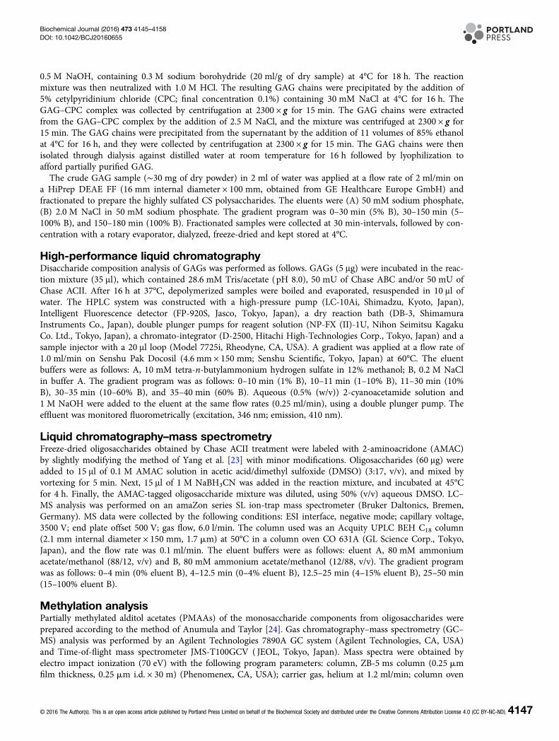
ResultsComposition of CSs derived from M. chinensisCrude GAGs were extracted from dry samples (30 g) from the viscera of M. chinensis by actinase E digestion,and recovered by ethanol precipitation. The dried pellet, including crude GAGs, was weighed after dialysis andfreeze-drying. As a result, 353.8 mg GAG/g of dry samples was recovered. The recovery of crude GAGs fromthe viscera of M. chinensis was confirmed repeatedly. Crude GAGs (30 mg) were further fractionated usinganion-exchange chromatography, and 18.5 mg of Fr. 3 and 3.73 mg of Fr. 4 were obtained. Disaccharide com-position analysis of each fraction was performed using Chase ABC treatment, and a significant amount ofΔDi-diSE was observed in Fr. 4 (Figure 1B). The disaccharide composition of clam CS in Fr. 4 was 2.1% ofΔDi-0S, 7.4% of ΔDi-4S, 30.7% of ΔDi-6S and 59.8% of ΔDi-diSE. However, recovery of total amount of CS dis-accharides in Fr. 3 and Fr.4 was very low, with clam CS contents in Fr. 3 and Fr. 4 of 2.97% (551 μg/18.5 mgdry powder) and 17.2% (640 μg/3.73 mg dry powder), respectively (Supplementary Figure S1). In addition, asmall unidentified peak eluting at 38.1 min was also found in Fr. 4 (Figure 1B). Thus, disaccharide analysis ofFr. 3 and Fr. 4 was carried out, using Chase ACI or Chase ACII. When CS in Fr. 3 or Fr. 4 was treated withChase ACII but not Chase ABC or ACI, a significant amount of four kinds of unidentified peaks were observed(Figure 1). In particular, unidentified peak (d) is a major component in clam CS, and detection of unidentifiedpeaks (a–c) is Chase ACII treatment-dependent. Next, clam CS was treated with Chase ACII at the specifiedconcentrations to characterize the unidentified structures. When a small amount (1.6 mU) of Chase ACII wasadded to the reaction mixture (17.5 μl) containing 2.5 μg of clam CS, well-known CS structures were com-pletely degraded to unsaturated disaccharides, including ΔDi-0S, ΔDi-4S, ΔDi-6S, ΔDi-diSE (Figure 2A). In con-trast, a higher concentration of Chase ACII (12.5 mU) was needed to detect the four kinds of unidentifiedpeaks. Unidentified peaks were also detectable at 230 nm (Supplementary Figure S2), suggesting the existenceof C4–C5 unsaturated uronate residue at the non-reducing end, and reducing end was GalNAc because ChaseACII can recognize the (1–4)-linkage between GalNAc and GlcA. Next, we tested whether unidentified
4148 © 2016 The Author(s). This is an open access article published by Portland Press Limited on behalf of the Biochemical Society and distributed under the Creative Commons Attribution License 4.0 (CC BY-NC-ND).
Biochemical Journal (2016) 473 4145–4158DOI: 10.1042/BCJ20160655

structures represented a consecutive sequence in clam CS. After the treatment of 1.6 mU of Chase ACII,degraded samples were separated by HiTrap desalting column, and monitored at 230 nm. As a result, singlepeak of remaining polysaccharide (unknown peak) was observed (Figure 2B). In addition, disaccharide analysiswith 12.5 mU of Chase ACII shows that unidentified peak (d) is the main component of remaining polysac-charides (Figure 2C). Thus, molecular masses of parent CS (Fr. 4 in Figure 1B) and remaining polysaccharideswere determined by GPC–HPLC analysis. The chromatogram of parent CS shows a high structural diversityand high polydispersity with the number average molecular mass of ∼12 kDa (Supplementary Figure S3).Interestingly, the remaining polysaccharides show a high structural diversity and high polydispersity with theaverage molecular mass of ∼8 kDa, suggesting a cluster of previously unidentified structures, such as DS havingIdoA-rich domains in the polysaccharide backbone [17,25]. The remaining polysaccharides with large chainlength heterogeneity consisted of more than 20 saccharides, given that the retention time of remaining polysac-charides (20.76 min) is shorter than that of dp20 (molecular mass 4960) from CS (21.84 min; SupplementaryFigure S3). Taken together, clam CS from M. chinensis has unknown consecutive repeating structures thatexhibit different chondroitinase susceptibilities when compared with other types of CS structures.
Analysis of CS oligosaccharides having unknown structures by UPLC–MS/MSUPLC–MS/MS analysis of AMAC-labeled sample was performed to obtain the mass of unidentified peaks (c)and (d) in Figure 2C. Before UPLC–MS/MS analysis, unidentified peaks (c) and (d) in Figure 2C were fractio-nated (Supplementary Figure S2) and reacted with AMAC according to the methods of Yang et al. [23]. When
Figure 1. Disaccharide composition of CS of M. chinensis after chondroitinase ABC, ACI and ACII treatment.
Chromatograms of unsaturated disaccharides of Fr. 3 (A) and Fr. 4 (B) of CS obtained by weak anion-exchange
chromatography (see Supplementary Figure S1). Unsaturated disaccharide analysis was performed as follows. CS (5 mg) were
incubated in the reaction mixture (35 ml), which contained 28.6 mM Tris/acetate (pH 8.0) and 25 mU of Chase ABC, ACI or
ACII. After incubation, depolymerized samples were submitted to gradient HPLC with fluorescence detection as described
previously [17]. Experiments were repeated in triplicate with reproducible results. Peaks: 1, ΔDi-0S; 2, ΔDi-4S; 3, ΔDi-6S; 4,
ΔDi-diSE; a–d, unknown peaks.
© 2016 The Author(s). This is an open access article published by Portland Press Limited on behalf of the Biochemical Society and distributed under the Creative Commons Attribution License 4.0 (CC BY-NC-ND).4149
Biochemical Journal (2016) 473 4145–4158DOI: 10.1042/BCJ20160655
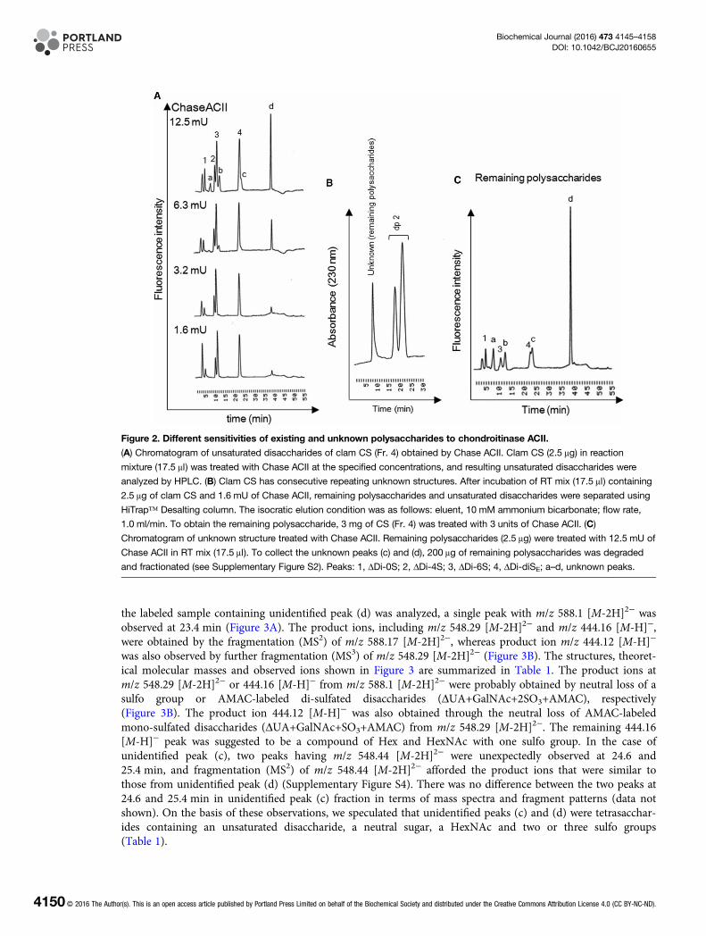
the labeled sample containing unidentified peak (d) was analyzed, a single peak with m/z 588.1 [M-2H]2− wasobserved at 23.4 min (Figure 3A). The product ions, including m/z 548.29 [M-2H]2− and m/z 444.16 [M-H]−,were obtained by the fragmentation (MS2) of m/z 588.17 [M-2H]2−, whereas product ion m/z 444.12 [M-H]−
was also observed by further fragmentation (MS3) of m/z 548.29 [M-2H]2− (Figure 3B). The structures, theoret-ical molecular masses and observed ions shown in Figure 3 are summarized in Table 1. The product ions atm/z 548.29 [M-2H]2− or 444.16 [M-H]− from m/z 588.1 [M-2H]2− were probably obtained by neutral loss of asulfo group or AMAC-labeled di-sulfated disaccharides (ΔUA+GalNAc+2SO3+AMAC), respectively(Figure 3B). The product ion 444.12 [M-H]− was also obtained through the neutral loss of AMAC-labeledmono-sulfated disaccharides (ΔUA+GalNAc+SO3+AMAC) from m/z 548.29 [M-2H]2−. The remaining 444.16[M-H]− peak was suggested to be a compound of Hex and HexNAc with one sulfo group. In the case ofunidentified peak (c), two peaks having m/z 548.44 [M-2H]2− were unexpectedly observed at 24.6 and25.4 min, and fragmentation (MS2) of m/z 548.44 [M-2H]2− afforded the product ions that were similar tothose from unidentified peak (d) (Supplementary Figure S4). There was no difference between the two peaks at24.6 and 25.4 min in unidentified peak (c) fraction in terms of mass spectra and fragment patterns (data notshown). On the basis of these observations, we speculated that unidentified peaks (c) and (d) were tetrasacchar-ides containing an unsaturated disaccharide, a neutral sugar, a HexNAc and two or three sulfo groups(Table 1).
Figure 2. Different sensitivities of existing and unknown polysaccharides to chondroitinase ACII.
(A) Chromatogram of unsaturated disaccharides of clam CS (Fr. 4) obtained by Chase ACII. Clam CS (2.5 μg) in reaction
mixture (17.5 μl) was treated with Chase ACII at the specified concentrations, and resulting unsaturated disaccharides were
analyzed by HPLC. (B) Clam CS has consecutive repeating unknown structures. After incubation of RT mix (17.5 μl) containing
2.5 μg of clam CS and 1.6 mU of Chase ACII, remaining polysaccharides and unsaturated disaccharides were separated using
HiTrap™ Desalting column. The isocratic elution condition was as follows: eluent, 10 mM ammonium bicarbonate; flow rate,
1.0 ml/min. To obtain the remaining polysaccharide, 3 mg of CS (Fr. 4) was treated with 3 units of Chase ACII. (C)
Chromatogram of unknown structure treated with Chase ACII. Remaining polysaccharides (2.5 μg) were treated with 12.5 mU of
Chase ACII in RT mix (17.5 μl). To collect the unknown peaks (c) and (d), 200 μg of remaining polysaccharides was degraded
and fractionated (see Supplementary Figure S2). Peaks: 1, ΔDi-0S; 2, ΔDi-4S; 3, ΔDi-6S; 4, ΔDi-diSE; a–d, unknown peaks.
4150 © 2016 The Author(s). This is an open access article published by Portland Press Limited on behalf of the Biochemical Society and distributed under the Creative Commons Attribution License 4.0 (CC BY-NC-ND).
Biochemical Journal (2016) 473 4145–4158DOI: 10.1042/BCJ20160655
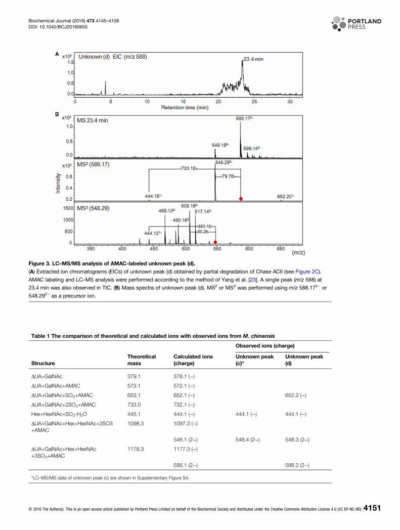
Figure 3. LC–MS/MS analysis of AMAC-labeled unknown peak (d).
(A) Extracted ion chromatograms (EICs) of unknown peak (d) obtained by partial degradation of Chase ACII (see Figure 2C).
AMAC labeling and LC–MS analysis were performed according to the method of Yang et al. [23]. A single peak (m/z 588) at
23.4 min was also observed in TIC. (B) Mass spectra of unknown peak (d). MS2 or MS3 was performed using m/z 588.172− or
548.292− as a precursor ion.
Table 1 The comparison of theoretical and calculated ions with observed ions from M. chinensis
StructureTheoreticalmass
Calculated ions(charge)
Observed ions (charge)
Unknown peak(c)*
Unknown peak(d)
ΔUA+GalNAc 379.1 378.1 (−)
ΔUA+GalNAc+AMAC 573.1 572.1 (−)
ΔUA+GalNAc+SO3+AMAC 653.1 652.1 (−) 652.2 (−)
ΔUA+GalNAc+2SO3+AMAC 733.0 732.1 (−)
Hex+HexNAc+SO3-H2O 445.1 444.1 (−) 444.1 (−) 444.1 (−)
ΔUA+GalNAc+Hex+HexNAc+2SO3+AMAC
1098.3 1097.3 (−)
548.1 (2−) 548.4 (2−) 548.3 (2−)
ΔUA+GalNAc+Hex+HexNAc+3SO3+AMAC
1178.3 1177.3 (−)
588.1 (2−) 588.2 (2−)
*LC–MS/MS data of unknown peak (c) are shown in Supplementary Figure S4.
© 2016 The Author(s). This is an open access article published by Portland Press Limited on behalf of the Biochemical Society and distributed under the Creative Commons Attribution License 4.0 (CC BY-NC-ND).4151
Biochemical Journal (2016) 473 4145–4158DOI: 10.1042/BCJ20160655
Methylation analysisGC–MS analysis of the reaction products of PMAA derived from the polysaccharides has been shown to representa powerful tool to investigate the glycosidic linkages. On the basis of this technique, fractionated unidentified peak(d) in Figure 2C was derivatized to PMAAs, and the resulting products were subsequently subjected to GC–MSanalysis. When the fragment ion at m/z 233 from PMAAs was monitored, a single peak was observed at 28.68 min(Figure 4A). The mass spectrum of the single peak at 28.68 min is shown in Figure 4B. On the basis of the observa-tion of certain fragment ions (m/z 45, 117, 161, 233) and earlier reported [26], it is suggested that the peak at28.68 min is 1,3,5-tri-O-acetyl-2,4,6-tri-O-methyl-galactitol [→3) Galactose (Gal) (1→]. Because KS consists of adistinctive repeating disaccharide unit [→3)-β-D-Gal-(1→4)-β-D-GlcNAc-(1→]n [27], PMAAs of KS from bovinecornea were analyzed as a control to confirm the PMAA of [→3) Gal (1→] at 28.68 min. When the fragment ionat m/z 233 from PMAAs was monitored, four peaks eluted at 28.62, 29.64, 39.65 and 50.55 min were observed(Supplementary Figure S5A). The fragment ion pattern of eluted peak at 28.62 min resembles that of Figure 4B(Supplementary Figure S5B), confirming that unidentified peak (d) was [→3) Gal (1→]. The peak correspondingto the 1,4,5-tri-O-acetyl-2-(acetylmethylamino)-2-deoxy-3,6-di-O-methyl-D-glucitol [→4) GlcNAc (1→] was also
Figure 4. Methylation analysis of unknown (d) peak.
PMAA from unknown (d) peak were performed as described in the Materials and methods section. To obtain PMAAs, 100 μg of
unknown (d) peak was used. (A) EIC (m/z 233) of unknown (d) peak. (B) Mass spectrum of PMAA at 28.68 min. (C) Theoretical
mass fragment pattern of 1,3,5-tri-O-acetyl-2,4,6-tri-O-methyl-galactitol [26].
4152 © 2016 The Author(s). This is an open access article published by Portland Press Limited on behalf of the Biochemical Society and distributed under the Creative Commons Attribution License 4.0 (CC BY-NC-ND).
Biochemical Journal (2016) 473 4145–4158DOI: 10.1042/BCJ20160655
observed at 50.55 min in PMAA analysis of cornea KS (Supplementary Figure S5A); however, there was no peak of[GlcNAc (1→] in PMAA analysis of fractionated unidentified peak (d) (Figure 4A).
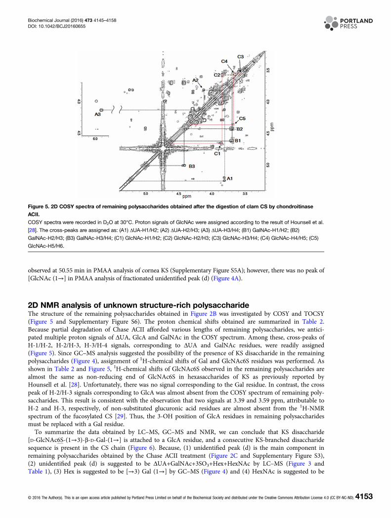
2D NMR analysis of unknown structure-rich polysaccharideThe structure of the remaining polysaccharides obtained in Figure 2B was investigated by COSY and TOCSY(Figure 5 and Supplementary Figure S6). The proton chemical shifts obtained are summarized in Table 2.Because partial degradation of Chase ACII afforded various lengths of remaining polysaccharides, we antici-pated multiple proton signals of ΔUA, GlcA and GalNAc in the COSY spectrum. Among these, cross-peaks ofH-1/H-2, H-2/H-3, H-3/H-4 signals, corresponding to ΔUA and GalNAc residues, were readily assigned(Figure 5). Since GC–MS analysis suggested the possibility of the presence of KS disaccharide in the remainingpolysaccharides (Figure 4), assignment of 1H-chemical shifts of Gal and GlcNAc6S residues was performed. Asshown in Table 2 and Figure 5, 1H-chemical shifts of GlcNAc6S observed in the remaining polysaccharides arealmost the same as non-reducing end of GlcNAc6S in hexasaccharides of KS as previously reported byHounsell et al. [28]. Unfortunately, there was no signal corresponding to the Gal residue. In contrast, the crosspeak of H-2/H-3 signals corresponding to GlcA was almost absent from the COSY spectrum of remaining poly-saccharides. This result is consistent with the observation that two signals at 3.39 and 3.59 ppm, attributable toH-2 and H-3, respectively, of non-substituted glucuronic acid residues are almost absent from the 1H-NMRspectrum of the fucosylated CS [29]. Thus, the 3-OH position of GlcA residues in remaining polysaccharidesmust be replaced with a Gal residue.To summarize the data obtained by LC–MS, GC–MS and NMR, we can conclude that KS disaccharide
[D-GlcNAc6S-(1→3)-β-D-Gal-(1→] is attached to a GlcA residue, and a consecutive KS-branched disaccharidesequence is present in the CS chain (Figure 6). Because, (1) unidentified peak (d) is the main component inremaining polysaccharides obtained by the Chase ACII treatment (Figure 2C and Supplementary Figure S3),(2) unidentified peak (d) is suggested to be ΔUA+GalNAc+3SO3+Hex+HexNAc by LC–MS (Figure 3 andTable 1), (3) Hex is suggested to be [→3) Gal (1→] by GC–MS (Figure 4) and (4) HexNAc is suggested to be
Figure 5. 2D COSY spectra of remaining polysaccharides obtained after the digestion of clam CS by chondroitinase
ACII.
COSY spectra were recorded in D2O at 30°C. Proton signals of GlcNAc were assigned according to the result of Hounsell et al.
[28]. The cross-peaks are assigned as: (A1) ΔUA-H1/H2; (A2) ΔUA-H2/H3; (A3) ΔUA-H3/H4; (B1) GalNAc-H1/H2; (B2)
GalNAc-H2/H3; (B3) GalNAc-H3/H4; (C1) GlcNAc-H1/H2; (C2) GlcNAc-H2/H3; (C3) GlcNAc-H3/H4; (C4) GlcNAc-H4/H5; (C5)
GlcNAc-H5/H6.
© 2016 The Author(s). This is an open access article published by Portland Press Limited on behalf of the Biochemical Society and distributed under the Creative Commons Attribution License 4.0 (CC BY-NC-ND).4153
Biochemical Journal (2016) 473 4145–4158DOI: 10.1042/BCJ20160655
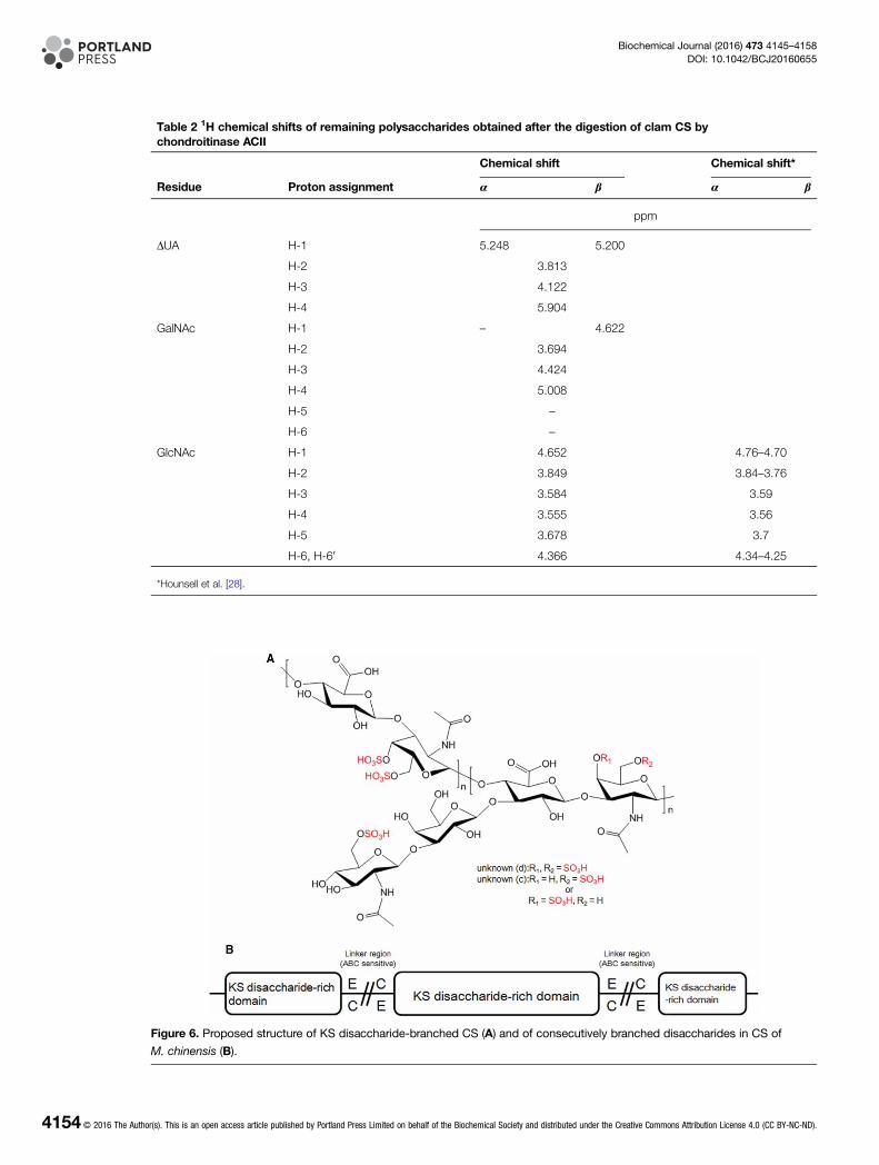
Figure 6. Proposed structure of KS disaccharide-branched CS (A) and of consecutively branched disaccharides in CS of
M. chinensis (B).
Table 2 1H chemical shifts of remaining polysaccharides obtained after the digestion of clam CS bychondroitinase ACII
Residue Proton assignment
Chemical shift Chemical shift*
α β α β
ppm
ΔUA H-1 5.248 5.200
H-2 3.813
H-3 4.122
H-4 5.904
GalNAc H-1 – 4.622
H-2 3.694
H-3 4.424
H-4 5.008
H-5 –
H-6 –
GlcNAc H-1 4.652 4.76–4.70
H-2 3.849 3.84–3.76
H-3 3.584 3.59
H-4 3.555 3.56
H-5 3.678 3.7
H-6, H-60 4.366 4.34–4.25
*Hounsell et al. [28].
4154 © 2016 The Author(s). This is an open access article published by Portland Press Limited on behalf of the Biochemical Society and distributed under the Creative Commons Attribution License 4.0 (CC BY-NC-ND).
Biochemical Journal (2016) 473 4145–4158DOI: 10.1042/BCJ20160655

[GlcNAc6S (1→] by COSY experiment (Figure 5). From these observations, we refer to the remaining polysac-charides as KS-branched polysaccharides.
Stimulation of neurite outgrowth by clam CSThe effect of CS on neurite outgrowth-promoting (NOP) activity was examined, using E16 embryonic mousehippocampal neurons, because M. chinensis CS contained significant amount of E-type units (Figure 1B). Thus,hippocampal neurons were cultured with clam CS (Fr. 4, see Figure 1B), and KS-branched polysaccharide(remaining polysaccharides, see Figure 2C), CS-E (a positive control) and CS-A (a negative control) that wereeach immobilized onto coverslips precoated with poly-D,L-ornithine. As a result, CS-E, clam CS (Fr. 4) andKS-branched polysaccharides stimulated neurite outgrowth, and their NOP activities were completely disap-peared by the Chase ABC and ACII treatment (Figure 7).
DiscussionIt has been known that commercially available CS, including CS-A (50–80% of A-type unit), CS-C (50–70% ofC-type unit), CS-D (20–40% of D-type unit) and CS-E (63.6% of E-type unit), is sensitive to digestion byChase ABC or ACII, while DS and CS-K (13.9% of K-type unit) are resistant to Chase ACII [16,17]. In con-trast, fucosylated CS exhibits resistance to Chase ABC and ACII, respectively [13]. In this study, we identifiednew CS from M. chinensis that lacks chondroitinase susceptibility. M. chinensis CS having KS disaccharideunits [D-GlcNAc6S-(1→3)-β-D-Gal-(1→] showed the resistance to Chase ABC and lower concentrations ofChase ACII, and higher concentrations of Chase ACII were required to obtain KS-branched disaccharide units(Figures 1 and 2). In addition, KS-branched disaccharide units represent consecutive sequences, such asIdoA-rich domains like CS/DS from shark fin or DS from porcine skin [17,25], because the remaining polysac-charides were obtained by the partial degradation at a lower concentration of Chase ACII (Figure 2Supplementary Figure S3).
Figure 7. Effect of clam CS (Fr. 4) or KS-branched polysaccharides on neurite outgrowth of hippocampal neurons.
(A) Representative morphological features of E16 hippocampal neurons cultured with clam CS (Fr.4) or KS-branched
polysaccharides. E16 hippocampal neuronal cells (16 000 cells/cm2) were cultured for 18 h on various substrates coated on
poly-D,L-ornithine, fixed and immunostained as described in the Materials and methods section. (B) The length of the longest
neurite of the randomly selected 50–100 individual neurons was measured. The values obtained from the three separate
experiments are expressed as the means ± SEM. Mann–Whitney’s U-test was used to evaluate the significance of differences
between means (**P < 0.01).
© 2016 The Author(s). This is an open access article published by Portland Press Limited on behalf of the Biochemical Society and distributed under the Creative Commons Attribution License 4.0 (CC BY-NC-ND).4155
Biochemical Journal (2016) 473 4145–4158DOI: 10.1042/BCJ20160655
Unfortunately, determination of the structure of unidentified peaks (a) to (b) failed, because their contentswere very low in remaining polysaccharides (Figure 2C and Supplementary Figure S2). However, LC–MSn ana-lysis showed that the difference between the unidentified peaks (c) and (d) is the number of sulfo groups onthe unsaturated disaccharide unit. On the basis of fragmentation patterns of each ion, unidentified peak (d)contains an E-type unit, because the major component in the CS from M. chinensis was an E-type unit(Figure 1). In contrast, there were two distinguishable peaks in the (c) fraction in LC–MS experiments(Supplementary Figure S4). Because unidentified (c) peaks contain mono-sulfated CS disaccharide, these peaksare likely to result from tetrasaccharide having ΔDi-4S or ΔDi-6S. This idea can be supported by the retentiontime of each peak observed in HPLC, that is, (d) peak which has three sulfo groups elutes close to ΔDi-TriS(data not shown), while (c) peak which has two sulfo groups appeared beside ΔDi-diSE (Figure 1B). For thisreason, we speculate that unidentified peak (b) is ΔDi-0S plus KS disaccharide unit, and (a) is ΔDi-0S plus KSdisaccharide unit (‒SO3), respectively. Further analysis is required to elucidate the complete CS structure of M.chinensis.E- and K-type units are likely to be subject to further modifications in marine organisms. For instance,
almost all CS from sea cucumbers are fucosylated [30], and a D-glucose branch at C-3 position of GalNAc wasalso found in CS-E from squid cartilage [31]. K-type units of CS from the cartilage of king crab and octopuswere also fucosylated [4,16]. Thus, lower marine organisms are rich sources of CS-E and CS-K having theseunique structures; however, biological functions of unique CS in individual organisms remain unclear. Vieiraet al. [13] proposed that these branches serve to prevent digestion of polysaccharide by microorganisms.Considering that KS-branched CS was abundant in the gut of the M. chinensis, the previous assumption isquite reasonable because Chase ABC and ACII produced by bacteria fail to completely degrade it.CS plays an important role in enhancing or preventing the elongation of axons [5,6]. Recent studies have
focused on the molecular mechanism by which CS PGs stimulate or prevent axonal elongation, and severalreceptors for CS have been identified at the neuronal cell surface. In the case of E16 embryonic mouse hippo-campal neurons, there are several reports that NOP activities of highly sulfated CS are observed when highlysulfated CS is pre-coated [4,8,14,16,17]. Because the barrier effect of KS PG to neurite outgrowth wasreported [32], the effect of clam CS, having KS disaccharide unit, was examined (Figure 7). However, NOPactivities of KS-branched polysaccharides were moderately stimulated when compared with those of CS-E andclam CS (Fr. 4), and it was also observed that Chase ABC and ACII treatment diminished its NOP activities,suggesting the importance of consecutive E-type unit sequence but not the KS disaccharide unit inKS-branched polysaccharides. Experiments are in progress to identify the specific biological activities of M.chinensis CS.
AbbreviationsAMAC, 2-aminoacridone; Chase, chondroitinase; COSY, correlation spectroscopy; CPC, cetylpyridinium chloride;CS, chondroitin sulfate; DMSO, dimethyl sulfoxide; DS, dermatan sulfate; GAGs, glycosaminoglycans;Gal, galactose; IdoA, iduronic acid; KS, keratan sulfate; NOP, neurite outgrowth-promoting; PG, proteoglycan;PMAAs, partially methylated alditol acetates; TIC, total ion chromatography; TOCSY, total correlationspectroscopy; UST, uronyl 2-O-sulfotransferase; ΔDi-0S, deoxy-α-L-threo-hex-4-enopyranosyluronic acid (ΔUA)(1→3) N-acetylgalactosamine (GalNAc); ΔDi-4S, ΔUA (1→3) GalNAc4S, where S is sulfo, ΔDi-6S is ΔUA (1→3)GalNAc6S; ΔDi-diSB, ΔUA2S (1→3) GalNAc4S; ΔDi-diSD, ΔUA2S (1→3) GalNAc6S; ΔDi-diSE, ΔUA (1→3)GalNAc4S6S; ΔDi-TriS, ΔUA2S (1→3) GalNAc4S6S; ΔDi-UA2S, ΔUA2S (1→3) GalNAc.
Author ContributionK.H. and T.T. designed the experiments. K.H., K.T., R.J.L. and T.T. interpreted data and wrote the manuscript.K.T. performed much of experiments. K.H., K.T., A.M., Y.O. and S.M. performed the biochemical and theanalytical experiments.
FundingA part of the present study was supported by the Grant-in-aid for Scientific Research from the Ministry of Education,Culture, Sport, Science and Technology of Japan (TT) and the Inohana Foundation, Chiba University (K.H.).
Competing InterestsThe Authors declare that there are no competing interests associated with the manuscript.
4156 © 2016 The Author(s). This is an open access article published by Portland Press Limited on behalf of the Biochemical Society and distributed under the Creative Commons Attribution License 4.0 (CC BY-NC-ND).
Biochemical Journal (2016) 473 4145–4158DOI: 10.1042/BCJ20160655
References1 Roden, L. (1980) Structure and Metabolism of Connective Tissue Proteoglycans, Plenum Press, New York2 Volpi, N.E. (2006) Chondroitin Sulfate: Structure, Role and Pharmacological Activity, Elsevier, London, UK3 Higashi, K., Okamoto, Y., Mano, T., Wada, T. and Toida, T. (2014) A simple HPLC method for identification of the origin of chondroitin sulfate in health
food. Jpn. J. Food Chem. Saf. 21, 187–1944 Fongmoon, D., Shetty, A.K., Basappa, Yamada, S., Sugiura, M., Kongtawelert, P. et al. (2007) Chondroitinase-mediated degradation of rare 3-O-sulfated
glucuronic acid in functional oversulfated chondroitin sulfate K and E. J. Biol. Chem. 282, 36895–36904 doi:10.1074/jbc.M7070822005 Mizumoto, S., Yamada, S. and Sugahara, K. (2015) Molecular interactions between chondroitin-dermatan sulfate and growth factors/receptors/matrix
proteins. Curr. Opin. Struct. Biol. 34, 35–42 doi:10.1016/j.sbi.2015.06.0046 Maeda, N. (2015) Proteoglycans and neuronal migration in the cerebral cortex during development and disease. Front. Neurosci. 9, 98
doi:10.3389/fnins.2015.000987 Malmström, A., Bartolini, B., Thelin, M.A., Pacheco, B. and Maccarana, M. (2012) Iduronic acid in chondroitin/dermatan sulfate: biosynthesis and
biological function. J. Histochem. Cytochem. 60, 916–925 doi:10.1369/00221554124598578 Bao, X., Nishimura, S., Mikami, T., Yamada, S., Itoh, N. and Sugahara, K. (2004) Chondroitin sulfate/dermatan sulfate hybrid chains from embryonic pig
brain, which contain a higher proportion of L-iduronic acid than those from adult pig brain, exhibit neuritogenic and growth factor binding activities. J.Biol. Chem. 279, 9765–9776 doi:10.1074/jbc.M310877200
9 Coles, C.H., Shen, Y., Tenney, A.P., Siebold, C., Sutton, G.C., Lu, W. et al. (2011) Proteoglycan-specific molecular switch for RPTPσ clustering andneuronal extension. Science 332, 484–488 doi:10.1126/science.1200840
10 Pavão, M.S.G., Mourão, P.A.S., Mulloy, B. and Tollefsen, D.M. (1995) A unique dermatan sulfate-like glycosaminoglycan from ascidian: its structure andthe effect of its unusual sulfation pattern on anticoagulant activity. J. Biol. Chem. 270, 31027–31036 doi:10.1074/jbc.270.52.31027
11 Pavão, M.S.G., Aiello, K.R.M., Werneck, C.C., Silva, L.C.F., Valente, A.-P., Mulloy, B. et al. (1998) Highly sulfated dermatan sulfates from Ascidians.Structure versus anticoagulant activity of these glycosaminoglycans. J. Biol. Chem. 273, 27848–27857 doi:10.1074/jbc.273.43.27848
12 Vilela-Silva, A.C., Werneck, C.C., Valente, A.P., Vacquier, V.D. and Mourão, P.A.S. (2001) Embryos of the sea urchin Strongylocentrotus purpuratussynthesize a dermatan sulfate enriched in 4-O- and 6-O-disulfated galactosamine units. Glycobiology 11, 433–440 doi:10.1093/glycob/11.6.433
13 Vieira, R.P., Mulloy, B. and Mourão, P.A. (1991) Structure of a fucose-branched chondroitin sulfate from sea cucumber. Evidence for the presence of3-O-sulfo-beta-D-glucuronosyl residues. J. Biol. Chem. 266, 13530–13536 PMID:1906878
14 Hikino, M., Mikami, T., Faissner, A., Vilela-Silva, A.-C.E.S., Pavão, M.S.G. and Sugahara, K. (2003) Oversulfated dermatan sulfate exhibits neuriteoutgrowth-promoting activity toward embryonic mouse hippocampal neurons: implications of dermatan sulfate in neuritogenesis in the brain. J. Biol.Chem. 278, 43744–43754 doi:10.1074/jbc.M308169200
15 Sugahara, K., Tanaka, Y., Yamada, S., Seno, N., Kitagawa, H., Haslam, S.M. et al. (1996) Novel sulfated oligosaccharides containing 3-O-sulfatedglucuronic acid from king crab cartilage chondroitin sulfate K. Unexpected degradation by chondroitinase ABC. J. Biol. Chem. 271, 26745–26754PMID:8900154
16 Higashi, K., Okamoto, Y., Mukuno, A., Wakai, J., Hosoyama, S., Linhardt, R.J. et al. (2015) Functional chondroitin sulfate from Enteroctopus dofleinicontaining a 3-O-sulfo glucuronic acid residue. Carbohydr. Polym. 134, 557–565 doi:10.1016/j.carbpol.2015.07.082
17 Higashi, K., Takeuchi, Y., Mukuno, A., Tomitori, H., Miya, M., Linhardt, R.J. et al. (2015) Composition of glycosaminoglycans in elasmobranchs includingseveral deep-sea sharks: identification of chondroitin/dermatan sulfate from the Dried Fins of Isurus oxyrinchus and Prionace glauca. PLoS One 10,e0120860 doi:10.1371/journal.pone.0120860
18 Ha, Y.W., Jeon, B.T., Moon, S.H., Toyoda, H., Toida, T., Linhardt, R.J. et al. (2005) Characterization of heparan sulfate from the unossified antler ofCervus elaphus. Carbohydr. Res. 340, 411–416 doi:10.1016/j.carres.2004.11.011
19 Kim, Y.S., Aln, M.Y., Wu, S.J., Kim, D.-H., Toida, T., Teesch, L.M. et al. (1998) Determination of the structure of oligosaccharides prepared fromacharan sulfate. Glycobiology 8, 869–877 doi:10.1093/glycob/8.9.869
20 Sakai, S., Kim, W.S., Lee, I.S., Kim, Y.S., Nakamura, A., Toida, T. et al. (2003) Purification and characterization of dermatan sulfate from the skin of theeel, Anguilla japonica. Carbohydr. Res. 338, 263–269 doi:10.1016/S0008-6215(02)00442-1
21 Warda, M., Mao, W., Toida, T. and Linhardt, R.J. (2003) Turkey intestine as a commercial source of heparin? Comparative structural studies of intestinalavian and mammalian glycosaminoglycans. Comp. Biochem. Physiol. B, Biochem. Mol. Biol. 134, 189–197 doi:10.1016/S1096-4959(02)00250-6
22 Warda, M., Gouda, E.M., Toida, T., Chi, L. and Linhardt, R.J. (2003) Isolation and characterization of raw heparin from dromedary intestine: evaluation ofa new source of pharmaceutical heparin. Comp. Biochem. Physiol. C, Toxicol. Pharmacol. 136, 357–365 doi:10.1016/j.cca.2003.10.009
23 Yang, B., Chang, Y., Weyers, A.M., Sterner, E. and Linhardt, R.J. (2012) Disaccharide analysis of glycosaminoglycan mixtures by ultra-high-performanceliquid chromatography–mass spectrometry. J. Chromatogr. A. 1225, 91–98 doi:10.1016/j.chroma.2011.12.063
24 Anumula, K.R. and Taylor, P.B. (1992) A comprehensive procedure for preparation of partially methylated alditol acetates from glycoproteincarbohydrates. Anal. Biochem. 203, 101–108 doi:10.1016/0003-2697(92)90048-C
25 Zhao, X., Yang, B., Solakyildirim, K., Solakylidirim, K., Joo, E.J., Toida, T. et al. (2013) Sequence analysis and domain motifs in the porcine skin decoringlycosaminoglycan chain. J. Biol. Chem. 288, 9226–9237 doi:10.1074/jbc.M112.437236
26 Björndal, H., Hellerqvist, C.G., Lindberg, B. and Svensson, S. (1970) Gas-liquid chromatography and mass spectrometry in methylation analysis ofpolysaccharides. Angew. Chem. Int. Ed. Engl. 9, 610–619 doi:10.1002/anie.197006101
27 Pomin, V.H. (2015) Keratan sulfate: an up-to-date review. Int. J. Biol. Macromol. 72, 282–289 doi:10.1016/j.ijbiomac.2014.08.02928 Hounsell, E.F., Scudder, P., Tang, P.W., Feizi, T. and Feeney, J. (1986) 1H-NMR studies at 500 MHz of a neutral disaccharide and sulphated di-, tetra-,
hexa- and larger oligosaccharides obtained by endo-β-galactosidase treatment of keratan sulphate. Eur. J. Biochem. 157, 375–384doi:10.1111/j.1432-1033.1986.tb09679.x
29 Mourão, P.A.S., Pereira, M.S., Pavão, M.S.G., Mulloy, B., Tollefsen, D.M., Mowinckel, M.-C. et al. (1996) Structure and anticoagulant activity of afucosylated chondroitin sulfate from echinoderm. Sulfated fucose branches on the polysaccharide account for its high anticoagulant action. J. Biol.Chem. 271, 23973–23984 doi:10.1074/jbc.271.39.23973
© 2016 The Author(s). This is an open access article published by Portland Press Limited on behalf of the Biochemical Society and distributed under the Creative Commons Attribution License 4.0 (CC BY-NC-ND).4157
Biochemical Journal (2016) 473 4145–4158DOI: 10.1042/BCJ20160655
30 Myron, P., Siddiquee, S. and Al Azad, S. (2014) Fucosylated chondroitin sulfate diversity in sea cucumbers: a review. Carbohydr. Polym. 112, 173–178doi:10.1016/j.carbpol.2014.05.091
31 Habuchi, O., Sugiura, K. and Kawai, N. (1977) Glucose branches in chondroitin sulfates from squid cartilage. J. Biol. Chem. 252, 4570–4576PMID:873906
32 Cole, G.J. and McCabe, C.F. (1991) Identification of a developmentally regulated keratan sulfate proteoglycan that inhibits cell adhesion and neuriteoutgrowth. Neuron 7, 1007–1018 doi:10.1016/0896-6273(91)90345-
4158 © 2016 The Author(s). This is an open access article published by Portland Press Limited on behalf of the Biochemical Society and distributed under the Creative Commons Attribution License 4.0 (CC BY-NC-ND).
Biochemical Journal (2016) 473 4145–4158DOI: 10.1042/BCJ20160655