the synchronyand cyclicity of developmental...
TRANSCRIPT

The Synchrony and Cyclicity ofDevelopmental Events
Yumiko Saga
Mammalian Development Laboratory, National Institute of Genetics, Yata 1111, Mishima, Shizuoka 411-8540, Japan
Correspondence: [email protected]
SUMMARY
Many of the morphogenetic processes that occur during development in the mouse are basedon cyclic events with defined time intervals, as exemplified by somitogenesis (every 2 h), haircycles (every 25 d), and spermatogenesis (every 35 d). Among these events, somitogenesis isthe most dynamic morphogenetic mechanism showing clear cyclicity during embryogenesisand is therefore a good system with which to review the synchronous and cyclic characteristicsof developmental pathways. The metameric properties of the somites underpin the segmentalproperties along the anterior–posterior (AP) axis of the body. The periodicity of somites is con-trolled by the so-called segmentation clock operating in the presomitic mesoderm (PSM). Thistissue contains the somite precursor cells that exist only during embryonic development. Boththeoretical and experimental approaches have contributed to the understanding of the mech-anism of somite segmentation. This article focuses on how the segmentation clock functions toorganize the collective behavior of cells and how this information is translated into the spatialpatterning of segmental somites. The interplay between signaling molecules that provides po-sitional information and the transcription factors that respond to such positional cues are crit-ical to the role of the segmentation clock and are discussed.
Outline
1 General aspects of somitogenesis
2 The temporal control of somitogenesis viathe segmentation clock
3 Translation of clock information
4 The mechanism to initiate and terminate cyclicgene expression: How is the somite numberdetermined?
5 Concluding remarks
References
Editors: Patrick P.L. Tam, W. James Nelson, and Janet Rossant
Additional Perspectives on Mammalian Development available at www.cshperspectives.org
Copyright # 2012 Cold Spring Harbor Laboratory Press; all rights reserved; doi: 10.1101/cshperspect.a008201
Cite this article as Cold Spring Harb Perspect Biol 2012;4:a008201
1
on May 10, 2018 - Published by Cold Spring Harbor Laboratory Press http://cshperspectives.cshlp.org/Downloaded from

In the early stages of vertebrate development, just after gas-trulation, the body is primarily composed of head and tailstructures. The intermediate part of the body is subse-quently filled with repeat blocks of cells called somites, ina process known as somitogenesis (Fig. 1). The primitivestreak and the tail bud at later developmental stages aresources of new cells that comprise the presomitic mesoderm(PSM). These cells have mesenchymal properties and arelaid down on both sides of the neural tube (Pourquie2003). The presomitic mesoderm is then sequentially sub-divided into blocks of epithelial cells to form somites, pro-gressively from the anterior end of the PSM. The timerequired for individual somite formation and the numberof somites formed differs in each species (Gomez et al.2008; Gomez and Pourquie 2009). In mice, this time frameis about 2 h commencing on embryonic day 8 (E8.0) andcontinues to E13.5 leading to the formation of about 65 so-mites (Saga and Takeda 2001). The somite differentiatesinto three distinct cell lineages to produce the adult tissues.These are the dermatome (dermis), myotome (skeletalmuscle), and sclerotome (vertebral bones) (Saga and Take-da 2001). The initial segmental border is defined when thecells are still in the PSM. In addition to the border betweeneach somite, the sclerotome is subdivided into rostral andcaudal halves to generate different vertebral components,a process referred to as resegmentation (Goldstein and Kal-cheim 1992). Importantly, this rostral–caudal boundary ispredetermined when the somite is born. Thus, the proper-ties of the somites are defined when the cells are still in anunsegmented structure, i.e., as PSM cells. The most intrigu-ing phenomenon during somitogenesis is the formation ofclear straight segmental or intersomitic borders across thedorso–ventral axis. This kind of coordinated cell behaviorcannot be achieved without synchrony among the PSMcells. In this article, the molecular mechanisms underly-ing the formation and regulation of synchrony and cyclicityduring somitogenesis are discussed with a particular focuson the mouse embryo, but with reference also to other ver-tebrate systems.
1 GENERAL ASPECTS OF SOMITOGENESIS
1.1 Derivation of the Paraxial Mesodermduring Embryogenesis
The origin of the somites is the paraxial mesoderm, whichis derived from the epiblast during gastrulation. The par-axial mesoderm is bilaterally located on both sides of themidline structures, neural tube and axial mesoderm. Afterformation of the paraxial mesoderm via ingression ofthe epiblast, the most posterior parts of the tail regionmaintain their undifferentiated state and retain the potency
to generate either the neural tube or paraxial mesoderm.The Wnt signaling molecules and a T-box transcriptionfactor are implicated in these fate decision processes (Fig.1A). In the absence of the Wnts (Wnt1 and Wnt3a) or ofTbx6, cells choose the neural tube fate over the paraxialmesoderm, which results in a triple neural tube formation(Chapman and Papaioannou 1998; Yamaguchi et al. 1999).Recently, it has been reported that the Tbx6-dependent reg-ulation of Sox2 determines the fate of the axial stem cells(Takemoto et al. 2011). Paraxial mesoderm-specific misex-pression of a Sox2 transgene in wild-type mouse embryosresults in ectopic neural tube development. Thus, Tbx6may repress Sox2 and thereby inhibit neural development,an essential step for the specification of the paraxial meso-derm from the axial stem cells. Hence, the tail bud regionacts as a signaling center for normal mesodermal tissues in-cluding somites.
1.2 The Derivation of the PSM and SomiteProperties Provide a Base for MetamericStructures
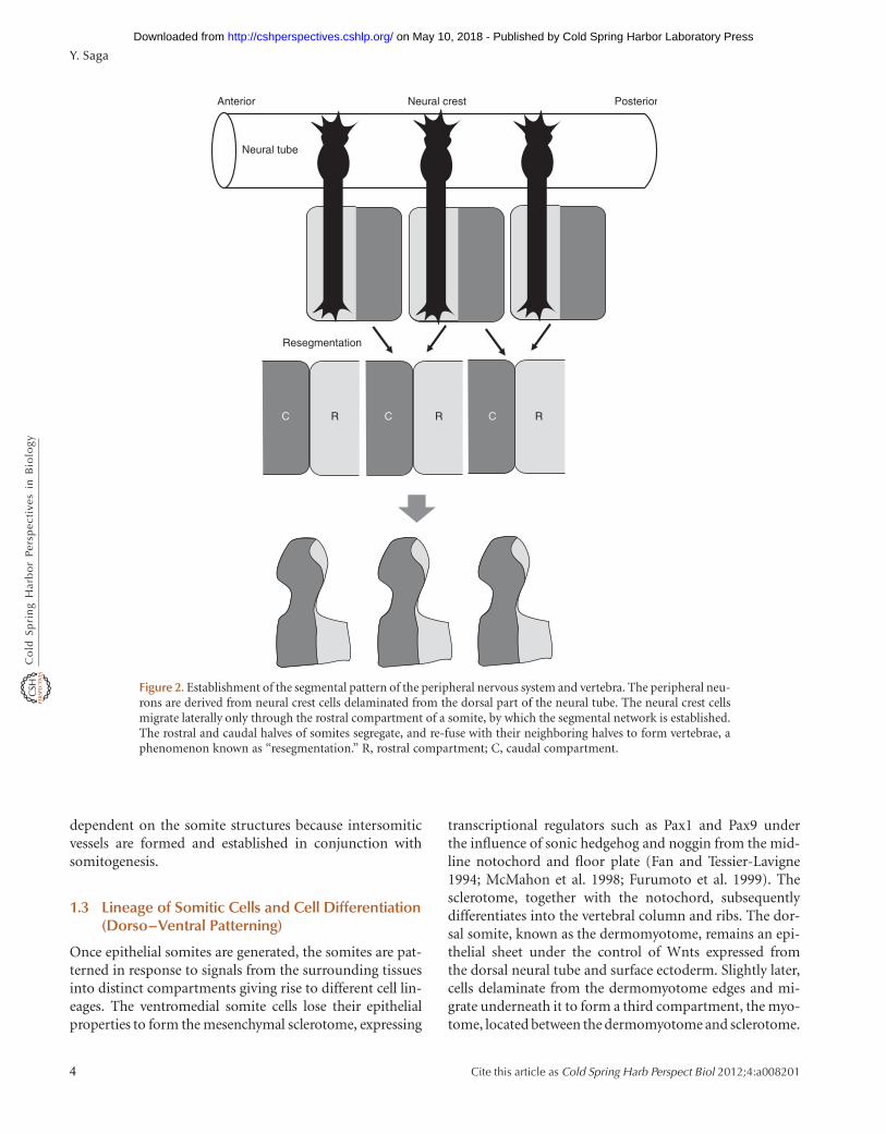
The paraxial mesoderm derived from the tail bud showsmesenchymal and immature characteristics as comparedwith the mature somite, and is thus referred to as thePSM. The tail bud provides a stem cell-like population sup-plying emerging new cells at the posterior end of the PSM,while cells are pushed anteriorly and finally bud off as asomite from the anterior part of the PSM. This is accompa-nied by a mesenchymal-epithelial transformation. The so-mites therefore consist of spherical epithelial cell massesthat differentiate into dermis, muscle, and skeleton accord-ing to their position within the somite (Fig. 1B). The moststriking feature of somite formation is the periodic gener-ation of metameric structures. The human body is com-posed of segmental units, although this is not obviousfrom its external structure. Internally, these are easily recog-nizable. The most prominent structures in this regard arethe vertebra and ribs, which are directly derived from so-mites. The nervous system and vasculature networks alsoexist in tandem and the major networks are segmental.These structures are established through the behavior ofprecursor cells. Because the neural crest cells derived fromthe dorsal part of the neural tube migrate laterally throughthe rostral compartment of the somite only and never enterthe caudal half, the metameric arrangement of the periph-eral nervous system is necessarily determined by the meta-meric nature and arrangement of the somites (Bronner-Fraser 1986; Teillet et al. 1987; Keynes and Stern 1988).This differential cell behavior reflects the different gene ex-pression patterns between the rostral and caudal halves ofthe somites (Fig. 2). The initial vascular network is also
Y. Saga
2 Cite this article as Cold Spring Harb Perspect Biol 2012;4:a008201
on May 10, 2018 - Published by Cold Spring Harbor Laboratory Press http://cshperspectives.cshlp.org/Downloaded from

Segmented somites Presomitic mesoderm (PSM) Tail bud
Tbx6Wnt3a
FGF
Sox2
Epaxial
Hypaxial
Dermomyotome
Myotome
Sclerotome
Notochord
Neural tube
Dermomyotome
Sclerotome
Myotome
A
B
C
Paraxial mesoderm
Neural tube
Paraxial mesoderm
Sox2
Figure 1. General description of somitogenesis. (A) A schematic representation of somitogenesis. The tail bud con-tains a stem cell population that gives rise to either the paraxial mesoderm or neural tube compartment. Wnt3a andTbx6 promote paraxial mesoderm formation, whereas cells expressing Sox2 choose a neural fate. Paraxial mesoder-mal cells maintain their immature state before the segmental border is formed and are referred to as the presomiticmesoderm (PSM). Cells located at the anteriormost part of the PSM are ready to form the next somite that is sepa-rated into two compartments with rostral and caudal properties. (B) After budding off from the PSM, somites start todifferentiate into dermomyotome and sclerotome according to the signals along the dorsal–ventral axis. (C) Differ-ent myotomal compartments derived from different regions of the dermomyotome contribute to different parts ofthe body muscle. The major signals come from the neural tube, notochord, and epidermis.
Somitogenesis
Cite this article as Cold Spring Harb Perspect Biol 2012;4:a008201 3
on May 10, 2018 - Published by Cold Spring Harbor Laboratory Press http://cshperspectives.cshlp.org/Downloaded from
dependent on the somite structures because intersomiticvessels are formed and established in conjunction withsomitogenesis.
1.3 Lineage of Somitic Cells and Cell Differentiation(Dorso–Ventral Patterning)
Once epithelial somites are generated, the somites are pat-terned in response to signals from the surrounding tissuesinto distinct compartments giving rise to different cell lin-eages. The ventromedial somite cells lose their epithelialproperties to form the mesenchymal sclerotome, expressing
transcriptional regulators such as Pax1 and Pax9 underthe influence of sonic hedgehog and noggin from the mid-line notochord and floor plate (Fan and Tessier-Lavigne1994; McMahon et al. 1998; Furumoto et al. 1999). Thesclerotome, together with the notochord, subsequentlydifferentiates into the vertebral column and ribs. The dor-sal somite, known as the dermomyotome, remains an epi-thelial sheet under the control of Wnts expressed fromthe dorsal neural tube and surface ectoderm. Slightly later,cells delaminate from the dermomyotome edges and mi-grate underneath it to form a third compartment, the myo-tome, located between the dermomyotome and sclerotome.
Resegmentation
Neural crest
Neural tube
Anterior Posterior
R RC CC R
Figure 2. Establishment of the segmental pattern of the peripheral nervous system and vertebra. The peripheral neu-rons are derived from neural crest cells delaminated from the dorsal part of the neural tube. The neural crest cellsmigrate laterally only through the rostral compartment of a somite, by which the segmental network is established.The rostral and caudal halves of somites segregate, and re-fuse with their neighboring halves to form vertebrae, aphenomenon known as “resegmentation.” R, rostral compartment; C, caudal compartment.
Y. Saga
4 Cite this article as Cold Spring Harb Perspect Biol 2012;4:a008201
on May 10, 2018 - Published by Cold Spring Harbor Laboratory Press http://cshperspectives.cshlp.org/Downloaded from
As the somite matures, the somitic compartments arefurther divided into subdomains with unique fates (Fig.1C). At the dorsomedial edge or lip (DML) of the dermo-myotome, cells migrate underneath to generate the epaxialmyotome, which then differentiates rapidly into backmuscle. Central dermomyotome cells de-epithelialize toform the dorsal dermis, and at the limb bud levels, cellsdelaminate from the ventrolateral lip (VLL) of the dermo-myotome to migrate into the lateral plate mesoderm, wherethey develop into limb and limb girdle muscle. At the inter-limb levels, the cells from the VLL of the dermomyotometranslocate underneath, producing the hypaxial myotome.The ventrolateral dermomyotome and hypaxial myotomeinvade the lateral plate mesoderm together as a somiticbud, from which the body wall and abdominal muscleemerge. Finally, within the sclerotome, the ventromedialcells give rise to the vertebral bodies, intervertebral discsand proximal ribs; the lateral cells, to the neural archesand distal ribs; and the dorsomedial cells, to the spinousprocesses (Brand-Saberi and Christ 2000; Brent and Tabin2002).
1.4 Regionalization along the AP Axis
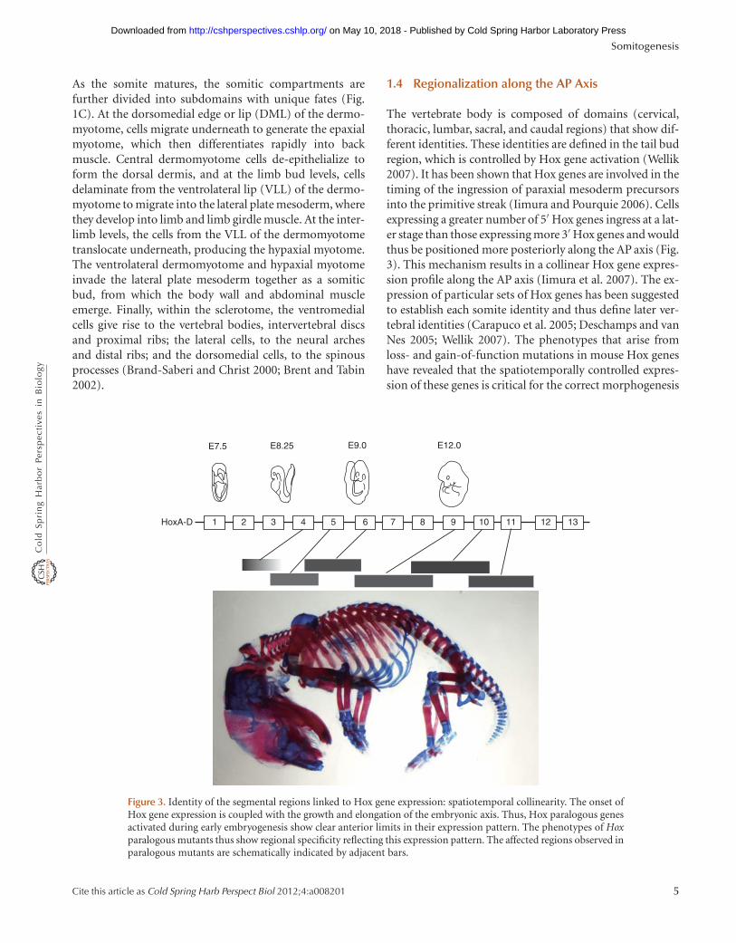
The vertebrate body is composed of domains (cervical,thoracic, lumbar, sacral, and caudal regions) that show dif-ferent identities. These identities are defined in the tail budregion, which is controlled by Hox gene activation (Wellik2007). It has been shown that Hox genes are involved in thetiming of the ingression of paraxial mesoderm precursorsinto the primitive streak (Iimura and Pourquie 2006). Cellsexpressing a greater number of 5′ Hox genes ingress at a lat-er stage than those expressing more 3′ Hox genes and wouldthus be positioned more posteriorly along the AP axis (Fig.3). This mechanism results in a collinear Hox gene expres-sion profile along the AP axis (Iimura et al. 2007). The ex-pression of particular sets of Hox genes has been suggestedto establish each somite identity and thus define later ver-tebral identities (Carapuco et al. 2005; Deschamps and vanNes 2005; Wellik 2007). The phenotypes that arise fromloss- and gain-of-function mutations in mouse Hox geneshave revealed that the spatiotemporally controlled expres-sion of these genes is critical for the correct morphogenesis
HoxA-D 1 2 3 4 6 7 8 9 10 11 12 135
E7.5 E8.25 E9.0 E12.0
Figure 3. Identity of the segmental regions linked to Hox gene expression: spatiotemporal collinearity. The onset ofHox gene expression is coupled with the growth and elongation of the embryonic axis. Thus, Hox paralogous genesactivated during early embryogenesis show clear anterior limits in their expression pattern. The phenotypes of Hoxparalogous mutants thus show regional specificity reflecting this expression pattern. The affected regions observed inparalogous mutants are schematically indicated by adjacent bars.
Somitogenesis
Cite this article as Cold Spring Harb Perspect Biol 2012;4:a008201 5
on May 10, 2018 - Published by Cold Spring Harbor Laboratory Press http://cshperspectives.cshlp.org/Downloaded from
of embryonic axial structures. For example, simultaneousinactivation of all three Hox paralogous group 10 genes re-sults in mice in which the prospective lumbo-sacral regionacquires thoraciclike characteristics whereby these mutantvertebrae display associated ribs all along the thoracolum-bar region (Wellik and Capecchi 2003).
2 THE TEMPORAL CONTROL OF SOMITOGENESISVIA THE SEGMENTATION CLOCK
2.1 The Clock and Wavefront Model(Classical Model)
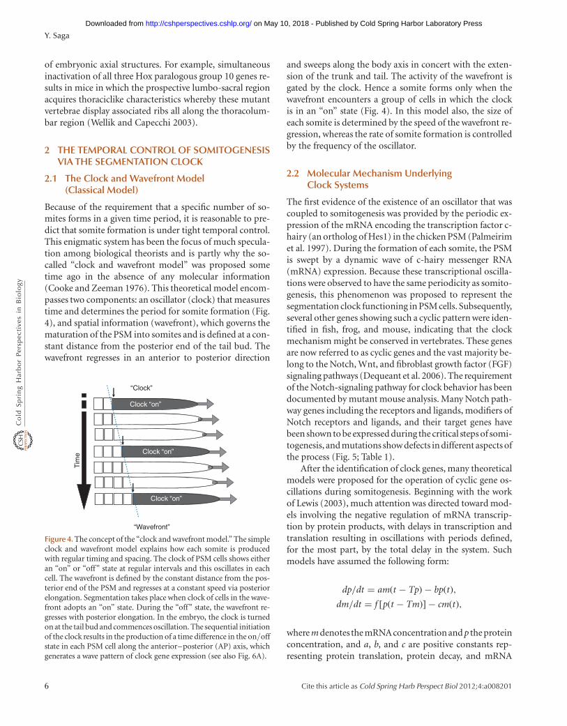
Because of the requirement that a specific number of so-mites forms in a given time period, it is reasonable to pre-dict that somite formation is under tight temporal control.This enigmatic system has been the focus of much specula-tion among biological theorists and is partly why the so-called “clock and wavefront model” was proposed sometime ago in the absence of any molecular information(Cooke and Zeeman 1976). This theoretical model encom-passes two components: an oscillator (clock) that measurestime and determines the period for somite formation (Fig.4), and spatial information (wavefront), which governs thematuration of the PSM into somites and is defined at a con-stant distance from the posterior end of the tail bud. Thewavefront regresses in an anterior to posterior direction
and sweeps along the body axis in concert with the exten-sion of the trunk and tail. The activity of the wavefront isgated by the clock. Hence a somite forms only when thewavefront encounters a group of cells in which the clockis in an “on” state (Fig. 4). In this model also, the size ofeach somite is determined by the speed of the wavefront re-gression, whereas the rate of somite formation is controlledby the frequency of the oscillator.
2.2 Molecular Mechanism UnderlyingClock Systems
The first evidence of the existence of an oscillator that wascoupled to somitogenesis was provided by the periodic ex-pression of the mRNA encoding the transcription factor c-hairy (an ortholog of Hes1) in the chicken PSM (Palmeirimet al. 1997). During the formation of each somite, the PSMis swept by a dynamic wave of c-hairy messenger RNA(mRNA) expression. Because these transcriptional oscilla-tions were observed to have the same periodicity as somito-genesis, this phenomenon was proposed to represent thesegmentation clock functioning in PSM cells. Subsequently,several other genes showing such a cyclic pattern were iden-tified in fish, frog, and mouse, indicating that the clockmechanism might be conserved in vertebrates. These genesare now referred to as cyclic genes and the vast majority be-long to the Notch, Wnt, and fibroblast growth factor (FGF)signaling pathways (Dequeant et al. 2006). The requirementof the Notch-signaling pathway for clock behavior has beendocumented by mutant mouse analysis. Many Notch path-way genes including the receptors and ligands, modifiers ofNotch receptors and ligands, and their target genes havebeen shown to be expressed during the critical steps of somi-togenesis, and mutations show defects in different aspects ofthe process (Fig. 5; Table 1).
After the identification of clock genes, many theoreticalmodels were proposed for the operation of cyclic gene os-cillations during somitogenesis. Beginning with the workof Lewis (2003), much attention was directed toward mod-els involving the negative regulation of mRNA transcrip-tion by protein products, with delays in transcription andtranslation resulting in oscillations with periods defined,for the most part, by the total delay in the system. Suchmodels have assumed the following form:
dp/dt = am(t − Tp) − bp t( ),dm/dt = f [p t − Tm)( ] − cm t( ),
where m denotes the mRNAconcentration and p the proteinconcentration, and a, b, and c are positive constants rep-resenting protein translation, protein decay, and mRNA
Tim
e
“Wavefront”
Clock “on”
“Clock”
Clock “on”
Clock “on”
Figure 4. The concept of the “clock and wavefront model.” The simpleclock and wavefront model explains how each somite is producedwith regular timing and spacing. The clock of PSM cells shows eitheran “on” or “off” state at regular intervals and this oscillates in eachcell. The wavefront is defined by the constant distance from the pos-terior end of the PSM and regresses at a constant speed via posteriorelongation. Segmentation takes place when clock of cells in the wave-front adopts an “on” state. During the “off” state, the wavefront re-gresses with posterior elongation. In the embryo, the clock is turnedon at the tail bud and commences oscillation. The sequential initiationof the clock results in the production of a time difference in the on/offstate in each PSM cell along the anterior–posterior (AP) axis, whichgenerates a wave pattern of clock gene expression (see also Fig. 6A).
Y. Saga
6 Cite this article as Cold Spring Harb Perspect Biol 2012;4:a008201
on May 10, 2018 - Published by Cold Spring Harbor Laboratory Press http://cshperspectives.cshlp.org/Downloaded from

decay rates, respectively. Tm and Tp represent the delays inmRNA transcription and protein translation, and the func-tion f represents the negative regulation of mRNA transcrip-tion by promoter binding (Lewis 2003). From this basicmodel of Lewis, many subsequent models were devised toexplain several aspects of clock oscillation and to predictmutant phenotypes. Thus far, the Notch-signaling pathwayhas been shown to be a common mechanism used in severalmodel animals, and a number of proposed models of howthis mechanism works in zebrafish and mouse are outlinedin the following sections.
2.2.1 Clock System in Zebrafish
The cyclic genes identified so far belong to the Notch path-way, and are the Notch downstream targets her1 (hairy andenhancer of split-related 1), her7, her11, her12, and her15(which are homologous to chicken HES1), and also includethe Notch ligand DeltaC (Holley et al. 2000; Henry et al.2002; Oates and Ho 2002). A simple oscillator model hasbeen proposed to explain the oscillation mechanism. Themodel indicates that oscillations are generated by a nega-tive feedback loop in which the Her1/7 genes are directly
Pofut1
Dll3
RBP-jk
MamL1/2
Hes7
Hes7
Notch1
ER-Golgi
Nucleus
Psen1/2
L-fng
L-fng
L-fng
Figure 5. Schematic depiction of the localization and function of the Notch-signaling pathway gene products in-volved in mouse somitogenesis. Once Notch receptor interacts with the Dll1 expressed in neighboring cells, it iscleaved by Psen1/2. The cleaved Notch intracellular domain is known as active Notch and enters the nucleus to in-teract with RBP-jk and MamL1 and thereby activate downstream target genes such as Hes7 and L-fringe. Hes7 is atranscriptional repressor and regulates its own and L-Fng transcription. The extracellular domain of Notch receptoris modified by two glycosyltransferases, Pofut1 and L-fng, in the ER-Golgi pathway before being exported to the cellmembrane. Dll3 is a member of the Notch ligand family, DSL. However, it may function in the cytoplasm, most no-tably in the Golgi.
Somitogenesis
Cite this article as Cold Spring Harb Perspect Biol 2012;4:a008201 7
on May 10, 2018 - Published by Cold Spring Harbor Laboratory Press http://cshperspectives.cshlp.org/Downloaded from
repressed by their own protein products. In addition, themodel takes into account a defined time delay from the be-ginning of transcription of the Her gene until the proteinproduct binds to the Her gene promoter (Lewis 2003).This Her1/7 intracellular oscillator was proposed to belinked to an intercellular oscillator involving the Notch-signaling pathway (Horikawa et al. 2006). Her1/7 nega-tively regulate DeltaC, thus potentially triggering oscilla-tions of this Notch ligand that should, in turn, result inperiodic Notch activation in neighboring cells. This cou-pling provides a basis for maintaining synchrony (see be-low). The Her1–Her7 oscillations require the function ofHer13.2 that is regulated by FGF signaling (Kawamuraet al. 2005). Her13.2 can form a heterodimer with Her1, en-hancing the ability of Her1 to negatively regulate its ownpromoter. Thus, although the Her1–Her7 negative feed-back loop constitutes the core of the segmentation clock,other signaling pathways may be additionally involved.
2.2.2 Clock System in Mice
The first cyclic genes identified in mice are also targets ofthe Notch-signaling pathway, such as Hes7 and L-fng (Bes-sho et al. 2001a; Cole et al. 2002). Furthermore, periodic ac-tivation of Notch signaling was detected in the mouse PSMusing antibodies raised against the cleaved intracellular do-main of the Notch1 receptor, providing direct evidence forthe rhythmic activation of the Notch-signaling pathway(Morimoto et al. 2005). As a mechanism to generate geneexpression oscillation, a negative feedback mechanism sim-ilar to that operating in zebrafish has also been proposed inmice. Hes7, a transcriptional repressor, is initially activatedby FGF signaling in the tail bud and then comes under thecontrol of Notch signaling (Fig. 6A) (Niwa et al. 2007).Transcription of the Hes7 gene and accumulation of Hes7protein occur in a mutually exclusive manner, indicatingthat Hes7 protein accumulation is substantially delayed
relative to Hes7 gene transcription (Fig. 6B,C). Inactivationof Hes7 in mutant mice results in an up-regulation of theHes7 transcription because of the lack of the Hes7 repres-sive activity (Bessho et al. 2003). Furthermore, stabilizationof Hes7 protein in vivo disrupts Notch-signaling oscilla-tions (Hirata et al. 2004). This negative feedback-mediatedoscillatory expression has been mathematically simulatedusing differential equations. These models have predictedthat a delay from transcription to protein expression is re-quired to achieve negative autoregulation. The importanceof the processing pathway involved in the transcription andsplicing of intronic sequences was recently tested by gener-ating mice carrying the Hes7 locus from which the intronshad been removed (Takashima et al. 2011). Hes7 expressiondid not oscillate in these mice but was steady, leading tosevere segmentation defects. These results indicate that in-trons are indeed required for Hes7 oscillations and high-light the significance of intronic delays in dynamic geneexpression (Fig. 6B).
In mice, Hes7 also suppresses another component ofthe Notch pathway, a gene known as lunatic fringe (L-fng)(Bessho et al. 2003). L-fng is a glycosyltransferase thatcan modify the extracellular domain of Notch receptorsand has been shown to repress Notch activation in boththe chick and mouse (Dale et al. 2003; Morimoto et al.2005). This may elicit Notch activity oscillation within am-niotes. However, L-fng oscillation is not observed in zebra-fish somitogenesis. The dynamic oscillations of clock genesare visualized in vivo using several reporter activities underthe control of Hes1, Hes7, and L-fng promoter or gene cas-settes (Masamizu et al. 2006; Aulehla et al. 2008). Therereporters faithfully reproduced oscillating expression pat-terns of these genes in the PSM, which confirmed the pe-riodicity of the clock system. Intriguingly, and unlike ze-brafish, some of the target genes of FGF and Wnt signal-ing pathways also show periodic activity in the mousePSM, although these signals themselves show a noncyclic
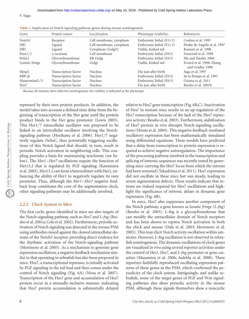
Table 1. Implication of Notch-signaling pathway genes during mouse somitogenesis
Gene Protein nature Localization Phenotype (viability) References
Notch1 Receptor Cell membrane, cytoplasm Embryonic lethal (E11.5) Conlon et al. 1995Dll1 Ligand Cell membrane, cytoplasm Embryonic lethal (E11.5) Hrabe de Angelis et al. 1997Dll3 Ligand Cytoplasm (Golgi?) Viable, kinked tail Kusumi et al. 1998Psen1/2 g secretase Cell membrane Embryonic lethal (E9.5) Donoviel et al. 1999Pofut1 Glycosyltransferase ER-Golgi Embryonic lethal (E9.5) Shi and Stanley 2006Lunatic fringe Glycosyltransferase Golgi Viable, kinked tail Evrard et al. 1998; Zhang
and Gridley 1998Mesp2 Transcription factor Nucleus Die just after birth Saga et al. 1997RBP-jk Transcription factor Nucleus Embryonic lethal (E9.5) de la Pompa et al. 1997Mastermind1/3 Transcription factor Nucleus Embryonic lethal (E9.5) Oyama et al. 2011Hes7 Transcription factor Nucleus Die just after birth Bessho et al. 2001b
Because all mutants show defective somitogenesis, the viability is indicated as the phenotype.
Y. Saga
8 Cite this article as Cold Spring Harb Perspect Biol 2012;4:a008201
on May 10, 2018 - Published by Cold Spring Harbor Laboratory Press http://cshperspectives.cshlp.org/Downloaded from

Delta
Notch
Hes7
AAAAmG
Hes7 gene
Primary transcript
Spliced transcript AAAAmG
Transcription
Splicing
Translation
Delay
Delay
Delay
AAAA
Delay
A
B
C
Nucleus
Cytoplasm
FGF-dependent
Notch-dependent
NICD Hes7 mRNA Hes7 protein
Time
Exp
ress
ion
leve
l
Figure 6. The mechanism generating the Hes7 clock gene oscillation wave during somitogenesis. (A) The expressionpattern of Hes7 at different stages is shown. This expression is activated in the tail bud by an FGF signal but once ini-tiated comes under the control of Notch signaling. The expression levels in individual cells serve as segmentationclocks and the states are synchronized with the neighbors via as yet unknown mechanisms. (B) The primary tran-scripts of Hes7 are processed in the nucleus and transported to the cytoplasm for translation. The delay in producingmature transcript and protein and the stability of this protein are strictly regulated. (C) Thus the time delay is animportant factor in the appropriate operation of the clock during somitogenesis.
Somitogenesis
Cite this article as Cold Spring Harb Perspect Biol 2012;4:a008201 9
on May 10, 2018 - Published by Cold Spring Harbor Laboratory Press http://cshperspectives.cshlp.org/Downloaded from

gradient along the AP axis (Dequeant et al. 2006; Dequeantand Pourquie 2008). The significance of oscillation of thesesignaling pathways remains elusive. In addition, the syn-chronization mechanism in amniotes has not yet been clari-fied (see below).
2.3 Synchronized Oscillation
The oscillations of clock genes are regulated by the segmen-tation clock and the underlying molecular mechanism iswell established. However, individual PSM cells will behaveas noisy autonomous oscillators with a range of differentperiods if the embryo is dissociated and the gene expressionis monitored in individual cells. Even within the embryo,the oscillation phase of individual oscillators must fluctu-ate owing to developmental noise imposed by stochasticgene expression and active cell proliferation. Hence, theremust be a mechanism that synchronizes the oscillation ofactive genes in the embryo, by which clear waves of geneexpression sweep along the AP axis. How this collective be-havior is established has been a key question in furtheringour understanding of segmentation clocks. It is now knownin zebrafish that the segmentation clock behaves as acoupled oscillator, via Notch-dependent intercellular com-munication (Jiang et al. 2000; Horikawa et al. 2006). Be-cause DeltaC expression is also under the control of theHer1/7 transcriptional repressor and also oscillates, indi-vidual cells can communicate with neighboring cells bytransmitting Notch signals, which enable a coupling ofthe oscillations in individual cells. This coupling processplays a crucial role in minimizing the effects of develop-mental noise to maintain coherent oscillation. In addition,it has been shown that Delta-Notch coupling influences thecollective period of the segmentation clock because disrup-tion of this mechanism results in an extended period of ze-brafish somitogenesis. It has thus been suggested thatcollective control of this period via delayed coupling maybe a general feature of biological clocks. However, themechanism by which synchronized oscillation is regulatedin the mouse PSM remains to be elucidated.
3 TRANSLATION OF CLOCK INFORMATION
As predicted by the clock and wavefront model, the clock(temporal) information must be translated into spatial in-formation to generate a morphologically distinct somite. Inzebrafish embryos, it has been suggested that the functionof Notch signaling is only to synchronize the oscillationsamong PSM cells, and that this pathway has no other func-tion during segmentation (Horikawa et al. 2006; Ozbudakand Pourquie 2008). However, Notch acts as an impor-tant output signal of the segmentation clock in mouse
somitogenesis. In mice, the Notch signal wave travels in aposterior to anterior direction and stops once it reachesthe wavefront where FGF and Wnt signals are below athreshold level (Oginuma et al. 2008). As a result, a segmen-tation program is initiated through the activation of a geneencoding the transcription factor Mesp2, the expression ofwhich oscillates in the anterior PSM as a response to thesegmentation clock (Morimoto et al. 2005).
3.1 Translating Oscillations into Segmentsin the Wavefront
The mechanism involved in the transcriptional activationof Mesp2 is directly involved in the mechanism by whichtemporal clock information is translated into spatial pat-terning. The analyses of factors involved in the dynamicchanges to the Mesp2 expression pattern have revealedthat the timing of Mesp2 activation depends on Notch activ-ity, whereas the positioning of the expression domain is de-fined by both Tbx6 and FGF signaling (Yasuhiko et al. 2006;Oginuma et al. 2008). The anterior border of the Mesp2 ex-pression domain accords with the anterior border of Tbx6,where Mesp2 transcription is triggered on Notch activation.However, once Mesp2 is activated and translated into itsprotein product, it destabilizes Tbx6 protein expression(Fig. 7B). The downstream target genes Ripply1/2 may beresponsible for the degradation of Tbx6 protein becauseknocking out these genes results in the anterior expansionof Tbx6 (Takahashi et al. 2010). Thus, once Mesp2 is ex-pressed, the next anterior border of Tbx6 expression is de-fined, which becomes the next somite border. Mesp2 isexpressed as a stripe in the anterior part of the Tbx6 expres-sion domain of about one somite in length. The questionthen arises as to how the posterior border of Mesp2 expres-sion is defined. Intriguingly, the posterior limit accordswith the limit of the FGF signaling gradient, which showsits highest concentration at the tail bud. In the absence ofFGF signaling, the Mesp2 expression domain shifts intothe posterior PSM, indicating that Mesp2 expression is sup-pressed in the posterior PSM via FGF. In addition, Wnt sig-naling may also work in conjunction with FGF to define theMesp2 expression domain. In a mouse embryo that showsconstitutive Wnt activity (activated b-catenin), the PSMregion is extended anteriorly and the oscillation of clockgenes such as L-fng persists for longer in the extendedPSM, which is accompanied by the additional formationof Mesp2 stripes (Aulehla et al. 2008).
The evidence shows therefore that both FGF and Wntare factors responsible for defining the maturation wave-front that facilitates a proper segmentation process. How-ever, the molecular identity of the wavefront has long beena controversial issue. Initially, Fgf8 was proposed to encode
Y. Saga
10 Cite this article as Cold Spring Harb Perspect Biol 2012;4:a008201
on May 10, 2018 - Published by Cold Spring Harbor Laboratory Press http://cshperspectives.cshlp.org/Downloaded from

wavefront activity because experimental manipulation ofFGF8 levels caused corresponding shifts in the position ofthe determination front in cultured chick and zebrafishembryos. However, mouse embryos lacking Fgf8 in thePSM still undergo somitogenesis (Perantoni et al. 2005).Mice homozygous for null mutations in other FGF ligandgenes (Fgf3, Fgf5, Fgf15, Fgf17, and Fgf18) also show no
early somitogenesis defects, or die before somitogenesis in-itiates (Fgf4) (Itoh and Ornitz 2008). Wnt has also beenproposed to contribute to wavefront activity, because themanipulation of canonical Wnt signaling also causes corre-sponding shifts in the determination front (Aulehla et al.2003; Aulehla et al. 2008). However, in Wnt loss-of-func-tion embryos, FGF signaling is also affected. Thus, the
FGFFGF
Mesp2
Tbx6
NICD
Notch
NICD
FGF
Mesp2 Tbx6
NICD
FGF
Mesp2Tbx6
Tbx6
FGF
Mesp2
Tbx6
NICD
Notch
NICD
NICD
NICD
NICDL-Fng
Mesp2 Hes7Hes7
Mesp2
Somite boundary
Presumptivesomite boundary
Tbx6
Tbx6
A
B
FGF
Tim
e
Figure 7. The mechanism of segment border formation. (A) A summary of the clock regulation leading to Mesp2activation on Notch signaling. (B) A detailed depiction of the interplay between Mesp2 and Tbx6, which is requiredfor segmental border formation. Mesp2 is activated in cells expressing Tbx6 and receiving a high level of Notch-signaling activity in the absence of FGF signaling (spanning one somite length). Once Mesp2 protein is produced,Tbx6 protein is quickly degraded and a new Tbx6 anterior border is thereby established. Because Notch intracellulardomain (NICD) signals move anteriorly, a Mesp2 expression gradient will be generated and its protein expressiondomain ultimately restricted to the rostral compartment of a somite. Mesp2 also has the ability to repress the Notch-signaling pathway and rostral–caudal polarity is established via the rostrally restricted function of Mesp2.
Somitogenesis
Cite this article as Cold Spring Harb Perspect Biol 2012;4:a008201 11
on May 10, 2018 - Published by Cold Spring Harbor Laboratory Press http://cshperspectives.cshlp.org/Downloaded from

observed somitogenesis defects may be primarily owing tothe loss of Wnt signaling or to secondary effects on FGF sig-naling. To better understand the actual role of FGF signal-ing in somitogenesis, a conditional strategy was used toinactivate both Fgf4 and Fgf8 in the PSM. This resulted indramatic shifts in the determination front and the prema-ture differentiation of the PSM (Naiche et al. 2011). More-over, the restoration of Wnt signaling to these mutants didnot restore the determination front, demonstrating thatthese two FGFs constitute the proposed wavefront activitythat maintains the PSM in an undifferentiated state.
3.2 The Mechanism of SegmentalBorder Formation
The segmental border is positioned through the coopera-tive function of Tbx6 and Mesp2. However, the nature ofhow the morphological segregation of the somite is regu-lated was a crucial question that remained. The anteriorborder of the segmented somite is perfectly matched withthe Mesp2 expression domain. Several target genes ofMesp2 have now been identified, including L-fng, EphA4,and Ripply2 (Morimoto et al. 2005; Nakajima et al. 2006;Morimoto et al. 2007). The transcription of L-fng is underthe strict control of Notch signaling in the posterior PSM,but subsequently comes under the control of Mesp2 inthe anterior PSM (Morimoto et al. 2005). Because Notchactivity can be repressed by L-fng function, it was reason-able to predict that L-fng represses Notch activity in theMesp2-expressing domain and forms a boundary for thenext presumptive somite. This idea was challenged, how-ever, by the impact on somite boundary formation by theuniform expression of NICD in the entire PSM and somiticregion (Feller et al. 2008). These results indicated that so-mite boundaries are generated even in the absence of anNICD border, although they are irregularly shaped and ofvariable size, and the resulting somites are completely cau-dalized. This result is further supported by the results of res-cue experiments using transgenic mice that express L-fngunder the control of Hes7 in the absence of endogenousL-fng expression (L-fng-null mouse) in which the NICDboundary was not generated in the presumptive segmentalborder (Oginuma et al. 2010). Nevertheless, the segmentedsomites were found to be normally generated, indicatingthat L-fng under the control of Mesp2 is dispensable for so-mite border formation. This being the case, the identity ofthe event that is actually required for segmental borderformation downstream from Mesp2 was a key issue to be re-solved. One candidate protein is EphA4, a transmembraneprotein kinase, implicated in the formation of the repulsiveresponse between cells via its interaction with ephrin ex-pressed in juxtaposed cells (Fig. 8). Although loss-of-
function experiments in the mouse have thus far shownno direct evidence for positive involvement of any particu-lar Eph or ephrin molecule in segmental border formation,the ephrin reverse signaling pathway that activates on EphAbinding was implicated in this process in chick (Watanabeet al. 2009).
3.3 The Mechanism of Somite Patterning(Rostro–Caudal Polarity)
One of major features of the segmented somite is its com-partmentalization into rostral and caudal parts, which isaccompanied by differential gene expression patterns andlineage restriction on differentiation. This compartmental-ization is required for the segmental patterning of spinalcord development because the somite compartment con-tributes to the pathway selection of neural crest cells. Howthis intrasomitic patterning is established during somito-genesis then became an important question. It is now clearthat Notch-signaling activity is a required determinant ofthe caudal identity of the somite and underlies why em-bryos that ectopically express active Notch in the entirePSM show completely caudalized somite properties (Felleret al. 2008). A similar phenotype is also observed in Mesp2-null embryos (Saga et al. 1997). Based on the fact that Mesp2expression is finally restricted to the rostral compartment ofa somite, it appears that Mesp2 suppresses Notch activity inthe rostral compartment (Takahashi et al. 2000). Initially itwas thought that L-fng induced by Mesp2 is involved in thesuppression of NICD formation in the rostral compart-ment. However, as mentioned above, the functional signifi-cance of L-fng in this regard has been discounted. Recently,the instructive role of Mesp2 in the suppression of Notchsignaling was revealed in analyses of a knockin mouse con-taining a dominant-negative form of RBP-jk, the mediatorof canonical Notch signaling in the Mesp2 locus (Sasakiet al. 2011). The resulting phenotype was quite surprisingbecause almost all of the defects observed in the Mesp2-nullembryo were completely rescued in the DN-RBP-jk knock-in mouse. A subsequent study indicated that Mesp2 sup-presses Notch activity by destabilizing Mastermind, oneof the core components of the nuclear NICD complex(Fig. 8) (Sasaki et al. 2011).
4 THE MECHANISM TO INITIATE AND TERMINATECYCLIC GENE EXPRESSION: HOW IS THESOMITE NUMBER DETERMINED?
Each vertebrate species has a defined numberof somites andso it was of central importance to determine how the somitenumber is controlled, beginning with an understandingof how the clock system is set up to start somite formation.
Y. Saga
12 Cite this article as Cold Spring Harb Perspect Biol 2012;4:a008201
on May 10, 2018 - Published by Cold Spring Harbor Laboratory Press http://cshperspectives.cshlp.org/Downloaded from

Tbx6
Mesp2
Ripply2
Groucho
MamL1
RBP-jk
L-fng
EphA4
Ripply2
Tbx6
Mesp2
Mesp2
Mesp2
Mesp2
MamL1
EphA4
L-fng
Dll1 Ephrin
PSM
S0S+1
Ros
tral
Cau
dal
RostralCaudal
Uncx4.1Dll1 Uncx4.1Dll1
RBP-J
NICDMam
CoR
Destabilization
Mesp2Mesp2
NucleusNucleus
Notch signaling ON Notch signaling OFF
Futuresegmental border
S–1
Mesp2Mesp2proteinprotein
A
B
Nucleus
Cytoplasm
Figure 8. Genetic pathways involved in the regulation of the Mesp2 pathway. (A) Mesp2 expression is activated byNotch signaling and Tbx6. Mesp2 then activates its downstream target genes such as Ripply2, EphA4, and L-fng. Rip-ply2 is involved in the suppression of Tbx6, which also leads to the suppression of Mesp2 transcription. EphA4 isimplicated in the segregation process in the segmental border. L-fng acts on the Notch receptor, resulting in the re-pression of NICD. Mesp2 also acts on Maml1 to destabilize this protein. (B) The molecular mechanism of rostral–caudal patterning. Notch activity is required to confer caudal property on a somite. In the rostral compartment,Notch activation is suppressed by a Mesp2 function that destabilizes MamL1. S + 1, somite number 1; S0, formingsomite; S 2 1, somite number 21; PSM, presomitic mesoderm, CoR, transcriptional repressor complex; Mam,mastermindlike proteins.
Somitogenesis
Cite this article as Cold Spring Harb Perspect Biol 2012;4:a008201 13
on May 10, 2018 - Published by Cold Spring Harbor Laboratory Press http://cshperspectives.cshlp.org/Downloaded from

In the primitive streak before the formation of somites,strong FGF activity and Tbx6 expression are observed inthe entire streak. On streak elongation, an FGF gradient isproduced along the AP axis. Notch signal oscillation thenappears to commence after a specifically determined peri-od. Once Notch activity oscillation becomes obvious, thefirst Mesp2 stripe emerges in a 7.5 days postcoitum (dpc)embryo (Oginuma et al. 2008). In principle, somites con-tinue to be formed as long as the PSM acts as a reservoirthat supplies new cells. Thus, the number of somites willdepend on the PSM life span during embryogenesis, de-fined by the balance between the speed of somitogenesisand the speed of axis elongation. If the speed of somitogen-esis is faster than the speed of axis elongation, then the PSMwill shrink quickly and presumably, somitogenesis will ar-rest when the PSM is finally exhausted. Conversely, if thespeed of axis elongation is faster than or equal to the speedof somitogenesis, the PSM size will theoretically increase orremain steady and an indefinite number of segments will beadded until the end. The speed of somitogenesis progres-sion therefore essentially depends on the parameters thatcontrol the regression speed of the wavefront, and henceon the rate of decay of the Wnt and FGF signals. In contrast,the speed of axis elongation depends on the proliferationrate in the tail bud, which may also depend on FGF andWnt signals, but likely involves other as yet unknown fac-tors. The mechanism by which the PSM stem cell systemis regulated during embryogenesis remains an attractive is-sue to be solved in the near future.
5 CONCLUDING REMARKS
The mechanisms leading to the formation of the somitehave long fascinated many researchers because they providean ideal model system for both mathematicians and devel-opmental biologists. Extensive analyses using mainly themouse and zebrafish model animals, and with the adoptionof mathematical modeling, we now understand how themetameric formation of somites occurs via the clock mech-anism operating in unsegmented immature PSM cells. Theconserved Notch-signaling pathway and the negative feed-back mechanism of the Hes family of transcriptional re-pressors take central roles in this process. Another factor,Mesp2, plays a crucial role in the translation of clock infor-mation into the spatial patterning of the somites. However,there are several important aspects of this system thatremain elusive. One is the significance of the oscillatingcomponents of the Wnt and FGF signaling pathways. Math-ematical models and some experimental evidence have in-dicated that these are integral parts of the clock system.However, the zebrafish appears not to use such processesto control its segmentation clock. There could be some
evolutional reasons for this. Each animal has its own clockperiod and somite numbers. Somitogenesis is an integralpart of embryogenesis and must be coupled with the timingof other developmental stages. The collective behavior ofPSM cells is a hallmark of somitogenesis. However, we stilldo not understand the molecular mechanism leading tothe synchronized oscillation that underpins mouse somito-genesis. Mouse genetics has proved to be a strong tool foruncovering the molecular events that govern embryogene-sis. However, new imaging technologies are needed in addi-tion to genetic manipulations to visualize and quantify themolecular events that underlie somitogenesis.
REFERENCES
Aulehla A, Wehrle C, Brand-Saberi B, Kemler R, Gossler A, Kanzler B,Herrmann BG. 2003. Wnt3a plays a major role in the segmentationclock controlling somitogenesis. Dev Cell 4: 395–406.
Aulehla A, Wiegraebe W, Baubet V, Wahl MB, Deng C, Taketo M, Lewan-doski M, Pourquie O. 2008. A b-catenin gradient links the clock andwavefront systems in mouse embryo segmentation. Nat Cell Biol 10:186–193.
Bessho Y, Sakata R, Komatsu S, Shiota K, Yamada S, Kageyama R. 2001a.Dynamic expression and essential functions of Hes7 in somite seg-mentation. Genes Dev 15: 2642–2647.
Bessho Y, Sakata R, Komatsu S, Shiota K, Yamada S, Kageyama R. 2001b.Dynamic expression and essential functions of Hes7 in somite seg-mentation. Genes Dev 15: 2642–2647.
Bessho Y, Hirata H, Masamizu Y, Kageyama R. 2003. Periodic repressionby the bHLH factor Hes7 is an essential mechanism for the somite seg-mentation clock. Genes Dev 17: 1451–1456.
Brand-Saberi B, Christ B. 2000. Evolution and development of distinctcell lineages derived from somites. Curr Top Dev Biol 48: 1–42.
Brent AE, Tabin CJ. 2002. Developmental regulation of somite deriva-tives: Muscle, cartilage and tendon. Curr Opin Genet Dev 12: 548–557.
Bronner-Fraser M. 1986. Analysis of the early stages of trunk neural crestmigration in avian embryos using monoclonal antibody HNK-1. DevBiol 115: 44–55.
Carapuco M, Novoa A, Bobola N, Mallo M. 2005. Hox genes specify ver-tebral types in the presomitic mesoderm. Genes Dev 19: 2116–2121.
Chapman DL, Papaioannou VE. 1998. Three neural tubes in mouse em-bryos with mutations in the T-box gene Tbx6. Nature 391: 695–697.
Cole SE, Levorse JM, Tilghman SM, Vogt TF. 2002. Clock regulatory ele-ments control cyclic expression of Lunatic fringe during somitogene-sis. Dev Cell 3: 75–84.
Conlon RA, Reaume AG, Rossant J. 1995. Notch1 is required for the co-ordinate segmentation of somites. Development 121: 1533–1545.
Cooke J, Zeeman EC. 1976. A clock and wavefront model for controlof the number of repeated structures during animal morphogenesis.J Theor Biol 58: 455–476.
Dale JK, Maroto M, Dequeant ML, Malapert P, McGrew M, Pourquie O.2003. Periodic notch inhibition by lunatic fringe underlies the chicksegmentation clock. Nature 421: 275–278.
de la Pompa JL, Wakeham A, Correia KM, Samper E, Brown S, AguileraRJ, Nakano T, Honjo T, Mak TW, Rossant J, et al. 1997. Conservation ofthe Notch signalling pathway in mammalian neurogenesis. Develop-ment 124: 1139–1148.
Dequeant ML, Pourquie O. 2008. Segmental patterning of the vertebrateembryonic axis. Nat Rev Genet 9: 370–382.
Dequeant ML, Glynn E, Gaudenz K, Wahl M, Chen J, Mushegian A,Pourquie O. 2006. A complex oscillating network of signaling genesunderlies the mouse segmentation clock. Science 314: 1595–1598.
Y. Saga
14 Cite this article as Cold Spring Harb Perspect Biol 2012;4:a008201
on May 10, 2018 - Published by Cold Spring Harbor Laboratory Press http://cshperspectives.cshlp.org/Downloaded from

Deschamps J, van Nes J. 2005. Developmental regulation of the Hoxgenes during axial morphogenesis in the mouse. Development 132:2931–2942.
Donoviel DB, Hadjantonakis AK, Ikeda M, Zheng H, Hyslop PS, Bern-stein A. 1999. Mice lacking both presenilin genes exhibit early embry-onic patterning defects. Genes Dev 13: 2801–2810.
Evrard YA, Lun Y, Aulehla A, Gan L, Johnson RL. 1998. Lunatic fringe isan essential mediator of somite segmentation and patterning. Nature394: 377–381.
Fan CM, Tessier-Lavigne M. 1994. Patterning of mammalian somites bysurface ectoderm and notochord: Evidence for sclerotome inductionby a hedgehog homolog. Cell 79: 1175–1186.
Feller J, Schneider A, Schuster-Gossler K, Gossler A. 2008. NoncyclicNotch activity in the presomitic mesoderm demonstrates uncouplingof somite compartmentalization and boundary formation. Genes Dev22: 2166–2171.
Furumoto TA, Miura N, Akasaka T, Mizutani-Koseki Y, Sudo H, FukudaK, Maekawa M, Yuasa S, Fu Y, Moriya H, et al. 1999. Notochord-dependent expression of MFH1 and PAX1 cooperates to maintainthe proliferation of sclerotome cells during the vertebral column devel-opment. Dev Biol 210: 15–29.
Goldstein RS, Kalcheim C. 1992. Determination of epithelial half-somites in skeletal morphogenesis. Development 116: 441–445.
Gomez C, Pourquie O. 2009. Developmental control of segment num-bers in vertebrates. J Exp Zool B Mol Dev Evol 312: 533–544.
Gomez C, Ozbudak EM, Wunderlich J, Baumann D, Lewis J, Pourquie O.2008. Control of segment number in vertebrate embryos. Nature 454:335–339.
Henry CA, Urban MK, Dill KK, Merlie JP, Page MF, Kimmel CB, AmacherSL. 2002. Two linked hairy/Enhancer of split-related zebrafish genes,her1 and her7, function together to refine alternating somite bounda-ries. Development 129: 3693–3704.
Hirata H, Bessho Y, Kokubu H, Masamizu Y, Yamada S, Lewis J, Kageya-ma R. 2004. Instability of Hes7 protein is crucial for the somite seg-mentation clock. Nat Genet 36: 750–754.
Holley SA, Geisler R, Nusslein-Volhard C. 2000. Control of her1 expres-sion during zebrafish somitogenesis by a delta-dependent oscillatorand an independent wave-front activity. Genes Dev 14: 1678–1690.
Horikawa K, Ishimatsu K, Yoshimoto E, Kondo S, Takeda H. 2006. Noise-resistant and synchronized oscillation of the segmentation clock. Na-ture 441: 719–723.
Hrabe de Angelis M, McIntyre J III, Gossler A. 1997. Maintenance of so-mite borders in mice requires the Delta homologue DII1. Nature 386:717–721.
Iimura T, Pourquie O. 2006. Collinear activation of Hoxb genes during gas-trulation is linked to mesoderm cell ingression. Nature 442: 568–571.
Iimura T, Yang X, Weijer CJ, Pourquie O. 2007. Dual mode of paraxialmesoderm formation during chick gastrulation. Proc Natl Acad Sci104: 2744–2749.
Itoh N, Ornitz DM. 2008. Functional evolutionary history of the mouseFgf gene family. Dev Dyn 237: 18–27.
Jiang YJ, Aerne BL, Smithers L, Haddon C, Ish-Horowicz D, Lewis J.2000. Notch signalling and the synchronization of the somite segmen-tation clock. Nature 408: 475–479.
Kawamura A, Koshida S, Hijikata H, Sakaguchi T, Kondoh H, Takada S.2005. Zebrafish hairy/enhancer of split protein links FGF signaling tocyclic gene expression in the periodic segmentation of somites. GenesDev 19: 1156–1161.
Keynes RJ, Stern CD. 1988. Mechanisms of vertebrate segmentation.Development 103: 413–429.
Kusumi K, Sun ES, Kerrebrock AW, Bronson RT, Chi DC, Bulotsky MS,Spencer JB, Birren BW, Frankel WN, Lander ES. 1998. The mousepudgy mutation disrupts Delta homologue Dll3 and initiation of earlysomite boundaries. Nat Genet 19: 274–278.
Lewis J. 2003. Autoinhibition with transcriptional delay: A simple mech-anism for the zebrafish somitogenesis oscillator. Curr Biol 13: 1398–1408.
Masamizu Y, Ohtsuka T, Takashima Y, Nagahara H, Takenaka Y, Yoshika-wa K, Okamura H, Kageyama R. 2006. Real-time imaging of thesomite segmentation clock: Revelation of unstable oscillators in the in-dividual presomitic mesoderm cells. Proc Natl Acad Sci 103: 1313–1318.
McMahon JA, Takada S, Zimmerman LB, Fan CM, Harland RM, McMa-hon AP. 1998. Noggin-mediated antagonism of BMP signaling is re-quired for growth and patterning of the neural tube and somite.Genes Dev 12: 1438–1452.
Morimoto M, Takahashi Y, Endo M, Saga Y. 2005. The Mesp2 transcrip-tion factor establishes segmental borders by suppressing Notch activ-ity. Nature 435: 354–359.
Morimoto M, Sasaki N, Oginuma M, Kiso M, Igarashi K, Aizaki K, Kan-no J, Saga Y. 2007. The negative regulation of Mesp2 by mouse Ripply2is required to establish the rostro-caudal patterning within a somite.Development 134: 1561–1569.
Naiche LA, Holder N, Lewandoski M. 2011. FGF4 and FGF8 comprisethe wavefront activity that controls somitogenesis. Proc Natl AcadSci 108: 4018–4023.
Nakajima Y, Morimoto M, Takahashi Y, Koseki H, Saga Y. 2006. Identifi-cation of Epha4 enhancer required for segmental expression and theregulation by Mesp2. Development 133: 2517–2525.
Niwa Y, Masamizu Y, Liu T, Nakayama R, Deng CX, Kageyama R. 2007.The initiation and propagation of Hes7 oscillation are cooperativelyregulated by Fgf and notch signaling in the somite segmentation clock.Dev Cell 13: 298–304.
Oates AC, Ho RK. 2002. Hairy/E(spl)-related (Her) genes are centralcomponents of the segmentation oscillator and display redundancywith the Delta/Notch signaling pathway in the formation of anteriorsegmental boundaries in the zebrafish. Development 129: 2929–2946.
Oginuma M, Niwa Y, Chapman DL, Saga Y. 2008. Mesp2 and Tbx6 coop-eratively create periodic patterns coupled with the clock machineryduring mouse somitogenesis. Development 135: 2555–2562.
Oginuma M, Takahashi Y, Kitajima S, Kiso M, Kanno J, Kimura A, Saga Y.2010. The oscillation of Notch activation, but not its boundary, is re-quired for somite border formation and rostral-caudal patterningwithin a somite. Development 137: 1515–1522.
Oyama T, Harigaya K, Sasaki N, Okamura Y, Kokubo H, Saga Y, HozumiK, Suganami A, Tamura Y, Nagase T, et al. 2011. Mastermind-like 1(MamL1) and mastermind-like 3 (MamL3) are essential for Notch sig-naling in vivo. Development 138: 5235–5246.
Ozbudak EM, Pourquie O. 2008. The vertebrate segmentation clock: Thetip of the iceberg. Curr Opin Genet Dev 18: 317–323.
Palmeirim I, Henrique D, Ish-Horowicz D, Pourquie O. 1997. Avian hairygene expression identifies a molecular clock linked to vertebrate seg-mentation and somitogenesis. Cell 91: 639–648.
Perantoni AO, Timofeeva O, Naillat F, Richman C, Pajni-Underwood S,Wilson C, Vainio S, Dove LF, Lewandoski M. 2005. Inactivation ofFGF8 in early mesoderm reveals an essential role in kidney develop-ment. Development 132: 3859–3871.
Pourquie O. 2003. Vertebrate somitogenesis: A novel paradigm for ani-mal segmentation? Int J Dev Biol 47: 597–603.
Saga Y, Takeda H. 2001. The making of the somite: Molecular events invertebrate segmentation. Nat Rev Genet 2: 835–845.
Saga Y, Hata N, Koseki H, Taketo MM. 1997. Mesp2: A novel mouse geneexpressed in the presegmented mesoderm and essential for segmenta-tion initiation. Genes Dev 11: 1827–1839.
Sasaki N, Kiso M, Kitagawa M, Saga Y. 2011. The repression of Notch sig-naling occurs via the destabilization of mastermind-like 1 by Mesp2and is essential for somitogenesis. Development 138: 55–64.
Shi S, Stanley P. 2006. Evolutionary origins of Notch signaling in early de-velopment. Cell Cycle 5: 274–278.
Takahashi Y, Koizumi K, Takagi A, Kitajima S, Inoue T, Koseki H, Saga Y.2000. Mesp2 initiates somite segmentation through the Notch signal-ling pathway. Nat Genet 25: 390–396.
Takahashi J, Ohbayashi A, Oginuma M, Saito D, Mochizuki A, Saga Y, Ta-kada S. 2010. Analysis of Ripply1/2-deficient mouse embryos reveals a
Somitogenesis
Cite this article as Cold Spring Harb Perspect Biol 2012;4:a008201 15
on May 10, 2018 - Published by Cold Spring Harbor Laboratory Press http://cshperspectives.cshlp.org/Downloaded from

mechanism underlying the rostro-caudal patterning within a somite.Dev Biol 342: 134–145.
Takashima Y, Ohtsuka T, Gonzalez A, Miyachi H, Kageyama R. 2011. In-tronic delay is essential for oscillatory expression in the segmentationclock. Proc Natl Acad Sci 108: 3300–3305.
Takemoto T, Uchikawa M, Yoshida M, Bell DM, Lovell-Badge R, Pa-paioannou VE, Kondoh H. 2011. Tbx6-dependent Sox2 regulation de-termines neural or mesodermal fate in axial stem cells. Nature 470:394–398.
Teillet MA, Kalcheim C, Le Douarin NM. 1987. Formation of the dorsalroot ganglia in the avian embryo: Segmental origin and migratory be-havior of neural crest progenitor cells. Dev Biol 120: 329–347.
Watanabe T, Sato Y, Saito D, Tadokoro R, Takahashi Y. 2009. EphrinB2coordinates the formation of a morphological boundary and cell
epithelialization during somite segmentation. Proc Natl Acad Sci106: 7467–7472.
Wellik DM. 2007. Hox patterning of the vertebrate axial skeleton. DevDyn 236: 2454–2463.
Wellik DM, Capecchi MR. 2003. Hox10 and Hox11 genes are required toglobally pattern the mammalian skeleton. Science 301: 363–367.
Yamaguchi TP, Takada S, Yoshikawa Y, Wu N, McMahon AP. 1999. T(Brachyury) is a direct target of Wnt3a during paraxial mesodermspecification. Genes Dev 13: 3185–3190.
Yasuhiko Y, Haraguchi S, Kitajima S, Takahashi Y, Kanno J, Saga Y. 2006.Tbx6-mediated Notch signaling controls somite-specific Mesp2 ex-pression. Proc Natl Acad Sci 103: 3651–3656.
Zhang N, Gridley T. 1998. Defects in somite formation in lunatic fringe-deficient mice. Nature 394: 374–377.
Y. Saga
16 Cite this article as Cold Spring Harb Perspect Biol 2012;4:a008201
on May 10, 2018 - Published by Cold Spring Harbor Laboratory Press http://cshperspectives.cshlp.org/Downloaded from

2012; doi: 10.1101/cshperspect.a008201Cold Spring Harb Perspect Biol Yumiko Saga The Synchrony and Cyclicity of Developmental Events
Subject Collection Mammalian Development
Mouse EmbryoThe Dynamics of Morphogenesis in the Early
HadjantonakisJaime A. Rivera-Pérez and Anna-Katerina Development
Neural Progenitors during Mammalian Cortical Cell Division Modes and Cleavage Planes of
Fumio Matsuzaki and Atsunori ShitamukaimicroRNAs as Developmental Regulators
Kathryn N. Ivey and Deepak SrivastavaBlood and Lymphatic Vessel Formation
Victoria L. Bautch and Kathleen M. CaronDevelopment of the Endochondral Skeleton
Fanxin Long and David M. Ornitz DevelopmentTranscriptional Networks in Liver and Intestinal
Karyn L. Sheaffer and Klaus H. KaestnerAdipogenesis
Kelesha Sarjeant and Jacqueline M. StephensPluripotency in the Embryo and in Culture
Jennifer Nichols and Austin SmithMolecular Mechanisms of Inner Ear Development
Doris K. Wu and Matthew W. Kelley Development and RegenerationSignaling and Transcriptional Networks in Heart
Benoit G. BruneauPolarity in Mammalian Epithelial Morphogenesis
Julie Roignot, Xiao Peng and Keith Mostov Cell DifferentiationSignals and Switches in Mammalian Neural Crest
Shachi Bhatt, Raul Diaz and Paul A. TrainorEye Development and Retinogenesis
Whitney Heavner and Larysa PevnyHematopoiesis
Michael A. Rieger and Timm SchroederPrimordial Germ Cells in Mice
Mitinori Saitou and Masashi YamajiEmbryonic AxesEstablishing Blastocyst Cell Lineages and Intercellular Interactions, Position, and Polarity in
P.L. TamRobert O. Stephenson, Janet Rossant and Patrick
http://cshperspectives.cshlp.org/cgi/collection/ For additional articles in this collection, see
Copyright © 2012 Cold Spring Harbor Laboratory Press; all rights reserved
on May 10, 2018 - Published by Cold Spring Harbor Laboratory Press http://cshperspectives.cshlp.org/Downloaded from