pediatric blistering diseases - cdn.ymaws.com · epidermis and dermal melanophages •treatment:...
TRANSCRIPT

Pediatric Blistering DiseasesMSU/Beaumont Health Dermatology Residency Program
Matthew Laffer, DO & Dustin Portela, DO PGY-4
Chelsea Duggan, DO & Peter Jajou, DO PGY-3
Sonam Rama, DO & Rachel Cetta, DO PGY-2
Steven Grekin, DO Program Director

Disclosures
• No conflicts of interest to disclose

Overview
• Infectious Disease
• Drug Related Disorders
• Hereditary Disorders
• Autoimmune Conditions
• Miscellaneous Disorders

INFECTIOUS

Staphylococcus Scalded Skin Syndrome
• Clinical Presentation• Fever, skin tenderness and erythema in groin folds, axillary regions, nose, that
leads to a generalized superficial exfoliative dermatitis and bullae
• +Nikolsky sign. Perioral crusting and fissuring
http://medlibes.com/entry/staphylococcal-scalded-skin-syndrome

Staphylococcus Scalded Skin Syndrome
• Pathogenesis• Staphylococcus aureus group 2 phage (71) infects child at a distant site
• Exfoliative toxins/Epidermolytic toxins A and B (ET-A, ET-B) bind to desmoglein-1 superficial bullae within the granular layer (subcorneal)
• ET is a unique serine protease that shows lock and key specificity to desmoglein -11-2
• Diagnosis• Mainly clinical• Culture of the bullae will be NEGATIVE. Must culture site of initial infection
• Treatment• Supportive care: IVF!!
• Dicloxacillin

Bullous Impetigo
• Clinical Presentation• Flaccid bullae which rupture and leave honey colored crust behind
• No surrounding erythema
• Can have systemic manifestations such as malaise, diarrhea
http://www.adhb.govt.nz/newborn/TeachingResources/Dermatology/InfectiveLesions.htmhttp://dermatologyoasis.net/bullous-impetigo-2/

Bullous Impetigo
• Pathogenesis• Often secondary to Staphylococci group 2 phage type 71 at the site of the lesion
• Exfoliative toxins A and B bind to desmoglein 1 forming superficial bullae within the granular layer (subcorneal)
• Diagnosis
• Mainly clinical, but can culture site of infection
• Treatment
• Topical mupirocin
• There is no clear preference among systemic treatment: antistaphylococcalpenicillins (dicloxacillin), amoxicillin/clavulanate, cephalosporins (cephalexin), and macrolides, although resistance rates to erythromycin are rising3

Bullous Tinea
• Clinical Presentation• Tense multilocular bullae with surrounding erythema and scale often located
along the arch of the foot
• Common to see interdigital fungal involvement
• May serve as a portal of entry for superinfection and cellulitis
http://www.regionalderm.com/Regional_Derm/Tfiles/tinea_bullous.html http://www.regionalderm.com/Regional_Derm/Tfiles/tinea_bullous.html

Bullous Tinea
• Pathogenesis• Most common dermatophyte: Trichophyton mentagrophytes
• Most commonly due to chronic disease that has not been treated or incorrectly treated
• Diagnosis
• KOH scraping, fungal culture, biopsy4
• Treatment
• Topical antifungals
• If extensive, can use oral terbinafine up to 250 mg once daily for 2 weeks4

Eczema Herpeticum
• Clinical Presentation• AKA Kaposi varicelliform eruption
• Itchy, umbilicated vesiculopustules that erupt in a disseminated pattern. These often become hemorrhagic and crusted
• Seen in children with atopic dermatitis
http://www.pcds.org.uk/clinical-guidance/eczema-eczema-herpeticumhttp://healthh.com/eczema-herpeticum/

Eczema Herpeticum
• Pathogenesis • Reactivation of HSV 1 predominantly• Other viruses indicated in literature: Coxsackie A 16, vaccinia, and varicella
zoster5
• Diagnosis• Mainly clinical• Tzanck smear, viral culture, direct fluorescent antibody, biopsy
• Treatment• Acyclovir (10–15 mg/kg/day), intravenous or oral• Oral valacyclovir has a higher bioavailability than oral acyclovir5
• In acyclovir resistant cases, can use foscarnet or cidofovir

Blistering Distal Dactylitis
• Clinical Presentation• Tense vesicles filled with purulent fluid and surrounding erythema on the
dorsal surface of distal fingers and toes
• Can involve the proximal nail fold9
http://www.cram.com/flashcards/bacterial-infections-of-the-skin-5578577 http://www.liveinternet.ru/journalshowcomments.php?jpostid=45192638&journalid=1693906&go=next&categ=0

Blistering Distal Dactylitis
• Pathogenesis• Group A β-hemolytic Streptococcus, S. aureus, S. epidermidis are the
responsible organisms
• Diagnosis• Rare, but mainly a clinical diagnosis. Can culture
• Treatment• I&D
• Dicloxicillin or first generation cephalosporin (cephalexin)

Bullous Scabies
• Clinical Presentation • Erythematous papules and burrows are seen in finger and toe webspaces,
nipples, axilla, male genitalia and the umbilicus
• Very pruritic leading to excoriations
• Must be considered in ddx for diaper rash ages 3-6 mon
http://www.jaad.org/cms/attachment/2005281640/2022604688/gr1.jpg
http://www.rash-pictures.com/images/4/455.jpg

Bullous Scabies
• Pathogenesis• The adult scabies mite burrows within the stratum corneum resting in the
upper granular layer.
• Diagnosis• Mineral oil scraping from burrow
• Treatment• Permethrin, the recommended first-line agent; with two applications one
week apart
• Lindane has become unavailable due to concerns regarding neurotoxicity, particularly in pediatric populations

Varicella Virus
• Clinical Presentation• A congenital infection of the newborn
• Greatest risk at first 20 weeks gestation
• Presents as cicatricial erythematous vesicles with crust
• Chorioretinitis, cataracts, cortical atrophy, psychomotor retardation and hypoplastic limbs
http://www.6minutes.com.au/news/features/vaccination-eliminates-congenital--varicella http://indianpediatrics.net/feb2010/feb-181.htm

Varicella Virus
• Pathogenesis• Transplacental infection
• Maternal rash within 5 days before to 2 days after delivery, varicella-associated antibodies are not transferred to the baby severe rash6
• Diagnosis• Clinical
• Can get viral titer to confirm
• Treatment• Live attenuated vaccine has lowered incidence
• Acyclovir (10–15 mg/kg/day)

Herpes Simplex Virus
• Clinical Presentation• Erythematous vesicles with erosions and scarring in a localized or
disseminated manner
• Temporal encephalitis
http://rickshawmag.com/tag/congenital/ http://www.ultramag50.com/page/321/

Herpes Simplex Virus
• Pathogenesis• Results from fetus contact with active maternal genital herpes lesions during
delivery
• Majority HSV 2
• Diagnosis• Clinical
• Can swab maternal lesions prior to delivery to check for viral shedding
• Treatment• Acyclovir (10–15 mg/kg/day)
• Mothers with active HSV-2 genital lesions should have c-section

Drug Related

Stevens Johnson Syndrome & Toxic Epidermal Necrolysis
• Clinical Presentation• Patients with fever, severe mucosal/conjunctival ulcerations and occasional GI
and/or GU involvement
• Dusky erythematous or purpuric macules, papules, patches or plaques flaccid bullae with epidermal detachment and necrosis • +Nikolsky and +Asboe-Hansen seen in most cases
http://www.bestonlinemd.com/stevens-johnson-syndrome-rare-but-serious-problem/
http://emedicine.medscape.com/article/1197450-clinical

Stevens Johnson Syndrome & Toxic Epidermal Necrolysis
• Pathogenesis• Almost always drug related, rarely infection (Mycoplasma pneumoniae most
common) or immunization• Anticonvulsants (#1), antibiotics and non steroid anti-inflammatory drugs7
• Diagnosis• Clinically, eruption begins 7-21 days after initiation of drug• Bx: full epidermal thickness necrosis
• Treatment• D/C offending agent!!• Supportive care in ICU or Burn unit: IVF, wound care, electrolyte and
nutritional supplement• IVIG

Acute Generalized Exanthematous Pustulosis
• Clinical Presentation• An eruption of multiple sterile pustules with surrounding erythema,
appearing first on face and intertriginous areas, then spreading to trunk and extremities
• Edema of the face and sometimes hands
http://jamanetwork.com/journals/jamadermatology/fullarticle/423839http://www.e-ijd.org/article.asp?issn=0019-5154;year=2014;volume=59;issue=2;spage=210;epage=210;aulast=Shingade

AGEP
• Pathogenesis• Most frequently mentioned drugs antibiotics (beta lactams, macrolides); antifungal
agents (terbinafine and nystatin); anticonvulsants (carbamazepine and phenobarbital) 8
• Diagnosis• Mainly clinical • Biopsy will show spongiosis with dermal eosinophilic and neutrophilic infiltrate and a
subcorneal pustule• Part of subcorneal pustule differential diagnosis
• Treatment• Resolves in 1-2 weeks with d/c of drug• Can use topical steroids for symptomatic relief

Bullous Fixed Drug Eruption• Pathogenesis: Lesions appear 1-2 weeks after first exposure. Found
anywhere on the body. With re-administration of the drug, lesions recur at the same site.
• Clinical Presentation: Sharply circumscribed erythematous to violaceouspatches with central vesicles and bullae

Bullous Fixed Drug• Causes: Sulfa, NSAIDs, Tetracyclines,
Aspirin, Acetaminophen, Metronidazole
• Histopathology: Superficial and deep interstitial and perivascular infiltratewith lymphocytes. There may be necrotic keratinocytes in the epidermis and dermal melanophages
• Treatment: Discontinue offending agent

Hereditary Bullous Disorders

Epidermolysis Bullosa
• Four main types based on cleavage zones• Epidermolysis Bullosa Simplex
• Blistering in epidermis within basal keratinocytes
• Junctional Epidermolysis Bullosa• Blistering within laminia lucida
• Dystrophic Epidermolysis Bullosa• Blistering in the dermis/ sublamina densa
• Kindler Syndrome• Multiple cleavage planes possible
• All types characterized by skin fragility and blister formation with mechanical traction
Ioffreda M. Histology of the skin. 31 Jul. 2016. http://plasticsurgerykey.com/histology-of-the-skin/

Epidermolysis Bullosa Simplex (EBS)• EBS is mainly associated with autosomal dominant inheritance
• Exceptions: EBS with muscular dystrophy, EBS with pyloric atresia, and EBS-autosomal recessive type
• Subtypes with mutations in Keratins 5 and 14 (located in basal keratinocytes) • Dowling-Meara
• Onset at birth • Herpetiform, widespread bullae • Mucosal involvement• Confluent palmoplantar keratoderma • Early death• Classic clumped tonofilaments seen on EM
• Weber-Cockayne (localized) • Palmoplantar bullae in childhood
• Koebner (generalized)• Generalized bullae at birth• Heal without scarring• Mucosal involvement
Intong LR, Murrell DF. Inherited epidermolysis bullosa: new diagnostic criteria and classification. Clin Dermatol. 2012 Jan-Feb;30(1):70-7. doi: 10.1016/j.clindermatol.2011.03.012.

Epidermolysis Bullosa Simplex (EBS)
• Keratin 5/14 subtypes continued:• EBS with mottled pigmentation
• mainly associated with KRT 5 mutation
• Autosomal recessive EB • Mutation in KRT 14 or occasionally BPAG1
• Subtypes with mutations in Plectin (links filaments to the plasma membrane and crosslinks hemidesmosomal proteins) • EBS with muscular dystrophy
• EBS with pyloric atresia
• EBS ogna type• Acral blistering, generalized bruising
Sprecher E. Epidermolysis bullosa simplex. DermatolClin. 2010 28(1): 23-32. doi: 10.1016/j.det.2009.10.003.

Junctional Epidermolysis Bullosa (JEB)• Autosomal recessive inheritance• Enamel hypoplasia is linked with multiple types of JEB • Subtypes
• JEB non-Herlitz type • Laminin 5 or BPAG2 mutation • Bullae heal with atrophic scarring, may also have scarring alopecia and nail dystrophy• Normal lifespan• Condition improves over time
• JEB Herlitz type• Laminin 5 mutation• Diffuse non-healing bullae• Early death• Perioral granulation tissue
• JEB with pyloric atresia• Integrin α6β4 mutation Intong LR, Murrell DF. Inherited epidermolysis bullosa: new diagnostic criteria and classification. Clin Dermatol. 2012 Jan-Feb;30(1):70-7.
doi: 10.1016/j.clindermatol.2011.03.012.

Dystrophic Epidermolysis Bullosa (DEB)
• All caused by mutation in collagen VII
• Recessive DEB:• Hallopeau-Siemens:
• Severe bullae that heal with scarring• Mitten deformity• Fatal squamous cell carcinomas
• Non-Hallopeau- Siemens: • Localized to acral prominences• Less severe than above
• Dominant DEB:• Cockayne- Touraine
• Bullae on extremities• Resolve with milia and scarring
• Pasini variant• Hypopigmented perifollicular papules
Intong LR, Murrell DF. Inherited epidermolysis bullosa: new diagnostic criteria and classification. Clin Dermatol. 2012 Jan-Feb;30(1):70-7. doi: 10.1016/j.clindermatol.2011.03.012.

Epidermolysis Bullosa Continued
• Treatment: • Day to day management includes avoiding mechanical trauma, loose clothing,
non-adhesive dressings, bleach baths
• Prompt treatment of secondary infections
• Close monitoring for squamous cell carcinoma development
• Multispecialty involvement
• Dystrophic Epidermolysis Bullosa Research Association of America (DEBRA) • http://www.debra.org/research-trials
• Information on the latest stem cell transplant, protein replacement and gene therapies that are currently being tested

Kindler Syndrome• Rare autosomal recessive inherited subtype of epidermolysis bullosa
• Usually caused by a loss of function mutation in the gene FERMT1 (KIND1) • Encodes a protein kindlin-1, involved in keratinocyte adhesion
• Characteristics:• Congenital blistering • Trauma induced skin fragility• Atrophic/wrinkled skin• Photosensitivity• Poikiloderma• Palmoplantar hyperkeratosis• Pseudosyndactyly• Periodontal disease • Phimosis • Pseduoainhum
Image: Youssefian L, Vahidnezhad H, Uitto J. Kindler Syndrome. 2016 Mar 3 [Updated 2016 Dec 1]. In: Pagon RA, Adam MP, Ardinger HH, et al., editors. GeneReviews® [Internet]. Seattle (WA): University of Washington, Seattle; 1993-2017. Available from: https://www.ncbi.nlm.nih.gov/books/NBK349072/

Kindler Syndrome
• Histology: • Atrophic epidermis• Vacuolar changes at basement membrane• Sparse infiltrate• Decreased immunostaining of anti-kindlin-1 antibody
• Treatment:• Need multidisciplinary team (dentist, urologist, ophthalmologist) • Skin protection from trauma and sun • Close monitoring early actinic keratosis and squamous cell carcinomas • Prompt treatment of secondary infections • Blistering improves with age, but poikiloderma is progressive
Burch JM, Fassihi H, Jones CA, Mengshol SC, Fitzpatrick JE, McGrath JA. Kindler SyndromeA New Mutation and New Diagnostic Possibilities. Arch Dermatol. 2006;142(5):620-624. doi:10.1001/archderm.142.5.620

Bullous Congenital IcthyosiformErythroderma (BCIE) • AKA Epidermolytic Hyperkeratosis or Epidermolytic Ichthyosis
• Autosomal dominant inheritance
• Gene defect in Keratins 1 (KRT1) and 10 (KRT10)• Expressed in spinous and granular layer of epidermis
• Presents with diffuse erythema at birth in addition to vesicles and erosions
• Overtime has less erythema and more verrucous plaques, especially over flexural areas
• Other characteristics: • Malodorous plaques • Palmoplantar keratoderma (KRT1)• Nail dystrophy
Images: Sung JY. Oh SW, Kim SE, Kim SC. Mild phenotype of epidermolytic hyperkeratosis mimicking ichthyosis bullosa of Siemens is related to specific mutation in 2B domain of KRT1 . J Dermatol Sci. 2013 Jun;70(3):220-2. doi: 10.1016/j.jdermsci.2013.03.001.

Bullous Congenital IcthyosiformErythroderma (BCIE)
• Histology:• Acanthosisis, hyperkeratosis• Expanded granular layer• Large keratohyalin granules • Vacuolar change and intracellular edema at spinous/granular layers • Clumped keratin filaments seen on electron microscopy
• A patient with epidermal nevi with above histology changes may have a postzygotic mutation and are at risk for having a child with BCIE.
• Treatment: • Mainly supportive, wound care and emollients for blistering• Use of antiseptic soap to decrease bacterial colonization • Oral or topical retinoids can be of benefit
Image: Sung JY. Oh SW, Kim SE, Kim SC. Mild phenotype of epidermolytic hyperkeratosis mimicking ichthyosis bullosa of Siemens is related to specific mutation in 2B domain of KRT1 . J Dermatol Sci. 2013 Jun;70(3):220-2. doi: 10.1016/j.jdermsci.2013.03.001.

Incontinentia Pigmenti (Bloch-Sulzberger Syndrome) • Inherited X-linked dominant
• usually lethal in males
• Gene mutation of NEMO (NF-kB essential modulator); also known as IKBKG gene• Normal function is to protect against apoptosis
• Four stages follow the lines of Blaschko• Vesiculobullous lesions
• Neonatal period
• Verrucous and hyperkeratotic lesions • 2-6 months
• Hyperpigmented patches • Toward the end of infancy
• Hypopigmentation and atrophic lesions • Early adulthood
Image: Batson R, Keeling BH, Diaz LZ. Incontinentia Pigmenti. J. Pediatr. 2016; 176:218-218. DOI: 10.1016/j.jpeds.2016.05.081
Image: Zafeiriou DI, Vargiami E, HatzidimitriouV, Kyriazi M. Incontinentia pigmenti: A Skin, Brain, and Eye Matter. J. Pediatr. 2013; 163(5):1520. DOI:10.1016/j.jpeds.2013.06.029
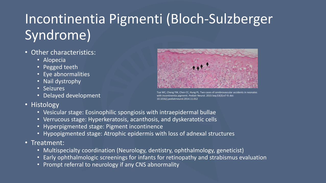
Incontinentia Pigmenti (Bloch-Sulzberger Syndrome) • Other characteristics:
• Alopecia• Pegged teeth• Eye abnormalities• Nail dystrophy• Seizures• Delayed development
• Histology• Vesicular stage: Eosinophilic spongiosis with intraepidermal bullae• Verrucous stage: Hyperkeratosis, acanthosis, and dyskeratotic cells • Hyperpigmented stage: Pigment incontinence • Hypopigmented stage: Atrophic epidermis with loss of adnexal structures
• Treatment:• Multispecialty coordination (Neurology, dentistry, ophthalmology, geneticist) • Early ophthalmologic screenings for infants for retinopathy and strabismus evaluation• Prompt referral to neurology if any CNS abnormality
Tsai WC, Cheng YW, Chen CC, Hung PL. Two cases of cerebrovascular accidents in neonates with incontinentia pigmenti. Pediatr Neurol. 2015 Sep;53(3):e7-9. doi: 10.1016/j.pediatrneurol.2014.11.012

Linear IgA Bullous Dermatosis of Childhood
• Immune-mediated, subepidermal, vesiculobullous dermatosis with clinically unique features in children. Also called “chronic bullous disease of childhood”
• Epidemiology: Mean age of 4.5 years
• Autoantigen: 97 kDa Ag (LAD-1/LABD97)
• Histology: subepidermal bullae with neutrophils in dermal papillae
• DIF: linear IgA (+/- C3) deposition at BMZ
• Treatment: dapsone (first line) 1-2 mg/kg or sulfapyridine. Second line agents include; oral corticosteroids, colchicine, and nicotinamide.
• Childhood disease remits within 2-4 years

Linear IgA Bullous Dermatosis of Childhood
Clinical: Annular and herpetiform bullae with preference for flexural areas of lower trunk, extensors, thigh, and groin. “Crown of Jewels” configuration.
http://escholarship.org/
uc/item/1379s6t6/1.jpg

Dermatitis Herpetiformis• Cutaneous manifestation of gluten sensitivity, (90% of DH patients have gluten-sensitive
enteropathy, 20% have intestinal symptoms of celiac disease)
• F>M in children. HLA-DQ2 (strongest association)
• Autoantigen: epidermal transglutaminase (TG-3), tissue transglutaminase (endomysial)
• Chronic pruritic erythematous grouped papules/vesicles on elbows, knees, and buttocks. Excoriations of vesicles common.
• Associated with gluten-sensitive enteropathy (wheat, barley, rye). NOT in rice, corn, oats
• Labs: anti-gliadin (DH)/anti-endomysial (celiac disease)
• Associations: Increased risk of enteropathy-associated T cell lymphoma (refer to GI), Hashimoto’s thyroiditis, insulin dependent diabetes mellitus.
• Treatment: Dapsone (skin improvement 24-48 hrs) 0.5 mg/kg, gluten-free diet

Dermatitis Herpetiformis
http://www.pcds.org.uk/ee/images/made/ee/images/uploads/clinical/DH_2_800_600_70_http:www.pcds.org.ukeeassetsimgwatermark.gif_0_0_80_r_b_-5_-5_.jpg
http://www.humpath.com/IMG/jpg/d_herpetiformis_40xhe2.jpg
Histology: neutrophilic microabscesses in dermal papillae
DIF (normal appearing skin): granular IgA > C3 in dermal papillae

Bullous Systemic Lupus Erythematosus• Rare blistering eruption that consists of vesicles and bullae. Clinically may look like
BP or EBA with histopathology usually resembling DH. May be more common in African Americans.
• Antigen: Type VII collagen
• Clinical: Non-inflamed bullae/vesicles (hx of SLE usually)
• Based on 5 main criteria: (1) acquired vesiculobullous eruption, (2) subepidermal bullae with neutrophils, (3) DIF with linear or granular deposition of IgG (+/- IgA, IgM, etc), (4) type VII antibodies, (5) response to dapsone
• Should also fulfill criteria for American College of Rheumatology for SLE ( positive ANA, decreased complement, anemia, leukopenia, proteinuria, thrombocytopenia, elevated ESR may be seen).
• Treatment: Dapsone, PO corticosteroids, anti-malarials, azathioprine, mycophenolate mofetil

Bullous Systemic Lupus Erythematosus
http://www.ijpd.in/articles/2016/17/2/images/IndianJPaediatrDermatol_2016_17_2_139_172468_f1.jpg
Histology: subepidermal bullae neutrophils in dermal papillae (similar to DH)
DIF/SSS: Linear IgG, IgM,IgA, C3, fibrinogen at BMZ, dermal side (SSS)

Epidermolysis Bullosa Acquisita
• Rare, acquired, subepidermal immune-mediated bullous disease with antibodies directed against Type VII collagen (290 kDa)
• Rarest subepidermal bullous diseases in Western Europe (may be more common in Korean/Asian and African-American populations)
• Treatment: Unsatisfactory and difficult to treat. Dapsone and prednisolone may be of benefit for the childhood form.

Epidermolysis Bullosa Acquisita
http://dermaamin.com/site/images/clinical-pic/e/epidermolysis_bullosa_acquisita/epidermolysis_bullosa_acquisita11.jpg
Non-inflammatory bullae with fragile skin in areas easily traumatized (elbows, hands, knees, toes).
Heals with scarring +/- mucous membrane involvement.
Childhood EBA overlaps with childhood BP and linear IgA bullous dermatosis.
Systemic diseases may occur in association (inflammatory bowel disease, SLE, RA, thyroiditis, and DM).

Epidermolysis Bullosa Acquisita
https://www.researchgate.net/profile/Michael_Camilleri/publication/24177073/figure/download/fig5/AS:277351515607040@1443137162174/Figure-7-Immunoglobulin-deposition-on-the-base-of-sodium-chloride-induced-split-human.png
http://dermaamin.com/site/images/histo-pic/e/epidermolysis-bullosa-acquisita/epidermolysis-bullosa-acquisita4.jpg
Histology: subepidermal separation without acantholysis, variable infiltrate (neutrophils or non-inflammatory)
DIF/IIF: linear IgG (-/+ C3, fibrinogen, IgA, IgM) at BMZ, binds dermal side of SSS (floor)

Bullous Pemphigoid• Rarely occurs in children
• Autoantigen: BPAG2 (collagen XVII) 180 kDa-NC16A domain, BPAG1 230kDa, cytoplasmic plaque protein
• Clinical
• IIF: SSS shows binding to epidermal side of split (roof)
• Treatment: Oral/topical corticosteroids, steroid-sparing (azathioprine, mycophenolate mofetil) tetracycline +/- nicotinamide, dapsone.

Bullous Pemphigoid
http://clinicalgate.com/wp-content/uploads/2015/03/c00024_f024-002-9781455708413.jpg
Often with initial urticarial lesions which evolveinto tense bullae, +/- pruritus.
Individual lesions in infants and older children look like elderly but sites of involvement may vary.
Infants-bullae first appear acrally, then generalize (including face). Involvement of the genital region and other mucosal sites may occur in children.

Bullous Pemphigoid
http://www.pcds.org.uk/download/2208
http://plaza.umin.ac.jp/pathol2/photo-library/system/data/image_data/11348999517711.jpg
Histology: subepidermal bulla with eosinophils and lymphocytes in papillary dermis, +/- neutrophils
DIF: linear C3 and IgG at BMZ

Pemphigus• Group of autoimmune blistering diseases of the skin and mucous
membranes
• Includes:• Vulgaris:
• potentially fatal disease, can be seen in newborns (neonatal pemphigus) but rare.
• Foliaceous: • more superficial and less severe than PV
• Endemic (Fogo selvagem)
• Drug Induced: • sulfhydryl group containing drugs are common offenders
• Parneoplastic: • less commonly due to an underlying malignancy in children
• IgA: • characteristic histologic deposit

Pemphigus Vulgaris (PV)• Rare in pediatric patients, but can be
seen in the newborns (neonatal pemphigus) whose mothers have pemphigus vulgaris.• Due to passive transfer of maternal IgG
to fetus
• Autoantibody: • Desmoglein 3 (mucosal) • Desmoglein 1 (mucocutaneous)
• Clinical• vesicles and erosions trunk and
extremities, mucosal involvement uncommon in neonatal form, (+) Nikolsky and Asboe-Hansen sign

PV• Histology
• “tomb stone row” suprabasalsplit with acantholytickeratinocytes (possible follicular involvement), perivascular lymphocytes and eosinophils
• DIF: intercellular IgG4 > C3, netlike pattern in epidermis
• IIF: monkey esophagus, correlates with disease severity
• Prognosis and Treatment • Self resolving within weeks

Pemphigus Foliaceous (PF) • Endemic (fogo selvagem) is more
common in pediatric patients • Seen in rural Brazil after a bite from black fly
(Simulium nigrimanum)
• Sporadic PF is rare in children, but if present, shows a dramatic pattern of crusted plaques and erosions with an arcuate, circinate, or polycyclic morphology on the face and scalp.
• Autoantibody: • Desmoglein 1
• Clinical• Crusted plaques and erosions in polycyclic
distribution on face and scalp

PF• Histology
• Subcorneal acantholysis with acantholytic cells seen on blister roof (cling-ons), neutrophils can be seen in blister cavity (resembling impetigo)
• DIF : same as PV, however, more pronounced in upper layers
• IIF : best substrate is guinea pig esophagus
• Treatment:• First line: Systemic corticosteroids• Steroid-sparing therapies reported:
Azathioprine, mycophenolate mofetil, IVIG, plasmapheresis, methotrexate, cyclophosphamide
• Biologics: Rituximab

Drug Induced Pemphigus • Particularly rare in children.
• Sulfhydryl group containing medicines are implicated, most common being penicillamine and captopril.• Sulfhydryl groups thought to interact with the sulfhydryl groups in desmogleins 1
and 3, thus modifying the antigenicity of the desmoglein.
• Clinical:• Initially, a nonspecific morbilliform, annular, or urticarial eruption may be seen,
eventually evolving after a variable latency period into the blistering process.
• Treatment:• Removal of offending agent if known
• Corticosteroids if necessary

Parneoplastic Pemphigus• General:
• In adults, commonly due to underlying malignancy (non-Hodgkin’s lymphoma most common)• In children, the most common causes include:
• Castleman’s disease
• Giant lymph node hyperplasia• Benign giant lymphoma
• Sarcoma
• Autoantibody: • desmoglein 3, periplakin, envoplakin, desmoplakin ½, BPAG1, plectin, 170 kDa Ag, rarely desmoglein 1
• Clinical• Intractable stomatitis, ocular involvement common, bullous or lichenoid lesions

Parneoplastic Pemphigus • Histology
• suprabasilar acantholysis, ± v acuolar basal layer damage associated with lichenoid dermal lymphocytic infiltrate, and necrotic keratinocytes
• DIF : intercellular + linear IgG/C3 along BMZ
• IIF : intercellular IgG, best substrate is rat bladder epithelium
• Prognosis & Treatment • Poor prognosis; respiratory failure due to
bronchiolitis obliterans more common
• Treatment of malignancy, palliative immunosuppression

IgA Pemphigus• Blistering disease with intraepidermal IgA deposits
• Two types: Subcorneal pustular dermatosis (SPD) & Intraepidermal neutrophilic type (IEN)
• There is no difference in the clinicopathologic features and prognosis of the disease between adults and children
• Autoantibody:• desmocollin 1 (SPD), ± desmoglein 1/3 (IEN)
• Clinical• SPD: serpiginous vesicles or pustules; may be associated
with underlying IgA gammopathy• IEN: flaccid pustules and bullae involving intertriginous
locations which enlarge forming annular or polycyclic (sunflower-like) arrangement
• Histology• intraepidermal pustule or vesicles containing neutrophils, no
acantholysis• DIF: intercellular IgA deposition• IIF: (+) in 50%, intercellular IgA
• Treatment: Dapsone, oral corticosteroids

Herpes Gestationis• Aka Pemphigoid gestationis (PG), is an autoimmune bullous dermatosis of pregnancy.
• Common in the 2nd or 3rd trimester, or immediately postpartum
• Neonatal disease is observed in up to 10% of children born to mothers with PG.
• Autoantibody• anti-BPAG2 (BP180, NC16A site)
• Clinical:• Erythematous urticarial or vesicular rash, but yellowish plaques on erythematous base and frank bullae
have also been reported.• Cutaneous lesions in the newborn are due to a passive transfer of Ig.
• Fetal risks: • prematurity & small for gestational age in 20%, also, a borderline increase in the rate of
spontaneous abortions has also been reported
• Histology:• papillary dermal edema resulting in subepidermal bulla with eosinophil rich infiltrate, ± keratinocyte
necrosis, perivascular infiltrate• DIF: linear C3 deposition ± IgG at basement membrane• IIF: epidermal base (roof of blister like BP)
• Treatment: • The disease is generally mild and resolves spontaneously in the course of days to weeks after onset.

Subcorneal Pustular Dermatosis• Aka Sneddon-Wilkinson
• Etiology still unknown, but certain associations have been seen including a benign monoclonal IgA gammopathy and pyoderma gangrenosum
• Clinical• annular or polycyclic lesions, usually commencing in
the flexures, with hallmark superficial (subcorneal) sterile pustules
• Has a cyclic course, as the pustules resolve they are replaced by superficial scaling and then new pustules form again
• Histology• Perivascular inflammatory infiltrate with neutrophils and
occasional eosinophils.• Neutrophils migrate through the epidermis, without
forming spongiform pustules, to collect beneath the stratum corneum in subcorneal pustules
Treatment: Dapsone

Chilblains • Abnormal inflammatory response to cold, damp, non-freezing
conditions
• Common in the UK and northwestern Europe, particularly in those whose homes lack central heating
• Unknown pathogenesis, but thought to have a vascular origin
• Clinical:• lesions are red-purple and usually macular, papular
or nodular • develop symmetrically on acral skin, in particular the
fingers and toes, but can occur elsewhere• Histology:
• Nonspecific, consisting of dermal edema plus a superficial and deep lymphohistiocytic infiltrate (mainly T cells) with peri-eccrine accentuation
• Treatment:• Adequate clothing and avoidance of cold, damp
conditions are important preventive measures, as are keeping feet dry and avoidance of smoking.
• In a Double-blind, placebo-controlled trial, nifedipine(20 to 60 mg daily) was found to be efficacious for the treatment of pernio
Phytophotodermatitis
• Pathogenesis: A phototoxic eruption, commonly caused by a topical or oral photosensitizing agent followed by exposure to UVR.
• Members of 2 plant families are common causes: Apiaceae and Rutaceae. The common photosensitizer is furocoumarins.
• Clinical presentation: Random configurations of erythema, edema and bullae after sun exposure.

Phytophotodermatitis

Erythema Multiforme
• Clinical Presentation: Typical target lesions with at least three distinct zones. There is usually a central bullae and the lesion resembles a target.
• Pathogenesis: Occurs in predisposed individuals, often in the setting of HSV or Mycoplasma Pneumonia infection.
• Histopathology: Spongiosis and focal vacuolar degeneration of basal keratinocytes with dermal edema and a perivascular infiltrate of mononuclear leukocytes and T lymphocytes

Erythema Multiforme

Bullous Mastocytosis
• Some infants with urticaria pigmentosa or diffuse cutaneous mastocytosis may develop bullous lesions
• Thought to be due to increased mast cell serine proteases
• Often resolves by age 3-5y
• Manage as you would for other child mastocytoses with increased attention to wound care for eroded lesions

Diffuse cutaneous mastocytosis with theformation of vesicles and bullae
Tharp, MD. Mastocytosis in Dermatology 3rd Ed. Bolognia J et al. Elsevier 2012 p 1996

Porphyria• Inherited or acquired disordes due to enzyme deficiency causing
increased production of photosensitizing porphyrins or their precursors during heme synthesis
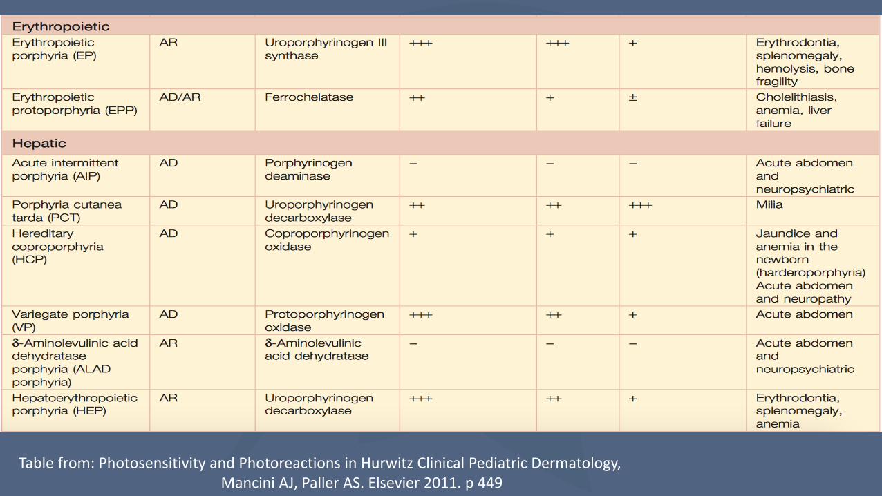
Table from: Photosensitivity and Photoreactions in Hurwitz Clinical Pediatric Dermatology,Mancini AJ, Paller AS. Elsevier 2011. p 449

Porphyria
• Congenital Erythropoeitic Porphyria (CEP) [AR]• Onset in infancy through 1st decade• Extreme photosensitivity, hypertrichosis, erythrodontia
• Erythropoeitic Porphyria [AD]• Onset between 1-4yo• Photosensitivity with burning, gallstones, hepatic damage
• Hepatoerythropoetic Porphyria [AR]• Usually manifest before 2 years of age• Dark urine is most common initial finding
• Acute Intermittent Porphyria [AD]• No skin findings• Neurologic and psychiatric findings with abdominal pain

Friction Blisters
• Occur most commonly on the soles and heels.
• Caused by repeated friction
• Blistering occurs just below the stratum granulosum

Burns and Scalds
• Can be accidental or intentional (child abuse)
• Careful history and physical examination with care to note child caregiver interactions
• In inflicted wounds there is often a delay in seeking care from the time of injury
• Look for lesions in various stages of healing

Raimer SS, Raimer-Goodman L, Raimer BG. Skin Signs of Abuse in Dermatology 3rd Ed.Bolognia J et al. Elsevier 2012 p 1523

References• Amagai M. Autoimmune and infectious skin diseases that target desmogleins. Proc Jpn Acad Ser B Phys Biol Sci. 2010;86(5):524-37.• Michal Bukowski, Benedykt Wladyka and Grzegorz Dubin. Toxins (Basel). 2010 May;2(5):1148-65. • Cole C and Gazewood J. Diagnosis and treatment of impetigo. Am Fam Physician. 2007 Mar 15;75(6):859-64• Neri I, Piraccini BM, Guareschi E et al. Bullous tinea pedis in two children. Mycoses. 2004 Dec;47(11-12):475-8.• Bruno Ferrari, Vanina Taliercio, Paula Luna et al. Kaposi's varicelliform eruption: A case series. Indian Dermatol Online J. 2015 Nov-Dec; 6(6):
399–402• Sharma C and Sharma D. A Classical Case of Neonatal Varicella. J Clin Neonatol. 2013 Oct-Dec; 2(4): 200• Çekiç Ş, Canıtez Y, Sapan N. Evaluation of the patients diagnosed with Stevens Johnson syndrome and toxic epidermal necrolysis: a single
center experience. Turk Pediatri Ars. 2016 Sep 1;51(3):152-158• Shingade P, Wankhede V, Kataria P et al. Rare case of phenytoin induced acute generalized exanthematous pustulosis with cerebellar
syndrome. Indian J Dermatol. 2014 Mar;59(2):210.• Cohen R, Levy C, Cohen J et al. Diagnostic of group A streptococcal blistering distal dactylitis. Arch Pediatr. 2014 Nov;21 Suppl 2:S93-6• Guergué D, González M, Díez B. Bullous Scabies in a 5-Year-Old Child. J Pediatr. 2016 Dec;179:270-270.e1• Gonzalez ME. Evaluation and treatment of the newborn with epidermolysisbullosa. Semin Perinatol. 2013; 37(1):32-9. doi:
10.1053/j.semperi.2012.11.004.• Intong LR, Murrell DF. Inherited epidermolysis bullosa: new diagnostic criteria and classification. Clin Dermatol. 2012 Jan-Feb;30(1):70-7.
doi: 10.1016/j.clindermatol.2011.03.012.• Falabella AF, Valencia IC, Eaglstein WH, Schachner LA. Tissue-Engineered Skin (Apligraf) in the Healing of Patients With Epidermolysis
Bullosa Wounds. Arch Dermatol. 2000;136(10):1225-1230. doi:10.1001/archderm.136.10.1225• Youssefian L, Vahidnezhad H, Uitto J. Kindler syndrome. 2016 Mar 3 [Updated 2016 Dec 1]. In: Pagon RA, Adam MP, Ardinger HH, et al.,
editors. GeneReviews® [Internet]. Seattle (WA): University of Washington, Seattle; 1993-2017. Available from: https://www.ncbi.nlm.nih.gov/books/NBK349072/
• Burch JM, Fassihi H, Jones CA, Mengshol SC, Fitzpatrick JE, McGrath JA. Kindler syndrome a new mutation and new diagnostic possibilities. Arch Dermatol. 2006;142(5):620-624. doi:10.1001/archderm.142.5.620

References• Kurosawa M, Takaqi A, Tamakoshi A, et al. Epidemiology and clinical characteristics of bullous congenital ichthyosiform
erythroderma (keratinolytic ichthyosis) in Japan: results from a nationwide survey. J Am Acad Dermatol. 2013 Feb;68(2):278-83. doi: 10.1016/j.jaad.2012.06.044.
• Sung JY. Oh SW, Kim SE, Kim SC. Mild phenotype of epidermolytic hyperkeratosis mimicking ichthyosis bullosa of Siemens is related to specific mutation in 2B domain of KRT1 . J Dermatol Sci. 2013 Jun;70(3):220-2. doi: 10.1016/j.jdermsci.2013.03.001.
• Li H, Torma H. Retinoids reduce formation of keratin aggregates in heat-stressed immortalized keratinocytes from an epidermolyticichthyosis patient with a KRT10 mutation*. Acta Derm Venereol 2013; 93: 44–49. doi: 10.2340/00015555-1368.
• Que SK, Weston G, Suchecki J, Ricketts J. Pigmentary disorders of the eyes and skin. Clin Dermatol. 2015; 33(2): 147-58. Doi: 10.1016/j.clindermatol.2014.10.007
• Batson R, Keeling BH, Diaz LZ. Incontinentia pigmenti. J. Pediatr. 2016; 176:218-218. DOI: 10.1016/j.jpeds.2016.05.081• Zafeiriou DI, Vargiami E, Hatzidimitriou V, Kyriazi M. Incontinentia pigmenti: A skin, brain, and eye matter. J. Pediatr. 2013; 163(5):1520.
DOI:10.1016/j.jpeds.2013.06.029• Treadwell PA. Systemic Conditions in children associated with pigmentary changes. Clind Dermatol. 2015; 33(3): 362-7. doi:
10.1016/j.clindermatol.2014.12.014.• Tsai WC, Cheng YW, Chen CC, Hung PL. Two cases of cerebrovascular accidents in neonates with incontinentia pigmenti. Pediatr Neurol.
2015 Sep;53(3):e7-9. doi: 10.1016/j.pediatrneurol.2014.11.012• Alajlan A, Al-Khawajah M, Al-Sheik O, Al-Saif F, Al-Rasheed S, Al-Hoqail I, Hamadah, IR. Treatment of linear IgA bullous dermatosis of
childhood with flucloxacillin. J Am Acad Dermatol 2006;54:652-6.• Cui YX, Yang BQ, Zhou GZ, Zhang FR. Childhood linear IgA bullous dermatosis successfully treated with oral nicotinamide. Clin Exp Dermatol.
2016 Oct;41(7):816-8.• De las Heras MN. Linear IgA bullous dermatosis of childhood: good response to antibiotic treatment. Clin Exp. Dermatol 2014
Apr;39(3):395-7.• Kong YL, Lim YL, Chandran NS. Retrospective study on autoimmune blistering disease in pediatric patients. Pediatr Dermatol. 2015 Nov-
Dec;32(6):845-52.• Boskovic A, Kitic I, Prokic D, Stankovic I. Cardiomyopathy associated with celiac disease in childhood. Case Report Gastrointest Med. 2012
Oct E-pub;170760.• Templet JT, Welsh JP, Cusack CA. Childhood dermatitis herepetiformis; a case report and review of the literature. Cutis 2007 Dec(80);6:473-
6.

References• Powel GR, Bruckner AL, Weston WL. Dermatitis herpetiformis presenting as chronic urticaria. Pediatr Dermatol. 2004 Sep-Oct;21(5):564-7• Kong YL, Lim YL, Chandran NS. Retrospective study on Autoimmune Blistering Disease in Pediatric Patients. Pediatr Dermatol. 2015 Nov-
Dec;32(6):845-52.• Tincopa M, Puttgen KB, Sule S, Cohen BA, Gerstenblith MR. Bullous lupus: an unusual initial presentation of systemic lupus erythematous in
an adolescent girl. Pediatr Dermal. 2010 Jul-Aug;27(4):373-6.• Kong YL, Lim YL, Chandran NS. Retrospective study on Autoimmune Blistering Disease in Pediatric Patients. Pediatr Dermatol. 2015 Nov-
Dec;32(6):845-52.• Goebeler M, Zillikens D. Blistering autoimmune diseases oaf childhood. Hautartz. 2003 Jan;54(1);14-24.• Kong YL, Lim YL, Chandran NS. Retrospective study on Autoimmune Blistering Disease in Pediatric Patients. Pediatr Dermatol. 2015 Nov-
Dec;32(6):845-52.• Atzori L, Pau M, Podda R, Manielie C, Aste N. A case of Bullous Pemphigoid treated with Local Corticosteroids.G Ital Dermatol Venereol.
2011 Dec;146(6):493-6.• Kalyani M. et al. Bullous diseases: Kids are not just little people. Clinics in Dermatology 2015 Nov-Dec: 33, 644–656.• Bolognia, Jean, et al. Dermatology. 3rd ed. Atlanta: Elsevier. 2014. Print. 461-473.• Paller, Amy, et al. Hurwitz’s clinical pediatric dermatology : a textbook of skin disorders of childhood and adolescence. 4th ed.
Atlanta: Elsevier. 2011. Print. 315.• Scalvenzi M. et al. Subcorneal Pustular Dermatosis in Childhood: A Case Report and Review of the Literature. Case Rep Dermatol Med. 2013
Jan: 424797.• Burns, Tony, et al. Fig 40.10. Rooks Textbook of Dermatology. 8th ed. Hoboken: Blackwell Publishing Ltd. 2010. Print. 40.21. • Rustin MH, Newton JA, Smith NP, Dowd PM. The treatment of chilblains with nifedipine: the results of a pilot study, a double-blind placebo-
controlled randomized study and a long-term open trial. Br J Dermatol. 1989; 120: 267–75.• Paller, Amy, et al. Fig 20.46. Hurwitz’s clinical pediatric dermatology : a textbook of skin disorders of childhood and adolescence. 4th ed.
Atlanta: Elsevier. 2011. Print. 476.• Bolognia, Jean, et al. Dermatology. 3rd ed. Atlanta: Elsevier. 2014. Print. 1493-1494.• Orkin M, Good RA, Clawson CC, Fisher I, Windhorst DB. Bullous Mastocytosis. Arch Dermatol. 1970;101(5):547-564. • Mancini AJ, Paller AS. Photosensitivity and Photoreactions in Hurwitz Clinical Pediatric Dermatology, Elsevier 2011• Badcock NR, O’Reilly DA, Zoanetti GD, Childhood Porphyrias: Implicationsand Treatments. CLIN. CHEM. 39/6, 1334-1340 (1993)

THANK YOU!