pediatric mandibular fracture therapy – a case report
TRANSCRIPT
Vojnosanit Pregl 2020; 77(6): 647–650. VOJNOSANITETSKI PREGLED Page 647
Correspondence to: Ivan Tušek, University of Novi Sad, Faculty of Medicine, Dental Clinic of Vojvodina, Hajduk Veljkova 12, 21 000 Novi Sad, Serbia. E-mail: [email protected]
C A S E R E P O R T S
UDC: 616.716.4–001.5-053.2 https://doi.org/10.2298/VSP180328110T
Pediatric mandibular fracture therapy – A case report Lečenje preloma donje vilice kod dece
Ivan Tušek*†, Miroslav P. Ili憇§, Jasmina Tušek║, Stojan Ivić*†, Branislav Tušek¶
University of Novi Sad, *Faculty of Medicine, †Dental Clinic of Vojvodina, ‡Clinical Center of Vojvodina, §Clinic for Maxillofacial Surgery, Novi Sad, Serbia; ║Private
Dental Practice “Palmadent”, Novi Sad, Serbia; ¶Institute for Pulmonary Diseases of Vojvodina, Sremska Kamenica, Serbia
Abstract Introduction. Frequency of pediatric mandibular fractures is relatively uncommon. Apart from rare exceptions, there is minimal invasive access in the treatment of those injuries in order to avoid the future developmental disorders. Case report. During the game with a colt, a 6-year-old boy was kicked by hoof in the chin. The child did not lose con-sciousness and did not experience nausea or vomiting. Ac-cording to clinical examination and radiological analysis, di-agnosis was assigned as dislocated mandibular fracture in the parasymphysis part of the jaw and luxation injury of teeth 31 and 72. The surgical treatment under general anes-thesia encompassed reduction and bimanual manipulation of bone fragments up to the optimal restoration of the den-tal occlusion, along with osteosynthesis with titanium mini-plates. Luxated deciduous tooth 72 at the fracture line was extracted and luxated permanent tooth 31 was fixed to tooth 41 with wire. The patient was given antibiotic therapy. Additional immobilization of the luxated tooth 31 and mandibular fracture was performed after surgery by com-posite resin splint. During five-month follow-up period there were no signs of pathological movements in the frac-ture line, no luxation of tooth 31 and no restriction in mouth opening. Conclusion. Osteosynthesis with mini-plates is adequate and very efficient treatment method in dislocated mandibular fracture that is recommended in chil-dren with both deciduous and mixed dentition. It is neces-sary to remove miniplates after fracture consolidation. Key words: child, preschool; fracture fixation, internal; mandible; mandibular fractures; oral surgical procedures; treatment outcome.
Apstrakt Uvod. Rasprostranjenost preloma donje vilice je relativno mala kod dece. Da bi se izbegli naknadni poremećaji u raz-voju vilice, sem retkih izuzetaka, u lečenju se primenjuje mi-nimalno invazivni pristup. Prikaz bolesnika. U toku igre sa ždrebetom, šestogodišnji dečak je dobio udarac kopitom u bradu. Nije gubio svest, niti je imao gađenje i povraćanje. Na osnovu kliničkog pregleda i rendgen analize postavljena je dijagnoza dislokovanog preloma donje vilice u parasimfi-znoj regiji i luksacija zuba 31 i 72. U opštoj anesteziji je izvr-šena hirurška repozicija, uz bimanuelnu manipulaciju košta-nih fragmenata do uspostavljanja optimalne dentalne okluzi-je, a zatim je izvršena osteosinteza donje vilice sa titanijum-skim mini pločicama. Luksirani mlečni donji levi lateralni sekutić (72), koji se nalazio u liniji preloma, je ekstrahovan, a luksirani donji levi centralni sekutić (31) fiksiran čeličnom žicom za donji desni centralni sekutić (41). Ordinirana je an-tibiotska terapija. Dodatna imobilizacija luksiranog zuba (31) i frakture donje vilice izvršena je postoperativno, po-moću kompozitnog splinta. Posle petomesečnog opservaci-onog perioda nije bilo znakova patološkog pomeranja u predelu frakturne linije, luksacije zuba 31, kao ni ograniče-nog otvaranja usta. Zaključak. Osteosinteza mini pločica-ma pokazala se kao adekvatna i vrlo efikasna metoda lečenja i preporučuje se u slučajevima dislokovanog preloma donje vilice kod dece sa mlečnom i mešovitom denticijom. Neop-hodno je ukloniti mini pločice posle konsolidacije preloma. Ključne reči: deca, predškolska; prelomi, fiksacija, unutrašnja; mandibula; mandibula, prelomi; hirugija, maksilofacijalna, procedure; lečenje, ishod.
Page 648 VOJNOSANITETSKI PREGLED Vol. 77, No 6
Tušek I, et al. Vojnosanit Pregl 2020; 77(6): 647–650.
Introduction
The mandible fractures frequency is low in children, and occurs in 5% of all maxillofacial traumas 1. The most of pediat-ric mandible fractures are not dislocated because of the bone elasticity and existing tooth buds that holds firmly the fragments together “like glue” 1. Frequency of the mandible injury is more commonly present in boys than girls by a ratio of 2 : 1 2. Treat-ment of pediatric mandible fractures is different in relation to that in adults, concerning the age of a child, the level of tooth development along with the teeth that start to grow-ups, or oth-ers still unerupted 3. Fracture treatment is basically more diffi-cult concerning deciduous teeth as their roots size is not enough strong to support fixation of mandible fragments with maxillary-mandibular fixation (MMF) 4. Younger patients also have better potential for restitution and remodeling comparing to the scle-rotic type of remodeling seen in adults 2. The principal condi-tions for successful bone healing are: early specific treatment, morphological reduction of bone fragments, immobilization and prevention of the infection. In the case of displaced bone frag-ments the use of closed reduction and immobilization are carried out a priori to avoid future functional disorders 2, 3. Most frac-tures in children without dislocation of the fragments have been treated conservatively by dental splints, occlusal splint with cir-cum-mandible wires, or absorbable plates and screws, all of them being well eligible and quite effective 1.
In this paper we described a case of rare pediatric para-symphysis mandible fracture with large dislocation of frag-ments that was successfully treated by a rigid internal fixa-tion with titanium miniplate system.
Case report
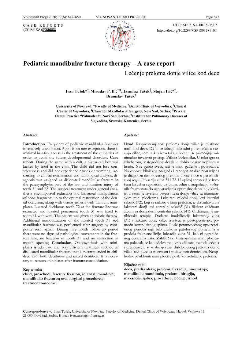
During the game with a colt, a 6-year-old male child was hit by hoof in the chin. After the injury, the patient came to the General Hospital where he got the first aid, and his soft-tissue wounds were thoroughly debrided. There was no history of bleeding from nose, ears or the head injury. The father reported no syncope, vomiting or drowsiness in the child. The patient was sent to the Dentistry Clinic of Vojvodina, Novi Sad, Serbia, where the further injury management was organized after pedi-atric dentist examination by joint work of surgeon, pediatric dentist and orthodontist. Extraoral examination revealed an one inch lacerated wound on the chin with gaping borders but ho-meostasis had already been achieved. The child had swelling and bruising in the submental region and mouth floor. There were also facial asymmetry, restricted mouth opening, deviation of the mandible to the affected side, incorrect speech and pain in the left part of the chin during the examination. An intraoral ex-amination, revealed laceration presented in lower labial vesti-bule, luxation injury of the central mandible incisors (31, 41) and lateral deciduous incisor (72). Fractured segments mobility, step defect and tenderness were observed in the left parasym-physis region. Radiological examination showed left parasym-physis fracture between left mandible, central displaced perma-nent incisior (31) and lateral deciduous incisior (72), with frac-ture line runs downward and backward (Figure 1). There was a large (7 mm) dislocation of fragments which it is not very com-
mon type of mandibular fracture at this young age. Usually, this injury is associated with unilateral or bilateral condylar fracture but not in this case.
Fig. 1 – Preoperative orthopantomograph view of left
parasymphyseal fracture.
Management
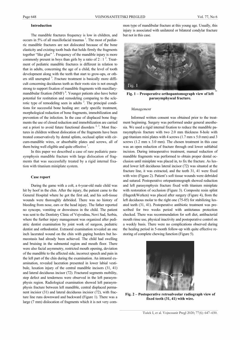
Informed written consent was obtained prior to the treat-ment beginning. Surgery was performed under general anesthe-sia. We used a rigid internal fixation to reduce the mandible pa-rasymphysis fracture with two 2.0 mm thickness 8˗hole with gap titanium mini plates with 4 screws (1.7 mm x 5.0 mm) and 3 screws (1.2 mm x 3.0 mm). The chosen treatment in this case was an open reduction of fracture through oral lower sublabial incision. During intraoperative treatment, manual reduction of mandible fragments was performed to obtain proper dental oc-clusion until miniplate was placed in, to fix the fracture. As lux-ated lower left deciduous lateral incisor (72) was situated at the fracture line, it was extracted, and the teeth 31, 41 were fixed with wire (Figure 2). Patient’s soft tissue wounds were debrided and sutured. Postoperative ortopantomograph showed reduction and left parasymphysis fracture fixed with titanium miniplate with restoration of occlusion (Figure 3). Composite resin splint (Hager&Werken) was placed after surgery (Figure 4), from the left deciduous molar to the right one (75˗85) for stabilizing lux-ated teeth (31, 41). Postoperative antibiotic treatment was pre-scribed for two weeks period with antitetanus protection checked. There was recommendation for soft diet, antibacterial mouth rinse use, physical inactivity and postoperative control on a weekly basis. There were no complications observed during the healing period in 5-month follow-up with quite effective re-storing of complete chewing function (Figure 5).
Fig. 2 – Postoperative retroalveolar radiograph view of
fixed teeth (31, 41) with wire.

Vol. 77, No 6 VOJNOSANITETSKI PREGLED Page 649
Tušek I, et al. Vojnosanit Pregl 2020; 77(6): 647–650.
Fig. 3 – The reduction and rigid internal fixation of left
parasymphysis fracture with a titanium mini plate.
Fig. 4 – Composite resin splint (Hager&Werken) from
the left deciduous molar to the right one (75˗85).
Fig. 5 – Postoperative photograph showing increased
mouth opening at the fifth month of follow-up.
Discussion
Very young age, a rare type of injury, the animal as a rare etiological factor for mandible trauma, different and contradictory opinions concerning therapy management, no consent concerning the matter of the appropriate time for plates removal in our case, incertitude therapy outcome es-pecially in small children with deciduous dentition, the lack of cooperation among surgeon, pediatric dentist and ortho-dontist, as well as no unique methods presented for treatment in such young age, gave us the reason to show this case re-port. It also can be very helpful, especially to pediatric den-tists because they are in the position to give the first aid to the young patient before oral surgeons, and also dentists are the ones who will further follow-up injured children.
Analysis of the literature indicates the lower prevalence of pediatric mandible fractures comparing to adult popula-tion. The highest frequency of mandible fractures occurred in
younger patients, 6˗12 years old (0.6%˗1.2%) 5. Kicks from animals as the etiological factor for mandible fracture was relatively rare, with 3.3% up to 6.0% of all maxillofacial fractures 2. Various management protocols of mandible frac-ture treatment are discussed in the literature 1. Nevertheless, some techniques may be better than others, but no one tech-nique can be used in all situations. The treatment of fractured pediatric mandible differs from that of adults, because of anatomic variation, rapidity of healing, degree of cooperation and the potential for interference with the mandible growth 6. Children have great ability for healing with few possible complications, aided by well blood supplied tissues with greater osteogenic potential than adults. Anatomic reduction in children should be accomplished earlier and immobiliza-tion time should be shorter ie. 2˗3 weeks as compared to 4˗6 weeks in adults 7, 8. Although there is no clear consensus about optimal method for fixation of mandible parasymphy-sis fractures, the most effective and the less invasive method is the best one. Using conservative therapy in the majority of cases of “greenstick” or minimally displaced fractures in children with a short period of MMF is satisfiying. Addition-ally, there are many treatments of those fractures, and some of them are: acrylic splints, circumferential wiring, the skele-tal fixation through the skin, compressive and noncompres-sive plates, isolated screws, absorbable plates and screws, open reduction, modified orthodontic brackets, etc. 9. The applied treatment of displaced fractures mostly varied from MMF to cap splints and either regular or absorbable mini-plates insert. A lot of serious mandible fractures demands the wide range of therapeutic approaches from open to close re-duction and rigid or non rigid fixation along with or without MMF. In our case, the reason for the pediatric mandible trauma was an accident at home. For treatment of these inju-ries, Davison et al. 10 said that the risks of facial growth dis-turbance in open reduction and internal fixation (ORIF) has not been supported. In contrast to that opinion, unappropriate treatment in unrecognized mandible fractures leads to high incidence of orthognathic surgery. Technique utilized to re-pair the fractured mandible parasymphysis, in our case, was the conventional approach of ORIF with titanium miniplates and screws. Intraoral approach through an oral mucosal inci-sion, allow direct control of appropriate occlusion during the incorporation of the titanium miniplates which stabilize the fracture site. The use of miniplates changed the treatment of mandible fractures in the past twenty years, with varying de-grees of success 11. Koshy et al. 12 reported that ORIF is not commonly performed until late mixed dentition, but may be indicated in the early mixed dentition in severely dislocated fractures. In our case the treatment is complicated by the presence of teeth (31, 41) instability and a lot of the teeth that did not grow yet. The potential damage to tooth roots and follicles can be minimized with careful technique, which places monocortical screws in the lower mandible edge. In the majority of cases of minimally displaced fragments of the pediatric mandible fracture, using conservative methods with MMF during a short period is generally satisfying. If surgical treatment is indicated, occlusal acrylic splints, interdental wiring, and monocortical plates and screws are all eventual

Page 650 VOJNOSANITETSKI PREGLED Vol. 77, No 6
Tušek I, et al. Vojnosanit Pregl 2020; 77(6): 647–650.
option 2. The use of titanium mini plates systems in relation to absorbable plates permits a stable rigid or semirigid fixa-tion that may eliminate the necessity for MMF 2. The mono-cortical bone plates are smaller in size and easily adaptable for application to any type of fractured site. Zimmerman et al. 6 consider that ORIF insures stable three dimensional re-constructions, encourages the primary bone healing, reduces the treatment time and eliminates the need for MMF. On the other hand, closed treatment of the parasymphysis fractures usually demands extended periods of MMF from 3 to 5 weeks. This can become an extremely important factor when it comes to the treatment of pediatric patients, since the level of cooperation is greatly reduced. A patient has to stay on the liquid diet, hospitalized for a longer period of time, with dif-ficulties in regular oral hygiene, and speech is also affected 2. Application of fabricated acrylic splints is more reliable than ORIF or MMF techniques with regard to the cost/ effective-ness ratio, ease of use and removal, reduced operation time, maximum stability during healing period, minimal trauma for surrounded anatomic structures etc. 1, but it was not suit-able in our case. Based on the literature data we can clearly recommend “minimally invasive” internal fixation by means of the monocortical plate and screws, as reported Cole et al. 7. We believe that choice of ORIF should always be rec-ommended to treat children younger than 6 years of age. With this system of fixing, we obtained the same success re-ported by the other authors 8. This method provides better
stability of fractured fragments, primary bone healing, the low infection rate and possibility to avoid MMF 11. However, this system has an important disadvantage, because plate and screws removal is recommended in order to minimize the risk of interference with normal growth of the mandible. There is a great possibility, as Bos et al. 13 reported, that met-al implants may cause stress shielding with local osteoporo-sis after later removal. Certainly, ORIF could have a nega-tive effect on skeletal growth and unerupted teeth because there is a need of plate removal after complete healing 2, 3, 8, 12. Hogg and Horswell 14 has not seen any growth disturbances caused by miniplate osteosynthesis when they remove it after a period of 6 months. The use of absorbable plates is less likely to disturb facial skeletal growth but is still associated with risk of damaging the teeth that have not yet erupted even when using monocortical plates and screws 2.
Conclusion
ORIF treatment is suggested in large dislocated pediat-ric mandible fractures and must be carefully done, because of rapid growing up and developmental phenomenon that con-tinues in children. Plates system should be removed as soon as healing period is over. Minimized invasive therapy should always be the choice, especially in children younger than 6 years of age. Joint work of surgeons, pediatric dentists and orthodontists is needed during the whole recovery period.
R E F E R E N C E S
1. Sodhi SPS, Brar G, Brar RS, Bhardwaj J, Jain A. Modified cir-cummandibular wiring fixation using acrylic splint for the treatment of displaced mandibular parasymphysis fracture: A case report. J Stomatognathic Sci 2015; 5(1): 10–3.
2. Marano R, de Oliveira Neto P, Oliveira Oliveira Sakugawa K, Zanetti SSL, de Moraes M. Mandibular fractures in children under 3 years: a rare case report. Rev Port Estomatol Med Dent Cir Maxilofac 2013; 54(3):166–70. (English, Portuguese)
3. Jain P, Yeluri R, Gupta S, Lumbini P. Management of pediatric mandibular parasymphyseal fracture with acrylic closed cap splint: a case report. Ann Dent Spec 2015; 3(1): 45–7.
4. Samad S, Priyanto W. Early treatment of symphysis mandibular fracture in 12 years old children using Erich arch bar: a case report. J Dentomaxillofac Sci 2017; 2(1): 45–8.
5. Agrawal RM, Yeluri R, Singh C, Chaudhry K, Munshi AK. Man-agement of pediatric mandibular fracture: a Case Series. Com-pend Contin Educ Dent 2014; 35(8): 578–82.
6. Zimmermann CE, Troulis MJ, Kaban LB. Pediatric facial frac-tures: recent advances in prevention, diagnosis and manage-ment. Int J Oral Maxillofac Surg 2006; 35(1): 2–13.
7. Cole P, Kaufman Y, Izaddoost S, Hatef DA, Hollier L. Principles of pediatric mandibular fracture management. Plast Reconstr Surg 2009; 123(3): 1022–4.
8. Shunmugavelu K, Subramaniam K. Fracture of medial pole of right condyle and symphysis of mandible in a 6-year-old male: A conservative approach. Sudan Med Monit 2016; 11(4): 133–6.
9. Madan N, Bajaj N. Conservative treatment of pediatric man-dibular fracture with removable acrylic splint. Indian J Dent Sci 2010; 2(4): 22–4.
10. Davison PS, Clifton MS, Davison MN, Hedrick M, Sotereanos G. Pediatric mandibular fractures: a free hand technique. Arch Facial Plast Surg 2001; 3(3): 185–9; discussion 190.
11. Sauerbier S, Schön R, Otten JE, Schmelzeisen R, Gutwald RJ. The development of plate osteosynthesis for the treatment of frac-tures of the mandibular body and a literature review. J Cra-niomaxillofac Surg 2008; 36(5): 251–9.
12. Koshy JC, Evan M, Feldman EM, Chike-Obi CJ, Bullocks JM. Pearls of Mandibular Trauma Management. Semin Plast Surg 2010; 24(4): 357–74.
13. Bos RR. Treatment of pediatric facial fractures: the case for metallic fixation. J Oral Maxillofac Surg 2005; 63(3): 382–4.
14. Hogg NJ, Horswell BB. Hard tissue pediatric facial trauma: a re-view. J Can Dent Assoc 2006; 72(6): 555–8.
Received on March 28, 2018. Revised on May 11, 2018.
Accepted on June 2, 2018. Online First June, 2018.