pekka keski-rahkonen new lc-ms assays for drugs and · pdf file ·...
TRANSCRIPT

Publications of the University of Eastern Finland
Dissertations in Health Sciences
isbn 978-952-61-0598-7
Publications of the University of Eastern FinlandDissertations in Health Sciencesd
issertation
s | 084 | P
ekk
a Kesk
i-Ra
hk
on
en | N
ew L
C-M
S A
ssays for Dru
gs and E
ndogen
ous C
omp
ounds in S
mall-V
olume B
iological Sam
ples
Pekka Keski-RahkonenNew LC-MS Assays for Drugs
and Endogenous Compounds in Small-Volume Biological
SamplesPekka Keski-Rahkonen
New LC-MS Assays for Drugs and Endogenous Compounds in Small-Volume Biological Samples
This thesis presents alternative
approaches for bioanalytical
method development using liquid
chromatography–mass spectrometry.
It explores the feasibility of using
atmospheric pressure chemical
ionization without corona discharge,
the use of this technique on a
microchip scale, derivatization of
non-polar analytes for improved
electrospray ionization, and different
pre-processing methods for sub-150 µl
biological samples.

PEKKA KESKI-RAHKONEN
New LC–MS Assays for Drugs and
Endogenous Compounds in Small-
Volume Biological Samples
To be presented by permission of the Faculty of Health Sciences, University of Eastern
Finland for public examination in auditorium ML2, Medistudia building, University of
Eastern Finland, Kuopio, on Friday January 27th 2012, at 12 noon
Publications of the University of Eastern Finland
Dissertations in Health Sciences
84
School of Pharmacy, Faculty of Health Sciences,
University of Eastern Finland
Kuopio
2012

Kopijyvä
Kuopio, 2012
Series Editors:
Professor Veli-Matti Kosma, M.D., Ph.D.
Institute of Clinical Medicine, Pathology
Faculty of Health Sciences
Professor Hannele Turunen, Ph.D.
Department of Nursing Science
Faculty of Health Sciences
Professor Olli Gröhn, Ph.D.
A.I. Virtanen Institute for Molecular Sciences
Faculty of Health Sciences
Distributor:
University of Eastern Finland
Kuopio Campus Library
P.O. Box 1627
FI-70211 Kuopio, Finland
http://www.uef.fi/kirjasto
ISBN (print): 978-952-61-0598-7
ISBN (pdf): 978-952-61-0599-4
ISSN (print): 1798-5706
ISSN (pdf): 1798-5714
ISSN-L: 1798-5706

III
Author’s address: School of Pharmacy
Faculty of Health Sciences
University of Eastern Finland
KUOPIO
FINLAND
Supervisors: Professor Seppo Auriola, Ph.D.
School of Pharmacy
University of Eastern Finland
KUOPIO
FINLAND
Timo Mauriala, Ph.D.
Orion Pharma
ESPOO
FINLAND
Reviewers: Docent Ari Tolonen, Ph.D.
Admescope Ltd.
OULU
FINLAND
Docent Tiina Kauppila, Ph.D.
Faculty of Pharmacy
Division of Pharmaceutical Chemistry
University of Helsinki
HELSINKI
FINLAND
Opponent: Professor Ilkka Ojanperä, Ph.D.
Hjelt Institute
Department of Forensic Medicine
University of Helsinki
HELSINKI
FINLAND

IV

V
Keski-Rahkonen, Pekka
New LC–MS Assays for Drugs and Endogenous Compounds in Small-Volume Biological Samples,
69 p.
University of Eastern Finland, Faculty of Health Sciences, 2011
Publications of the University of Eastern Finland. Dissertations in Health Sciences 84. 2011. 69 p.
ISBN (print): 978-952-61-0598-7
ISBN (pdf): 978-952-61-0599-4
ISSN (print): 1798-5706
ISSN (pdf): 1798-5714
ISSN-L: 1798-5706
ABSTRACT
This thesis, which consists of four original publications and a summary, explores the feasibility of combining different unconventional ionization techniques and sample preparation methods to improve the sensitivity of liquid chromatography–mass spectrometry (LC–MS) based bioanalytical assays. It addresses important sensitivity-limiting factors, such as the weak electrospray ionization (ESI) of non-polar analytes, ion-suppressing effect of ion-pairing mobile phase additives, and the loss of analytes during sample pre-processing. The study demonstrates the suitability of using an atmospheric pressure chemical ionization (APCI) source without a corona discharge for the efficient ionization of selected analytes in the presence of ion-pairing agents. This rarely used technique, termed atmospheric pressure thermospray ionization or no-discharge APCI, is also shown to be suitable for use with a novel microchip heated nebulizer. Moreover, oxime derivatization is presented as a simple and efficient method to improve the proton affinity and thus the ESI ionization of non-polar ketosteroids. Small-scale liquid-liquid extraction and direct injection can be utilized to increase the recovery in the sample preparation process. By combining the above techniques, three different assays were developed and their performance was evaluated by validation. The assays were also used for the analysis of samples from clinical and animal studies. Taken together, this thesis presents alternative or complementary approaches for bioanalytical LC–MS method development. The assays developed represent significant improvements in sensitivity, dynamic range, and robustness and the novel findings regarding ionization in particular provide important new information about some rarely used analytical techniques. National Library of Medical Classification: QU 25, QV 25, QV 26, QV 744, QY 25, QY 90
Medical Subject Headings: Chemistry Techniques, Analytical; Chemistry, Clinical/methods;
Chromatography, Liquid; Mass Spectrometry; Analytic Sample Preparation Methods;
Sulfonamides/analysis; Labetalol/analysis; Eye; Aqueous Humor; Serum; Acetylcholine/analysis;
Microdialysis; Steroids/analysis; Sensitivity and Specificity; Validation Studies as Topic

VI

VII
Keski-Rahkonen, Pekka
Uusia LC–MS-menetelmiä lääkeaineiden ja endogeenisten yhdisteiden analysointiin pienikokoisista
biologisista näytteistä, 69 s.
Itä-Suomen yliopisto, terveystieteiden tiedekunta, 2011
Publications of the University of Eastern Finland. Dissertations in Health Sciences 84. 2011. 69 s.
ISBN (print): 978-952-61-0598-7
ISBN (pdf): 978-952-61-0599-4
ISSN (print): 1798-5706
ISSN (pdf): 1798-5714
ISSN-L: 1798-5706
TIIVISTELMÄ
Tässä neljästä osajulkaisusta ja niiden yhteenvedosta koostuvassa väitöskirjatyössä tutkittiin erilaisten näytteenkäsittely- ja ionisaatiotekniikoiden soveltuvuutta bioanalyyttisten nestekromatografia–massaspektrometriamenetelmien (LC–MS) suorituskyvyn lisäämiseen. Tutkimuksessa pyrittiin löytämään ratkaisuja tunnettuihin mittausmenetelmän herkkyyttä heikentäviin tekijöihin, kuten poolittomien yhdisteiden heikkoon ionisaatioon sähkösumutuksessa (ESI), ionipari-reagenssien aiheuttamaan ionisupressioon sekä analysoitavien yhdisteiden puutteelliseen saantoon näytteen esikäsittelyn aikana. Tutkimuksen mukaan eräiden lääkeaineiden ja endogeenisten yhdisteiden analysoinnissa voidaan hyödyntää ilmanpaine-kemiallinen ionisaatio (APCI) tekniikkaa täysin ilman sen toimintaan olennaisesti liittyvää koronapurkausta. Tuloksena on ESI:n kaltainen ionisaatio, joka toisin kuin ESI-tekniikka yleensä, mahdollistaa ioniparireagenssien käytön ajoliuoksessa ilman merkittävää ionisupressiota. Tutkimuksen mukaan tekniikka soveltuu käytettäväksi normaalin APCI-ionilähteen lisäksi myös kuumasumutus-mikrosirulla, mikä mahdollistaa mikrofluidististen analyysitekniikoiden, kuten kapillaari-nestekromatografian, hyödyntämisen. Lisäksi tutkimuksessa kyettiin parantamaan nestefaasissa ionisoitumattomien steroidien ESI-ionisaatiota kemiallisella derivatisoinnilla sekä näytteenkäsittelyn saantoa pienen mittakaavan neste-neste uuton ja näytteen suoran injektion avulla. Edellä mainittuja tekniikoita yhdistämällä kehitettiin kolme uutta mittausmenetelmää, joiden käytännön suorituskyky arvioitiin validoinnilla sekä biologisten tutkimusnäytteiden analysoinnilla. Väitöskirjatyön tulokset tarjoavat uusia, vaihtoehtoisia lähestymistapoja bioanalyyttisten LC–MS menetelmien kehitykseen. Kehitetyillä analyysimenetelmillä on huomattavia herkkyyteen, pitoisuusalueeseen sekä luotettavuuteen liittyviä etuja. Erityisesti ionisaatiotekniikoihin liittyvät tulokset sisältävät merkittävää uutta tietoa harvoin käytetyistä analyysitekniikoista. Luokitus: QU 25, QV 25, QV 26, QV 744, QY 25, QY 90
Yleinen Suomalainen asiasanasto: analyyttinen kemia; analyysimenetelmät; kvantitatiivinen analyysi;
kliininen kemia; nestekromatografia; massaspektrometria; tamsulosiini; labetaloli; asetyylikoliini;
steroidit; validointi

VIII

IX
Acknowledgements
The work included in this thesis was carried out in the Department of
Pharmaceutical Chemistry, University of Kuopio, subsequently named the School of
Pharmacy, University of Eastern Finland, during the years 2005–2010. I would like to
express my gratitude and thanks to my principal supervisor Professor Seppo Auriola
for all the guidance, support, and trust during these past years. I also want to thank
my other supervisor Dr. Timo Mauriala, as well as Professor Tomi Järvinen and Mr.
Marko Lehtonen for their encouragement at the start of these studies.
I am grateful to Professor Ilkka Ojanperä for accepting the invitation to serve as
an opponent. Docent Ari Tolonen and Docent Tiina Kauppila are warmly
acknowledged for the pre-examination review of the thesis and Dr. Ewen
MacDonald for proofreading the final version of the manuscript. Thanks to Professor
Markus Forsberg, Dr. Elina Jarho, and Dr. Veli-Pekka Ranta for their helpful
comments about the research plan.
I have had the privilege to work with many experts from different scientific
backgrounds and I would like to thank all my co-authors: Professor Seppo Auriola,
Professor Sami Franssila, Professor Risto Kostiainen, Professor Tapio Kotiaho,
Professor Matti Poutanen, Dr. Markus Haapala, Dr. Kaisa Huhtinen, Dr. Jouni
Ihalainen, Dr. Esa Leppänen, Dr. Timo Mauriala, Dr. Olavi Pärssinen, Dr. Ville
Saarela, Dr. Timo Sarajärvi, and Mr. Marko Lehtonen. Special thanks to Marko for
introducing me to laboratory quality.
All the current and former Deans of the Faculty and Heads of the Department
are acknowledged for providing excellent facilities in which to work. I wish to
express a big thank you to all the personnel, colleagues and friends in the School of
Pharmacy for creating such a superb working environment.
Grants were received from the Faculty of Health Sciences and the Graduate
School in Pharmaceutical Research. The study related to steroids was part of a project
on steroid metabolomics, funded by the Finnish Funding Agency for Technology and
Innovation. A great amount of work was also done during the time I was working
for the Drug Research and Development Centre in the School of Pharmacy. I am
grateful for all the grants and funding that enabled this study to be carried out.
I would also like to thank my family and friends for all the support that I have
received outside the laboratory.

X
Finally, I owe my warmest thanks to my wife, whose boundless patience and
encouragement constituted priceless support during this project.
December 14, 2011. Sydney, Australia
Pekka Keski-Rahkonen

XI
List of the original publications
This dissertation is based on the following original publications, hereafter referred to
by their Roman numerals (I-IV):
I Keski-Rahkonen P, Pärssinen O, Leppänen E, Mauriala T, Lehtonen M,
Auriola S. Determination of tamsulosin in human aqueous humor and
serum by liquid chromatography–electrospray ionization tandem mass
spectrometry. Journal of Pharmaceutical and Biomedical Analysis 43: 606–612,
2007.
II Keski-Rahkonen P, Lehtonen M, Ihalainen J, Sarajärvi T, Auriola S.
Quantitative determination of acetylcholine in microdialysis samples using
liquid chromatography/atmospheric pressure spray ionization mass
spectrometry. Rapid Communications in Mass Spectrometry 21: 2933–2943,
2007.
III Keski-Rahkonen P, Haapala M, Saarela V, Franssila S, Kotiaho T,
Kostiainen R, Auriola S. Atmospheric pressure thermospray ionization
using a heated microchip nebulizer. Rapid Communications in Mass
Spectrometry 23: 3313–3322, 2009.
IV Keski-Rahkonen P, Huhtinen K, Poutanen M, Auriola, S. Fast and sensitive
liquid chromatography-mass spectrometry assay for seven androgenic and
progestagenic steroids in human serum. Journal of Steroid Biochemistry and
Molecular Biology 127: 396–404, 2011.
The publications were adapted with the permission of the copyright owners. Some
unpublished results are also presented.

XII
XIII
Contents
1 INTRODUCTION......................................................................................................... 1
2 LC–MS IN QUANTITATIVE BIOANALYSIS........................................................ 5
2.1. Chromatographic separation................................................................................. 5
2.2. Analyte ionization................................................................................................... 8
2.3. Mass spectrometric detection................................................................................ 13
2.4. Sample preparation techniques............................................................................ 16
2.5. Quantification, validation, and reliability of results......................................... 20
3 TOWARDS MAXIMUM ASSAY SENSITIVITY................................................... 25
4 AIMS OF THE STUDY................................................................................................ 27
5 EXPERIMENTAL.......................................................................................................... 29
5.1. Instruments and supplies...................................................................................... 29
5.2. Chemicals and reagents......................................................................................... 30
6 RESULTS AND DISCUSSION.................................................................................. 35
6.1. Increasing the analyte recovery............................................................................ 35
6.1.1. Direct sample injection................................................................................ 35
6.1.2. Sample concentration and recovery maximization................................. 38
6.2. Increasing the mass spectrometric response....................................................... 40
6.2.1. Atmospheric pressure thermospray ionization....................................... 40
6.2.2. Derivatization of the analytes for increased proton affinity.................. 45
6.3. Developed assays.................................................................................................... 47
7 SUMMARY AND CONCLUSIONS........................................................................ 53
8 REFERENCES.............................................................................................................. 57
APPENDIX: ORIGINAL PUBLICATIONS

XIV

XV
Abbreviations
ACh Acetylcholine
AH Aqueous humor
APCI Atmospheric pressure chemical ionization
API Atmospheric pressure ionization
APPI Atmospheric pressure photoionization
APTSI Atmospheric pressure thermospray ionization
CID Collision induced dissociation
CSF Cerebrospinal fluid
ESI Electrospray ionization
FDA Food and Drug Administration
FT–ICR Fourier transform ion cyclotron resonance
GC Gas chromatography
HFBA Heptafluorobutyric acid
HILIC Hydrophilic interaction liquid chromatography
HPLC High performance liquid chromatography
ISO International Organization for Standardization
IT Ion trap
LC Liquid chromatography
LLE Liquid-liquid extraction
LLOQ Lower limit of quantification
LPME Liquid phase microextraction
MEPS Microextraction in packed syringe
MRM Multiple reaction monitoring
MS Mass spectrometer/spectrometry
MS/MS Tandem mass spectrometry
MSPD Matrix solid phase dispersion
MTBE Methyl tert-butyl ether
m/z Mass-per-charge
PPT Protein precipitation

XVI
QC Quality control
QQQ Triple quadrupole
RAM Restricted access media
RP Reversed phase
RSD Relative standard deviation
SBSE Stir-bar sorptive extraction
S/N Signal-to-noise
SPE Solid phase extraction
SPME Solid phase microextraction
SRM Selected reaction monitoring
TEA Triethyl amine
TFA Trifuoroacetic acid
TSI Thermospray ionization
TOF Time-of-flight
UV Ultraviolet

XVII

XVIII

1 Introduction
Quantitative analysis of small organic compounds in biological samples is an
integral aspect of research in many fields, such as pharmaceutical development,
clinical diagnosis, forensic and environmental toxicology, as well as in studying
biological organisms. The analytes consist of different exogenous and endogenous
compounds together with their metabolites, while the samples are usually different
bodily fluids or tissues. Due to the complexity of the biological sample material and
the often low sample volumes and analyte concentrations, highly selective and
sensitive analytical methods are needed. Currently, the combination of
chromatography and mass spectrometry is one of the most commonly used
techniques. Although gas chromatography–mass spectrometry (GC–MS) has existed
for decades (Ryhage, 1964), the amount of published work based on liquid
chromatography–mass spectrometry (LC–MS) has increased rapidly, particularly
during the last decade (Figure 1).
Figure 1. The number of articles related to liquid chromatography–mass spectrometry.
ISI Web of Knowledge, Thomson Reuters. Search string: Topic=("liquid
chromatography–mass spectrometry") OR Topic=("lc-ms").
0
500
1000
1500
2000
2500
3000
1980
1981
1982
1983
1984
1985
1986
1987
1988
1989
1990
1991
1992
1993
1994
1995
1996
1997
1998
1999
2000
2001
2002
2003
2004
2005
2006
2007
2008
2009
2010
Num
ber
of art
icle
s/y
ear

2
The popularity of LC–MS is often attributed to its suitability for the analysis of polar
compounds without derivatization, soft ionization conditions, fast chromatography,
and the straightforward sample preparation techniques that can be used (Niessen,
1999). However, despite the advantages of LC–MS, insufficient sensitivity, selectivity,
or some other performance-related aspect often limits the use of these assays.
Sensitivity and selectivity not only depend greatly on the mass spectrometer in use,
but also on several other factors including sample preparation, type and conditions
of the separation stage, and choice of ionization method.
Since the amount of analytes in biological samples is usually low, improving
the sensitivity has long been one of the main objectives in developing new
bioanalytical assays, being also a strong driving force in the introduction of new MS
instruments. However, in addition to adequate sensitivity, a method has to be
reliable and reproducible for its intended purpose. Moreover, depending on the
purpose of the method, several other factors may need to be considered. As a
summary, an ideal bioanalytical assay would be:
Sensitive enough for its intended purpose
Accurate and precise across the required concentration range
Robust in use
Fast
Easy to operate
Safe for its operators
Environmentally friendly
Economical
In practice, it may be difficult to achieve all of these requirements in a single method.
The work described in the present thesis concentrates on increasing the sensitivity of
bioanalytical methods, but also addresses the above-mentioned factors that are
related to the assay usability. The sensitivity issue is approached by exploiting
different sample preparation techniques to maximize the analyte recovery (I, II, IV),
together with new ionization techniques (II, III) and a derivatization method (IV) to
improve the analyte recovery and response at the ionization stage. These two stages,
sample preparation and ionization, were selected for their significance in the loss of
analyte molecules during sample analysis.
Chapter 2 reviews the LC–MS instrumentation and bioanalytical methodology
with an emphasis on sample preparation, ionization and quantification aspects.

3
Chapter 3 presents the specific challenges arising from low analyte amounts,
dissecting an assay into stages where loss of analytes occurs and offering
methodological solutions for the analyte preservation. Aims of the study are given in
Chapter 4 and the experimental details in Chapter 5. The results are divided into
three sections in Chapter 6, the first section discussing the sample preparation stage,
the second describing the results achieved by modifying the ionization stage, and
the third summarizing the developed assays. The overall results of the study are
summarized and concluded in Chapter 7.

4

5
2 LC–MS in Quantitative Bioanalysis
2.1 CHROMATOGRAPHIC SEPARATION
High performance liquid chromatography (HPLC) has become a major analytical
technique, particularly in the field of pharmaceutical and biomedical analysis (Unger
et al., 2010; Görög, 2007). Despite the selectivity of the MS detection system, liquid
separation is also a fundamental part of LC–MS assays for biological samples. The
main reason for this is the complexity of the biological sample material. The
presence of isobaric compounds is obvious, as is the possibility of similar product
ions from the isobaric compounds. Without differences in the mass-to-charge (m/z)
values either at MS or MSn levels, no mass spectrometric selectivity can be obtained.
In addition, ionization enhancement or suppression due to coeluting sample
components is also possible (Matuszewski et al., 1998; Matuszewski et al., 2003). If
the compounds of interest are susceptible to this phenomenon, it can have a
detrimental impact on assay sensitivity or reliability. Moreover, depending on the
efforts made during sample pre-processing, the sample material can contaminate the
ion source over time, although this can be avoided if selected chromatographic
fractions can be diverted away from the mass spectrometer inlet.
Chromatographic techniques
In its modern form, the chromatographic separation of small organic compounds is
most often performed with reversed phase (RP) columns (Majors, 2009). Since many
analytes of biological activity are relatively polar, other forms of liquid
chromatography can also be used, such as hydrophilic interaction chromatography
(HILIC) (Alpert, 1990; Alpert, 2011), ion exchange chromatography, or techniques
that utilize mixed column functionalities, such as reversed phase with embedded ion
exchange capabilities (Nogueira at al., 2005; Ma et al., 2008). The dimensions of the
chromatographic column used in LC–MS methods have been mostly dictated by
different practical aspects such as the resolution needs and the mode of ionization in
use. Electrospray ionization (ESI) is a concentration-sensitive process that benefits
from the use of low eluent flow rates, which are best combined with sub-3 mm (i.d.)
columns, while atmospheric pressure chemical ionization (APCI) and atmospheric
pressure photoionization (APPI) are considered as mass-flow sensitive techniques
that allow much higher flow rates and thus the use of wider column diameters

6
(Kebarle and Ho, 1997; Voyksner, 1997). However, since high assay sensitivity is
usually desirable, column diameter can be also used to control the sample dilution
within the column. For instance, decreasing the diameter may be used to increase
the MS response obtained from a certain amount of analytes being injected, or to
preserve the response when the injection volume is decreased. Small-diameter
columns have several other attractive features, which were noticed very early in the
development of HPLC; research on the miniaturization of the column dimensions
was started already in the late 1960’s, with the first international meeting on
microcolumn separation methods held in 1982 (Takeuchi, 1990). However, due to
difficulties in the operation of the capillary LC systems and the limited detection
techniques, columns with internal diameters between 2 and 5 mm have remained
more popular in routine use. Recently there has been increasing interest in
microfabricated devices and other microanalytical systems that can manipulate
extremely small sample volumes. With these techniques, capillary- and microfluidic
LC is usually employed to maintain sensitivity (Yin and Killeen, 2007; West et al.,
2008).
Current status
The development of LC column technology has been largely focused on RP columns,
with silica as a major base material for the stationary phase, despite its limited pH
stability (Unger et al., 2008). Monolith columns have been available for more than a
decade, but despite the advantages of monoliths over particles, they have become
overtaken by the recent advances in the particle technology (Majors, 2011).
Decreasing the particle size, while maintaining the quality of the packing, has been
an area of particular interest. The reason for the use of particles with sub-2 µm mean
diameter in place of the previously utilized 3–5 µm can be seen from the following
theoretical resolution equation (Snyder et al., 2010):
RS = (1/4) (α - 1) N1/2 {k/(1 + k)} (1)
where the retention factor k, selectivity α, and the number of theoretical plates N
(column efficiency) contribute to the chromatographic separation. Since N is
inversely proportional to particle diameter (Snyder et al., 2010), by decreasing the
particle size by a factor of three, from 5 µm to 1.7 µm, N is also changed by three and
the RS by the square root of three, or 1.7. As N is also inversely proportional to the
square of the peak width (Snyder et al., 2010), the resulting narrower peaks improve

7
the separation, resulting also in higher peaks, as the peak area remains the same.
Thus, in theory, the separation efficiency of a column that has certain internal
dimensions can be improved by decreasing the particle size of the packing.
Alternatively, the column can be shortened to achieve faster separations, without
affecting the separation efficiency. The small particles can also offer greater
flexibility in selecting the optimal mobile phase flow rate, which can be predicted
using van Deemter calculations (van Deemter et al., 1956). The advantages of the
described columns are obvious and their use has been rapidly increasing (Guillarme
et al., 2010). However, one downside of the sub-2 µm particles is that the column
backpressure ( p) increases at a greater rate than N when the particle size (dp) is
decreased ( p 1/dp2) (Meyer, 2010). The resulting increase in the operational
pressures has had an profound effect on the entire HPLC instrument development
and to enable the efficient use of the new small-particle columns, systems capable of
operating at and beyond 1000 bar (14000 psi) have been introduced (Nováková and
Vlcková, 2009).
Despite the new generation of HPLC instruments, the full theoretical advances
from the use of small particles are still not always realized (Fekete et al., 2010;
Petersson et al., 2011) and the very high pressures and the increased susceptibility to
plugging of the small-pore column frits have contributed to another field of column
development: by using slightly bigger particles (2–3 µm) with a solid core and
porous surface, several advantages, such as lower backpressures, can be realized
without losing the improvements in separation efficiency (Abrahim et al., 2010; Oláh
et al., 2010). Since 2011, these superficially porous particles have also been
commercially available in 1.7 µm size and experimentally synthesized in even
smaller diameters (Blue and Jorgenson, 2011). The porous surface layer of these
particles can be as little as 0.1 µm, which provide a significantly lower diffusion path
and higher rate of mass transfer in comparison to the fully porous particles of the
same diameter.
Liquid chromatography–Mass spectrometry
Despite the recent improvements in the field, achieving the full potential of HPLC is
limited in LC–MS due to specific needs associated with the two techniques, as noted
by the pioneers of LC–MS already in 1975: “The chief problem at this time is that of
achieving a useful balance between retaining the advantages of LC separation methods and
accepting the requirement for vaporization of the solute” (Carroll et al., 1975). Certain
mobile phase additives are usually required in the LC stage to achieve or improve

8
the separation, while at the ion source, the analyte molecules must be transferred
from the condensed phase into the gas phase. The required phase-transfer
necessitates the use of fully volatile eluents, precluding some of the most useful
buffers, such as phosphate, that are commonly used to control analyte ionization in
HPLC. It can be difficult to find substitutive volatile additives with adequate buffer
capacity working in an appropriate pH range. In addition, many ion-pairing
additives are problematic for MS ionization, particularly with ESI. It is well known
that halogenated carboxyl acids, such as trifluoroacetic acid (TFA) and
heptafluoroacetic acid (HFBA), usually produce severe ion suppression with ESI
(Kuhlmann et al., 1995). Some ion pairing agents, such as triethylamine (TEA), are
also known to strongly contaminate the MS instrument through surface adsorption,
regardless of the ionization mode (Trufelli et al., 2011). In bioanalytical assays, high
sensitivity is often required, and thus ion-pairing agents are usually avoided or used
at low concentrations.
Sometimes the most efficient ionization is achieved with an eluent that is
incompatible with the chromatography being employed. This is common with APPI
that typically requires the use of a dopant solvent, which mediates the ionization
process (Kauppila et al., 2002). Since the dopant cannot be added to the mobile phase
without affecting the separation, it is infused post-column using a separate pump,
which adds complexity to the instrumentation.
In some cases, derivatization is an efficient way to improve analyte ionization
(Gao et al., 2005; Iwasaki et al., 2011). Particularly with ESI, this can be used to
improve the gas-phase proton affinity of non-polar compounds (Higashi and
Shimada, 2006; Liu et al., 2000). However, derivatization reactions can produce a
variety of isomeric products from a single precursor that may be separated
chromatographically, dividing the analyte response into separate chromatographic
peaks, or otherwise complicating the separation (Kalhorn et al., 2007).
2.2 ANALYTE IONIZATION
As mentioned in the previous section, the early difficulties of combining LC and MS
arose from the two instruments’ different requirements for the sample (Arpino, 1982).
However, after several different approaches in combining LC and MS (Niessen,
2006), the development of APCI (Horning et al., 1974a; Horning et al., 1974b; Carroll
et al., 1975) and ESI (or ionspray) (Dole et al., 1968; Yamashita and Fenn, 1984a,
Yamashita and Fenn, 1984b; Bruins et al., 1987; Fenn et al., 1989) has enabled the
simple and efficient coupling of the instruments. In addition to the above methods,

9
many other ionization techniques and their modifications have been described later,
but with the exception of APPI (Robb et al., 2000; Syage et al., 2000), they have
remained marginal players and are not commercially available from the major MS
instrument producers. In this section, the three most commonly employed
techniques will be shortly presented.
Electrospray ionization
The basis of the ESI is the spraying of the mobile phase from the tip of a needle with
the help of potential difference between the needle and the mass spectrometer inlet.
The applied electric field leads to the formation of a Taylor cone at the needle tip
(Taylor, 1964). The charged liquid escapes through the cone apex as small droplets,
with charge imbalance generated by the electrical current applied to the needle.
When the droplets shrink by evaporation, the charge density within the droplets
increases. The increasing charge repulsion at the droplet surface finally exceeds the
Rayleigh limit, leading to the formation of new droplets by Coulombic fission
(Gomez and Tang, 1994). The process continues until all solvent molecules have
been evaporated, or the droplet radius is sufficiently small to allow the charged
analyte molecules to escape the droplet by field desorption. Modern ion sources
employ heat and gas flow to assist the ESI process (Kebarle and Verkerk, 2009).
Atmospheric pressure chemical ionization
APCI is a chemical ionization process. The mobile phase is vaporized in the ion
source with the help of heat and gas flow. A sharp needle with a high voltage is
positioned in the forming gas cloud. A corona discharge at the needle tip generates
high-energy ions from air, water, and solvents. Since the mobile phase is usually a
mixture of water and an organic solvent, such as methanol or acetonitrile, the
secondary chemical reagents are protonated or deprotonated ions of these solvents
(e.g. H3O+ or OH-) that can react with the analytes, leading to analyte ionization
(Carroll et al., 1981; Dzidic et al., 1974).
Atmospheric pressure photoionization
APPI is another form of chemical ionization that is based on similar ion source
architecture as APCI. The main difference is that an ultraviolet (UV) lamp is used in
place of a corona discharge needle. Most often, post-column infusion of suitable
dopant solvent is employed. When the ionization energy of the dopant is sufficiently
low, it can start the ionization process by absorbing photons generated by the UV

10
lamp. In the process, the formed dopant radical cation either loses a proton to the
analyte with a higher proton affinity, or accepts an electron from a species with
lower electron affinity, in which case the latter will be seen as a radical cation (Robb
and Blades, 2006; Kauppila et al., 2002; Kauppila et al., 2004a; Robb et al., 2008; Robb
and Blades, 2005).
The suitability of these ionization techniques for the analysis of different compounds
is often classified by their applicability to analytes with different polarities and
molecular weights (Figure 2).
Figure 2. Approximation for the suitability of ESI, APCI, and APPI ionization techniques
for analytes with different polarity and molecule weight
ESI is thought to be a softer ionization technique than APCI and APPI, being suitable
for the analysis of macromolecules such as proteins (Mann et al., 1989). It also results
in an efficient ionization of compounds with structures that can be ionized in
solution, such as many drugs. However, ESI is associated with low mobile phase
flow rates and stronger dependency on the eluent composition than APCI and APPI
(Kostiainen and Kauppila, 2009), and is also more susceptible to matrix effects (ME)
(Enke et al., 1997; Souverain et al., 2004a). For the ionization of non-polar
compounds, APCI and APPI are more feasible and APPI in particular has been
found to be superior for the analysis of non-polar compounds in complex biological
matrices, such as endogenous steroids present at low concentrations (Harwood and

11
Handelsman, 2009). In an attempt to combine the advantages of the mentioned
ionization techniques and to cover broader range of analytes, such as in non-
targeted metabolomic studies (Nordström et al., 2008), combinatory or multimode
techniques (e.g. ESI and APCI or APPI in the same ion source) have also been
developed (Gallagher et al., 2003; Short and Syage, 2008). Some of these multiple
ionization techniques are commercially available from different MS manufacturers.
Current use of different ionization techniques
ESI continues to be the most widely used ionization technique. A search made in the
Web of Science (ISI Web of Knowledge, Thomson Reuters) reveals the role of ESI in
the field of LC–MS (Figure 3). The number of publications describing ESI–MS has
continuously increased over the past 20 years, while APCI has remained as a
complementary technique. APPI appears for the first time in the year 2000 (Robb et
al., 2000; Syage et al., 2000) with 79 reports in 2010. Older techniques, such as
thermospray ionization (TSI) (Blakley et al., 1980; Blakley and Vestal, 1983) have
become obsolete at the turn of the millennia.
Figure 3. The different ionization techniques referred to in the published articles during
the last 20 years. ISI Web of Knowledge, Thomson Reuters. Representative search string:
Topic=("electrospray ionization") OR Topic=("electrospray ionisation") AND
Topic=("mass spectrometry").
0
500
1000
1500
2000
1990
1991
1992
1993
1994
1995
1996
1997
1998
1999
2000
2001
2002
2003
2004
2005
2006
2007
2008
2009
2010
Num
ber
of art
icle
s/y
ear
ESI
APCI
APPI
Thermospray

12
In addition to ESI, APCI, and APPI, several other atmospheric pressure ionization
techniques or methods have been described in recent years. Many of these are based
on the modifications or the unconventional use of existing ion sources, including
sonic spray ionization (Hirabayashi et al., 1994), no-discharge APCI (Cristoni et al.,
2002), zero needle voltage ESI (Sørensen et al., 2008), photon independent ionization
(Hommerson et al., 2007), atmospheric pressure laser ionization (Constapel et al.,
2005), and cold-spray ionization (Sakamoto et al., 2000). The majority of the
mentioned techniques rely on the use of ESI, APCI, or APPI source, which however
is operated only as a pneumatic nebulizer or thermal vaporizer, without applying
any electrical potential, discharge, or photo irradiation for the generation of ions.
Although these ionization techniques have some advantages over the more
established counterparts, they have not generated widely implemented commercial
solutions.
Some inherent drawbacks of ESI, mostly its susceptibility to ion suppression
(Enke et al., 1997; King et al., 2000) and dependency on the flow rate have been
reduced by the recent advances in ion source design. This has mainly been achieved
by addition of heating capabilities and through the improvements in the use of
drying and nebulizing gases (Ikonomou and Kebarle, 1994; Kebarle and Verkerk,
2009).
The decrease in the dimensions of the chromatographic columns has created a
need for smaller ion sources to reduce sample dilution after the separation stage. A
number of techniques are described that combine ESI with microfluidic separations,
often spraying directly from a capillary column (Koster and Verpoorte, 2007). ESI in
itself has also some advantages when operated at very low flow rates and with small
diameter electrospray emitters (Schmidt et al., 2003; Marginean et al., 2008), so the
efforts in downscaling the dimensions of this ionization technique are not always
dictated by the dimensions of the LC. The combination of an entire separation
system and an ESI ion source within a single microchip has been available from
Agilent Technologies since 2005. In addition, several different micro- and nano-ESI
appliances also exist that can be used to improve the interfacing of LC and MS.
Challenges related to ionization
In bioanalytical method development, the choice of ionization technique is usually
based on the nature of the analyte. For compounds that can be charged in solution,
ESI is a straightforward choice, leaving APCI or APPI usually as a second alternative,
often used for less polar analytes. However, in addition to the ionization capabilities

13
of the analyte, the sample matrix and the chromatographic conditions have an effect
on the suitability of the ionization mode. As noted earlier, ESI is sensitive to the
mobile phase constituents, flow rate, and the presence of matrix based compounds
that co-elute with the analyte (Kostiainen and Kauppila, 2009). The pH of the mobile
phase, for instance, has a strong effect on the ionization of cationic and anionic
analytes. Furthermore, if the chromatographic separation necessitates the use of an
ion pairing agent, such as TFA, to generate retention for polar cationic compounds,
some degree of ion suppression is usually inevitable. This can also result from the
co-elution of matrix compounds, often of an unknown nature. The actual
mechanisms of ion suppression are debated, but in the case of ESI, it appears to be
related to the processes taking place at the droplet surface (Enke et al., 1997). Since
APCI and APPI do not involve any competition between analytes to enter the gas
phase from the surface of the shrinking droplets, they can, in theory, produce a
better analyte response in the presence of ion suppressing agents (Marchi et al., 2007;
Souverain et al., 2004a). However, some compounds, quaternary ammonium ions
being a good example, cannot be detected with APCI (Sakairi and Kato, 1998) or
APPI (Robb and Blades, 2009; Syage et al., 2004), leaving ESI as the only choice,
despite its limitations.
Derivatization has been particularly useful for the formation of [M]+, [M+H]+, or
[M-H]- ions of compounds that are poorly ionized in their native form (Higashi and
Shimada, 2006; Singh et al., 2000). However, it is usually avoided in the development
of quantitative LC–MS assays, as it makes the sample preparation more laborious
and is a potential source of measurement errors.
Lastly, as mentioned above, the use of capillary and nanoscale columns has
created a demand for miniaturized ion sources. However, the majority of the
compatible designs have been based solely on ESI, which has constrained the
development of microscale LC–MS assays (Wood et al., 2003; Koster and Verpoorte,
2007). Only recently, an experimental heated microchip nebulizer suitable for APCI
and APPI ionization was described (Östman et al., 2004; Kauppila et al., 2004b),
which triggered great interest in the development of microchip based ion sources
that could be used for ionization techniques other than ESI (Sikanen et al., 2010).
2.3 MASS SPECTROMETRIC DETECTION
Within the mass spectrometer, analyte ions generated in the ion source are directed
into a system that consists mainly of mass analyzers and a detector. The most widely
used mass analyzers are quadrupole mass filters, different types of ion traps (IT) and
14
orbitraps, magnetic sectors, Fourier transform ion cyclotron resonance spectrometers
(FT–ICR or FTMS), and time-of-flight (TOF) analyzers. Different hybrid instruments
also exist, including quadrupole–quadrupole (or triple quadruple; QQQ),
quadrupole–time-of-flight (Q–TOF), ion trap–time-of-flight (IT–TOF), time-of-flight–
time-of-flight (TOF–TOF), and different combinations of trap analyzers, such as
quadrupole–ion trap (Q–Trap). When used for quantitative analysis in combination
with LC, the type of MS is usually based on quadrupole, TOF, or IT analyzers.
Depending on the intention of the analytical work, the mass analyzers’ different
characteristics regarding sensitivity, scan speed, dynamic range, resolving power,
mass accuracy, and mass range can be exploited. The highest possible mass accuracy,
resolving power and the constant acquisition of spectra are seldom essential for
quantitative bioanalysis, but sensitivity, dynamic range, and speed are important.
These properties are usually considered best obtained with quadrupoles, certain IT
analyzers, and hybrid instruments based on their combinations. However, whereas
triple quadrupole technology is considered fairly mature (Bennett, 2011), recent
instrument developments have improved the quantitative capabilities of other
analyzers, particularly of the TOF-type, that now have a dynamic range of around
four orders of magnitude, in addition to their inherently high speed and good
resolving power (Williamson et al., 2008; Bristow et al,. 2008; Pelander et al., 2011;
Fung et al., 2011). Moreover, as the quantitative performance of orbitrap based
instruments approaches or even matches that of QQQ (Zhang et al., 2009; Kaufmann
et al., 2011; Romero-González et al., 2011), the use of high resolution MS instruments
is likely to become more wide-spread in quantitative bioanalysis (Ramanathan et al.,
2011).
Tandem mass spectrometry (MS/MS) is usually employed for increased analyte
selectivity in quantitative bioanalysis. With QQQ instruments, this is achieved by
operating the first quadrupole as an m/z selective filter, fragmenting the ions
through collision-induced dissociations (CID) in the second quadrupole that acts as
a collision cell, and transferring the product ions on to a third quadrupole that is
used similarly to the first one (selected reaction monitoring or multiple reaction
monitoring, SRM or MRM, respectively). Although the collision cells in the modern
QQQ instruments may not be of quadrupole design, the instruments are still
commonly known as “triple quadrupole”. MS/MS can also be performed with IT
analyzers, but whereas QQQ instruments achieve MS/MS in space, an IT mass
spectrometer performs MS/MS in time. This is achieved by injecting ions into a trap
for a certain time, destabilizing the trap for all except the selected m/z ions, applying

15
molecule-fragmenting conditions and passing the formed ions out in a scanning
function. These multiple steps usually limit the achievable scan speeds, making
QQQ instruments more favorable for LC–MS use if fast data acquisition of
numerous MS/MS transitions with maximum sensitivity is needed. However,
especially the more recent linear quadrupole IT instruments (Douglas et al., 2005)
can perform well in many quantitative applications requiring high sensitivity
(Schwartz et al., 2002) and a CID process that is distinct from that of QQQ
instruments, may also result in improved sensitivity (Shipkova et al., 2008).
Notes on MS/MS detection
Despite the compound selectivity available with the MS/MS technique, some
detection-related challenges in bioanalytical LC–MS still remain. As noted earlier,
isobaric compounds must have either chromatographic resolution or different
product ions, in order to be separated. However, as a result of a cross-talk effect,
selectivity issues can be experienced even if the compounds have different precursor
ions. In QQQ instruments, this occurs when the dwell times of individual MS/MS
transitions are short enough to enable the fragment ions from a previous transition
to be monitored. The effect has particular importance with assays employing fast
chromatography and numerous MS/MS transitions, as adequate cycle times can
usually be obtained only by decreasing the dwell times of individual transitions
(Tong et al., 1999). Developments in collision cell technology have reduced the
possibility of cross-talk (Loboda et al., 2000), but it must be taken into account if very
short dwell times are being used (Gergov et al., 2003).
Another issue affecting the use of MS/MS is related to the analysis of
compounds with low proton affinities. Especially with ESI, the formation of different
adducts, such as [M+Na]+ and [M+K]+, is sometimes favored over protonation of the
analyte molecules. These adducts may be used for single ion monitoring (SIM) and
their formation can be even promoted by using different mobile phase additives.
However, these adducts are generally not considered suitable as precursors for
MS/MS, since structurally relevant fragment ions may not be produced in the CID
process of the alkali-metal adducts of non-ionizable compounds due to the
dissociation of the complexes to bare alkali-metal ions (Maleknia and Brodbelt, 1992).
Some adducts however, such as lithium adduct of vitamin D, are reported to be
sufficiently stable for quantitative MS/MS (Casetta et al., 2010; Yuan et al., 2011).
Finally, with MS/MS, the fragmentation characteristics of the analyte and the
number of different product ions resulting from the dissociation of the precursor ion

16
are reflected on the limits of detection and quantification of an assay – distribution of
the charge to a great number of different product ions leads to difficulties in
selecting a single high-intensity product ion. However, the overall sensitivity of the
LC–MS/MS assay depends also to a great extent on the MS instrument in use.
2.4 SAMPLE PREPARATION TECHNIQUES
A search made in Web of Science for different sample matrices using the words
"liquid chromatography-mass spectrometry" as a publication topic reveals the
significance of blood sample analysis (usually in form of plasma and serum) in
bioanalytical research (Figure 4).
Figure 4. Occurrence of some commonly analyzed biological sample matrices in the
articles associated with LC–MS during the years 2006–2010. ISI Web of Knowledge,
Thomson Reuters. Representative search string: Topic=("liquid chromatography–mass
spectrometry") AND Topic=("plasma").
However, even if sample preparation is a potential source of measurement errors
(Skonberg et al., 2010), direct injection of plasma, serum, or some other commonly
analyzed sample matrix into the separation column is rarely feasible. Even if most
sample types are aqueous in nature, they usually contain material that will not be
dissolved in the mobile phase and thus can obstruct the chromatographic column.
160.8
83.8
66.0
46.2 40.4
6.6 6.4 5.8 4.8
0
20
40
60
80
100
120
140
160
180
Avera
ge n
um
ber
of
art
icle
s/y
ear

17
The sample may also contain ion-pairing substances that change the retention
behavior of the column by accumulating in the stationary phase. Depending on the
ionization technique employed, MS detection is also sensitive to ion suppression, for
example due to phospholipids, nonvolatile material, different extractables from
plastics, and other sample-related, often unknown, factors (Souverain et al., 2004a).
In addition to ion suppression, any non-volatile material in the sample solution will
accumulate on the ion source surfaces. Moreover, despite the combined selectivity of
chromatography and MS, in some cases, the sample may need additional
fractionation before the LC–MS analysis.
Current use of different sample preparation techniques
There are several different sample preparation techniques (Kole et al., 2011). For the
LC–MS analysis of drugs and endogenous compounds, the main objective is
removal of the abundant proteins. The protein content in biological matrices is high;
in human serum, for instance, the normal albumin concentration is around 3–5
grams per 100 ml (Rustad et al., 2004). However, even after the protein removal,
additional clean-up may be needed, depending on the sample and the LC–MS
instrumentation in use. To gain some insight into the current use of different
methods, 21 recent issues of the Journal of Chromatography B and the Journal of
Pharmaceutical and Biomedical Analysis were reviewed. In these issues, 141 LC–MS
assays for human or animal samples were presented (Table 1).
Table 1. Current use of different sample preparation techniques. Number of times
described in 21 recent issues of J Chromatogr B (Volume 878, issues 15–25) and J
Pharm Biomed Anal (Volumes 51–52).
Sample matrix PPT LLE SPE Direct
injection Other
Plasma 43 32 18 0 4
Urine 4 3 6 1 0
Tissue 6 2 3 0 0
Feces 3 2 0 0 0
Serum 1 2 2 0 0
Whole blood 2 1 1 0 0
CSF 0 0 1 1 1
Microdialysate 0 0 0 1 1
Total 59 42 31 3 6
PPT: protein precipitation
LLE: liquid-liquid extraction
SPE: solid phase extraction

18
Of all the described methods, the vast majority were employing protein precipitation
(PPT) with miscible organic solvents, liquid-liquid extraction (LLE) with immiscible
organic solvents, or solid phase extraction (SPE) with various stationary phases.
Only three methods described direct injection of samples without any pre-
treatments. The number of sample preparation techniques other than PPT, LLE, and
SPE was less than 5 percent. These techniques included solid phase microextraction
(SPME) (Arthur and Pawliszyn, 1990), stir-bar sorptive extraction (SBSE) (Baltussen
et al., 1999) matrix solid phase dispersion (MSPD) (Long et al., 1990) and the use of
an on-line column based on restricted access media (RAM) (Hagestam and
Pinkerton, 1985; Šatínský and Solich, 2007).
Although not present in the reviewed articles, several additional sample
preparation techniques such as microextraction in packed syringe (MEPS) (Abdel-
Rehim, 2004) and liquid phase microextraction (LPME) (Jinno et al., 1996) are
available. A significant new addition to the repertoire of commercially available
sample preparation products is an SPE-type cartridge that combines PPT with
specific removal of the ion-suppressing phospholipids (Pucci et al., 2009). However,
despite these new techniques, LLE, PPT, and SPE have retained their popularity,
most probably due to their simplicity. In particular, PPT and LLE are
straightforward procedures that can be performed in basic test tubes or centrifuge
tubes (Figure 5). They are also relatively easy to scale down, and performing PPT or
LLE well plates has gained popularity. Even though usually relying on the use of
commercial supplies, SPE can offer more variability in analyte selectivity due to the
great number of different packings currently available.
Challenges related to sample preparation
Despite their popularity, LLE, PPT, and SPE all retain some negative aspects.
Samples that are prepared with PPT are typically associated with stronger ME than
those made using LLE and SPE (Souverain et al., 2004a). This is significant, as PPT
and the ion-suppression prone ESI are the two most widely used techniques. With
LLE, high recoveries cannot be realized for hydrophilic compounds, as the
extraction process is based on the use of solvents that are immiscible with the
aqueous sample media. SPE is an efficient technique, but the method development
can be time consuming, especially if the physicochemical properties of the analytes
are diverse (Marchi et al., 2009). Moreover, all these techniques are usually
performed off-line, which limits the sample throughput. Instruments for automated
sample preparation are available and there is increasing interest in on-line

19
Transfer sample into a
test tube
Remove the precipitate
by filtration or
centrifugation
Add solvent and mix
Dilute the filtrate/
supernatant if needed
Transfer sample into a
test tube
Add solvent and mix
Separate the two
phases; collect the
solvent into another
tube
Evaporate to dryness
Transfer to sample vial,
inject for analysis
Condition an SPE
cartridge
Transfer sample into the
cartridge
Wash out unwanted
sample components
with suitable solution
Elute the analytes with
a suitable solution
Dilute the solution if
needed
Reconstitute in mobile
phase
techniques (Mitchell et al., 2010). However, as evidenced by Table 1, this interest has
not yet challenged the popularity of the traditional techniques, as only four of the
141 presented methods employ on-line sample preparation.
Protein precipitation Liquid-liquid extraction Solid phase extraction
Figure 5. Principles of the three most commonly used sample preparation techniques
Lastly, in addition to influencing the throughput, clean-up efficiency, recovery, and
precision of the method, the sample preparation technique contributes significantly
to the total cost and environmental impact of the analyses. In the ideal case, the
biological sample would be injected as such, without a separate sample preparation
stage. Certain commercial instruments are available that do achieve this possibility,
incorporating on-line SPE or RAM in the analytical instrumentation (Souverain et al.,
2004b). For a bioanalytical laboratory, this is an attractive approach, but its
implementation requires considerable investments, compared to the traditional
methods.

20
2.5 QUANTIFICATION, VALIDATION, AND RELIABILITY OF
RESULTS
A bioanalytical LC–MS assay is built on the same principles as a typical quantitative
HPLC method. Chromatographic peak areas are usually taken as the index of
analyte response and the concentrations of the unknowns are determined by using
the response-concentration equation of the calibration curve. Due to the complexity
of biological matrices, the calibration standards are usually prepared by adding the
analyte to a blank matrix and processing it similarly to the so-called incurred
samples (samples taken from human or animal subject after administration of the
analyte). To compensate for the variable loss of analyte in the different stages of
sample preparation and analysis, internal standards (IS) are often employed. In this
case, peak area ratios of the analyte to the IS are calculated as a function of the
concentration of the analyte.
Assay validation
In addition to the technical aspects of a LC–MS assay, it must fulfill its intended
purpose, which is usually the sufficiently accurate determination of the analyte
concentration in a sample with long-term reproducibility. Exactly what is sufficient
is dictated by the intended purpose for which the results will be used (Lee et al.,
2006). According to the International Organization for Standardization (ISO), method
validation is required to “confirm the fitness for the purpose of an analytical method.”
This is to verify that the “defined method protocol, applicable to a specified type of test
material and to a defined concentration range of the analyte is fit for a particular analytical
purpose” (ISO/IEC 17025:2005). Demonstrating the validity of bioanalytical assays is
not a trivial task and it has been the subject of much debate over the past 15 years.
As a result, there are now reasonably well-established principles for the validation,
with different authorities, having slightly different guidelines for the method
validation, governing many analytical laboratories (European Commission, 2002;
Torbeck, 2002). However, guidance by the U.S Food and Drug Administration (FDA)
on bioanalytical method validation (FDA, 2001) with supplemental material from the
3rd AAPS/FDA Bioanalytical Workshop (Viswanathan et al., 2007), has been of
particular value to practitioners of quantitative bioanalytical LC–MS, especially for
drug analysis. The FDA guidance covers the following aspects of method validation:

21
Construction of the calibration/standard curve
Qualities of the calibration samples
Amount of concentration levels and blanks
Criteria for the lowest standard of the calibration curve
Requirements for the concentration-response curve
Selectivity
Lack of interference from biological matrix; matrix effect should not
compromise method performance
Determination of accuracy, precision, and recovery
Analysis of multiple spiked samples across the expected range
Criteria for the variability of the measurement results
Criteria for the closeness of the measured and calculated concentrations
Requirements for the analyte recovery during sample preparation
Instructions for studying analyte stability
Long term storage stability
Stability during multiple freeze and thaw cycles
Short-term stability after thawing
Stability after sample preparation
Stability of the standard and IS stock solutions
In addition, it sets recommendations for the application of the method for routine
analysis. However, the guidance is not a comprehensive collection of all relevant
issues that must be taken into account when the validity of the measurement is of
concern. It is also intended solely for the drug analysis and cannot be directly
applied for the analysis of endogenous compounds.
Quantitative bioanalysis
The prevailing practice of comparing the instrument response between spiked and
incurred samples involves the assumption that the analyte is extracted similarly
from both samples. However, depending on the sample material, the analyte may be
distributed in the samples differently, for example due to the different nature of
protein binding. The determination of method accuracy according to FDA may not

22
fully reveal this, being determined by analyzing spiked matrix against a calibration
curve of spiked matrix.
For endogenous analytes, the accurate quantification is more complicated. As
an analyte-free sample matrix is usually not available, alternative strategies for
calibration have to be used. Two techniques exist: the spiking of surrogate matrix
with authentic analyte, or spiking of authentic, analyte-containing matrix with
surrogate analyte (van de Merbel, 2008). The first approach assumes that the
solubility and extractability from the surrogate matrix is comparable to the sample
matrix. The second assumes that the physicochemical properties of the surrogate
analyte are identical to the analyte of interest. For this, stable isotope labeled
analogues are usually employed (Petucci et al., 2010).
Matrix effects and quantification of low concentrations
Ion suppression can prevent the analysis of compounds for which there is a low
response, but in many cases, the sensitivity of the assay can be adequate despite the
presence of ME. However, the variability in the quantity of ME between samples
and species leads to the variability in analytical results if adequate IS is not used.
There is debate whether matrix matching is necessary or if surrogate standards are
acceptable when isotope labeled IS is used (Jacobson et al., 2011, Hewavitharana,
2011). Despite its positive effect on accuracy, the use of IS cannot compensate for the
loss of detector response due to ion suppression.
Quantification at the sensitivity limits of the method has been addressed in the
FDA guideline by setting several requirements for the lowest concentration level of
the calibration curve (lower limit of quantification, LLOQ). With these conditions,
higher LLOQ concentrations are usually obtained in comparison to the values based
on S/N ratios or response linearity (ICH, 2005). However, when constructing a
calibration curve for bioanalysis using standards in a surrogate matrix, even the
FDA-defined LLOQ can represent an overestimate. This is due to the need to use the
surrogate matrix for both standards and the samples that are used for the estimation
of LLOQ. If surrogate standards are employed, the absolute response from a
standard at LLOQ has to be reasonably strong to leave some headroom for the
possible ion suppression in the samples at the same concentration level. Otherwise,
false negative results will be reported if ion suppression is strong enough to
diminish the analyte response below the detection limit. In addition, the RSD values
gathered from the analysis of samples in surrogate matrix at LLOQ can overestimate
the method precision for analyzing biological samples at the same concentration

23
level. For this reason, when using surrogate standards for biological samples at
LLOQ levels, lack of ME should be assured, or its actual percentage amount should
be determined (Viswanathan et al., 2007), and its effect on the results taken into
consideration.
There is also a selectivity-related issue in the LC–MS analysis of samples near
to the LLOQ. A common practice to increase the confidence of the analyte
identification is to monitor a second, qualifier MS/MS transition in addition to that
being used for quantification. A branching ratio of the precursor ion fragmentation
to the qualifier and quantifier ions is usually used as an acceptance criterion for the
identification (Kushnir et al., 2006). However, if the qualifier ion response is lower
than that of the quantifier, the qualifier signal may be absent at the LLOQ.

24

25
3 Towards maximum assay sensitivity
Despite the low amount or poor ionization of the analytes, injection of the sample
has to result in an adequate detector response to enable successful quantification. As
reviewed in the previous sections, the detector response is related to the employed
ionization technique, the type of mass spectrometer in use, and the dimensions and
operational conditions of the chromatographic separation. Furthermore, the sample
preparation can have a strong effect on the response, determining the recovery of the
analyte and the amount of possible ion suppression, sometimes described as
“process efficiency” of the sample preparation (Matuszewski et al., 2003).
In this study, before setting the aims and objectives for the method
development, all of the above-mentioned aspects that contribute to the LC–MS/MS
assay sensitivity were considered. As a result, the following list of conditions was
compiled, which summarizes the theoretical criteria for achieving maximum
detector response:
1. All available sample material is used
2. All analyte molecules are recovered during the sample preparation process
3. All of the prepared sample is injected
4. Chromatographic peak volume is infinitesimally small
5. All the analyte molecules are ionized to a single form of ions that enter the gas
phase and remain ionized
6. All the analyte ions are taken into the MS – simultaneously
7. If tandem mass spectrometry is being used, the amount of monitored product
ions is equal to the number of precursor ions
8. All of the analyte ions are preserved during the transfer, focusing, selection,
and fragmentation within the MS
9. Every analyte ion contributes to the mass spectrometric signal; the detector
has an unlimited dynamic range
10. The instrument creates noiseless data
In reality, few of the above conditions can be realized. In particular, the loss of
analyte ions during the MS stage is largely related to the MS instrument design, and
thus out of the control of the analyst. However, some stages before the MS analysis

26
are critical for preserving the analyte and preventing the sample dilution. Sample
preparation should be performed using techniques that cause a minimum amount of
analyte loss. If the sample size is significantly larger than the injection volume used
for the method, sample preparation may be used to concentrate the analyte in the
injection solvent. Further concentration can usually be made in the column head
using gradient elution, before the chromatographic separation. During the
separation, minimal peak volumes should be generated by employing suitable
chromatography and downscaling of the internal volumes of the column and the
connections. The most efficient ionization technique and ion source conditions
should be used. Analyte structure may be manipulated to improve its ionization
properties. If possible, the dimensions and positioning of the ion source should be
optimized to minimize sample dilution and analyte loss during the ionization
process, which is particularly important if very low flow rates are used in
conjunction with capillary columns.

27
4 Aims of the study
The aim of this study was to develop new approaches to overcome three major
sensitivity-limiting factors of bioanalytical LC–MS assays: low analyte
concentrations, small sample volumes, and poor ionization. The work was focused
on sample preparation and ionization. Overall, the aim was to combine different
techniques and demonstrate their feasibility by developing complete, validated
assays that were applied for routine sample analysis. The following cases were
selected for the experimental work:
Quantitative analysis of α1-adrenoceptor antagonist drug, tamsulosin, in human
serum and 30 µl of aqueous humor (AH). The objective was to maximize the analyte
recovery during the sample preparation and enable the analysis of AH and serum
samples within a single analytical sequence (I).
Quantitative analysis of the neurotransmitter acetylcholine in 15 µl of rat brain
microdialysate. The objective was to achieve efficient analyte ionization, despite the
presence of an ion-suppressing mobile phase, and maximum analyte recovery
during the sample preparation (II).
Miniaturization of the atmospheric pressure thermospray ionization technique
(APTSI) that was employed in the second case. The objective was to study the
feasibility of the technique for use with microscale separation systems using a novel
microchip nebulizer (III).
Simultaneous analysis of seven endogenic steroids in 150 µl of human serum. The
objective was to improve the analyte ionization through derivatization, and to
achieve maximum analyte recovery during sample preparation (IV).

28
29
5 Experimental
5.1 INSTRUMENTS AND SUPPLIES
The analytical instruments and other supplies used for the study are listed in
Table 2.
Table 2. A list of instruments and supplies
HPLC and MS Manufacturer Paper
Thermo LTQ linear ion trap (ESI, APCI) Thermo Scientific (San Jose, CA, USA) I, II, III
Agilent 6410 Triple Quadrupole (ESI) Agilent Technologies (Palo Alto, CA, USA) IV
Microchip-APCI Custom, described in the paper III
ADPC-IMS PicoFrit ion source adapter New Objective Inc. (Woburn, MA, USA) III
Surveyor HPLC system Thermo Scientific I, II, III
Agilent 1200 Series RRLC Agilent Technologies IV
Xcalibur 1.4 SR1 Acquisition software Thermo Scientific I, II, III
LCquan 2.0 Quantification software Thermo Scientific I, II
MassHunter Acquisition software B.01.04 Agilent Technologies IV
Quantitative Analysis software B.04.00 Agilent Technologies IV
HPLC columns Manufacturer Paper
Zorbax SB-C18 (50×2.1 mm; 1.8 µm) Agilent Technologies IV
Zorbax SB-Aq (100×2.1 mm; 3.5 µm) Agilent Technologies II
Zorbax XDB-C8 Guard Column Agilent Technologies I
Zorbax SB-Aq Guard Column Agilent Technologies II
Atlantis HILIC Silica (50×2.1 mm; 3 µm) Waters (Milford, MA, USA) II
Oasis WCX (20×2.1 mm; 30 µm) Waters II
Waters XTerra C8 (50 2.1 mm; 3.5 µm) Waters I
polyHYDROXYETHYL A (100×2.1 mm; 5 µm) PolyLC (Columbia, MD, USA) II
General supplies Manufacturer Paper
Water purification system, Milli-Q Gradient Millipore (Milford, MA, USA) All
DC power supply, Iso-Tech IPS-603 RS Components (Northants, England) III
Analytical balance, AX205
Mass flow controller, Aalborg GFC-17
Mettler-Toledo (Greifensee, Switzerland)
Aalborg Instruments & Controls, Inc.,
Orangeburg, NY, USA
All
Test tube shaker, Multi Reax Heidolph (Schwabach, Germany) IV
Laboratory oven, ULE500 Memmert (Schwabach, Germany) IV
Nitrogen evaporator, N-EVAP 112 Organomation Assoc. (Berlin, MA, USA) IV

30
Mass spectrometer tuning and calibration
The mass spectrometer used in papers I, II, and III (Thermo LTQ) was tuned by
infusing the analyzed compounds and employing the semi-automatic tuning
function of the instrument software. The same tuning was used in papers II and III.
The mass spectrometer used in paper IV (Agilent G6410A) was calibrated and tuned
by using the instrument built-in automatic tuning function and the associated
tuning solution.
5.2 CHEMICALS AND REAGENTS
The chemicals and reagents used in the work are listed in Table 3. The analyzed
compounds are presented in Figure 6.
Table 3. A list of analytical instruments and supplies
Chemicals Manufacturer/Supplier Paper
17α-hydroxypregnenolone Sigma-Aldrich Chemie (Steinheim, Germany) IV
17α-hydroxyprogesterone Sigma-Aldrich Chemie IV
2,2′-bipyridyl Sigma-Aldrich Chemie III
6-ketocholestanol Sigma-Aldrich Chemie III
Acetonitrile, HPLC Ultra Gradient Mallinckrodt Baker (Deventer, Netherlands) II, III
Acetonitrile, HPLC-grade Rathburn (Walkerburn, UK) I
Acetylcholine iodide Fluka (Buchs, Switzerland) II, III
Acetyl-β-methylcholine Sigma-Aldrich Chemie III
Aldosterone Sigma-Aldrich Chemie IV
Ammonium acetate Sigma-Aldrich Chemie III
Ammonium formate Sigma-Aldrich Chemie III
Ammonium hydroxide solution Sigma-Aldrich Chemie III
Androstanedione Steraloids (Newport, RI, USA) IV
Androstenedione Sigma-Aldrich Chemie IV
Androsterone Sigma-Aldrich Chemie IV
Angiotensin I Sigma-Aldrich Chemie III
Arginine Sigma-Aldrich Chemie III
Bovine serum albumin (BSA) Sigma-Aldrich Chemie IV
Calcium chloride Merck (Darmstadt, Germany) II
Choline chloride Fluka (Buchs, Switzerland) II, III
D3-17α-hydroxypregnenolone C/D/N Isotopes (Quebec, Canada) IV
D3-testosterone Sigma-Aldrich Chemie IV
D4-pregnenolone C/D/N Isotopes IV
D6-dehydroepiandrosterone Sigma-Aldrich Chemie IV
D7-androstenedione Steraloids IV
D8-17α-hydroxyprogesterone C/D/N Isotopes IV
D9-acetylcholine chloride C/D/N isotopes II
D9-progesterone Steraloids IV
Dehydroepiandrosterone Sigma-Aldrich Chemie IV
(Table continues to the following page)

31
(Table continued from the previous page)
Chemicals Manufacturer/Supplier Paper
Dihydrotestosterone Steraloids IV
Estradiol Sigma-Aldrich Chemie IV
Estrone Sigma-Aldrich Chemie IV
Ethyl acetate LabScan (Dublin, Ireland) I
Etiocholanolone Gift from United Medix Laboratories Ltd. IV
Formic acid Sigma-Aldrich Chemie I, III
Formic acid, LC/MS grade Sigma-Aldrich Chemie IV
Glucose Sigma-Aldrich Chemie III
Heptafluorobutyric acid (HFBA) Fluka II
Hydroxylamine hydrochloride Sigma-Aldrich Chemie IV
Iron (II) sulphate Sigma-Aldrich Chemie III
Labetalol hydrochloride Sigma-Aldrich Chemie I
Lidocaine Sigma-Aldrich Chemie III
Magnesium chloride Riedel-de Haën (Seelze, Germany) II
Methanol, HPLC Gradient Grade Mallinckrodt Baker III
Methanol, LC/MS grade Sigma-Aldrich Chemie IV
Methyl tert-butyl ether (MTBE) Sigma-Aldrich Chemie IV
Neostigmine bromide Sigma-Aldrich Chemie II
Nitrogen, pharmacopeial grade Oy AGA Ab (Espoo, Finland) III
Ofloxacin Sigma-Aldrich Chemie III
Phenylalanine amide Sigma-Aldrich Chemie III
Potassium chloride Merck II
Pregnenolone Sigma-Aldrich Chemie IV
Progesterone Steraloids IV
Sodium bicarbonate Riedel-de Haën I
Sodium chloride Mallinckrodt Baker II
Sterofundin ISO B.Braun (Melsungen, Germany) IV
Tamsulosin hydrochloride Fermion Oy (Espoo, Finland) I

32
Paper I
Tamsulosin Labetalol (IS)
(408.2) (328.2)
Paper II
ON
+
O
Acetylcholine (146.1)
Paper III
OHN
+
O
N+
O
O
N+
O
NH2
N OH
O
NH2
NH
Choline Acetylcholine Acetyl-β-methylcholine Arginine
(104.1) (164.1) (160.1) (174.1)
OH
O
O
OH
OH
OH
OH
OH
OH
O
6-ketocholestanol Glucose Valproic acid
(402.4) (180.1) (144.1)
Figure 6. Structures and monoisotopic masses of compounds used in the study. Isotope
labeled internal standards are not included. Numbering of the steroid carbons and the
derivatization reaction (IV) are presented using DHEA.
(Figure continues to the following page)
OH
NH2
O
N
OH
S
N
O
O
O
O
NH2
O

33
(Figure continued from the previous page)
Paper III
F
N
O
O
N
OH
O
N
O
NN
NH2
O
NH2
Ofloxacin Lidocaine Phenylalanine amide
(361.1) (234.2) (164.1)
Paper IV
O
OH
O
O
O
O
O
OH Testosterone Androstenedione Progesterone Pregnenolone
(288.2) (286.2) (314.2) (314.2)
O
OH
OH
O
OH
O
O
OH
N
OH
OH
11
12
14
1316
17
158
7
6
5
4
3
2
1
10
9
18
19
17-OH-pregnenolone 17-OH-progesterone DHEA DHEA-oxime
(332.2) (330.2) (288.2) (303.2)
Figure 6. Continued

34

35
6 Results and discussion
This chapter is divided into three sections. In the first section (6.1), results of the
sample preparation stage of the developed assays are presented and discussed. The
second section (6.2) concentrates on the ionization stage. In the third section (6.3), the
developed assays are summarized and the contribution of the chosen approaches to
the assay performance and usability are critically assessed. The papers are referred
to by their Roman numerals.
6.1 INCREASING THE ANALYTE RECOVERY
Particularly with small-volume samples, a significant percentage of the analytes may
be lost during the various steps of the sample preparation process. The term
“analyte recovery” is used here to describe the percentage fraction of analytes in the
initial sample that were preserved during the sample preparation and injected into
the column. In each case of this study, the challenges in realizing maximum analyte
recovery were different: ionizable analyte of medium polarity in an aqueous
biological matrix (I), permanently ionized polar analyte in aqueous dialysis fluid (II),
and non-ionizable, non-polar analytes in serum (IV) (Figure 6). The results
demonstrate how analyte polarity can govern the later stages of an assay, including
the chromatographic technique, injection volume, and ionization.
6.1.1 Direct sample injection (I, II)
In the analysis of tamsulosin, the objective was to prevent unnecessary analyte loss
by keeping the sample preparation steps at minimum. AH is derived from plasma
within the ciliary body of the eye, containing primarily the substrates and by-
products of the metabolic processes of the vascular cornea and the lens (Goel et al.,
2010). The protein content of human AH is minimal, between 0.12 and 0.50 mg/mL
(Chowdhury et al., 2010). Since this is around 150−600 times less than in human
serum (Rustad et al., 2004), the typical sample preparation methods (PPT, LLE, SPE)
were not considered necessary for protein removal. It was also found that the
aqueous tamsulosin solution could be injected in high volumes using the developed
chromatographic method. Hence, direct injection of AH, subsequent to the IS
addition, was employed. A pre-column was included and provided sufficient

36
protection against possible protein precipitation on the column head. The maximum
amount of AH that could be accurately pipetted from all the sample tubes was 30 µl.
After adding the IS, the total volume was 50 µl, of which 35 µl could be injected. A
larger injection volume would have been desirable, but the lack of precision in the
autosampler needle height setting prevented injecting the entire sample. By using
special vials with a spring between the vial and the insert, higher sample recoveries
would have been attainable. However, since the sample was prepared directly in an
insert without any extraction steps, relatively high analyte recovery (70%) was
obtained even with the more economical standard vials and inserts.
Direct injection of the sample was also used for the analysis of ACh. However,
the assay development was not as straightforward as with tamsulosin, mainly due to
the very high polarity of the analyte. With RP columns, ion-pair reagents were
needed to generate sufficient retention, but the resulting mobile phase was
unfavorable for ESI ionization, both because of the ion-pair additives and the low
amount of organic solvent that had to be used (2% acetonitrile). APCI could not be
employed, since ACh was not detectable with this technique. Therefore, an attempt
was made to use HILIC, which employs a mobile phase with better compatibility
with ESI. With HILIC, adequate retention was achieved for ACh, but the retention of
the inorganic salts of the microdialysate buffer affected the chromatographic
selectivity. Attempts to remove these salts and to concentrate the sample with a
short on-line cation exchange column failed, most probably due to the strong salt
concentration of the dialysate sample. In addition, the use of HILIC required heavy
dilution of the microdialysate with acetonitrile to decrease the elution strength of the
injected solution, ruling out direct injection of the samples. Fortunately, ACh was
found to produce intense MS response when APCI source was used without the
corona needle, even if TFA was included in the mobile phase. The accidental
discovery of this special ionization technique, which will be discussed in more detail
in the next chapter, allowed the use of reversed phase separation with TFA and the
direct injection technique.
For the same reasons encountered with the tamsulosin assay, the microdialysis
sample was prepared by pipette-mixing the sample and the IS in a pulled-point vial
insert (Figure 7). Due to the high sampling frequency of the microdialysis
experiment, dialysate fractions of only around 20 µl were collected. Of these samples,
15 µl could be transferred to the vials and used for the analysis. After adding equal
volume of aqueous IS solution to the sample (bringing the total volume to 30 µl), 20
µl was injected. Using this method, analyte recovery of 67% was achieved (20 µl

37
injection of a 30 µl sample). Larger injection volumes could not be employed without
disturbing the chromatography, which was based on isocratic elution with a mobile
phase containing only 2% acetonitrile. For this reason, the maximum amount of
microdialysate was used and the total sample volume after IS addition was kept as
low as practical.
Figure 7. Sample solution levels at the pulled-point vial inserts used for the direct
injection analyses (I, II). Left: 30 µL (15 µL of microdialysate, 15 µL of internal
standard), of which 20 was injected. Right: 50 µL (35 µL of aqueous humor, 15 µL of
internal standard), of which 35 µL was injected. In the photograph, colored water was
used for illustrative purposes instead of real samples.
Based on the described studies, direct injection of small-volume samples with IS
dilution was shown to be an efficient way of delivering the maximum amount of
analytes into the column and should be used when sample pre-processing is not
absolutely necessary. Since it does not involve any extraction steps or transfers of the
sample, analyte recovery is theoretically the ratio of injection volume to volume of
the sample in the vial. In addition, variability in the extraction recovery and the
possible measurement errors arising from the multiple pre-processing steps are
avoided.
However, direct injection is limited to samples that are relatively free from
protein or particulate material that can obstruct the column. It is also necessary to
resolve any possible matrix effects during subsequent stages of an assay. In addition,
the sample volume has to be within the injection volume limits of the autosampler.
Depending on the sample composition, large injection volumes may also overload

38
the separation column, resulting in poor chromatography. This has to be considered
during the selection of the column dimensions and type; the new superficially
porous particles for instance have a lower mass loading capacity in comparison with
the fully porous ones. Moreover, the amount of sample that is left in the vial after
injection depends on the design of the vial and the instrument. In both discussed
studies, pulled-point vial inserts were used to maintain a high solution surface level
(Figure 6), but since the employed autosampler was not able to withdraw the sample
from the very bottom of the vials, some loss of sample solution was inevitable.
6.1.2 Sample concentration and recovery maximization (IV)
Concentrating the samples during the sample preparation process has been
mentioned as a means to increase the analyte recovery. This technique was applied
in the human serum steroid assay. Direct injection of serum is not feasible and hence
a different technique had to be employed. However, the aim was to maintain the
sample preparation as simple and robust as possible, while ensuring efficient clean-
up. In addition, analyte derivatization was known to be necessary and its suitability
for integration with the sample preparation was taken into account.
Protein precipitation with organic solvent was not considered, as the technique
leads to large volumes of partly aqueous solution, which cannot be easily
concentrated by evaporation. Moreover, PPT has been shown to be more susceptible
to ion suppression than some other popular techniques, like SPE and LLE. After
experimenting with LLE, it was found to produce high extracting recoveries when
diethyl ether or MTBE were used. These volatile organic solvents could also be
rapidly evaporated. However, since a typical LLE process involves the transfer of
the sample from the evaporation tube into the injection vial or well, the resulting
analyte loss can be significant, particularly if the volume of the reconstituted sample
is small. To ensure maximum analyte recovery during the LLE, a simplified method
was developed: the sample extraction, evaporation, derivatization, and injection
were performed using only two autosampler vials. The extraction was performed in
a standard 1.5 ml vial using 1 ml of MTBE. After the extraction, the organic phase
was transferred into a cone-bottomed vial, into which it was evaporated. The sample
was then redissolved in derivatization solution, the vial was capped, heated for
derivatization, and injected directly from the same vial (Figure 8).
With this technique, the initial serum volume of 150 µl was transformed into 50
µl of derivatization solution, of which 40 µl could be injected. This translates into

39
80% recovery of the original serum volume. The extraction recovery of the assay,
calculated as a mean of all the analytes from both male and female serum, was 85%.
Figure 8. Combined liquid-liquid extraction and oxime derivatization of serum steroids
using two autosampler vials (IV). (a) The sample preparation process. (b) A serum
sample spiked with IS solution, (c) after adding the MTBE, (d) after the extraction and
5-minute equilibration time. (e) The MTBE phase transferred to a cone-bottomed vial, (f)
the vial after solvent evaporation, (g) after reconstituting in the derivatization solution
and heating.
Thus, the overall mean analyte recovery (extraction recovery + percentage of the
sample injected) was 68%. Once again, injecting the entire contents of the vial would
have increased the recovery, but dispersion of the sample on the inside surface of the
a
b
c
d
e
f
g
Transfer 150 µl of serum
into a 1.5 ml
autosampler vial
Add 20 µl of IS solution,
mix shortly
Add 1 ml of MTBE, shake
for 10 minutes, transfer
the MTBE into another vial
Evaporate to dryness
Heat for 30 min at 60°C,
inject for analysis
Add 50 µl of 100 mM
NH2OH in 50% MeOH

40
vials reduced the volume of the injectable solution to around 40 µl. The extraction
recovery would also have been slightly higher if the samples were extracted more
than once. However, the resulting volume of the MTBE would have necessitated
either two evaporation cycles, or abandoning the concept of evaporation straight
into the injection vial. As the assay usability and reliability were strongly
emphasized, single extraction was employed. The described sample preparation
technique resulted in fully satisfactory sensitivity after the analyte derivatization
was supplemented into the extraction process.
6.2 INCREASING THE MASS SPECTROMETRIC RESPONSE
In this study, there were two major ionization-related challenges. In the first case,
the mobile phase composition did limit the response, even though the analyte was
permanently ionized (II). In this case, a special ionization technique was employed
to counteract the negative effects of the mobile phase. As this technique had never
been described for microscale analysis, an additional study was devoted to the
characterization of the technique in microchip scale (III). In the second case, the low
proton affinity of the analytes resulted in weak ionization and the formation of
different adducts, irrespective of the mobile phase composition (IV). In this case,
modification of the analyte structures was used to improve the proton affinity of the
compounds.
6.2.1 Atmospheric pressure thermospray ionization (II, III)
As discussed in chapter 6.1.1, the mass spectrometric response for ACh was
extensively affected by the chromatographic conditions that were needed for the
highly polar analyte. The mostly organic mobile phase of a HILIC separation
technique would have favored the ESI process, but due to the reasons discussed
above, this technique could not be employed, necessitating the use of an ion-pair
agent (TFA) and an almost completely aqueous mobile phase. Although ionization
of the permanently cationic ACh was not of concern, the TFA-containing mobile
phase resulted in a suboptimal ESI response. Interestingly, however, a clear ACh
peak was seen with APCI, when the corona discharge was accidentally set to zero.
When a discharge current was applied, the observed [M]+ ion of ACh disappeared. It
was speculated that by using the heated APCI vaporizer without any corona
discharge, more efficient desolvation and hence response for the analyte could be
realized than with the ESI source. This was indeed the case. Particularly with TFA-
containing mobile phase, this technique, which was later termed atmospheric

41
pressure thermospray ionization (APTSI) produced significantly higher ion counts
for acetylcholine than could be achieved with ESI (Figure 9).
Figure 9. Comparison of acetylcholine signal intensity using electrospray (ESI) and
atmospheric pressure thermospray ionization (APTSI). Mobile phase: 2% acetonitrile,
flow rate 0.3 mL/min.
Thus a novel sensitive assay for ACh was developed by combining APTSI with the
previously discussed ion-pair chromatography. The use of APCI source in this
unconventional manner has been previously reported in few papers (Turnipseed et
al., 2006; Andersen et al., 2009; Favretto et al., 2010). Its favorability for ACh analysis
or mobile phases containing TFA has not been previously described.
APTSI in microchip scale
Increasing the ACh assay sensitivity further to enable the analysis of samples
without acetylcholinesterase may be difficult, without changing the described
instrumentation. For instance, sensitivity improvements that could be achieved by
downscaling of the separation system volumes are limited by the size of the
employed ion source; as discussed earlier, only ESI sources are commercially
available in capillary or microchip scale. However, shortly after the ACh assay
development, the opportunity arose to use the experimental heated microchip
0
10000
20000
30000
40000
50000
0 0.025 0.050 0.075 0.100
Inte
nsity/c
ounts
(M
ean ±
CI9
5%
; n
=6)
Trifluoroacetic acid % (v/v)
APTSI
ESI

42
nebulizer mentioned in chapter 2.2 (Saarela et al., 2007). When attached to a nano-
ESI adapter probe originally designed to enable ESI spraying directly from a
capillary column (Figure 10), the microchip could be operated inside a closed ion
source housing as a miniature APCI source (Figure 11).
Figure 10. Photographs of the standard APCI ion source probe and the microchip
heated nebulizer used for the miniature APTSI experiments. (a) Electrical wires for the
resistive heating of the microchip, (b) fused silica sample inlet capillary, (c) platinum
heater wire, (d) vaporizer gas tubing, (e) vaporizer channel and the nozzle of the
microchip.
e
d
c
b
Standard Thermo APCI probe
Microchip nebulizer attached to a modified
New Objective PicoFrit probe
a

43
Figure 11. Positioning of the microchip nebulizer inside a Thermo ion source housing
Nozzle of the
microchip
Orifice of the ion
transfer capillary
Attachment for the APCI
corona discharge needle;
needle removed

44
As the first prototype of the microchip had been already successfully used for APCI
and APPI (Östman et al., 2004; Kauppila et al., 2004b), the feasibility of utilizing the
later version for APTSI was studied. Similarly to the previous experiments with a
standard APCI source, the [M]+ ion was observed for ACh when the corona
discharge was turned off. The most suitable flow rate was 2–20 µl/min in contrast
with the 200 µl/min that was used for the ACh assay.
In an attempt to further characterize this novel ionization technique, several
additional compounds were included in the study and the comparisons were made
with microchip-APCI and standard ESI. As a result, the microchip-APTSI (µAPTSI)
was found to produce ESI-like spectra, with weaker overall intensities but
remarkably good signal-to-noise ratios that were comparable with the standard-
sized ESI. For instance, for the test compound ofloxacin (Figure 6), similar S/N ratios
were observed, despite the 10-times greater response of ESI. The test compound
spectra were comparable with ESI, showing adduct ions and multiple charging of
peptides, as demonstrated with angiotensin II. In addition, similarly to the earlier
results with the standard-sized APTSI, the microchip-APTSI was found to tolerate
well the ion-suppressing mobile phase constituents, including TFA and ammonium
salts of formic and acetic acid. It also favored the use of a mobile phase with a high
organic content, like ESI, being suitable for use with HILIC separations.
Although the aim of the study was not to investigate the theoretical
mechanisms of the APTSI ionization process, the results suggest that at least in its
microchip variation, the ion formation proceeds through ion evaporation or charged
residue process. The inability of APTSI to ionize non-polar test compounds, such as
6-ketocholestanole, and the strong response for quaternary ammonium compounds
are comparable to ESI and in contrast with APCI. Moreover, the disappearance of
the signal when high nebulizer temperatures were applied implies that, after
reaching a certain temperature, complete vaporization of the solution occurs and the
ionization process driven by the shrinkage of droplets is no longer realized. The
subject is discussed in more detail in the paper III.
Overall, the results indicate that APTSI ionization can be used as an alternative
ionization technique also on a smaller scale, in combination with capillary
separation systems. With the studied microchip, the separation column could be
integrated into the fused silica inlet capillary, or within the chip structure. This
opens interesting new options especially for the development of more sensitive ACh
assays, since in theory the smaller dimensions of the microchip source would result

45
in sensitivity advances. However, direct comparison of the chip and a standard
APCI source was not made using the mobile phase composition of the ACh assay.
6.2.2 Derivatization of the analytes for increased proton affinity (IV)
All the analytes in the serum steroid assay were poorly ionized with ESI. Using
positive polarity, [M+H]+ ions were observed, but the adduct formation was
pronounced, even if the mobile phase consisted of solvents and reagents of MS
grade. This is a well known phenomenon for steroid compounds (Ma and Kim,
1997). However, it was not possible to use these adduct ions as precursors as the
product ions were left uncharged and were not detected. Loss of water was also
evident for compounds with a hydroxyl group in 3-position (pregnenolone, 17-OH-
pregnenolone, and DHEA), likely resulting in the formation of conjugated double
bond between carbons 3 and 4 (Figure 6).
In some cases, the ESI response of underivatized steroids can be improved by
adding ammonia to the mobile phase to suppress the adduct formation (Leinonen et
al. 2002; Saito et al., 2010). Although this can increase the amount of [M+H]+ ions, the
inherent weak gas phase proton affinities of steroids remain and many factors,
including the proton affinity of the organic solvent used for the mobile phase, have
a significant effect on the ion intensity (Ma and Kim, 1997).
With derivatization, however, the ionization efficiency of the analytes can be
modified, which has particular importance for ESI based methods, where the gas
phase proton affinities of all the sample and mobile phase components contribute to
the analyte ion intensity (Amad et al. 2000). In the present case, all the analytes had
ketone functionality, which was indicative of the suitability for the recently
introduced oxime derivatization technique to increase the proton affinity of the
analytes (Liu, Sjövall, Griffiths, 2000). Indeed, the reaction was fast and efficient and
had a substantial positive effect on the mass spectrometric response of all the
analyzed compounds, resulting in 1.7–16 fold increase in the analyte [M+H]+
intensities (Figure 12).

46
Figure 12. The effect of derivatization on the [M+H]+ ion intensity of ketosteroids
After derivatization, the steroids were detected as [M+H]+ ions, without adducts
(Figure 13), and with loss of water only evident for 17-OH-pregnenolone. The
derivatives were stable when stored in the reaction mixture at ambient temperature,
allowing the derivatization to be made up to 18 hours before the sample analysis.
Moreover, since the derivatization reaction mixture could be injected without any
additional clean-up procedures, it provided a simple and efficient method for
improving the analyte ionization, without adding unnecessary complexity to the
assay.
0,0E+00
5,0E+06
1,0E+07
1,5E+07
2,0E+07
2,5E+07
3,0E+07
Peak a
rea (
mean;
n =
5)
Underivatized, [M+H]+
Derivatized, [M+H]+

47
Figure 13. The effect of oxime derivatization on the spectra of selected ketosteroid
analytes. The spectra are marked (a) before and (b) after the derivatization. 1:
testosterone, 2: pregnenolone, 3: DHEA.
6.3 DEVELOPED ASSAYS
The next paragraphs provide a brief discussion of each developed assay regarding
the method development, validation, and routine use. The performance
characteristics related to the study objectives are presented in Table 4. More detailed
results of the method performance and validation results are described in the
individual papers.
[M+Na]+
1a
[M+H]+
[M+K]+
1b
[M+H]+
2b
[M+H]+
3b
[M+H]+
2a
[M+H]+ [M-H2O+H]+
[M+Na]+
3a
[M+H]+ [M-H2O+H]+
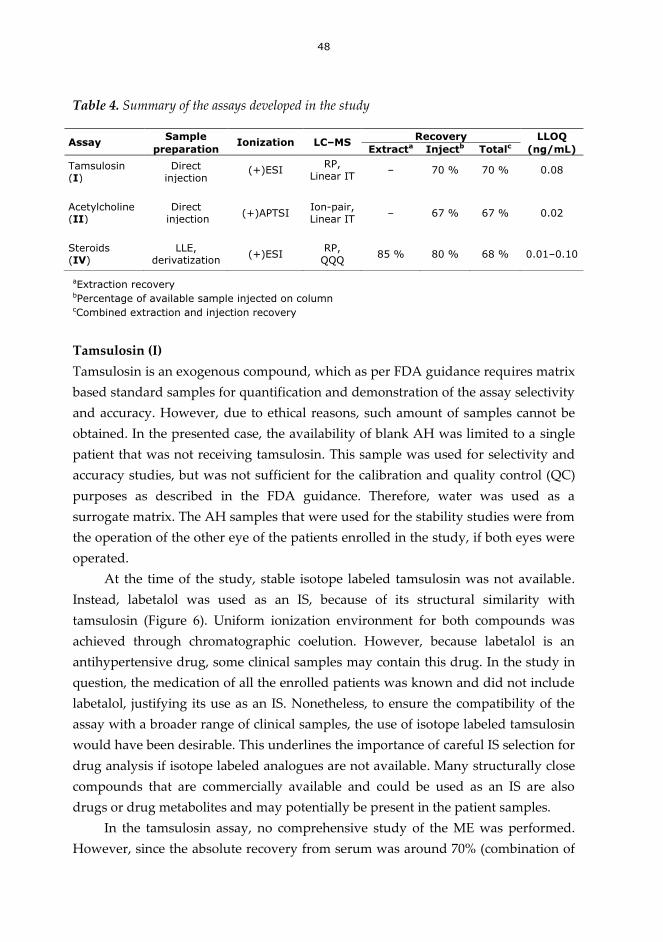
48
Table 4. Summary of the assays developed in the study
aExtraction recovery bPercentage of available sample injected on column cCombined extraction and injection recovery
Tamsulosin (I)
Tamsulosin is an exogenous compound, which as per FDA guidance requires matrix
based standard samples for quantification and demonstration of the assay selectivity
and accuracy. However, due to ethical reasons, such amount of samples cannot be
obtained. In the presented case, the availability of blank AH was limited to a single
patient that was not receiving tamsulosin. This sample was used for selectivity and
accuracy studies, but was not sufficient for the calibration and quality control (QC)
purposes as described in the FDA guidance. Therefore, water was used as a
surrogate matrix. The AH samples that were used for the stability studies were from
the operation of the other eye of the patients enrolled in the study, if both eyes were
operated.
At the time of the study, stable isotope labeled tamsulosin was not available.
Instead, labetalol was used as an IS, because of its structural similarity with
tamsulosin (Figure 6). Uniform ionization environment for both compounds was
achieved through chromatographic coelution. However, because labetalol is an
antihypertensive drug, some clinical samples may contain this drug. In the study in
question, the medication of all the enrolled patients was known and did not include
labetalol, justifying its use as an IS. Nonetheless, to ensure the compatibility of the
assay with a broader range of clinical samples, the use of isotope labeled tamsulosin
would have been desirable. This underlines the importance of careful IS selection for
drug analysis if isotope labeled analogues are not available. Many structurally close
compounds that are commercially available and could be used as an IS are also
drugs or drug metabolites and may potentially be present in the patient samples.
In the tamsulosin assay, no comprehensive study of the ME was performed.
However, since the absolute recovery from serum was around 70% (combination of
Assay Sample
Ionization LC–MS Recovery LLOQ
preparation Extracta Injectb Totalc (ng/mL)
Tamsulosin (I)
Direct injection
(+)ESI RP,
Linear IT – 70 % 70 % 0.08
Acetylcholine (II)
Direct injection
(+)APTSI Ion-pair, Linear IT
– 67 % 67 % 0.02
Steroids (IV)
LLE, derivatization
(+)ESI RP, QQQ
85 % 80 % 68 % 0.01–0.10

49
extraction recovery and the possible ME, calculated by comparing pure standard
solution with extracted sample), this points to the absence of strong ion suppression.
In the AH samples, ME was not studied due to the shortage of blank sample
material. However, assaying the spiked blank AH against water based standards
resulted in adequate accuracy, indicating that if present, the ion suppression was at
an acceptable level.
Using dedicated standard samples and separate quantification, both serum and
AH samples could be analyzed within the same run. During the tamsulosin
medication, the concentrations in the serum samples were expectedly high (mean
concentration 12.7 ng/ml or 31.1 nM), whereas the AH levels were low (mean
concentration 0.4 ng/ml or 0.98 nM). Since the LLOQs for serum and AH were 0.1
ng/ml (0.25 nM) and 0.08 ng/ml (0.20 nM), respectively, the concentrations of
tamsulosin were well within the sensitivity limits of the assay. After a pause of 7–28
days in the tamsulosin medication, most of the AH samples still had concentrations
above LLOQ (Pärssinen et al., 2006). However, to further study the kinetics and the
clearance of tamsulosin from the eye, more sensitive assay would be needed. It is
noteworthy that the sensitivity of the ion trap MS used for this study was not
comparable with the current QQQ instruments. However, this was the first
published assay for the determination of tamsulosin from the human AH, revealing
the ophthalmic penetration of the drug and its prolonged presence in the anterior
chamber of the eye, which had been recently found to complicate eye operations.
Acetylcholine (II)
At the time of its publication, the ACh assay was the first to use a stable isotope
labeled IS. Unlike the AH assay for tamsulosin, the availability of microdialysis
samples enabled the careful validation of the method. In this study, the absence of
sensitivity-affecting ME could be proved (Figure 14).
Probably due to the special ionization technique, the concentration range of the
assay was wider than in any of the previously published ESI based methods,
ranging from 0.15 nM (0.02 ng/ml) to 73 nM (10.7 ng/ml). The assay was used for the
determination of hippocampal levels of ACh in the rats treated to mimic Alzheimer’s
disease (Ihalainen et al, 2010; Ihalainen et al, 2011). All the study samples were
within the standard curves, requiring however, the full use of a wide concentration
range as the concentrations in the study samples were between 0.3 and 70 nM (0.04–
10.2 ng/ml).

50
Ion suppression time window
Figure 14. A post-column infusion chromatogram after injecting a rat brain
microdialysate sample. APTSI ionization, MRM transition of ACh monitored. Retention
time for ACh: 1.35 min.
Although the sensitivity of the developed assay enabled the collection of small-
volume samples, which enabled fine temporal resolution of the microdialysis
experiment, an acetylcholinesterase inhibitor was needed in the perfusion fluid to
prevent the enzymatic breakdown of ACh. This technique is commonly used to
increase the ACh levels in the microdialysis samples, but it is also controversial, as
the inhibitors may leak into the studied tissues and contribute to the biological
processes. For the measurement of ACh in the perfusates that do not contain
inhibitors, assays with significantly greater sensitivity are needed.
Steroids (IV)
In addition to the improvements in sensitivity in the ketosteroid analysis, the oxime
derivatization also improved the chromatographic selectivity, increasing the number
of analytes that could be included in the assay. Under identical chromatographic
conditions, the peaks of testosterone, 17-hydroxyprogesterone, and
dehydroepiandrosterone were overlapping, but became baseline-separated after
derivatization (Figure 15). This was important, as MS/MS selectivity could not be
obtained for these compounds due to their isobaric precursor ions or similar
fragmentation patterns.
RT: 0.00 - 5.00
0.0 0.5 1.0 1.5 2.0 2.5 3.0 3.5 4.0 4.5
Time (min)
0
10
20
30
40
50
60
70
80
90
100
Rela
tive A
bundance
NL: 3.12E5
m/z= 86.50-87.50 F: ITMS + c APCI !corona SRM ms2 [email protected] [ 84.80-89.80] MS 160407ACh17

51
Figure 15. Separation of testosterone, 17-hydroxyprogesterone, and
dehydroepiandrosterone after their derivatization to oximes. Identical chromatographic
method in both runs. Details of the separation system and conditions are described in
paper IV.
Depending on the analyte, the LLOQs were between 0.03 and 0.34 nM (0.01−0.10
ng/ml), enabling the analysis of all of the included steroids from adult female and
male serum. However, some important analytes had to be excluded due to their
insufficient response. Dihydrotestosterone could be measured from males, but not
from females. This compound, as well as androstanedione that could not be
detected from the serum of either sex, showed pronounced separation of the oxime
isomers, which resulted in decrease of peak height (Figure 16). Estrone was
detectable in the serum of both sexes, but its low levels resulted in poor precision.
Androsterone could not be chromatographically separated from its biologically
active isomer etiocholanolone. It also produced fragmentation that was identical
with etiocholanolone and hence these two coumpound could not be included in the
assay. Despite the shortcomings in the analysis of these compounds, the steroid
assay was the first to describe a simultaneous analysis of all of the main ketosteroid
metabolites of progestagen and androgen groups in both female and male serum.

52
Figure 16. Formation of chromatographically separable isomers during oxime
derivatization. Identical chromatographic method in both runs. Details of the separation
system and conditions are described in paper IV.
The ketosteroid assay was used for the serum steroid profiling of endometriosis
patients (n=155) and healthy controls (n=64). All the included analytes could be
reliably assayed with a typical throughput of 90–100 samples per day. The high
sensitivity of the assay was essential for the successful analysis of progesterone, 17-
hydroxyprogesterone, and 17-hydroxypregnenolone.

53
7 Summary and conclusions
In this thesis, different techniques to increase the sensitivity of bioanalytical LC–MS
assays were studied. The experimental work was focused on improving the sample
preparation and the analyte ionization, with two different approaches taken for both
stages of an assay. For the aqueous, protein-deficient samples with a volume below
50 µL, direct injection was employed. For the larger serum samples that could not be
injected as such, small-scale LLE technique was developed. Ionization of
permanently charged analyte ACh was improved by using a novel APTSI technique.
For poorly ionized ketosteroids, oxime derivatization was used in conjunction with
ESI. The techniques were implemented in three complete assays, with validation and
sample analysis data to support the evaluation of their performance. In addition, the
feasibility of using the APTSI technique at the microchip scale was evaluated in a
preliminary manner prior to further studies.
The assays demonstrate the feasibility of direct injection for aqueous samples
with a low protein content, such as AH or brain microdialysate. Diluting the sample
with IS increases the analyte recovery by diluting the solution that remains in the
vial after the injection. However, the injection volumes are limited by the analyte
polarity and column capacity. Increasing the injection volumes may not be a feasible
option when very small columns or particles with low mass loading capacities are
employed. Direct injection of aqueous samples also relies on RP or ion-pair
chromatography, since injection of aqueous samples into a HILIC column is not
feasible. For the serum samples that required sample pre-processing, LLE was used.
In the case of steroid analysis that required derivatization in addition to the
extraction, minimal sample loss was achieved by performing the entire process
using only two autosampler vials. This method is also feasible with large injection
volumes, though having the same limitations related to the capacity of the
separation column as the methods based on direct injection.
APTSI was found to be a remarkably suitable ionization technique for ACh
analysis, enabling the use of TFA in the mobile phase without ion-suppression.
Experiments with a heated microchip nebulizer highlighted the feasibility of using
APTSI with the microchip technique, enabling its use in combination with
microscale separation systems. For the first time, APTSI was compared against APCI
and ESI, and was found to result in ESI-like ionization of various compounds that

54
can be charged in solution. The low noise and the tolerance for ion-suppressing
mobile phase additions seem to be the major advantages of this ionization technique
over ESI. For the ketosteroid analytes with weak proton affinity, oxime
derivatization was used. This resulted in improved ionization efficiency, permitting
the use of ESI. The derivatization reaction could be efficiently incorporated into the
LLE process, maintaining the high analyte recovery of the sample preparation
method.
Future perspectives
The advantages of APTSI should be fully probed. The studies with APTSI and
different mobile phase additives suggest that APTSI may be less sensitive to matrix
based ion-suppression than ESI. In this case, APTSI could represent a feasible
alternative to ESI, when APCI and APPI cannot be used. In addition, a direct
comparison of standard-sized APTSI with ESI, APCI, and APPI would allow its
establishment among the other ionization techniques.
Due to the decreased dimensions, a microchip APTSI in combination with a
capillary column should offer significantly improved sensitivity for ACh analysis.
Performance characterization of the described setup in an analytical setup is needed
to allow comparisons against standard-sized systems. If the increase in sensitivity is
sufficient to allow the quantification of the baseline ACh levels in rat brain without
requiring the inclusion of acetylcholinesterase inhibitors in the perfusion fluid, it
would be the preferred technique. In addition, the smaller injection volumes
possible with the capillary systems would allow smaller sample volumes and
greater sampling frequency, increasing the temporal resolution of the microdialysis
experiments. However, it must be noted that the performance of the experimental
microchip used in this study was not optimal when the amount of organic solvent in
the mobile phase was below 10%. This indicates that the design of the microchips,
such as the efficiency of the heater and the type and dimensions of the vaporizer
may have strong effect on their performance.
In the present study, the IS dilution and mixing were performed manually in
the vials using a micropipette. To increase the method throughput, this step could be
performed using the autosampler functions. This would enable the collection of the
samples directly into the vials or well plates and their subsequent analysis without
any need to manually process the samples.
The described oxime derivatization method can only be used for compounds
containing a ketone functionality. Consequently, ionization of some steroids of

55
significant biological importance, such as estradiol, cannot be improved with this
technique. Other derivatization techniques exist, but they are not compatible with
the steroids described in this study. However, there is a great need for the
simultaneous measurement of estradiol and ketosteroids. To achieve this goal, an
approach different from the one described in this study would be needed. The use of
APPI is a promising alternative to older techniques in the analysis of underivatized
steroids, and together with suitable derivatization, further increase in the sensitivity
may be obtained, with the ability to analyze a greater range of steroids.

56

57
8 References
Abdel-Rehim M: New trend in sample preparation: on-line microextraction in
packed syringe for liquid and gas chromatography applications. I.
Determination of local anaesthetics in human plasma samples using gas
chromatography-mass spectrometry. J Chromatogr B 801: 317–321, 2004.
Abrahim A, Al-Sayah M, Skrdla P, Bereznitski Y, Chen Y, Wu N: Practical
comparison of 2.7 µm fused-core silica particles and porous sub-2 µm particles
for fast separations in pharmaceutical process development. J Pharm Biomed
Anal 51: 131–137, 2010.
Alpert AJ: Hydrophilic-interaction chromatography for the separation of peptides,
nucleic acids and other polar compounds. J Chromatogr 499: 177–196, 1990.
Alpert AJ: HILIC at 21: Reflections and perspective. J Chromatogr A 1218: 5879,
2011.
Amad MH, Cech NB, Jackson GS, Enke CG: Importance of gas-phase proton
affinities in determining the electrospray ionization response for analytes and
solvents. J Mass Spectrom 35: 784–789, 2000.
Andersen WC, Turnipseed SB, Karbiwnyk CM, Lee RH, Clark SB, Rowe WD,
Madson MR, Miller KE: Multiresidue method for the triphenylmethane dyes in
fish: Malachite green, crystal (gentian) violet, and brilliant green Anal Chim Acta
637: 279–289, 2009.
Arpino P: On-line liquid chromatography/mass spectrometry? An odd couple!
Trends Anal Chem 1: 154–158, 1982.
Baltussen E, Sandra P, David F, Cramers C: Stir bar sorptive extraction (SBSE), a
novel extraction technique for aqueous samples: Theory and principles. J
Microcolumn Sep 11: 737–747, 1999.
Bennett P: Future developments for triple quadrupole mass spectrometers: not just
hardware. Bioanalysis 3: 709–711, 2011.
Blakley CR, Carmody JJ, Vestal ML: Liquid chromatograph-mass spectrometer for
analysis of nonvolatile samples. Anal Chem 52: 1636–1641, 1980.
Blakley CR, Vestal ML: Thermospray interface for liquid chromatography/mass
spectrometry. Anal Chem 55: 750–754, 1983.

58
Blue LE, Jorgenson JW: 1.1 μm superficially porous particles for liquid
chromatography. Part I: Synthesis and particle structure characterization. J
Chromatogr 1218: 7989–7995, 2011.
Bristow T, Constantine J, Harrison M, Cavoit F: Performance optimisation of a new-
generation orthogonal-acceleration quadrupole-time-of-flight mass
spectrometer. Rapid Commun Mass Spectrom 22: 1213–1222, 2008.
Bruins AP, Covey TR, Henion JD: Ion spray interface for combined liquid
chromatography/atmospheric pressure ionization mass spectrometry. Anal
Chem 59: 2642–2646, 1987.
Carroll DI, Dzidic I, Stillwell RN, Haegele KD, Horning EC: Atmospheric pressure
ionization mass spectrometry: Corona discharge ion source for use in liquid
chromatograph-mass spectrometer-computer analytical system. Anal Chem 47:
2369–2373, 1975.
Carroll DI, Dzidic I, Horning EC, Stillwell RN: Atmospheric-pressure ionization
mass spectrometry. Appl Spectrosc Rev 17: 337–406, 1981.
Casetta B, Jans I, Billen J, Vanderschueren D, Bouillon R: Development of a method
for the quantification of 1 alpha,25(OH)(2)-vitamin D(3) in serum by liquid
chromatography tandem mass spectrometry without derivatization. Eur J Mass
Spectrom 16; 81–89, 2010.
Chowdhury UR, Madden BJ, Charlesworth MC, Fautsch MP: Proteome analysis of
human aqueous humor. Invest Ophthalmol Vis Sci 51: 4921–4931, 2010.
Constapel M, Schellenträger M, Schmitz OJ, Gäb S, Brockmann KJ, Giese R, Benter T:
Atmospheric-pressure laser ionization: a novel ionization method for liquid
chromatography/mass spectrometry. Rapid Commun Mass Spectrom 19: 326–
336, 2005.
Cristoni S, Bernardi LR, Biunno I, Guidugli F: Analysis of protein ions in the range
3000-12000 Th under partial (no discharge) atmospheric pressure chemical
ionization conditions using ion trap mass spectrometry. Rapid Commun Mass
Spectrom 16: 1153–1159, 2002.
Dole M, Mack LL, Hines RL, Mobley RC, Ferguson LD, Alice MB: Molecular beams
of macroions. J Chem Phys 49: 2240–2250, 1968.
Douglas DJ, Frank AJ, Mao DM: Linear ion traps in mass spectrometry. Mass
Spectrom Rev 24: 1–29, 2005.
Dzidic I, Carroll DI, Stillwell RN, Horning EC: Gas phase reactions. Ionization by
proton transfer to superoxide anions. J Am Chem Soc 96: 5258–5259, 1974.

59
Enke CG: A predictive model for matrix and analyte effects in electrospray
ionization of singly-charged ionic analytes. Anal Chem 69: 4885–4893, 1997.
European Commission: Commission Decision of 12 August 2002 implementing
Council Directive 96/23/EC concerning the performance of analytical methods
and the interpretation of results (2002/657/EC). Off J Eur Communities 221: 8–36,
2002.
Favretto D, Nalesso A, Frison G, Viel G, Traldi P, Ferrara SD: No-discharge
atmospheric pressure chemical ionization mass spectrometry of ethyl
glucuronide and ethyl sulfate. J Mass Spectrom 45: 121–124, 2010.
Fekete S, Ganzler K, Fekete J: Facts and myths about columns packed with sub-3 μm
and sub-2 μm particles. J Pharm Biomed Anal 51: 56–64, 2010.
Fenn, JB, Mann M, Meng CK, Wong SF, Whitehouse CM: Electrospray ionization for
mass spectrometry of large biomolecules. Science 246: 64–71, 1989.
FDA, Food and Drug Administration: Guidance for Industry. Bioanalytical method
validation. Rockville, MD: US Department of Health and Human Services, FDA,
CDER; 2001.
Fung EN, Xia YQ, Aubry AF, Zeng J, Olah T, Jemal M: Full-scan high resolution
accurate mass spectrometry (HRMS) in regulated bioanalysis: LC-HRMS for the
quantitation of prednisone and prednisolone in human plasma. J Chromatogr B
879: 2919–2927, 2011.
Gao S, Zhang Z-P, Karnes HT: Sensitivity enhancement in liquid
chromatography/atmospheric pressure ionization mass spectrometry using
derivatization and mobile phase additives. J Chromatogr B 825: 98–110, 2005.
Gallagher RT, Balogh MP, Davey P, Jackson MR, Sinclair I, Southern LJ: Combined
electrospray ionization-atmospheric pressure chemical ionization source for use
in high-throughput LC-MS applications. Anal Chem 75: 973–977, 2003.
Gergov M, Ojanperä I, Vuori E: Simultaneous screening for 238 drugs in blood by
liquid chromatography–ionspray tandem mass spectrometry with multiple-
reaction monitoring. J Chromatogr B 795: 41–53, 2003.
Goel M, Picciani RG, Lee RK, Bhattacharya SK: Aqueous humor dynamics: A review.
Open Ophthalmol J 4: 52–59, 2010.
Gomez A, Tang K: Charge and fission of droplets in electrostatic sprays. Phys Fluids
6: 404–414, 1994.

60
Görög S: The changing face of pharmaceutical analysis. Trends Anal Chem 26: 12–17,
2007.
Hagestam IH and Pinkerton TC: Internal surface reversed-phase silica supports for
liquid chromatography. Anal Chem 57: 1757–1763, 1985.
Harwood DT and Handelsman DJ: Development and validation of a sensitive liquid
chromatography–tandem mass spectrometry assay to simultaneously measure
androgens and estrogens in serum without derivatization. Clin Chim Acta 409:
78–84, 2009.
Hewavitharana AK: Matrix matching in liquid chromatography–mass spectrometry
with stable isotope labelled internal standards—Is it necessary? J Chromatogr A
1218: 359–361, 2011.
Higashi T and Shimada K: Derivatization of neutral steroids to enhance their
detection characteristics in liquid chromatography-mass spectrometry. Anal
Bioanal Chem 386: 658–665, 2006.
Hirabayashi A, Sakairi M, Koizumi H: Sonic spray ionization method for
atmospheric pressure ionization mass spectrometry. Anal Chem 66: 4557–4559,
1994.
Hommerson P, Khan AM, Bristow T, Niessen WMA, de Jong GJ, Somsen GW:
Photon-independent gas-phase-ion formation in capillary electrophoresis- mass
spectrometry using atmospheric pressure photoionization. Anal Chem 79: 5351–
5357, 2007.
Horning EC, Carroll DI, Dzidic I, Haegele KD, Horning MG, Stillwell RN: Liquid
chromatography-mass spectrometer-computer analytical systems: a continuous
flow system based on atmospheric pressure ionization mass spectrometry. J
Chromatogr 99: 13–21, 1974a.
Horning EC, Carroll DI, Dzidic I, Haegele KD, Horning MG, Stillwell RN:
Atmospheric pressure ionization (API) mass spectrometry. Solvent-mediated
ionization of samples introduced in solution and in a liquid chromatograph
effluent stream. J Chromatogr Sci 12: 725–729, 1974b.
ICH, The international conference on harmonisation of technical requirements for
registration of pharmaceuticals for human use: Validation of analytical
procedures: text and methodology Q2(R1), 2005.
Ihalainen J, Sarajärvi T, Kemppainen S, Keski-Rahkonen P, Lehtonen M, Tanila H: A
novel delayed non-match to sample object recognition task that allows
simultaneous in vivo microdialysis. J Neurosci Methods 189: 2010, 210–215.

61
Ihalainen J, Sarajärvi T, Rasmusson D, Kemppainen S, Keski-Rahkonen P, Lehtonen
M, Banerjee PK, Semba K, Tanila H: Effects of memantine and donepezil on
cortical and hippocampal acetylcholine levels and object recognition memory in
rats. Neuropharmacology 61: 2011, 891–899.
Ikonomou MG and Kebarle P: A heated electrospray source for mass spectrometry
of analytes from aqueous solutions. J Am Soc Mass Spectrom 5: 791–799, 1994.
ISO/IEC 17025:2005: General requirements for the competence of testing and
calibration laboratories, Geneva, 2005.
Iwasaki Y, Nakano Y, Mochizuki K, Nomoto M, Takahashi Y, Ito R, Saito K,
Nakazawa H: A new strategy for ionization enhancement by derivatization for
mass spectrometry. J Chromatogr B 879: 1159–1165, 2011.
Jacobson B-M, Olsson A, Fakt C, Öhman D: The use of human plasma as matrix for
calibration standards in pre-clinical LC–MS/MS methods—A way to reduce
animal use. J Pharm Biomed Anal 54: 826–829, 2011.
Jinno K, Muramatsu T, Saito Y, Kiso Y, Magdic S, Pawliszyn J: Analysis of pesticides
in environmental water samples by solid-phase micro-extraction-high-
performance liquid chromatography. J Chromatogr A 754: 137–144, 1996.
Kalhorn TF, Page ST, Howald WN, Mostaghel EA, Nelson PS: Analysis of
testosterone and dihydrotestosterone from biological fluids as the oxime
derivatives using high-performance liquid chromatography/tandem mass
spectrometry. Rapid Commun Mass Spectrom 21: 3200–3206, 2007.
Kauppila TJ, Kuuranne T, Meurer EC, Eberlin MN, Kotiaho T, Kostiainen R:
Atmospheric pressure photoionization mass spectrometry. Ionization
mechanism and the effect of solvent on the ionization of naphthalenes. Anal
Chem 74: 5470–5479, 2002.
Kauppila T, Kostiainen R, Bruins A: Anisole, a new dopant for atmospheric pressure
photoionization mass spectrometry of low proton affinity, low ionization energy
compounds. Rapid Commun Mass Spectrom 18: 808–815, 2004a.
Kauppila TJ, Östman P, Marttila SJ, Ketola RA, Kotiaho T, Franssila S, Kostiainen R:
Atmospheric pressure photoionization-mass spectrometry with a microchip
heated nebulizer. Anal Chem 76: 6797–6801, 2004b.
Kaufmann A, Butcher P, Maden K, Walker S, Widmer M: Development of an
improved high resolution mass spectrometry based multi-residue method for
veterinary drugs in various food matrices. Anal Chim Acta 700: 86–94, 2011.

62
Kebarle P and Ho Y: On the mechanism of electrospray mass spectrometry. In:
Electrospray ionization mass spectrometry: fundamentals, instrumentation, and
applications. John Wiley: New York, 1997, pp. 3–63.
Kebarle P and Verkerk UH: Electrospray: from ions in solution to ions in the gas
phase, what we know now. Mass Spectrom Rev 28: 898–917, 2009.
King R, Bonfiglio R, Fernandez-Metzler C, Miller-Stein C, Olah T: Mechanistic
investigation of ionization suppression in electrospray ionization. J Am Soc Mass
Spectrom 11: 942–950, 2000.
Kole PL, Venkatesh G, Kotechac J, Sheshalad R: Recent advances in sample
preparation techniques for effective bioanalytical methods. Biomed Chromatogr
25; 199–217, 2011.
Koster S, Verpoorte E: A decade of microfluidic analysis coupled with electrospray
mass spectrometry: An overview. Lab Chip 7: 1394–1412, 2007.
Kostiainen R, Kauppila TJ: Effect of eluent on the ionization process in liquid
chromatography-mass spectrometry. J Chromatogr A 1216: 685–699, 2009.
Kuhlmann FE, Apffel A, Fischer SM, Goldberg G, Goodley PC: Signal enhancement
for gradient reverse-phase high-performance liquid chromatography
electrospray ionization mass spectrometry analysis with trifluoroacetic and
other strong acid modifiers by postcolumn addition of propionic acid and
isopropanol. J Am Soc Mass Spectrom 6: 1221–1225, 1995.
Kushnir MM, Rockwood AL, Roberts WL, Pattison EG, Bunker AM, Fitzgerald RL,
Meikle AW. Performance characteristics of a novel tandem mass spectrometry
assay for serum testosterone. Clin Chem 52: 120–128, 2006.
Lee JW, Devanaryan V, Barrett YC, Weiner R, Allinson J, Fountain S, Keller S,
Weinryb I, Green M, Duan L, Rogers JA, Millham R, O’Brien, Sailstad J, Khan M,
Ray C, Wagner JA: Fit-for-purpose method development and validation for
successful biomarker measurement. Pharm Res 23: 312–328, 2006.
Leinonen A, Kuuranne T, Kostiainen R: Liquid chromatography/mass spectrometry
in anabolic steroid analysis—optimization and comparison of three ionization
techniques: electrospray ionization, atmospheric pressure chemical ionization
and atmospheric pressure photoionization. J Mass Spectrom 37: 693–698, 2002.
Liu S, Sjövall J, Griffiths WJ: Analysis of oxosteroids by nano-electrospray mass
spectrometry of their oximes. Rapid Commun Mass Spectrom 14: 390–400, 2000.

63
Loboda A, Krutchinsky A, Loboda O, McNabb J, Spicer V, Ens W, Standing KG:
Novel Linac II electrode geometry for creating an axial field in a multipole ion
guide. Eur J Mass Spectrom 6: 531–536, 2000.
Long AR, Hsieh LC, Malbrough MS, Short CR, Barker SA: Matrix solid-phase
dispersion (MSPD) isolation and liquid chromatographic determination of
oxytetracycline, tetracycline, and chlortetracycline in milk. J Assoc Off Anal
Chem 73: 379–384, 1990.
Ma Q, Chen M, Yin H-R, Shi Z-G, Feng Y-Q: Preparation of pH-responsive
stationary phase for reversed-phase liquid chromatography and hydrophilic
interaction chromatography. J Chromatogr A 1212: 61–67, 2008.
Ma Y-C and Kim H-Y: Determination of Steroids by Liquid Chromatography/Mass
Spectrometry. J Am Soc Mass Spectrom 8: 1010–1020, 1997.
Majors RE: Current Trends in HPLC Column Usage. LC GC N AM 27: 956–972, 2009.
Majors RE: Highlights of HPLC 2011. LC GC Eur 24: 468–480, 2011.
Maleknia S and Brodbelt J: High energy collision-induced dissociation of alkali-
metal ion adducts of crown ethers and acyclic analogs. Rapid Commun Mass
Spectrom 6: 376–381, 1992.
Mann M, Meng CK, Fenn JB: Interpreting mass spectra of multiply charged ions.
Anal Chem 61: 1702–1708, 1989.
Marchi I, Rudaz S, Selman M, Veuthey J-L: Evaluation of the influence of protein
precipitation prior to on-line SPE-LC-API/MS procedures using multivariate
data analysis. J Chromatogr B 845: 244–252, 2007.
Marchi I, Rudaz S, Veuthey J-L: Sample preparation development and matrix effects
evaluation for multianalyte determination in urine. J Pharm Biomed Anal 49:
459–467, 2009.
Marginean I, Kelly RT, Prior DC, LaMarche BL, Tang K, Smith RD: Analytical
Characterization of the electrospray ion source in the nanoflow regime. Anal
Chem 80: 6573–6579, 2008.
Matuszewski BK, Constanzer ML, Chavez-Eng CM: Matrix effects in quantitative
LC–MS–MS analysis of biological fluids: A method for determination of
finasteride in human plasma at pg/ml concentrations. Anal Chem. 70: 882–889,
1998.

64
Matuszewski BK, Constanzer ML, Chavez-Eng CM: Strategies for the assessment of
matrix effect in quantitative bioanalytical methods based on HPLC–MS–MS.
Anal Chem 75: 3019–3030, 2003.
Meyer VR: The 1000 bar and 24 hour limits of one-dimensional HPLC – graphical
representations. Anal Bioanal Chem 396: 1633–1640, 2010.
Mitchell RJ, Christian R, Hughes H, Miah A, Walker DK: The application of fully
automated on-line solid phase extraction in bioanalysis. J Pharm Biomed Anal
52: 86–92, 2010.
Niessen WMA: State-of-the-art in liquid chromatography–mass spectrometry. J
Chromatogr A 856: 179–197, 1999.
Niessen WMA: Liquid chromatography-mass spectrometry. CRC Press: Boca Raton,
2006.
Nogueira R, Lämmerhofer M, Lindner W: Alternative high-performance liquid
chromatographic peptide separation and purification concept using a new
mixed-mode reversed-phase/weak anion-exchange type stationary phase. J
Chromatogr A 1089: 158–169, 2005.
Nordström A, Want E, Northen T, Lehtiö J, Siuzdak G: Multiple ionization mass
spectrometry strategy used to reveal the complexity of metabolomics. Anal
Chem 80: 421–429, 2008.
Nováková L, Vlcková H: A review of current trends and advances in modern bio-
analytical methods: chromatography and sample preparation. Anal Chim Acta
656: 8–35, 2009.
Oláh E, Fekete S, Fekete J, Ganzler K: Comparative study of new shell-type, sub-2
micron fully porous and monolith stationary phases, focusing on mass-transfer
resistance. J Chromatogr A 1217: 3642–3653, 2010.
Pelander A; Decker P; Baessmann C; Ojanperä I: Evaluation of a high resolving
power time-of-flight mass spectrometer for drug analysis in terms of resolving
power and acquisition rate. J Am Soc Mass Spectrom 22: 379–385, 2011.
Petersson P, Forssen P, Edström L, Samie F, Tatterton S, Clarke A, Fornstedt T: Why
ultra high performance liquid chromatography produces more tailing peaks
than high performance liquid chromatography, why it does not matter and how
it can be addressed. J Chromatogr A 1218: 6914–6921, 2011.

65
Petucci C, Lloyd T, Harris HA, Zhang X, Chennathukuzhi VM, Mekonnen B, Cai Y:
Trace LC/MS/MS quantitation of 17β-estradiol as a biomarker for selective
estrogen receptor modulator activity in the rat brain. J Mass Spectrom 45: 65–71,
2010.
Pucci V, Di Palma S, Alfieri A, Bonelli F, Monteagudo E: A novel strategy for
reducing phospholipids-based matrix effect in LC-ESI-MS bioanalysis by means
of HybridSPE. J Pharm Biomed Anal 50; 867–871, 2009.
Pärssinen O, Leppänen E, Keski-Rahkonen P, Mauriala T, Dugue B, Lehtonen M:
Influence of tamsulosin on the iris and its implications for cataract surgery.
Invest Ophthalmol Vis Sci 47: 2006, 3766–3771.
Ramanathan R, Jemal M, Ramagiri S, Xia Y-Q, Humpreys WG, Olah T, Korfmacher
WA: It is time for a paradigm shift in drug discovery bioanalysis: from SRM to
HRMS. J Mass Spectrom 46: 595–601, 2011.
Robb DB and Blades MW: Factors affecting primary ionization in dopant-assisted
atmospheric pressure photoionization (DA-APPI) for LC/MS. J Am Soc Mass
Spectrom 17: 130–138, 2006.
Robb DB, Smith DR, Blades MW: Investigation of substituted-benzene dopants for
charge exchange ionization of nonpolar compounds by atmospheric pressure
photoionization. J Am Soc Mass Spectrom 19: 955–963, 2008.
Robb DB and Blades MW: Effects of solvent flow, dopant flow, and lamp current on
dopant-assisted atmospheric pressure photoionization (DA-APPI) for LC–MS.
Ionization via proton transfer. J Am Soc Mass Spectrom 16: 1275–1290, 2005.
Robb DB, Covey TR, Bruins AP: Atmospheric pressure photoionization: an
ionization method for liquid chromatography-mass spectrometry. Anal Chem
72: 3653–3659, 2000.
Robb DB, Blades MW: An electropneumatic-heated nebulizer for enhancing spray
ionization in PhotoSpray atmospheric pressure photoionization sources for
liquid chromatography/mass spectrometry. Rapid Commun Mass Spectrom 23:
3394–3400, 2009.
Romero-González R, Aguilera-Luiz MM, Plaza-Bolaños P, Garrido Frenich A,
Martínez Vidal JL: Food contaminant analysis at high resolution mass
spectrometry: Application for the determination of veterinary drugs in milk. J
Chromatogr A 1218: 9353–9365, 2011.

66
Rustad P, Felding P, Franzson L, Kairisto V, Lahti A, Mårtensson A, Hyltoft Petersen
P, Simonsson P, Steensland H, Uldall A: The nordic reference interval project
2000: recommended reference intervals for 25 common biochemical properties.
Scand J Clin Lab Invest 64: 271–284, 2004.
Ryhage R: Use of a mass spectrometer as a detector and analyzer for effluent
emerging from high temperature gas liquid chromatography columns. Anal
Chem 36: 759–764, 1964.
Saarela V, Haapala M, Kostiainen R, Kotiaho T, Franssila S. Glass microfabricated
nebulizer chip for mass spectrometry. Lab Chip 7:644–646, 2007.
Saito K, Yagi K, Ishizaki A, Kataoka H: Determination of anabolic steroids in human
urine by automated in-tube solid-phase microextraction coupled with liquid
chromatography–mass spectrometry. J Pharm Biomed Anal 52; 727–733, 2010.
Sakairi M, Kato Y: Multi-atmospheric pressure ionisation interface for liquid
chromatography-mass spectrometry. J Chromatogr A 794: 391–406, 1998.
Sakamoto S, Fujita M, Kim K, Yamaguchi K: Characterization of self-assembling
nano-sized structures by means of coldspray ionization mass spectrometry.
Tetrahedron 56: 955–964, 2000.
Šatínský D, Solich P: Using restricted-access materials and column switching in
high-performance liquid chromatography for direct analysis of biologically-
active compounds in complex matrices. Trends Anal Chem 26: 375–384, 2007.
Schmidt A, Karas M, Dulcks T: Effect of different solution flow rates on analyte ion
signals in nano-ESI MS, or: When does ESI turn into nano-ESI? J Am Soc Mass
Spectrom 14: 492–500, 2003.
Schwartz JC, Senk MW, Syka JEP: A two-dimensional quadrupole ion trap mass
spectrometer. J Am Soc Mass Spectrom 13: 659–669, 2002.
Shipkova P, Drexler DM, Langish R, Smalley J, Salyan ME, Sanders M: Application
of ion trap technology to liquid chromatography/mass spectrometry
quantitation of large peptides. Rapid Commun Mass Spectrom 22: 1359–1366,
2008.
Short LC and Syage JA: Electrospray photoionization (ESPI) liquid
chromatography/mass spectrometry for the simultaneous analysis of
cyclodextrin and pharmaceuticals and their binding interactions. Rapid
Commun Mass Spectrom 22: 541–548, 2008.
Sikanen T, Franssila S, Kauppila TJ, Kostiainen R, Kotiaho T, Ketola RA: Microchip
technology in mass spectrometry. Mass Spectrom Rev 29: 351–391. 2010.

67
Singh G, Gutierrez A, Xu KY, Blair IA: Liquid chromatography/electron capture
atmospheric pressure chemical ionization/mass spectrometry: Analysis of
pentafluorobenzyl derivatives of biomolecules and drugs in the attomole range.
Anal Chem 72: 3007–3013, 2000.
Skonberg C, Artmann A, Cornett C, Hansen SH, Hansen HS: Pitfalls in the sample
preparation and analysis of N-acylethanolamines. J Lipid Res 51: 3062–3073,
2010.
Snyder LR, Kirkland JJ, Dolan JW: Introduction to modern liquid chromatographty,
3rd ed., John Wiley & Sons, Inc. Hoboken, New Jersey 2010.
Souverain S, Rudaz S, Veuthey J-L: Matrix effect in LC-ESI-MS and LC-APCI-MS
with off-line and on-line extraction procedures. J Chromatogr A 1058: 61–66,
2004a.
Souverain S, Rudaz S, Veuthey JL: Restricted access materials and large particle
supports for on-line sample preparation: an attractive approach for biological
fluids analysis. J Chromatogr B 801: 141–156, 2004b.
Syage JA, Evans MD, Hanold KA: Photoionization mass spectrometry. Am Lab 32:
24–29, 2000.
Sørensen MB, Aaslo P, Egsgaard H, Lund T: Determination of D/L-amino acids by
zero needle voltage electrospray ionization. Rapid Commun Mass Spectrom 22:
455–461, 2008.
Syage JA, Hanold KA, Lynn TC, Horner JA, Thakur RA: Atmospheric pressure
photoionization. II. Dual source ionization. J Chromatogr A 1050: 137–149, 2004.
Takeuchi T: Modern aspects of micro-bore column HPLC. Fresenius J Anal Chem
337: 631–637, 1990.
Taylor GI: Disintegration of Water Drops in an Electric Field. Proc R Soc Lond A 280:
383–397, 1964.
Tong X, Ita IE, Wang J, Pivnichny JV: Characterization of a technique for rapid
pharmacokinetic studies of multiple co-eluting compounds by LC:MS:MS. J
Pharm Biomed Anal 20: 773–784, 1999.
Torbeck L: Complying with ICH and FDA Requirements for assay validation.
Suffield Press, Evanston, IL, USA, 2002.
Trufelli H, Palma P, Famiglini G, Cappiello A: An overview of matrix effects in
liquid chromatography-mass spectrometry. Mass Spectrom Rev 30: 491–509,
2011.

68
Turnipseed SB, Andersen WC, Karbiwnyk CM, Roybal JE, Miller KE: No-discharge
atmospheric pressure chemical ionization: evaluation and application to the
analysis of animal drug residues in complex matrices. Rapid Commun Mass
Spectrom 20: 1231–1239, 2006.
Unger KK, Ditz R, Machtejevas E, Skudas R: Liquid chromatography–its
development and key role in life science applications. Angew Chem Int Ed Engl
49: 2300–2312, 2010.
Unger KK, Skudas R, Schulte MM: Particle packed columns and monolithic columns
in high-performance liquid chromatography-comparison and critical appraisal. J
Chromatogr A 1184: 393–415, 2008.
van Deemter JJ, Zuiderweg FJ Klinkenberg A: Longitudinal diffusion and resistance
to mass transfer as causes of non ideality in chromatography. Chem Eng Sci 5:
271–289, 1956.
van de Merbel NC: Quantitative determination of endogenous compounds in
biological samples using chromatographic techniques. Trends Anal Chem 27:
924–933, 2008.
Viswanathan CT, Bansal S, Booth B, DeStefano AJ, Rose MJ, Sailstad J, Shah VP,
Skelly JP, Swann PG, Weiner R: Workshop/conference report—quantitative
bioanalytical methods validation and implementation: best practices for
chromatographic and ligand binding assays. AAPS J 9: E30–E42, 2007.
Voyksner RD: Combining liquid chromatography with electrospray mass
spectrometry. In: Electrospray Ionization Mass Spectrometry: Fundamentals,
Instrumentation, and Applications. John Wiley: New York, 1997, pp. 323–341.
West J, Becker M, Tombrink S, Manz A: Micro total analysis systems: Latest
achievements. Anal Chem 80: 4403–4419, 2008.
Williamson LN, Zhang G, Terry AV Jr, Bartlett MG: Comparison of time-of-flight
mass spectrometry to triple quadrupole tandem mass spectrometry for
quantitative bioanalysis: application toantipsychotics. J Liq Chrom Rel Tech 31:
2737–2751, 2008.
Wood TD, Moy MS, Dolan AR, Bigwarfe PM Jr, White TP, Smith DR, Higbee DJ:
Miniaturization of electrospray ionization mass spectrometry. Appl Spectrosc
Rev 38: 187–244, 2003.
Yamashita M, Fenn JB: Electrospray ion source. Another variation on the free-jet
theme. J Phys Chem 88: 4451–4459, 1984a.

69
Yamashita M, Fenn JB: Negative ion production with the electrospray ion source. J
Phys Chem, 88: 4671–4675, 1984b.
Yin HF, Killeen K: The fundamental aspects and applications of Agilent HPLC-Chip.
J Sep Sci 30: 1427–1434, 2007.
Yuan C, Kosewick J, He X, Kozak M, Wang SH: Sensitive measurement of serum
1α,25-dihydroxyvitamin D by liquid chromatography/tandem mass
spectrometry after removing interference with immunoaffinity extraction. Rapid
Commun Mass Spectrom 25; 1241–1249, 2011.
Zhang NR, Yu S, Tiller P, Yeh S, Mahan E, Emary WB: Quantitation of small
molecules using high-resolution accurate mass spectrometers - a different
approach for analysis of biological samples. Rapid Commun Mass Spectrom 23:
1085–1094, 2009.
Östman P, Marttila SJ, Kotiaho T, Franssila S, Kostiainen R: Microchip atmospheric
pressure chemical ionization source for mass spectrometry. Anal Chem 76: 6659–
6664, 2004.


Publications of the University of Eastern Finland
Dissertations in Health Sciences
isbn 978-952-61-0598-7
Publications of the University of Eastern FinlandDissertations in Health Sciences
dissertatio
ns | 0
84 | Pek
ka K
eski-R
ah
ko
nen
| New
LC
-MS
Assays for D
rugs an
d En
dogenou
s Com
poun
ds in Sm
all-Volum
e Biological S
amples
Pekka Keski-RahkonenNew LC-MS Assays for Drugs
and Endogenous Compounds in Small-Volume Biological
SamplesPekka Keski-Rahkonen
New LC-MS Assays for Drugs and Endogenous Compounds in Small-Volume Biological Samples
This thesis presents alternative
approaches for bioanalytical
method development using liquid
chromatography–mass spectrometry.
It explores the feasibility of using
atmospheric pressure chemical
ionization without corona discharge,
the use of this technique on a
microchip scale, derivatization of
non-polar analytes for improved
electrospray ionization, and different
pre-processing methods for sub-150 µl
biological samples.