pelvic limb alignment in small breed dogs: a … · of butorphanol (nargesic, acme srl, cavriago,...
TRANSCRIPT
45
Veterinaria Italiana 2015, 52 (1), 45-50. doi: 10.12834/VetIt.71.206.3Accepted: 05.08.2015 | Available on line: 17.12.2015
SummarySmall breed dogs are 12 times more likely to develop medial patellar luxation (MPL) than large breed dogs and breed predisposition has been reported. Many surgical techniques are available for correction of patellar luxation in dogs. However, recent studies reported an 8% incidence of reluxation when traditional techniques are used. The relatively high frequency of major complications and patellar reluxation may be partially caused by inadequate appreciation of the underlying skeletal deformity and subsequent incorrect selection and application of traditional techniques. The aims of this study were to report the normal values of the anatomic and mechanical joint angles of the femur and tibia in small breed dogs and to compare these data to a population of small breed dogs affected by different degrees of MPL. Normal values of the anatomic and mechanical angles of the femur are similar to the ones reported in literature in Pomeranian dogs. Normal values of the anatomic and mechanical angles of the tibia have been described for the first time. Significant differences were found between normal population and dogs affected by grade 4 MPL in relation to anatomical Lateral Distal Femoral Angle (aLDFA), mechanical Medial Proximal Tibial Angle (mMPTA), and mechanical Caudal Proximal Tibial Angle (mCaPTA).
RiassuntoI cani di piccola taglia sono 12 volte più predisposti a sviluppare lussazione mediale di rotula rispetto a quelli di taglia grande. Sono state proposte molte tecniche chirurgiche per il trattamento della lussazione di rotula, ma studi recenti riportano un’incidenza di recidiva del 8% quando viene eseguito il trattamento con tecniche tradizionali. Tale incidenza di recidiva, in seguito all’esecuzione di tecniche tradizionali, è imputabile all’inaccurata valutazione dell’allineamento dell’arto pelvico. Lo scopo di questo studio era: riportare i valori fisiologici dell’allineamento di femore e tibia nei cani sani di piccola taglia e di confrontare questi valori con quelli di pazienti affetti da diversi gradi di lussazione di rotula.
L’allineamento dell’arto pelvico nei cani di piccola taglia: una comparazione tra soggetti sani e affetti da lussazione mediale di rotula
Parole chiaveAllineamento dell’arto pelvico,Cani di piccola taglia,Lussazione mediale di rotula.
KeywordsMedial patellar luxation,Pelvic limb alignment,Small breed dogs.
Struttura Didattica Speciale Veterinaria, L.go P. Braccini, 10095 Grugliasco (TO), Italy.
* Corresponding author at: Struttura Didattica Speciale Veterinaria, L.go P. Braccini, 10095 Grugliasco (TO), Italy.Tel.: +39 011 6709061, e‑mail: [email protected].
Matteo Olimpo*, Lisa Adele Piras & Bruno Peirone
Pelvic limb alignment in small breed dogs:a comparison between affected and free subjects
from medial patellar luxation
LXVII Meeting of the Italian Society for Veterinary Sciences (SISVet) ‑ Selected papers
(Hayes and Boudreiau 1994, Hulse1981, LaFond et al. 1992, Piermattei and Flo 1997, Trotter 1980). It has been reported as the most common congenital pathology (7.2%) in 1679 immature dogs (Ruble and Hird 1993). In more details, small-breed dogs are 12 times more likely to develop MPL than large breed dogs (Hayes and Boudreiau
IntroductionMedial patellar luxation (MPL), described as the most common orthopaedic disease affecting canine stifle (Campbell et al. 1972, Denny and Minter 1973, Flo and Brinker1970, Priester 1972, Singleton 1969), occurs bilaterally in 52.4-65% of affected dogs
46 Veterinaria Italiana 2016, 52 (1), 45-50. doi: 10.12834/VetIt.71.206.3
Pelvic limb alignment and medial patellar luxation in small breed dogs Olimpo et al.
best surgical option while considering the severity and degree of clinical signs. While the prevalence of MPL is higher in small-breed dogs, the femoral angles are only reported in 2 articles, 1 delving into normal femora and 1 focusing on femora with MPL (Soparat et al. 2010, Fasanella et al. 2011). In this study we compare the normal values of the joint angles of the femur and tibia in small breed dogs with the data obtained by dogs affected by MPL.
Materials and methods
Inclusion criteriaNon-chondrodystrophic dogs weighting less than 7.5 kg were included in the study. Breed, age, weight, sex, MPL degree and the affected stifle were recorded for each dog.
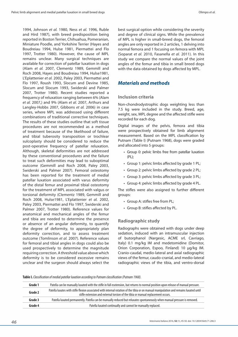
Digital images of the pelvis, femora and tibia were prospectively obtained for limb alignment measurement. Based on the MPL classification by Putnam (Table I) (Putnam 1968), dogs were graded and allocated into 5 groups:
• Group 0: pelvic limbs free from patellar luxation (PL);
• Group 1: pelvic limbs affected by grade 1 PL;
• Group 2: pelvic limbs affected by grade 2 PL;
• Group 3: pelvic limbs affected by grade 3 PL;
• Group 4: pelvic limbs affected by grade 4 PL.
The stifles were also assigned to further different groups:
• Group A: stifles free from PL;
• Group B: stifles affected by PL.
Radiographic studyRadiographs were obtained with dogs under deep sedation, induced with an intramuscular injection of butorphanol (Nargesic, ACME srl, Cavriago, Italy) 0.1 mg/kg IM and medetomidine (Domitor, Orion Corporation, Espoo, Finland) 10 μg/kg IM. Cranio-caudal, medio-lateral and axial radiographic views of the femur, caudo-cranial, and medio-lateral radiographic views of the tibia, and ventro-dorsal
1994, Johnson et al. 1980, Ness et al. 1996, Ruble and Hird 1987), with breed predisposition being reported in Boston Terrier, Chihuahua, Pomeranian, Miniature Poodle, and Yorkshire Terrier (Hayes and Boudreiau 1994, Hulse 1981, Piermattei and Flo 1997, Trotter 1980). However, the cause of MPL remains unclear. Many surgical techniques are available for correction of patellar luxation in dogs (Alam et al. 2007, Clementz 1989, Gemmill and Roch 2008, Hayes and Boudreiau 1994, Hulse1981, L’Eplattenier et al. 2002, Paley 2003, Piermattei and Flo 1997, Roush 1993, Slocum and Devine 1985, Slocum and Slocum 1993, Swiderski and Palmer 2007, Trotter 1980). Recent studies reported a frequency of reluxation ranging between 6% (Alam et al. 2007,) and 9% (Alam et al. 2007, Arthurs and Langley-Hobbs 2007, Gibbons et al. 2006) in case series, where MPL was addressed using different combinations of traditional corrective techniques. The results of these studies outline that soft tissue procedures are not recommended as a method of treatment because of the likelihood of failure, and tibial tuberosity transposition or trochlear sulcoplasty should be considered to reduce the post-operative frequency of patellar reluxation. Although, skeletal deformities are not addressed by these conventional procedures and the failure to treat such deformities may lead to suboptimal outcome (Gemmill and Roch 2008, Paley 2003, Swiderski and Palmer 2007). Femoral osteotomy has been reported for the treatment of medial patellar luxation associated with varus deformity of the distal femur and proximal tibial osteotomy for the treatment of MPL associated with valgus or torsional deformity (Clementz 1989, Gemmill and Roch 2008, Hulse1981, L’Eplattenier et al. 2002, Paley 2003, Piermattei and Flo 1997, Swiderski and Palmer 2007, Trotter 1980). Reference values for anatomical and mechanical angles of the femur and tibia are needed to determine the presence or absence of an angular deformity, to quantify the degree of deformity, to appropriately plan deformity correction, and to assess treatment outcome (Tomlinson et al. 2007). Reference values for femoral and tibial angles in dogs could also be used prospectively to determine the magnitude requiring correction. A threshold value above which deformity is to be considered excessive remains unclear and the surgeon should always select the
Table I. Classification of medial patellar luxation according to Putnam classification (Putnam 1968).
Grade 1 Patella can be manually luxated with the stifle in full esxtension, but returns to normal position upon release of manual pressure.
Grade 2 Patella luxates with stifle flexion associated with internal rotation of the tibia or on manual manipulation and remains luxated until stifle extension and external torsion of the tibia or manual replacement occurs.
Grade 3 Patella luxated permanently. Patella can be manually reduced but reluxates spontaneously when manual pressure is removed.
Grade 4 Patella luxated continually and cannot be manually replaced.
47Veterinaria Italiana 2016, 52 (1), 45-50. doi: 10.12834/VetIt.71.206.3
Olimpo et al. Pelvic limb alignment and medial patellar luxation in small breed dogs
each of the 5 MPL groups as measured by each of the 3 examiners. A Tukey-test was used after ANOVA to perform paired comparisons and detect the differences of the mean between each pair of groups.
The 95% confidence intervals of the mean values for the pelvic limb angles and correlation of degree of MPL were determined. A series of T-tests was run to assess significant differences for each outcome measures between subjects free from patellar luxation and subjects affected by patellar luxation. A T-test was performed to assess significant differences for all outcome measures between groups 0, 1, 2, 3, 4. The mean values ± Standard Deviation (SD) of the stifle free from MPL were used to determine the physiological angles for small breed dogs.
ResultsStifles of 48 small breed dogs (mean weight 4.5 kg, median 4 kg) were included. Mean age on admission was 31 months, median age was 24 months. The largest part of the sample was represented by 11 mixed breed dogs. Other breeds included Yorkshire terrier (6), Pinscher (6), Cavalier King Charles Spaniel (3), Jack Russell Terrier (3), Bolognese (3), Pomeranian (2), and 1 each of Chihuahua, Fox Terrier, Poodle and Maltese.
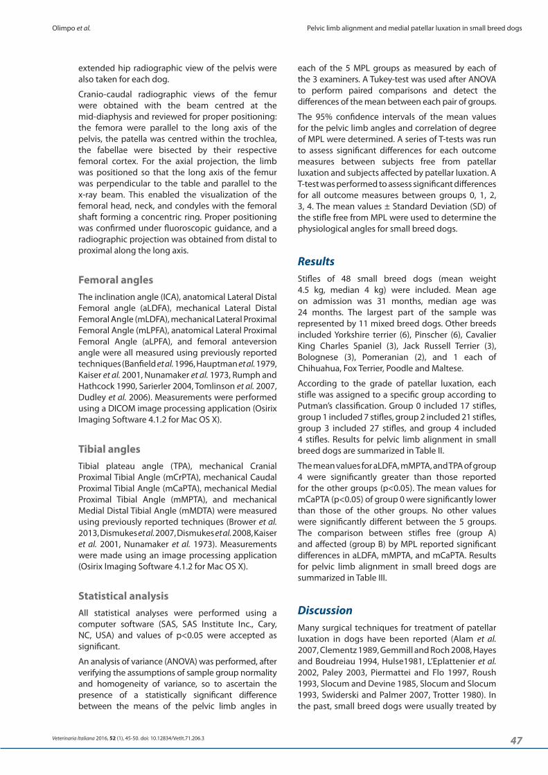
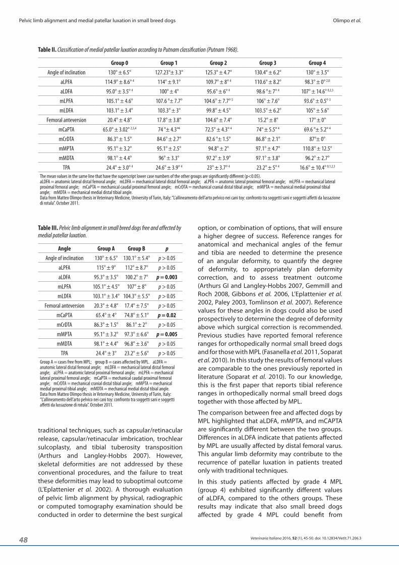
According to the grade of patellar luxation, each stifle was assigned to a specific group according to Putman’s classification. Group 0 included 17 stifles, group 1 included 7 stifles, group 2 included 21 stifles, group 3 included 27 stifles, and group 4 included 4 stifles. Results for pelvic limb alignment in small breed dogs are summarized in Table II.
The mean values for aLDFA, mMPTA, and TPA of group 4 were significantly greater than those reported for the other groups (p<0.05). The mean values for mCaPTA (p<0.05) of group 0 were significantly lower than those of the other groups. No other values were significantly different between the 5 groups. The comparison between stifles free (group A) and affected (group B) by MPL reported significant differences in aLDFA, mMPTA, and mCaPTA. Results for pelvic limb alignment in small breed dogs are summarized in Table III.
DiscussionMany surgical techniques for treatment of patellar luxation in dogs have been reported (Alam et al. 2007, Clementz 1989, Gemmill and Roch 2008, Hayes and Boudreiau 1994, Hulse1981, L’Eplattenier et al. 2002, Paley 2003, Piermattei and Flo 1997, Roush 1993, Slocum and Devine 1985, Slocum and Slocum 1993, Swiderski and Palmer 2007, Trotter 1980). In the past, small breed dogs were usually treated by
extended hip radiographic view of the pelvis were also taken for each dog.
Cranio-caudal radiographic views of the femur were obtained with the beam centred at the mid-diaphysis and reviewed for proper positioning: the femora were parallel to the long axis of the pelvis, the patella was centred within the trochlea, the fabellae were bisected by their respective femoral cortex. For the axial projection, the limb was positioned so that the long axis of the femur was perpendicular to the table and parallel to the x-ray beam. This enabled the visualization of the femoral head, neck, and condyles with the femoral shaft forming a concentric ring. Proper positioning was confirmed under fluoroscopic guidance, and a radiographic projection was obtained from distal to proximal along the long axis.
Femoral anglesThe inclination angle (ICA), anatomical Lateral Distal Femoral angle (aLDFA), mechanical Lateral Distal Femoral Angle (mLDFA), mechanical Lateral Proximal Femoral Angle (mLPFA), anatomical Lateral Proximal Femoral Angle (aLPFA), and femoral anteversion angle were all measured using previously reported techniques (Banfield et al. 1996, Hauptman et al. 1979, Kaiser et al. 2001, Nunamaker et al. 1973, Rumph and Hathcock 1990, Sarierler 2004, Tomlinson et al. 2007, Dudley et al. 2006). Measurements were performed using a DICOM image processing application (Osirix Imaging Software 4.1.2 for Mac OS X).
Tibial anglesTibial plateau angle (TPA), mechanical Cranial Proximal Tibial Angle (mCrPTA), mechanical Caudal Proximal Tibial Angle (mCaPTA), mechanical Medial Proximal Tibial Angle (mMPTA), and mechanical Medial Distal Tibial Angle (mMDTA) were measured using previously reported techniques (Brower et al. 2013, Dismukes et al. 2007, Dismukes et al. 2008, Kaiser et al. 2001, Nunamaker et al. 1973). Measurements were made using an image processing application (Osirix Imaging Software 4.1.2 for Mac OS X).
Statistical analysis All statistical analyses were performed using a computer software (SAS, SAS Institute Inc., Cary, NC, USA) and values of p<0.05 were accepted as significant.
An analysis of variance (ANOVA) was performed, after verifying the assumptions of sample group normality and homogeneity of variance, so to ascertain the presence of a statistically significant difference between the means of the pelvic limb angles in
48 Veterinaria Italiana 2016, 52 (1), 45-50. doi: 10.12834/VetIt.71.206.3
Pelvic limb alignment and medial patellar luxation in small breed dogs Olimpo et al.
option, or combination of options, that will ensure a higher degree of success. Reference ranges for anatomical and mechanical angles of the femur and tibia are needed to determine the presence of an angular deformity, to quantify the degree of deformity, to appropriately plan deformity correction, and to assess treatment outcome (Arthurs GI and Langley-Hobbs 2007, Gemmill and Roch 2008, Gibbons et al. 2006, L’Eplattenier et al. 2002, Paley 2003, Tomlinson et al. 2007). Reference values for these angles in dogs could also be used prospectively to determine the degree of deformity above which surgical correction is recommended. Previous studies have reported femoral reference ranges for orthopedically normal small breed dogs and for those with MPL (Fasanella et al. 2011, Soparat et al. 2010). In this study the results of femoral values are comparable to the ones previously reported in literature (Soparat et al. 2010). To our knowledge, this is the first paper that reports tibial reference ranges in orthopedically normal small breed dogs together with those affected by MPL.
The comparison between free and affected dogs by MPL highlighted that aLDFA, mMPTA, and mCAPTA are significantly different between the two groups. Differences in aLDFA indicate that patients affected by MPL are usually affected by distal femoral varus. This angular limb deformity may contribute to the recurrence of patellar luxation in patients treated only with traditional techniques.
In this study patients affected by grade 4 MPL (group 4) exhibited significantly different values of aLDFA, compared to the others groups. These results may indicate that also small breed dogs affected by grade 4 MPL could benefit from
traditional techniques, such as capsular/retinacular release, capsular/retinacular imbrication, trochlear sulcoplasty, and tibial tuberosity transposition (Arthurs and Langley-Hobbs 2007). However, skeletal deformities are not addressed by these conventional procedures, and the failure to treat these deformities may lead to suboptimal outcome (L’Eplattenier et al. 2002). A thorough evaluation of pelvic limb alignment by physical, radiographic or computed tomography examination should be conducted in order to determine the best surgical
Table III. Pelvic limb alignment in small breed dogs free and affected by medial patellar luxation.
Angle Group A Group B pAngle of inclination 130° ± 6.5° 130.1° ± 5.4° p > 0.05
aLPFA 115° ± 9° 112° ± 8.7° p > 0.05
aLDFA 95.3° ± 3.5° 100.2° ± 7° p = 0.003mLPFA 105.1° ± 4.5° 107° ± 8° p > 0.05
mLDFA 103.1° ± 3.4° 104.3° ± 5.5° p > 0.05
Femoral anteversion 20.3° ± 4.8° 17.4° ± 7.5° p > 0.05
mCaPTA 65.4° ± 4° 74.8° ± 5.1° p = 0.02mCrDTA 86.3° ± 1.5° 86.1° ± 2° p > 0.05
mMPTA 95.1° ± 3.2° 97.3° ± 6.6° p = 0.005mMDTA 98.1° ± 4.4° 96.8° ± 3.6° p > 0.05
TPA 24.4° ± 3° 23.2° ± 5.6° p > 0.05Group A = cases free from MPL; group B = cases affected by MPL. aLDFA = anatomic lateral distal femoral angle; mLDFA = mechanical lateral distal femoral angle; aLPFA = anatomic lateral proximal femoral angle; mLPFA = mechanical lateral proximal femoral angle; mCaPTA = mechanical caudal proximal femoral angle; mCrDTA = mechanical cranial distal tibial angle; mMPTA = mechanical medial proximal tibial angle; mMDTA = mechanical medial distal tibial angle.Data from Matteo Olimpo thesis in Veterinary Medicine, University of Turin, Italy: “L’allineamento dell’arto pelvico nei cani toy: confronto tra soggetti sani e soggetti affetti da lussazione di rotula”. October 2011.
Table II. Classification of medial patellar luxation according to Putnam classification (Putnam 1968).
Group 0 Group 1 Group 2 Group 3 Group 4Angle of inclination 130° ± 6.5° 127.23°± 3.3° 125.3° ± 4.7° 130.4° ± 6.2° 130° ± 3.5°
aLPFA 114.9° ± 8.6° 4 114° ± 9.1° 109.7° ± 8° 4 110.6° ± 8.2° 98.3° ± 0° 2,0
aLDFA 95.0° ± 3.5° 4 100° ± 4° 95.6° ± 6° 4 98.6 °± 7° 4 107° ± 14.6° 0,2,3
mLPFA 105.1° ± 4.6° 107.6 °± 7.7° 104.6° ± 7.7° 5 106° ± 7.6° 93.6° ± 0.5° 3
mLDFA 103.1° ± 3.4° 103.3° ± 3° 99.8° ± 4.5° 103.5° ± 6.2° 105° ± 5.6°
Femoral anteversion 20.4° ± 4.8° 17.8° ± 3.8° 104.6° ± 7.4° 15.2° ± 8° 17° ± 0°
mCaPTA 65.0° ± 3.02° 2,3,4 74 °± 4.3°4 72.5° ± 4.3° 4 74° ± 5.5° 4 69.6 °± 5.2° 4
mCrDTA 86.3° ± 1.5° 84.6° ± 2.7° 82.6 °± 1.5° 86.8° ± 2.1° 87°± 0°
mMPTA 95.1° ± 3.2° 95.1° ± 2.5° 94.8° ± 2° 97.1° ± 4.7° 110.8° ± 12.5°
mMDTA 98.1° ± 4.4° 96° ± 3.3° 97.2° ± 3.9° 97.1° ± 3.8° 96.2° ± 2.7°
TPA 24.4° ± 3.0° 4 24.6° ± 3.9° 4 23° ± 3.7° 4 23.2° ± 5° 4 16.6° ± 10.4° 0,1,2,3
The mean values in the same line that have the superscript lower case numbers of the other groups are significantly different (p<0.05).aLDFA = anatomic lateral distal femoral angle; mLDFA = mechanical lateral distal femoral angle; aLPFA = anatomic lateral proximal femoral angle; mLPFA = mechanical lateral proximal femoral angle; mCaPTA = mechanical caudal proximal femoral angle; mCrDTA = mechanical cranial distal tibial angle; mMPTA = mechanical medial proximal tibial angle; mMDTA = mechanical medial distal tibial angle.Data from Matteo Olimpo thesis in Veterinary Medicine, University of Turin, Italy: “L’allineamento dell’arto pelvico nei cani toy: confronto tra soggetti sani e soggetti affetti da lussazione di rotula”. October 2011.
49Veterinaria Italiana 2016, 52 (1), 45-50. doi: 10.12834/VetIt.71.206.3
Olimpo et al. Pelvic limb alignment and medial patellar luxation in small breed dogs
The data collected in this study do not allow us to ascertain if caudal tibial deformity is also linked to patellar luxation.
Recently, Erb and colleagues (Erb and Pfeil 2012) have described a treatment of patellar luxation in toy and small breed of dogs by means of torsional correction of the femur, reporting high complication rates but promising first results (Erb and Pfeil 2012). However, femoral torsion values between affected and unaffected dogs as measured in this study were not significantly different. No significant difference was found among dogs affected by different grades of patellar luxation.
The radiographic methodology established reference values for clinically unaffected small breed dogs and for dogs with MPL. Differences existed between femora with and without MPL for aLDFA, mMPTA, and mCaPTA. Therefore, our findings would suggest that also small breed dogs affected by grade 4 MPL could be eligible for distal femur corrective osteotomy.
corrective osteotomy, as reported for large breed dogs in the extant literature (Swiderski and Palmer 2007, Brower et al. 2013).
A significant difference was also observed in mMPTA and mCaPTA. The mMPTA measurement can be influenced by limb positioning (Lambert and Wendelburg 2010), patients affected by patellar luxation have a loss of the extensor mechanism and this can alter the radiographic view and mMPTA measurement. It has been speculated that tibial valgus, or proximal excessive tibial varus alone, does not cause patellar luxation because none of them affect the position of the tibial tuberosity in the frontal plane or in the transverse plane (Petazzoni 2012).
The dogs affected by MPL, evaluated in this study, were characterized by an excessive value of mCaPTA. These results support previous studies, which have shown that toy and small breed dogs exhibit a caudal deformity of the proximal tibia (Macias and McKee 2002). Caudal deformity of the proximal tibia in small breeds has been associated with an increased risk of cranial cruciate ligament rupture.
Alam M.R., Lee J.I. & Kang H.S. 2007. Frequency and distribution of patellar luxation in dogs, 134 cases (2000 to 2005). Vet Comp Orthop Traumatol, 1, 59-64.
Arthurs G.I. & Langley-Hobbs S.J. 2007. Patellar luxation as a complication of surgical intervention for the management of cranial cruciate ligament rupture in dogs. A retrospective study of 32 cases. Vet Comp Orthop Traumatol, 20, 204-210.
Banfield C.M., Bartels J.E., Hudson J.A., Wright J.C., Hathcock J.T. & Montgomery R.D. 1996. A retrospective study of canine hip dysplasia in 16 military working dogs part 1: angle measurements and Orthopedic Foundation for Animals (OFA) grading. J Am Anim Hosp Assoc, 32 (5), 413-422.
Brower B.E., Kowaleski M.P., Peruski A.M., Pozzi A., Dyce J., Johnson K.A. & Boudrieau R.J. 2013. Treatment of medial patellar luxation and distal femoral varus by distal femoral osteotomy in dogs. Veterinary Orthopaedic Society, 40th Annual Conference Abstracts, March 9th-16th Canyons Resort, Utah, USA.
Campbell J.R. & Pond M.J. 1972. The canine stifle joint II. Medial luxation of the patella. J Small Anim Pract, 13, 11-18.
Clementz B.G. 1989. Assessment of tibial torsion and rotational deformity with a new fluoroscopic technique. Clin Orthop Relat Res, 245, 199-209.
Denny H.R. & Minter H.M. 1973. Long term results of surgery of canine stifle disorders. J Small Anim Pract, 14, 695-713.
References
Dismukes D.I., Tomlinson J.L., Fox D.B., Cook J.L. & Song K.J. 2007. Radiographic measurement of the proximal and distal mechanical joint angles in the canine tibia. Vet Surg, 36 (7), 699-704 .
Dismukes D.I., Tomlinson J.L., Fox D.B., Cook J.L. & Witsberger T.H. 2008. Radiographic measurement of canine tibial angles in the sagittal plane. Vet Surg, 37 (3), 300-305. doi: 10.1111/j.1532-950X.2008.00381.x.
Dudley R.M., Kowaleski M.P., Drost W.T. & Dyce J. 2006. Radiographic and computed tomographic determination of femoral varus and torsion in the dog. Vet Radiol Ultrasound, 47 (6), 546-552.
Erb K. & Pfeil I. 2012. Treatment of grade 3-4 patellar luxation with correction of femoral torsion in small dogs. 16th ESVOT Congress 2012, 12th - 15th September. Bologna (Italy).
Fasanella F.J., Horstman C.L. & Mason D.R. 2011. Radiographic measurement of the femoral axes, joint angles, and varus angle in orthopedically normal small breed dogs and small breed dogs with medial patellar luxation. Veterinary Orthopedic Society 38th Annual Conference Abstracts March 5th-12th, Snowmass, Colorado, USA.
Flo G.F. & Brinker W.O. 1970. Fascia lata overlap procedure for surgical correction of recurrent medial luxation of the patella in the dog. J Am Vet Med Assoc, 156 (5), 595-599.
Gemmill T.J. & Roch S.P. 2008. Treatment of medial patellar luxation by femoral closing wedge ostectomy
50 Veterinaria Italiana 2016, 52 (1), 45-50. doi: 10.12834/VetIt.71.206.3
Pelvic limb alignment and medial patellar luxation in small breed dogs Olimpo et al.
Petazzoni M. 2012. Patellar luxation: when tibia is guilty. 16th ESVOT Congress 2012, 12th-15th September. Bologna (Italy).
Piermattei D.L. & Flo G.L. 1997. The stifle joint. In Handbook of small animal orthopedics and fracture repair. 3rd Ed. Philadelphia, PA Saunders, 516-534.
Priester W.A. 1972. Sex, size, and breed as risk factors in canine patellar dislocation. J Am Vet Med Assoc, 160, 740-742.
Putnam R.W. 1968. Patellar luxation in the dog. Master’s thesis, University of Guelph, Ontario, Canada 1968; 24, 234-240.
Roush J.K. 1993. Canine patellar luxation. Veterinary Clinics of North America and Small Animal Practice, 23, 855-868.
Ruble R.P. & Hird D.W. 1993. Congenital abnormalities in immature dogs from a pet store: 253 cases (1987-1988). J Am Vet Med Assoc, 202 (4), 633-636.
Rumph P.F. & Hathcock J.T. 1990. A Symmetric axis-based method for measuring the projected femoral angle of inclination in dogs. Vet Surg, 19, 328-333.
Sarierler M. 2004. Comparison of femoral inclination angle measurements in dysplastic and nondysplastic dogs of different breeds. Acta Vet Hung, 52 (2), 245-252.
Singleton W.B. 1969. Surgical correction of stifle deformities in the dog. J Small Anim Pract, 10 (2), 59-69.
Slocum B. & Devine T. 1985. Trochlear recession for correction of luxating patella in the dog. J Am Vet Med Assoc, 186, 365-369.
Slocum B. & Slocum T.D. 1993. Trochlear wedge recession for medial patellar luxation: an update. Veterinary Clinic of North America and Small Animal Practice, 23, 869-875.
Soparat C., Wangdee C., Chuthatep C. & Kalpravidh M. 2010. Deformity of the distal femur in pomeranians with patellar luxation. Thai Journal of Veterinary Medicine, 40, 119.
Swiderski J.K. & Palmer R.H. 2007. Long-term outcome of distal femoral osteotomy for treatment of combined distal femoral varus and medial patellar luxation: 12 cases (1999-2004). J Am Vet Med Assoc, 231, 1070-1075.
Tomlinsoon J., Fox D., Cook J.L. & Keller G.G. 2007.Measurement of femoral angles in four dog breeds. Vet Surg, 36 (6), 593-598.
Trotter E.J. 1980. Medial patellar luxation in the dog. Compendium Continuing Education for Veterinarians, 2, 58-67.
using a distal femoral plate in four dogs. J Small Anim Pract, 49, 152-158.
Gibbons S.E., Macias C., Tozing M.A., Pinchbeck G.L. & McKee W.M. 2006. Patellar luxation in 70 large breed dogs. J Small Anim Pract, 47, 3-9.
Hauptman J., Prieur W.D., Butler H.C. & Guffy M.M.1979. The angle of inclination of the canine femoral head and neck. Vet Surg, 8, 74-77.
Hayes A.G. & Boudreiau R.J. 1994. Frequency and distribution of medial and lateral patellar luxation in dogs: 124 cases. J Am Vet Med Assoc, 205, 716-720.
Hulse D.A. 1981. Pathophysiology and management of medial patellar luxation in the dog. Vet Med Small Anim Clin, 76 (1), 43-51.
Johnson J.A., Austin C. & Breur G.J. 1994. Incidence of canine appendicular musculoskeletal disorders in 16 veterinary teaching hospitals from 1980 through 1989. Vet Comp Orthop Traumatol, 7, 56-69.
Kaiser S., Cornely D., Goldner W., Garner M., Waibl H. & Brunnberg L. 2001. Magnetic resonance measurements of the deviation of the angle of force generated by contraction of the quadriceps muscle in dogs with congenital patellar luxation. Vet Surg, 30 (6), 552-558.
L’Eplattenier H. & Montavon P. 2002. Patellar luxation in dogs and cats: management and prevention. Compendium Continuing Education for Veterinarians, 24, 292-298.
LaFond E., Breur G.J. & Austin C.C. 2002. Breed susceptibility for developmental orthopedic diseases in dogs. J Am Anim Hosp Assoc, 38 (5), 467-477.
Lambert R.J. & Wendelburg K.L. 2010. Determination of the mechanical medial proximal tibial angle using a tangential radiographic technique. Vet Surg, 39, 181-186.
Macias C., McKee W.M. & May C. 2002. Caudal proximal tibial deformity and cranial cruciate ligament rupture in small-breed dogs. J Small Anim Pract, 43, 433-438.
Ness M.G., Abercromby R.H., May C., Turner B.M. & Carmichael S. 1996. A survey of orthopaedic conditions in small animal veterinary practice in Britain. Vet Comp Orthop Traumatol, 9, 43-52.
Nunamaker D.M., Beiry D.N. & Newton C.D. 1973. Femoral neck anteversion in the dog: its radiographic measurement. American Journal Veterinary Radiology, 14, 45-48.
Paley D. 2003. Frontal plane mechanical and anatomic axis planning. In Principles of deformity correction. (J.E. Herzenberg, ed). Berlin: Springer-Verlag, 61-97.