pemeriksaan neoplasma akibat virus dari aspek...
TRANSCRIPT

PEMERIKSAAN NEOPLASMA AKIBAT
VIRUS DARI ASPEK PATOLOGI ANATOMI
DEPARTEMENT ANATOMIC PATHOLOGY
MEDICAL FACULTY UNIVERSITY OF NORTH SUMATERA
MEDAN - 2009
DR. H. DELYUZAR, SP.PA (K)
DR. BETTY, SP.PA
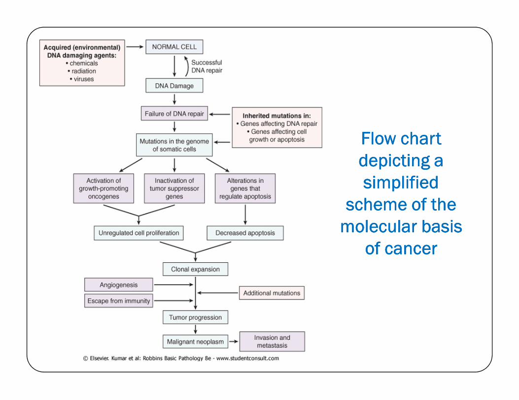
Flow chart Flow chart Flow chart Flow chart
depicting a depicting a depicting a depicting a
simplified simplified simplified simplified
scheme of the scheme of the scheme of the scheme of the scheme of the scheme of the scheme of the scheme of the
molecular basis molecular basis molecular basis molecular basis
of cancerof cancerof cancerof cancer
Immunohistochemistry

Immunohistochemistry
Merupakan suatu teknik untuk menentukan lokasi antigen (protein target) dalam jaringan/sel dengan menggunakan reaksi
antigen-antibodi.
Fluorescent dye
Enzyme
Radioactive element
Colloidal gold
Visualized by a marker such as :
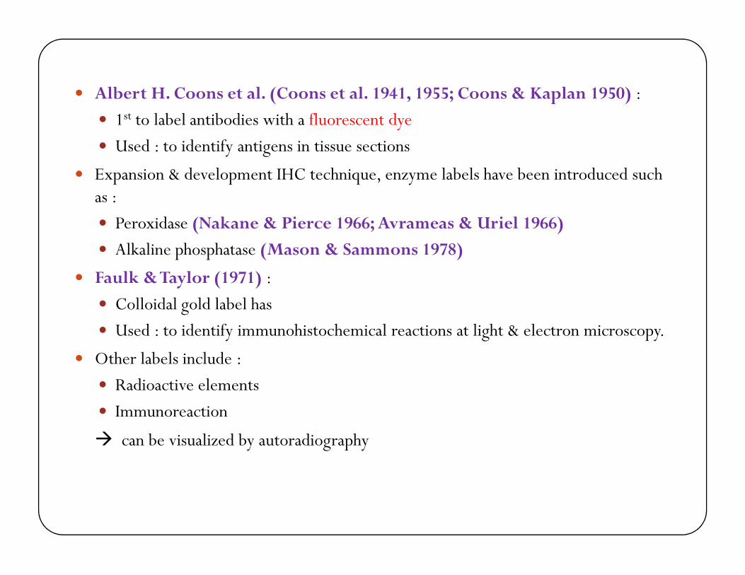
� Albert H. Coons et al. (Coons et al. 1941, 1955; Coons & Kaplan 1950) :
� 1st to label antibodies with a fluorescent dye
� Used : to identify antigens in tissue sections
� Expansion & development IHC technique, enzyme labels have been introduced such as :
� Peroxidase (Nakane & Pierce 1966; Avrameas & Uriel 1966)
� Alkaline phosphatase (Mason & Sammons 1978)
� Faulk & Taylor (1971) :Faulk & Taylor (1971) :
� Colloidal gold label has
� Used : to identify immunohistochemical reactions at light & electron microscopy.
� Other labels include :
� Radioactive elements
� Immunoreaction
� can be visualized by autoradiography
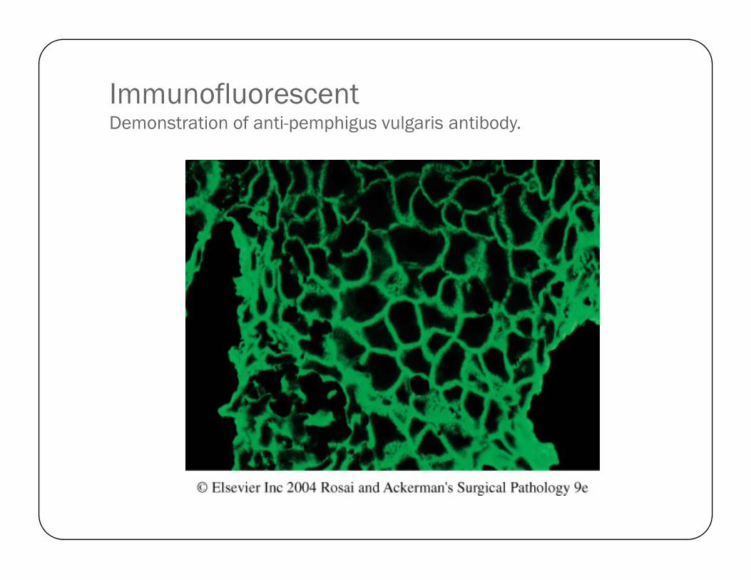
Immunofluorescent Demonstration of anti-pemphigus vulgaris antibody.
Immunohistochemistry� Involves : specific antigen-antibody reaction
� Advantage > traditionally (used special enzyme staining techniques) identify only a limited number of :
� Proteins
Enzymes � Enzymes
� Tissue structures
� Crucial technique & widely used in many medical research laboratories as well as clinical diagnostics.

Antibody types
Polyclonal Antibodies
The antibodies used for specific detection can be :
Monoclonal Antibodies
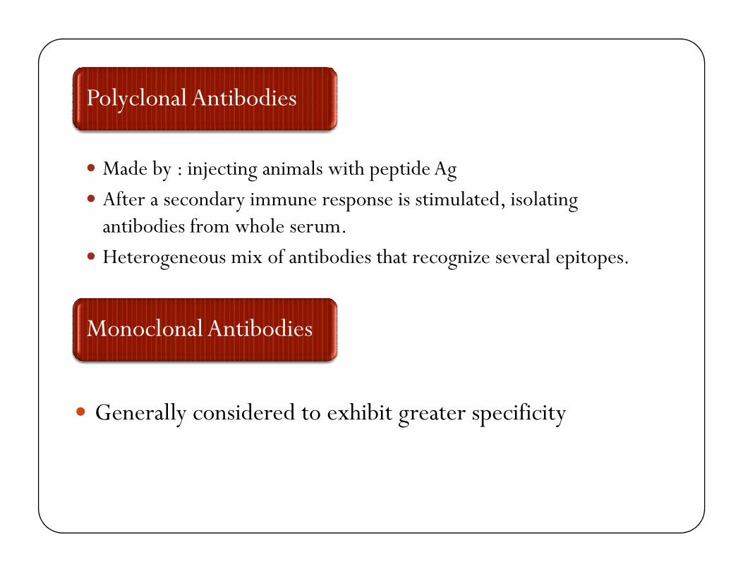
� Made by : injecting animals with peptide Ag
� After a secondary immune response is stimulated, isolating antibodies from whole serum.
� Heterogeneous mix of antibodies that recognize several epitopes.
Polyclonal Antibodies
� Generally considered to exhibit greater specificity
Monoclonal Antibodies

Immunohistochemistry techniques
Direct technique
Indirect technique

Antibodies reagents
Primary reagents
Can also be classified as:
Secondary reagents

Primary reagents
� Raised against an antigen of interest
� Typically unconjugated (unlabelled)� Typically unconjugated (unlabelled)
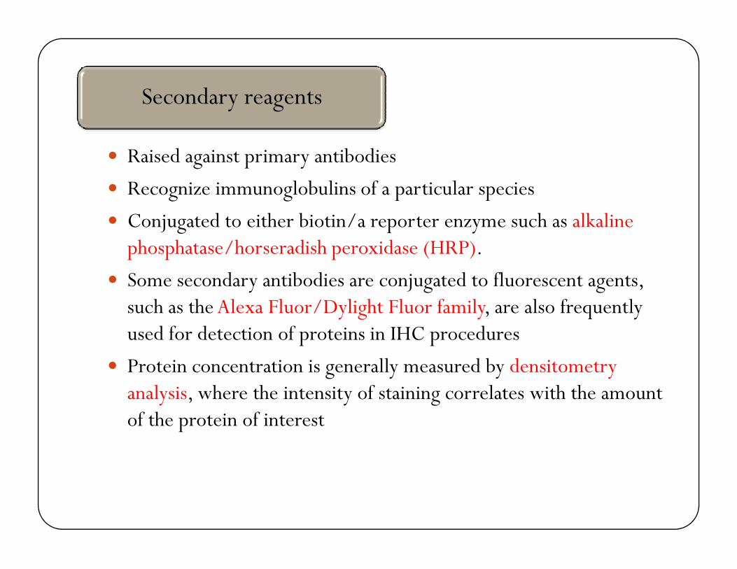
� Raised against primary antibodies
� Recognize immunoglobulins of a particular species
� Conjugated to either biotin/a reporter enzyme such as alkaline phosphatase/horseradish peroxidase (HRP).
� Some secondary antibodies are conjugated to fluorescent agents,
Secondary reagents
� Some secondary antibodies are conjugated to fluorescent agents, such as the Alexa Fluor/Dylight Fluor family, are also frequently used for detection of proteins in IHC procedures
� Protein concentration is generally measured by densitometry analysis, where the intensity of staining correlates with the amount of the protein of interest

Immunohistochemistry
� Several procedures are avaiable
� The 2 most commonly used :
1. Peroxidase-antiperoxidase immune complex method
2. The biotin-avidin immunoenzymatic technique
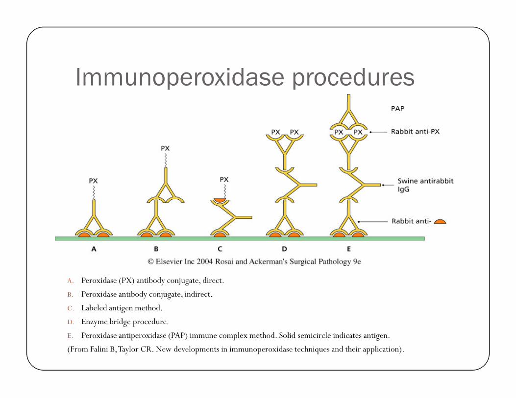
Immunoperoxidase procedures
A. Peroxidase (PX) antibody conjugate, direct.
B. Peroxidase antibody conjugate, indirect.
C. Labeled antigen method.
D. Enzyme bridge procedure.
E. Peroxidase antiperoxidase (PAP) immune complex method. Solid semicircle indicates antigen.
(From Falini B, Taylor CR. New developments in immunoperoxidase techniques and their application).

Biotin-avidin immunoenzymatic techniques. Solid semicircle, antigen; PX, peroxidase; *, biotin; shaded open
cross, avidin.
A. Biotinylated primary antibody method.
B. Biotinylated peroxidase method.
C. Avidin–biotin–peroxidase complex method.
(From Falini B, Taylor CR. New developments in immunoperoxidase techniques & their
application).
� Various methods � ↑ sensitivity of the procedure
� The aim :
� To expose antigenic sites (epitop) � that may unexposed (‘masked’) � ’antigenic unmasking’ /’antigen-retrieving’ techniques
� They include :
�Digestion with a variety of proteolytic enzym (e.g. �Digestion with a variety of proteolytic enzym (e.g. enzim trypsin)
� Treatment with microwave
� Exposure to the combined action of heat & pressure in a pressure cooker

The advent of monoclonal technology:
� Antibodies have become available for which the antigenic determinant is :� Chemically poorly defined, or
� Totally unknown

…. The advent of monoclonal technology:
� Marker located in:
� Nucleus
� Cytoplasm
� Cell membrane
� Extracellular space� Extracellular space
� The advantages offer is that
� Generally higher degree of specificity
� Practically no diffusion
� If the marker location is nuclear � stain can be combined with another with different chromogen aimed at cytoplasmic/cell membrane marker
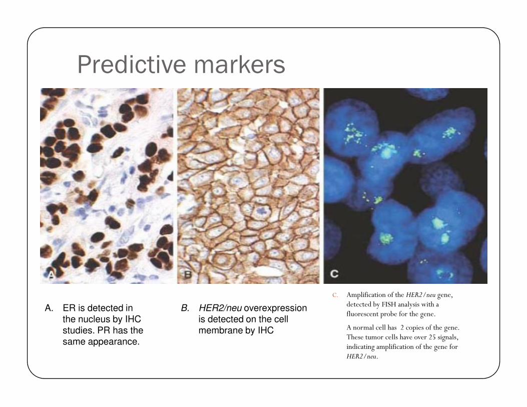
Predictive markers
C. Amplification of the HER2/neu gene, detected by FISH analysis with a fluorescent probe for the gene.
A normal cell has 2 copies of the gene. These tumor cells have over 25 signals, indicating amplification of the gene for HER2/neu.
A. ER is detected in the nucleus by IHC studies. PR has the same appearance.
B. HER2/neu overexpression is detected on the cell membrane by IHC

Hormone receptors
� The effect of hormones in target organs is medicated by intracellular (largely intranuclear) peptides known as hormons receptors.
� Monoclonal antibodies are available for :� Estrogen receptors
� Progesterone receptors � Progesterone receptors
� Androgen receptors
� Originally the technique worked reliably only in frozen section material � but now : the results in paraffin-embedded material
� They compare favorably with those obtained with the conventional biochemical assay.
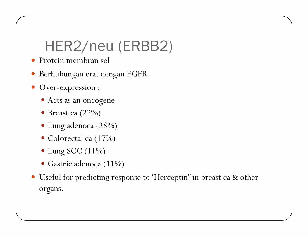
HER2/neu (ERBB2)� Protein membran sel
� Berhubungan erat dengan EGFR
� Over-expression :
� Acts as an oncogene
� Breast ca (22%)
� Lung adenoca (28%)
� Colorectal ca (17%)
� Lung SCC (11%)
� Gastric adenoca (11%)
� Useful for predicting response to ‘Herceptin” in breast ca & other organs.
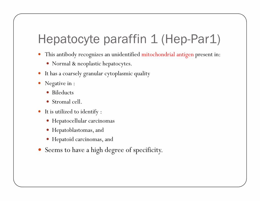
Hepatocyte paraffin 1 (Hep-Par1)� This antibody recognizes an unidentified mitochondrial antigen present in:
� Normal & neoplastic hepatocytes.
� It has a coarsely granular cytoplasmic quality
� Negative in :
� Bileducts
� Stromal cell. � Stromal cell.
� It is utilized to identify :
� Hepatocellular carcinomas
� Hepatoblastomas, and
� Hepatoid carcinomas, and
� Seems to have a high degree of specificity.

CD117� This antibody identifies c-KIT (a transmembrane
tyrosine kinase)
� Normal expressed by :
��Cajal’s interstitial cells � GIST tumor family
�Melanocytes � melanoma
� Mast cells � mastocytosis
�Germ cells � germ cell tumors

Desmin� This muscle type intermediate filament (MW 55.000) is found in
cells of :
� Smooth muscle � smooth muscle tumor
� Striated muscle � skeletal muscle tumor
� Primarily used for the identification tumor of :� Primarily used for the identification tumor of :
� Smooth muscle
� Skeletal muscle
� Desmin (+) associated with Actin (-) is a feature of :
� Subset cells of myofibroblastic appearance
� Hormone dependent stroma (vagina, breast0
� Desmoplastic small cell tumor

Keratin / cytokeratin� A family of water-insoluble, intracellular fibrous proteins
� Present in : almost all epithelia
� ± 20 sub-classes have been identified on basis :
� MW 40.000-68.000
� Isoelectric pH value (5-8)
� Keratins represent an excellent marker for epithelial differentiation regardless � Keratins represent an excellent marker for epithelial differentiation regardless of whether the tumor is of:
� Endodermal
� Neuroectodermal
� Mesenchymal, or
� Germ cell derivation
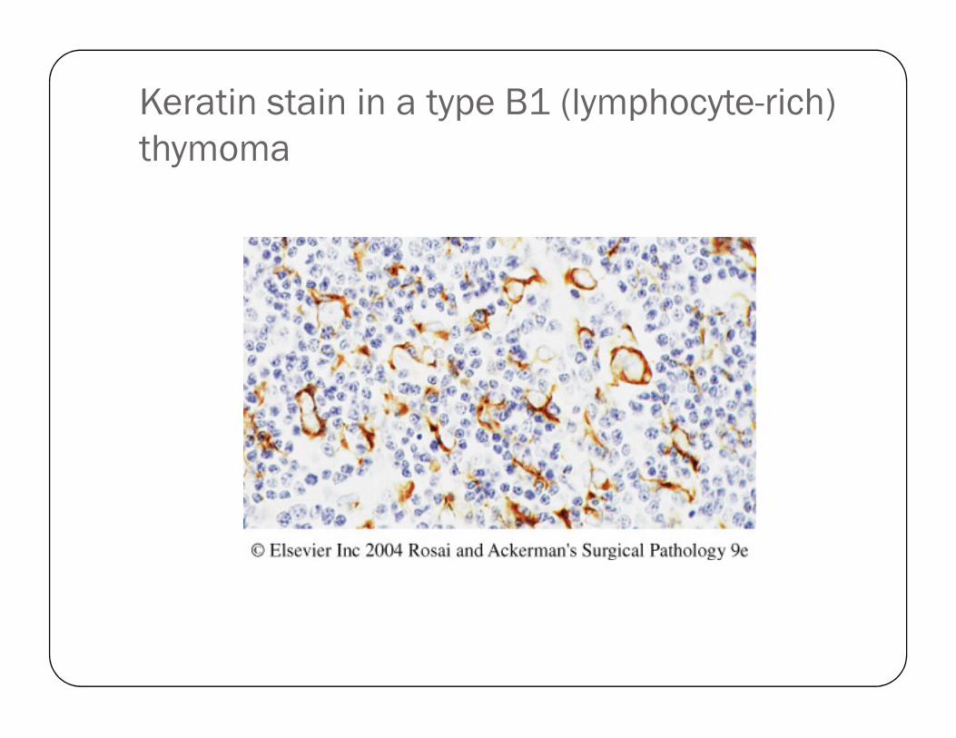
Keratin stain in a type B1 (lymphocyte-rich)
thymoma

Keratin stain.
Anaplastic carcinoma of pancreas

MBI-1 (Ki-67)
� An antigen corresponds to a nuclear nonhistone protein expressed by cells in proliferative phases G1, G2, M and S
� In general, there is a good correlation between Ki-67 staining & mitotic count

Strong immunostaining for MBI-1 in
germinal center of hyperplastic lymph node

Latent membrane protein 1 (LMP1)
� An EBV membrane protein expressed in latent infection
� Is considered to be ‘EBV oncoprotein’