penetrating neck injuries
TRANSCRIPT
Penetrating neck injuries
Dr. SoeDr. Bhebhe
A&EFamily Medicine
Introduction
Penetrating neck injuries are commonly seen in South Africa. Although many are minor injuries of no significance, they may be deceptive in appearance.
This presentation focuses on a logical approach to the accurate assessment and management of penetrating injuries to the neck, excluding non-penetrating neck injuries and injuries to the cervical spine.
Epidemiologic Features
Firearms are responsible for approximately 44%, stab wounds for approximately 40%, shotguns for approximately 4%, and other weapons for approximately 12% of all penetrating neck injuries in urban trauma centers in the United States.
Gunshot wounds are significantly more likely to be associated with large neck hematomas, hypotension on admission, and vascular or aerodigestive injuries than knife wounds.

Mechanism of penetrating neck injuries
This type of injury may be the result of interpersonal violence, for example, stab or gunshot wounds, or accidents due to foreign bodies, or iatrogenic incidents during endoscopy or surgery.
Classification of penetrating neck injuries
A penetrating neck injury is one that has penetrated platysma.
Probing of the wound with a finger or an instrument to determine the depth of the wound is absolutely contraindicated.
Classification of penetrating neck injuries (cont)
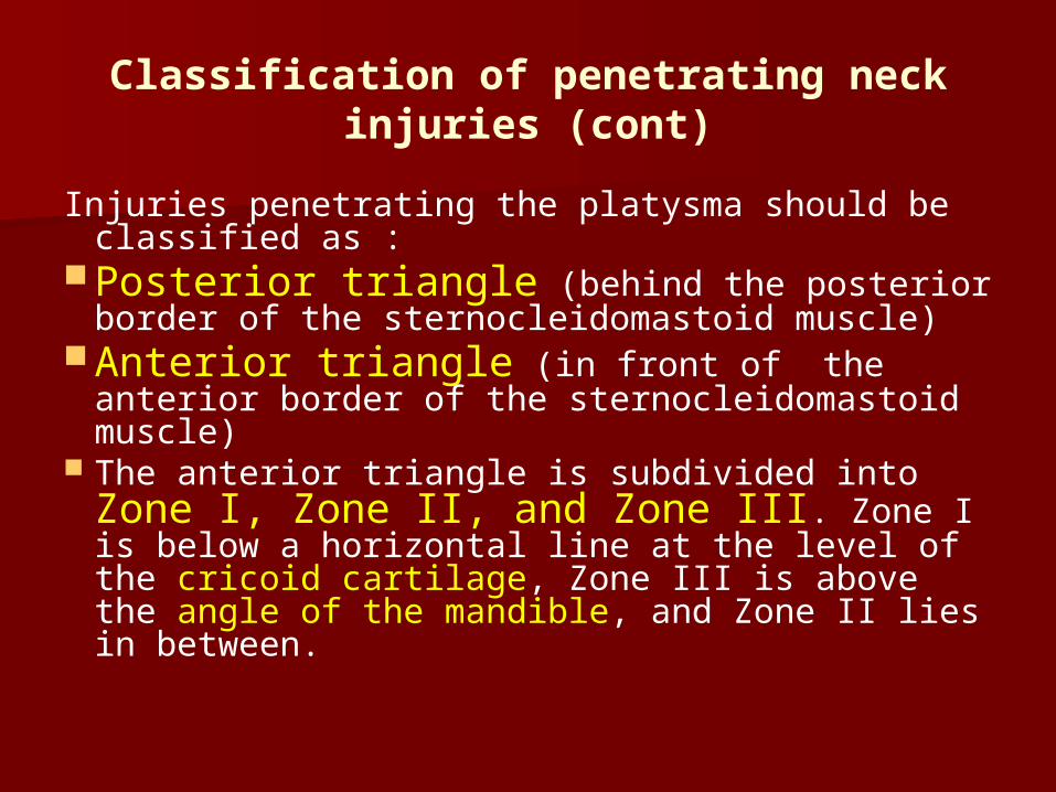
Injuries penetrating the platysma should be classified as :
Posterior triangle (behind the posterior border of the sternocleidomastoid muscle)
Anterior triangle (in front of the anterior border of the sternocleidomastoid muscle)
The anterior triangle is subdivided into Zone I, Zone II, and Zone III. Zone I is below a horizontal line at the level of the cricoid cartilage, Zone III is above the angle of the mandible, and Zone II lies in between.
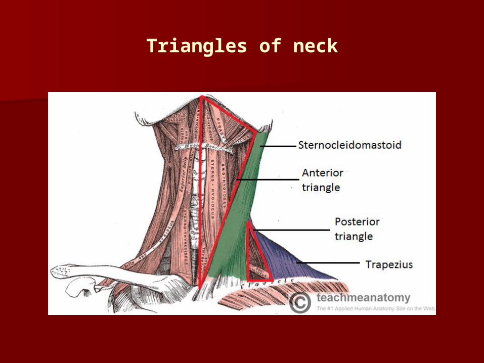
Triangles of neck
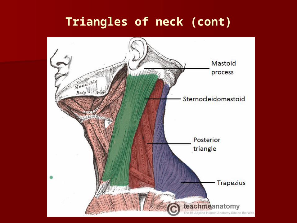
Triangles of neck (cont)
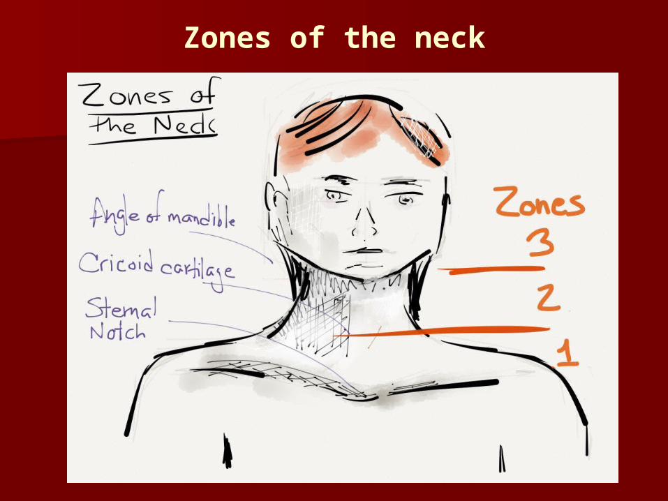
Zones of the neck
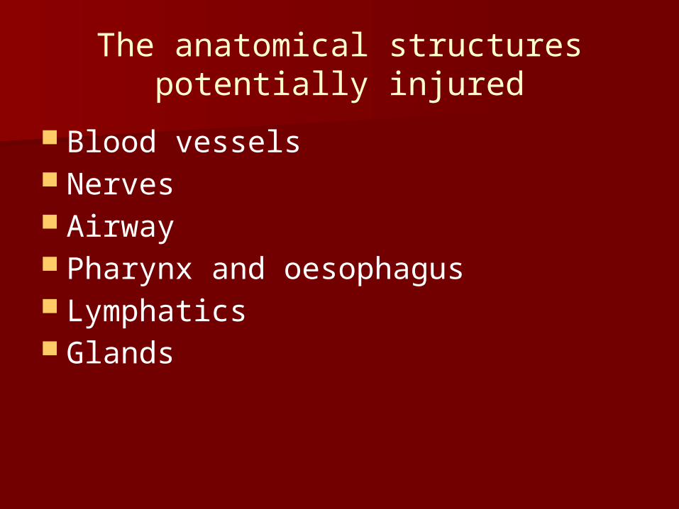
The anatomical structures potentially injured
Blood vessels Nerves Airway Pharynx and oesophagus Lymphatics Glands
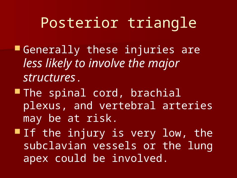
Posterior triangle
Generally these injuries are less likely to involve the major structures.
The spinal cord, brachial plexus, and vertebral arteries may be at risk.
If the injury is very low, the subclavian vessels or the lung apex could be involved.
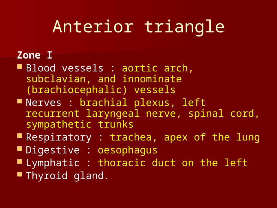
Anterior triangle
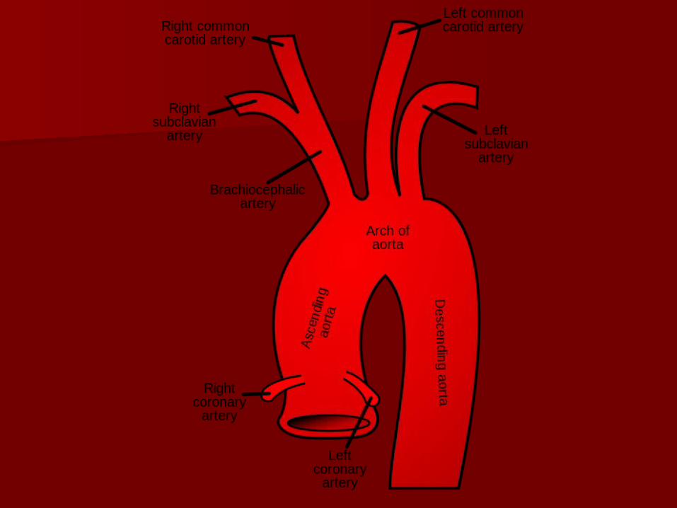
Zone I Blood vessels : aortic arch, subclavian,
and innominate (brachiocephalic) vessels Nerves : brachial plexus, left recurrent
laryngeal nerve, spinal cord, sympathetic trunks
Respiratory : trachea, apex of the lung Digestive : oesophagus Lymphatic : thoracic duct on the left Thyroid gland.
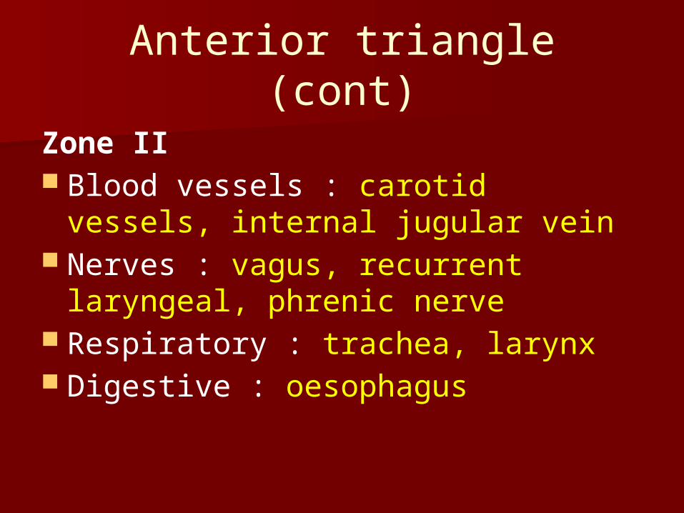
Anterior triangle (cont)
Zone II Blood vessels : carotid vessels,
internal jugular vein Nerves : vagus, recurrent laryngeal,
phrenic nerve Respiratory : trachea, larynx Digestive : oesophagus
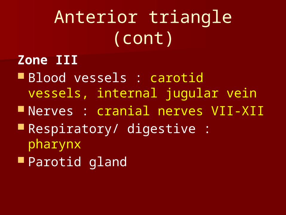
Anterior triangle (cont)
Zone III Blood vessels : carotid vessels,
internal jugular vein Nerves : cranial nerves VII-XII Respiratory/ digestive : pharynx Parotid gland
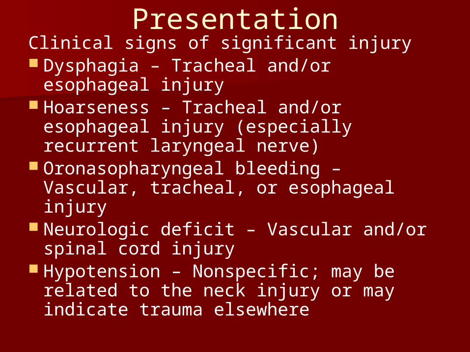
PresentationClinical signs of significant injury Dysphagia – Tracheal and/or esophageal
injury Hoarseness – Tracheal and/or
esophageal injury (especially recurrent laryngeal nerve)
Oronasopharyngeal bleeding – Vascular, tracheal, or esophageal injury
Neurologic deficit – Vascular and/or spinal cord injury
Hypotension – Nonspecific; may be related to the neck injury or may indicate trauma elsewhere
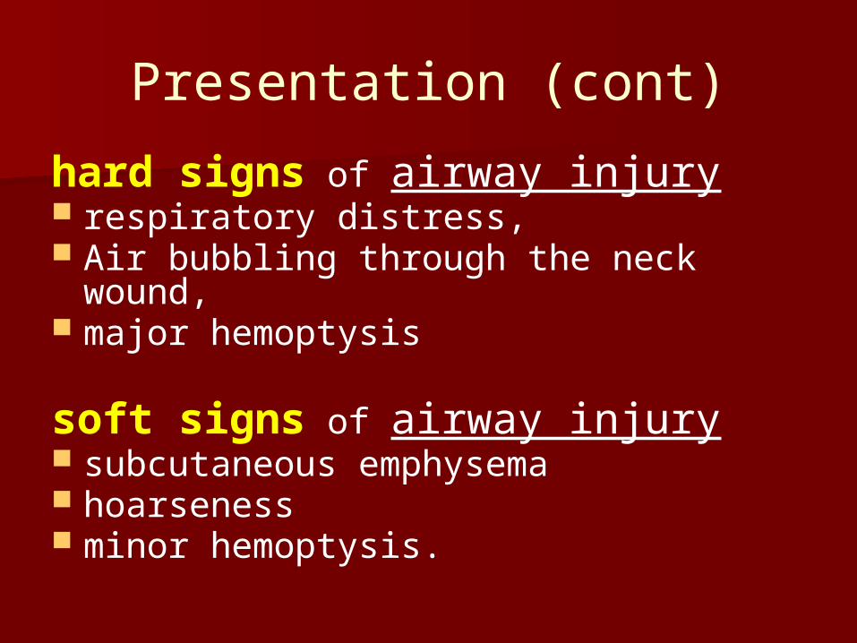
Presentation (cont)
hard signs of airway injury respiratory distress, Air bubbling through the neck wound, major hemoptysis
soft signs of airway injury subcutaneous emphysema hoarseness minor hemoptysis.
Presentation (cont)
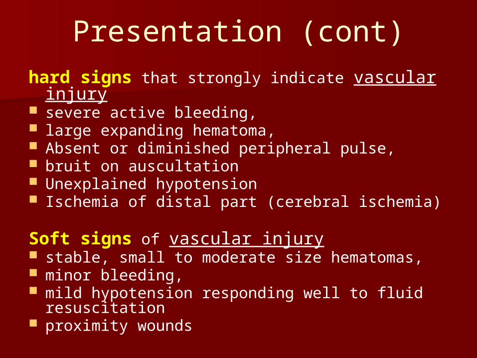
hard signs that strongly indicate vascular injury
severe active bleeding, large expanding hematoma, Absent or diminished peripheral pulse, bruit on auscultation Unexplained hypotension Ischemia of distal part (cerebral ischemia)
Soft signs of vascular injury stable, small to moderate size hematomas, minor bleeding, mild hypotension responding well to fluid
resuscitation proximity wounds
Presentation (cont)
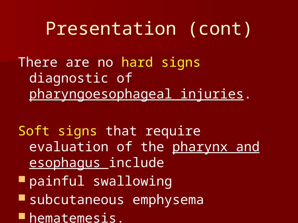
There are no hard signs diagnostic of pharyngoesophageal injuries.
Soft signs that require evaluation of the pharynx and esophagus include
painful swallowing subcutaneous emphysema hematemesis.
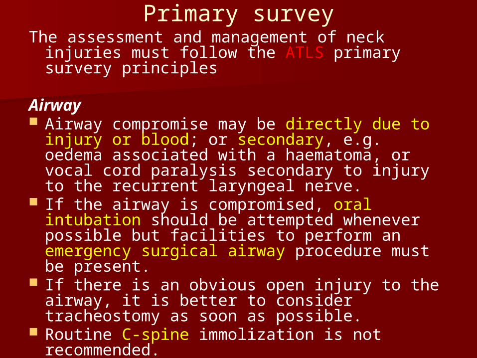
Primary surveyThe assessment and management of neck
injuries must follow the ATLS primary survery principles
Airway Airway compromise may be directly due to
injury or blood; or secondary, e.g. oedema associated with a haematoma, or vocal cord paralysis secondary to injury to the recurrent laryngeal nerve.
If the airway is compromised, oral intubation should be attempted whenever possible but facilities to perform an emergency surgical airway procedure must be present.
If there is an obvious open injury to the airway, it is better to consider tracheostomy as soon as possible.
Routine C-spine immolization is not recommended.
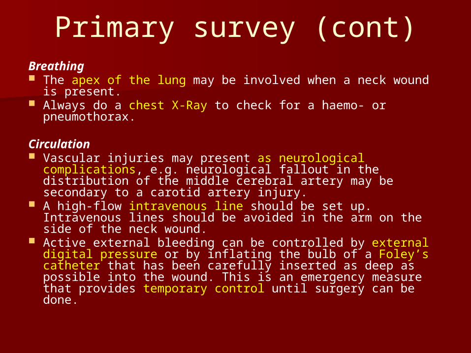
Primary survey (cont)Breathing The apex of the lung may be involved when a neck wound
is present. Always do a chest X-Ray to check for a haemo- or
pneumothorax.
Circulation Vascular injuries may present as neurological complications,
e.g. neurological fallout in the distribution of the middle cerebral artery may be secondary to a carotid artery injury.
A high-flow intravenous line should be set up. Intravenous lines should be avoided in the arm on the side of the neck wound.
Active external bleeding can be controlled by external digital pressure or by inflating the bulb of a Foley’s catheter that has been carefully inserted as deep as possible into the wound. This is an emergency measure that provides temporary control until surgery can be done.
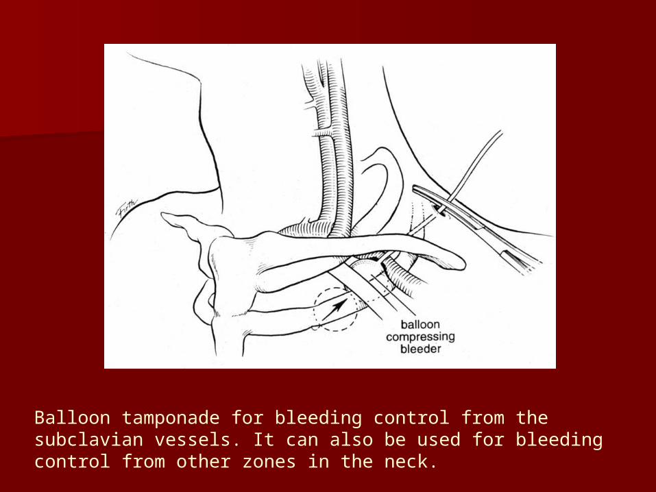
Balloon tamponade for bleeding control from the subclavian vessels. It can also be used for bleeding control from other zones in the neck.
Primary survey (cont)
Disability Neurological deficit may be secondary
to vascular injury; cranial nerve or spinal cord damage.
Exposure/ environment Look for other injuries – consider injury
patterns associated with the mechanism of injury, or the trajectory.
Secondary SurveyHistory Establish the mechanism of injury, note voice change, ask
about chest pain, dysphagia, haemoptysis, weakness, paresthaesia, or numbness in the arms.
ExaminationAssess for the presence of : Local bleeding, pulsation, bruit, absent pulses, expanding
haematoma Air in soft tissues, distended neck veins Fluid leaking from the wound (saliva, CSF, lymph) Cranial nerve deficit, particularly CN VII-XII, Horner’s
syndrome Loss of sensation and power in the upper limbs Loss of sensation and power in the lower limbs Pneumo-/ haemothorax, abnormal breathing pattern (e.g.
diaphragmatic breathing) Blood pressure difference of more than 10 mmHg in the 2
arms Frequent reassessment of the airway is mandatory to check
for impending obstruction due to oedema
Investigative management
The mechanism of injury and clinical examination should determine the need and type of specific investigations in the evaluation of PNI.
Patients with hard signs of major vascular or laryngotracheal injuries should undergo an operation without any delay for definitive investigations.
Investigative management (cont)
In the stable patient who has no immediate indication for surgery, the blood vessels, respiratory, and digestive systems should be investigated to rule out injury. This may be done primarily by surgical exploration, or by utilizing special investigations which may obviate the need for surgery.
Zone II injuries are readily exposed and accessed, and are therefore often surgically explored without preoperative investigations. The structures in Zone I or III are more difficult to visualize intraoperatively and need more preoperative planning and preparation.
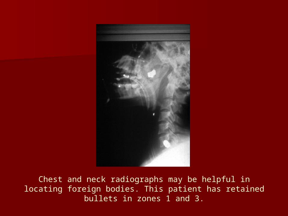
Chest and neck radiographs may be helpful in locating foreign bodies. This patient has retained bullets in zones 1 and 3.
Investigative management (cont)
Chest X-ray This is essential in all patients with neck injuries. Do not sit patient up; if there is an open wound, it may cause a
fatal air embolism or complicate a cervical spine injury.
Cervical spine X-ray Look for the presence of fractures, foreign bodies, or air in soft
tissues. CT scan or CT angiography In the stable patient, a spiral CT scan (if available) with
intravenous contrast will provide information on soft tissue, bony structures, wound trajectory, and vascular injuries.
Specifically look out for intimal injuries of the carotids. Oral contrast can be given if required to identify leaks.
Color Flow Doppler (CFD) Color flow Doppler has been suggested as a reliable alternative to
angiography in the evaluation of PNI.
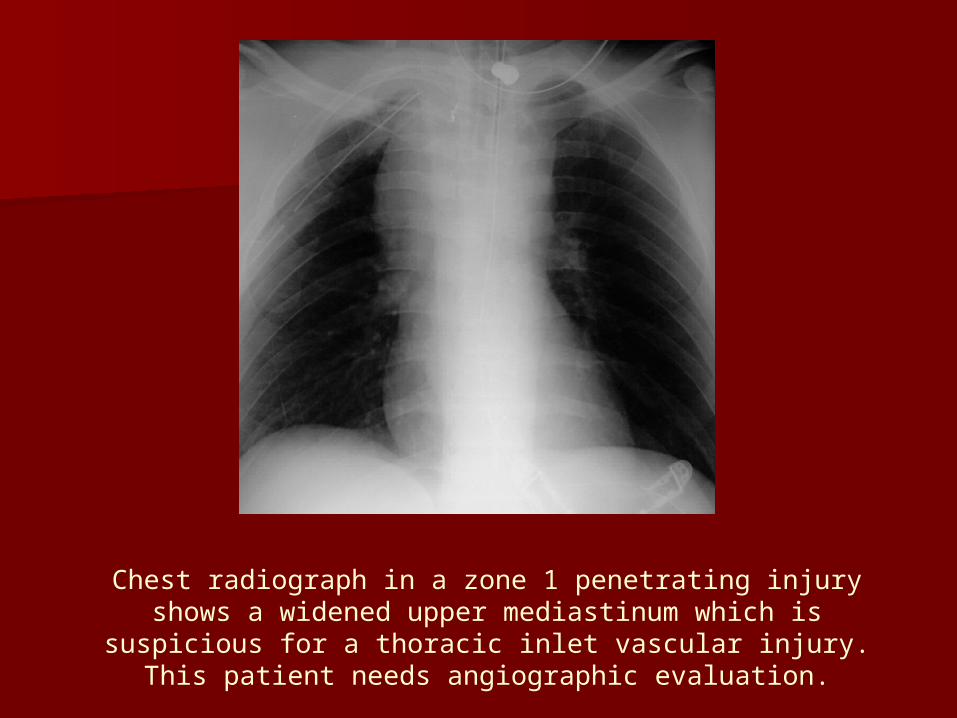
Chest radiograph in a zone 1 penetrating injury shows a widened upper mediastinum which is suspicious for a thoracic
inlet vascular injury. This patient needs angiographic evaluation.
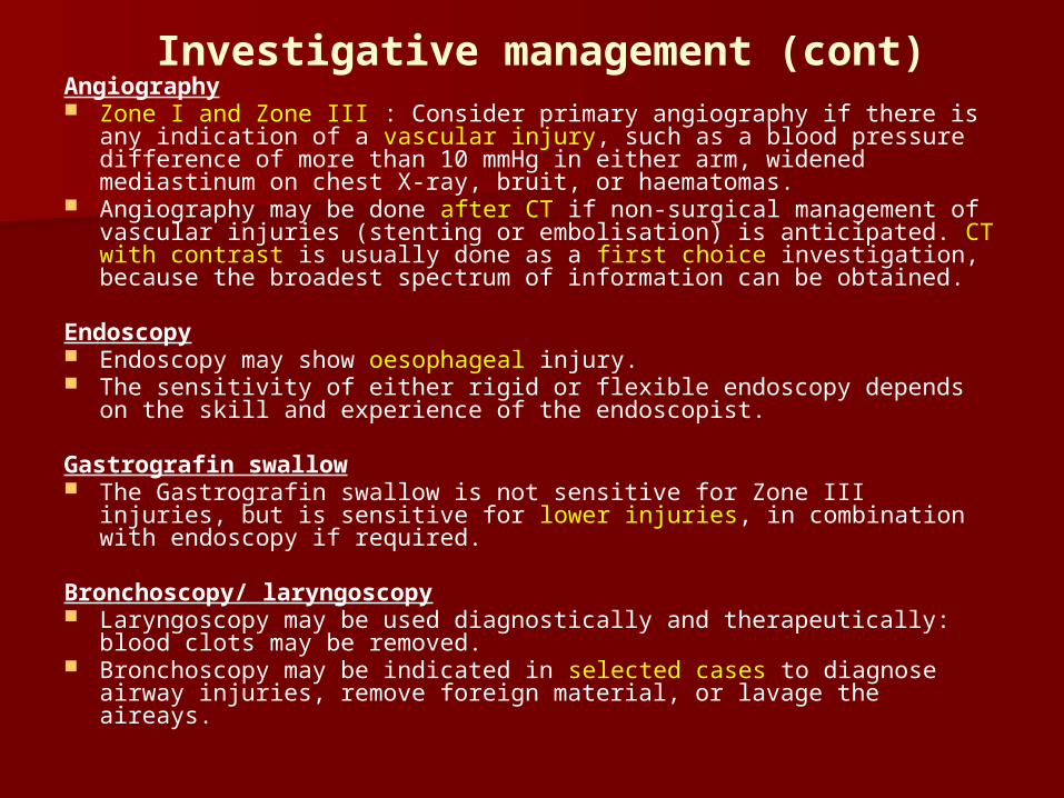
Investigative management (cont)Angiography Zone I and Zone III : Consider primary angiography if there is any
indication of a vascular injury, such as a blood pressure difference of more than 10 mmHg in either arm, widened mediastinum on chest X-ray, bruit, or haematomas.
Angiography may be done after CT if non-surgical management of vascular injuries (stenting or embolisation) is anticipated. CT with contrast is usually done as a first choice investigation, because the broadest spectrum of information can be obtained.
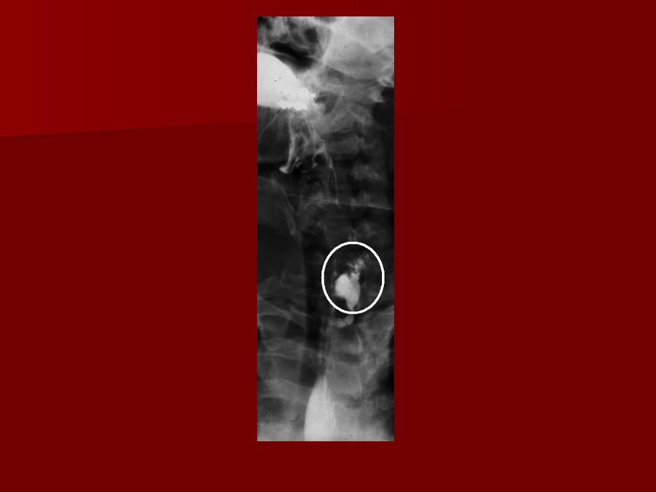
Endoscopy Endoscopy may show oesophageal injury. The sensitivity of either rigid or flexible endoscopy depends on the
skill and experience of the endoscopist.
Gastrografin swallow The Gastrografin swallow is not sensitive for Zone III injuries, but is
sensitive for lower injuries, in combination with endoscopy if required.
Bronchoscopy/ laryngoscopy Laryngoscopy may be used diagnostically and therapeutically: blood
clots may be removed. Bronchoscopy may be indicated in selected cases to diagnose
airway injuries, remove foreign material, or lavage the aireays.
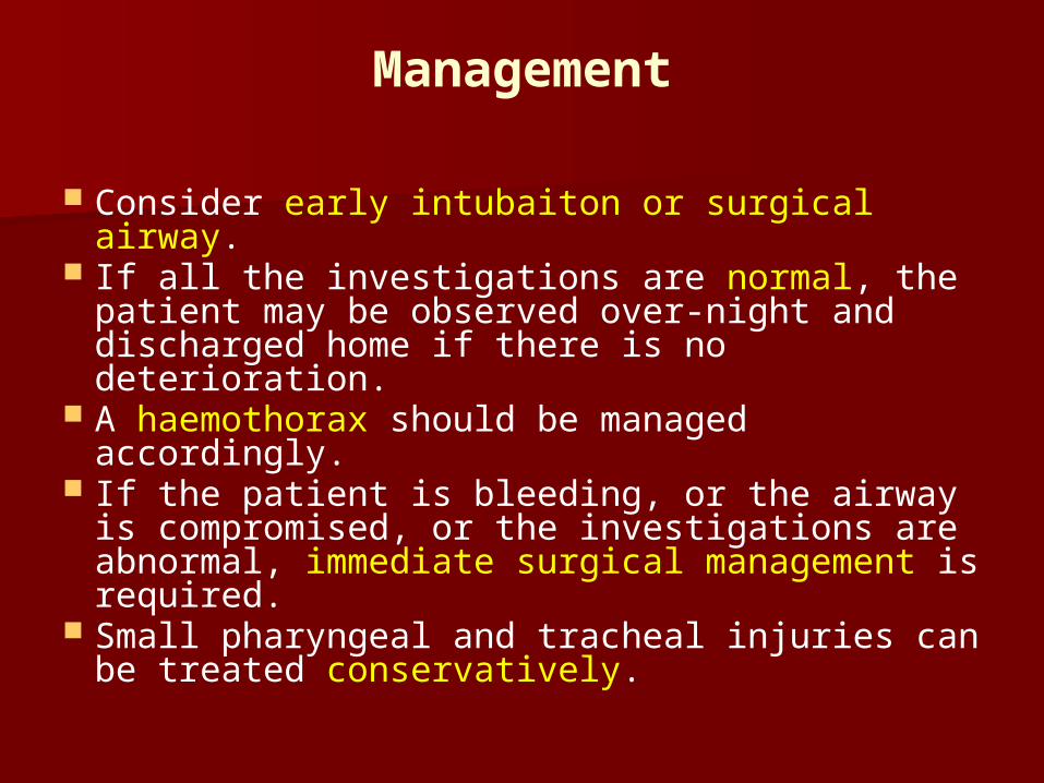
Management
Consider early intubaiton or surgical airway. If all the investigations are normal, the
patient may be observed over-night and discharged home if there is no deterioration.
A haemothorax should be managed accordingly.
If the patient is bleeding, or the airway is compromised, or the investigations are abnormal, immediate surgical management is required.
Small pharyngeal and tracheal injuries can be treated conservatively.
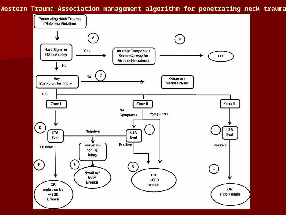
Western Trauma Association management algorithm for penetrating neck trauma.
Pitfalls
Always adhere to ABCDE for the initial management of the patient.
Always frequently reassess the airway in order to recognise airway problems that may develop over time.
Do a thorough assessment of platysmal penetration. The wound should never be probed as bleeding is sure to be precipitated.
Penetrating neck injuries may involve the lung or mediastinal structures. The chest should always be assessed.
Vascular injuries may cause neurological manifestations.

Questions !!!