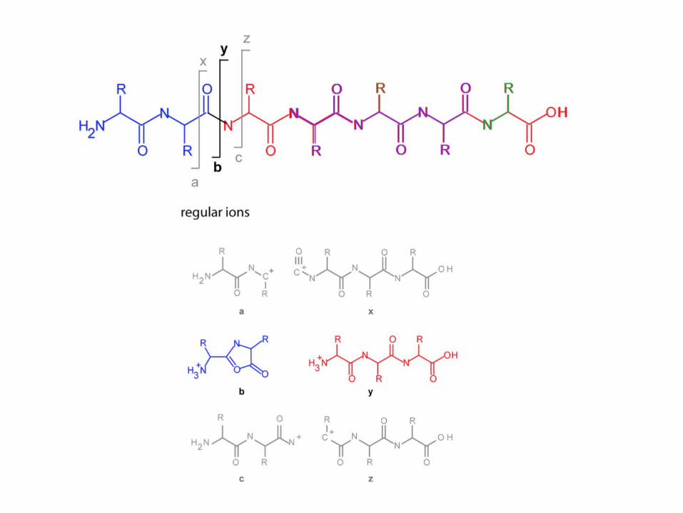
peptide ion fragmentation technologies cid = collision induced dissociation cad = collision...
TRANSCRIPT

Peptide Ion Fragmentation TechnologiesCID = collision induced dissociationCAD = collision activated disassociation

HCD = high energy collisional disassociation + high resolution fragments in MS2 spectra
e.g. +/- 0.01Da MS2 window or ~15ppmsCID/CAD = low resolution fragments +/- 0.5Da
+ can be acquired much more quickly


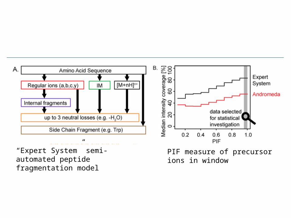
“Expert System” semi-automated peptide fragmentation model
PIF measure of precursor ions in window


Comparison of single-scan (500- to 600-msec) CAD and ETD mass spectra recorded during data-dependent analyses (nHPLC-μESI-MS/MS) of phosphopeptides generated in a tryptic
digest of human nuclear proteins.
Syka J E P et al. PNAS 2004;101:9528-9533
©2004 by National Academy of Sciences
ETD = Electron Transfer Disassociation
c and z* ions primarily
Can handle phosphorylation better.

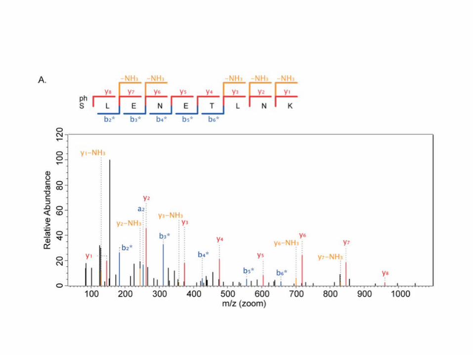
Get all c-ions (see next slide for question).

How would you determine sequence from these?


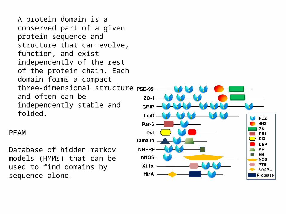
A protein domain is a conserved part of a given protein sequence and structure that can evolve, function, and exist independently of the rest of the protein chain. Each domain forms a compact three-dimensional structure and often can be independently stable and folded.
PFAM
Database of hidden markov models (HMMs) that can be used to find domains by sequence alone.
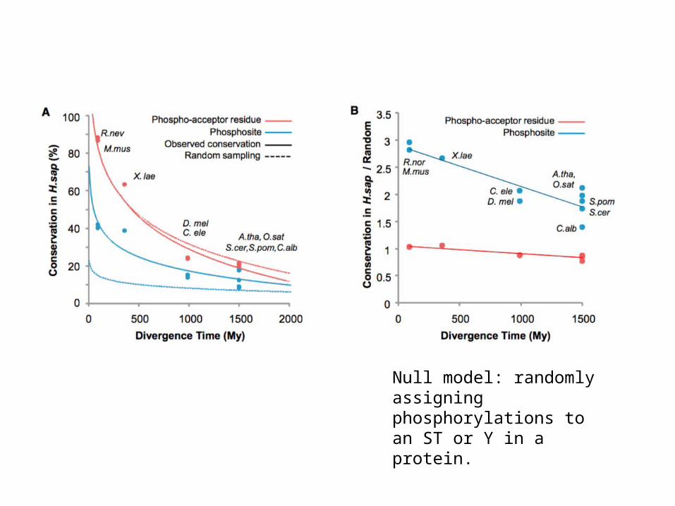
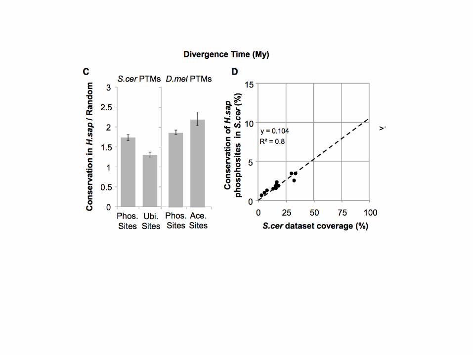
Null model: randomly assigning phosphorylations to an ST or Y in a protein.

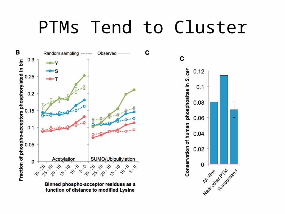
PTMs Tend to Cluster
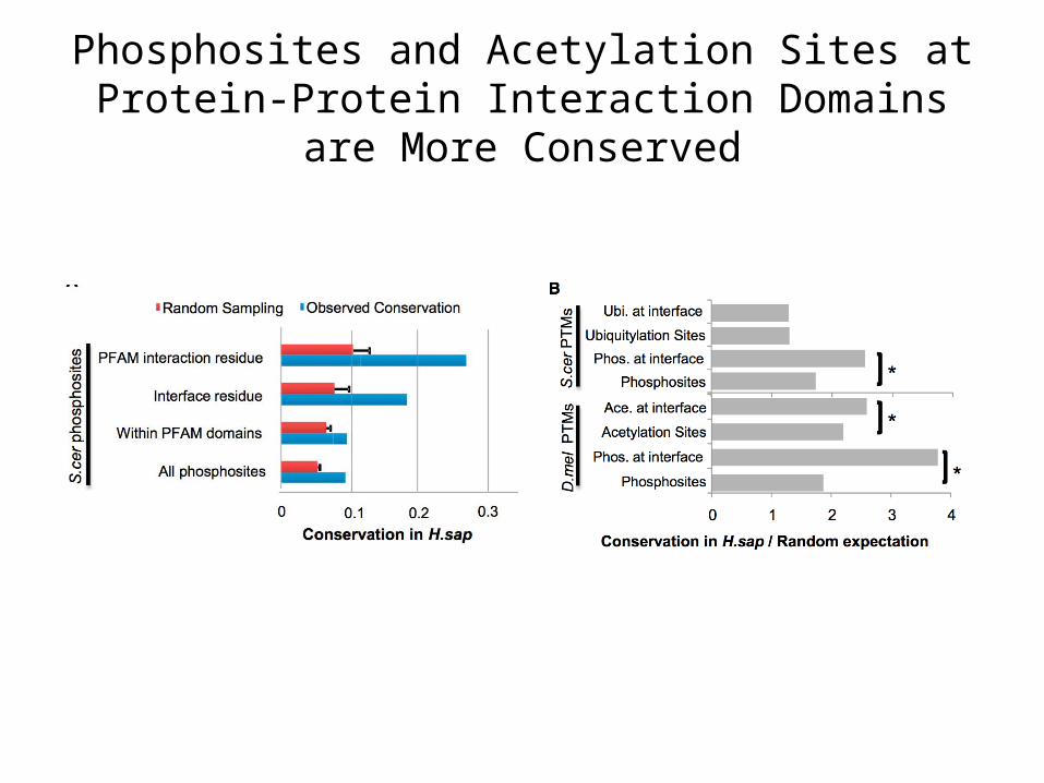
Phosphosites and Acetylation Sites at Protein-Protein Interaction Domains are More Conserved
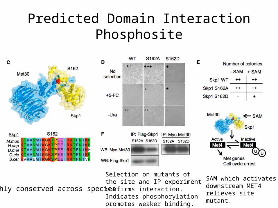
Predicted Domain Interaction Phosphosite
Highly conserved across species.
Selection on mutants of the site and IP experiment confirms interaction.Indicates phosphorylation promotes weaker binding.
SAM which activates downstream MET4 relieves site mutant.
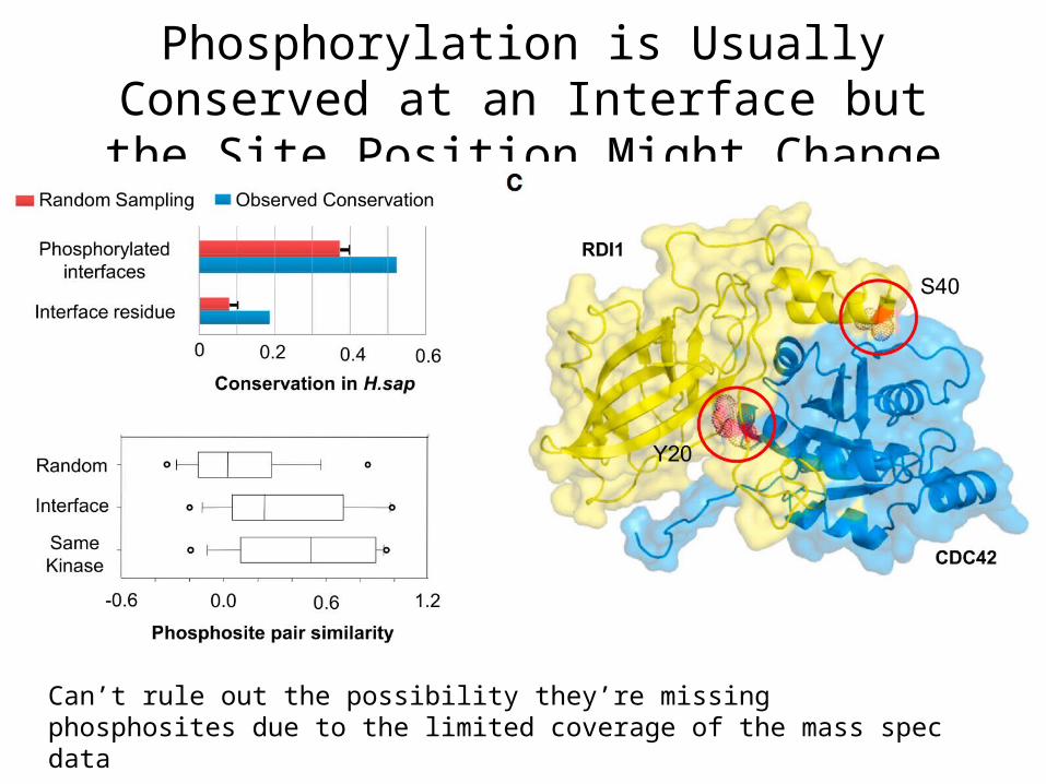
Phosphorylation is Usually Conserved at an Interface but the Site Position Might Change
Can’t rule out the possibility they’re missing phosphosites due to the limited coverage of the mass spec data

Using Conservation to IdentifyRegulatory “Hot spots”

SSA1 (HSP70 family protein) Highly abundant protein
A little strange they refer to HSP70 as a “domain”
Nevertheless in each region they
D mutants mimicthe phosphoyrlated state of theamino acid
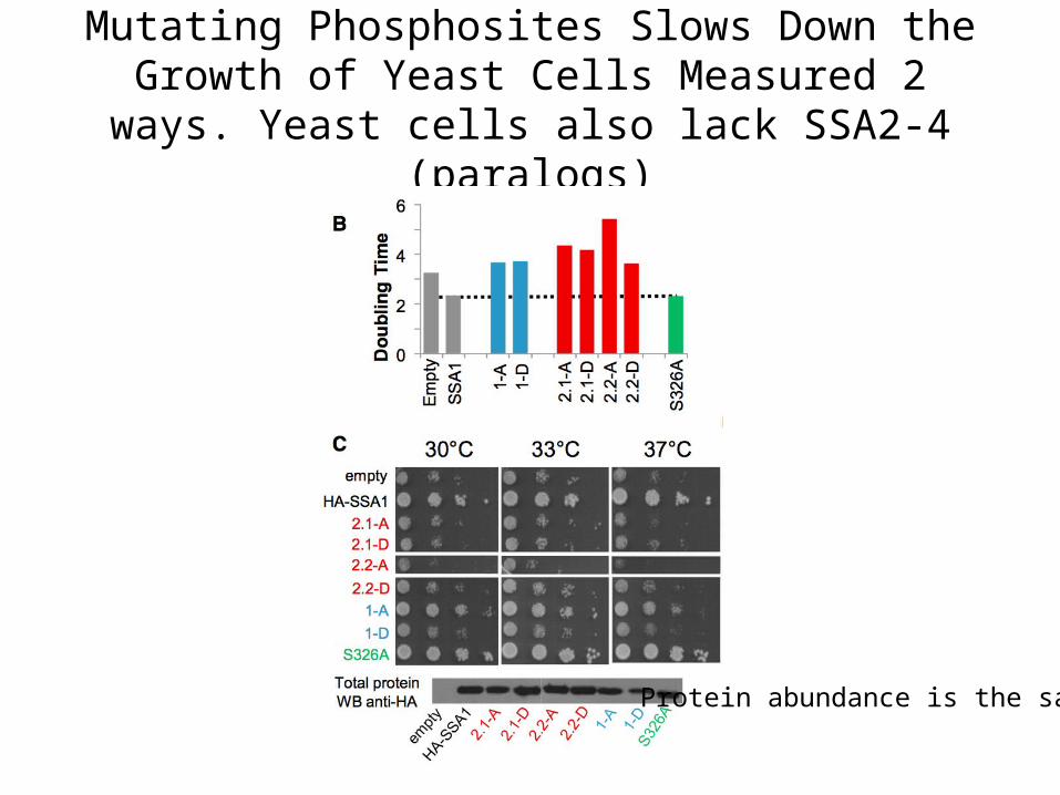
Mutating Phosphosites Slows Down the Growth of Yeast Cells Measured 2 ways. Yeast cells also lack SSA2-
4 (paralogs)
Protein abundance is the same.
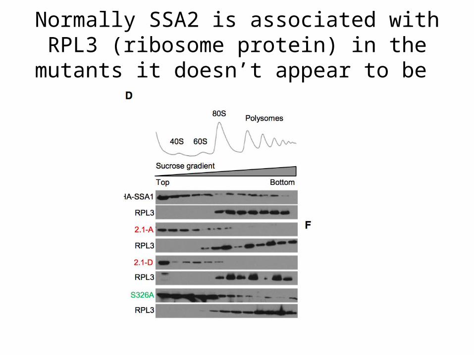
Normally SSA2 is associated with RPL3 (ribosome protein) in the mutants it doesn’t
appear to be
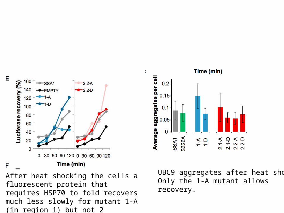
After heat shocking the cells a fluorescent protein that requires HSP70 to fold recovers much less slowly for mutant 1-A (in region 1) but not 2
UBC9 aggregates after heat shockOnly the 1-A mutant allows recovery.