percutaneous coronary intervention report
TRANSCRIPT

Percutaneous Coronary Intervention Report Form DOH-3331
Instructions and Data Element Definitions January 2003
NEW YORK STATE DEPARTMENT OF HEALTH BUREAU OF HOSPITAL & PRIMARY CARE SERVICES
CARDIAC SERVICES PROGRAM One University Place, Suite 209
Rensselaer, NY 12144-3455 Phone: (518) 402-1016 Fax: (518) 402-6992
CARDIAC SERVICES PROGRAM CONTACTS Donna R. Doran, Director, [email protected] Casey S. Roark MPH, Cardiac Databases Coordinator, [email protected] Kimberly S. Cozzens MA, PCI Coordinator, [email protected] Paula Waselauskas RN MS, Hospital Nursing Services Consultant, [email protected]

Table of Contents
Topic
Page
Revision Highlights and Coding Clarification
5-8
ITEM-BY-ITEM INSTRUCTIONS
PFI Number 9Sequence Number
9
I. Patient Information 9Patient’s Name 9Medical Record Number 9Social Security Number 9Age in Years 9Date of Birth 10Sex 10Ethnicity 10Race 10Residence Code 10Hospital Admission Date
10
II. Procedural Information
Hospital that Performed Diagnostic Cath 11Primary Physician Performing PCI 11Date of PCI 11Time at Start of Procedure 11Diagnostic cath during same lab visit 11Previous PCI this admission 12PCI prior to this admission at this hospital 12Procedure Related Medications Fractionated Heparin 12 Un-Fractionated Heparin 12 Direct Thrombin Inhibitors 12 If IV GPIIbIIIa Platelet Inhibitors 12 Indications for the use of IV GPIIbIIIa Platelet Inhibitors 12 Timing for the use of IV GPIIbIIIa Platelet Inhibitors 13 Thrombolytics
13
III. Lesion-Specific Information
IVUS Used 14Lesion Specific Information 14 Location 14 Bypassed (A or V) 14 Bypass Stenosis 14 Lesion Type 14 % Pre-op Stenosis 14
Form DOH-3331 (12/02) 2

Topic
Page
III. Lesion-Specific Information (Cont.)
Previous PCI 14 Primary and Secondary Device 15 Stent 15 Radiation 15 % Post-op Stenosis
15
IV. Acute MI Information
Cardiac Enzymes 16New Abnormal Wall Motion 16New Q Waves 16New ST Elevation 17New ST ↓ or T ↓ 17New Left Bundle Branch Block 17TIMI < II 17Ischemic Type Chest Pain 17Time from Onset of Chest Pain to Hospital Procedure 17Ongoing Ischemia at Time of Procedure
18
V. Pre-Intervention Risk Factors
Priority 18Height 18Weight 18Ejection Fraction and Measure 19Angina: CCS Functional Class 20Angina Type 20Vessels Diseased 21Pre-intervention Risk Factors (None) 22Previous PCIs 22Previous MI (most recent) 22Peripheral Vascular Disease Stroke 23-24 Carotid/Cerebrovascular Disease 23-24 Aortoiliac Disease 23-24 Femoral/Popliteal Disease 23-24Hemodynamic Instability at the time of the procedure Unstable 25 Shock 26 CPR 26More than One Previous MI 27Hypertension History 27Congestive Heart Failure, Current 27Congestive Heart Failure, Past 27Malignant Ventricular Arrhythmia 28Chronic Obstructive Pulmonary Disease 29Diabetes requiring medication 29Renal Failure, creatinine > 2.5 mg/dl 30
Form DOH-3331 (12/02) 3

Topic
Page
V. Pre-Intervention Risk Factors (continued)
Renal Failure, dialysis 30IABP required at the start of procedure 30Previous CABG Surgery 31Immune System Deficiency 31Smoking History, in past 2 weeks 31Smoking History, in past year 31Emergency PCI due to DX cath complication 31Stent Thrombosis 31Any Previous Organ Transplant
32
VI. Major Events Following Intervention None 32Stroke (new neurological deficit) 24 hours or less 32Stroke (new neurological deficit) over 24 hours 33Transmural MI (New Q Waves) 33Non-Transmural MI (No New Q Waves) 33Acute Occlusion in the Targeted Lesion 33Acute Occlusion in a Significant Side Branch 34A/V Injury at Cath Entry Site, requiring intervention 34Renal Failure 34Emergency Cardiac Surgery 35Stent Thrombosis 35Emergency Return to Cath Lab for PCI
35
VII. Discharge Information
Discharged Alive to 36Died in 36Hospital Discharge Date
36
VIII. Person Completing Report
36
Attachments
A: PFI Numbers for Cardiac Diagnostic and Surgical Centers 37-39B: Residence Codes 40C: Definitions of CCS Functional Classes 41D: Codes for Location of Lesions 42E: Characteristics of Type A, B, and C Lesions 43F: Procedure/Device List 44
Form DOH-3331 (12/02) 4

Revision Highlights and Coding Clarification
Patient Information Race: In addition to collecting information on White, Black, and Other this data element has been expanded to include Native American, Asian, and Pacific Islander. If Other is checked you must specify the race.
Procedural Information Time at Start of Procedure: This is a new code in 2003. It should be the time that the first balloon was inflated or when the first stent was deployed. It should be reported using Military Time. Procedure Related Medicines: Heparin: A bolus of heparin at the start of the intervention should NOT be reported. IV GPIIbIIIa Platelet Inhibitors: The Indication for Use of IV GPIIbIIIa Platelet Inhibitors will now be collected by two variables: one to indicate WHY the medicine was given and one to indicate WHEN the medicine was given.
Lesion Specific Information IVUS: The use of intravascular ultrasound (IVUS) is now collected with a yes/no option. Anytime IVUS is used for any reason during the PCI, code YES. Device Code Changes: Additional codes have been added or returned to the system in 2003, please see Attachment F for details. Brachytherapy should be coded as whatever Primary Device was used to open the vessel (i.e. Balloon, Cutting Balloon), Secondary Device: Brachytherapy Catheter, and Radiation. If the radiation is delivered in a separate Cath Lab visit and no device was used to open the vessel code Primary Device: Brachytherapy Catheter and Radiation. Form DOH-3331 (12/02) 5

Revision Highlights and Coding Clarification (Cont.)
Lesion Specific Information (Cont.) Stent Code Changes: Additional codes have been added to the system in 2003, please see Attachment F for details. These codes will be used to collect more detailed information on new stent technologies.
Acute MI Information This section combines the Cardiac Enzyme and Peri-Procedural sections of the reporting system. All information collected in this section should be limited to patients who have a documented MI within 24 hours prior to the PCI. In addition, all information is limited to 24 hours prior to the procedure. The time of onset of chest pain to procedure is the only variable in this section that should be looked at for more than 24 hours pre-procedure. If the patient has had chest pain for 3 days before the procedure, then the time of onset of chest pain to procedure would be 72. Even though the Post-PCI information is no longer reported on the PCIRS form, the Cardiac Advisory Committee recommendations regarding enzyme monitoring remain unchanged.
Pre-Intervention Risk Factors Previous MI < 24 hours: Documentation MUST be present in the medical record that reports the date and time the patient was ruled in with an MI, along with the date and time of the intervention. Congestive Heart Failure, Current and Past: These have new definitions as of 2001. It is important to recognize that it is now possible to code BOTH risk factors. This is due to the fact that the definitions are within “2 weeks” and “2 weeks to 6 months”, respectively. It is also important to note that the definitions are no longer subjective NYHA classifications, they have specific requirements that need to be met for coding.
Form DOH-3331 (12/02) 6
Revision Highlights and Coding Clarification (Cont.)
Pre-Intervention Risk Factors (Cont.) Renal Failure, Creatinine > 2.5 mg/dl: This risk factor should only be coded is the creatinine level goes above 2.5 mg/dl at least one time PRIOR to the start of the procedure. If the creatinine is equal to 2.5 mg/dl DO NOT CODE. In addition, it is appropriate to code renal failure, creatinine > 2.5 mg/dl even if renal failure, dialysis is also coded. Previous CABG Surgery: This was previously called “Previous Open Heart Surgery”. The definition has been limited to Coronary Artery Bypass Graft (CABG) Surgery ONLY. Any Previous Organ Transplant: This risk factor is new to the reporting system in 2003. It should be coded anytime the patient has had any organ transplant prior to the current PCI. This includes, but is not limited to, heart, lung, kidney, and liver transplants.
Major Events Following PCI New Major Events: The following major events are being added in 2003: Transmural MI (new Q waves) Non-Transmural MI (no new Q waves) Emergency Return to the Cath Lab for PCI Please make note of these new events and their definitions (pages 33-35). Acute Occlusion at the Site of Intervention: This major event has been separated into two separate events: Acute Occlusion in the Targeted Lesion and Acute Occlusion in a Significant Side Branch. The definition for each event will be the same as the original definition with the exception that the location of the occlusion determines which event is coded.
Form DOH-3331 (12/02) 7

Revision Highlights and Coding Clarification (Cont.)
Major Events Following PCI (Cont.) Emergency Cardiac Surgery: This event is replacing two previously collected events: (Emergency Bypass Surgery, Hemodynamically Stable and Unstable). This event will not be separated out by hemodynamic stability and will include any cardiac surgery instead of being limited to bypass surgery. Stent Thrombosis: Stent Thrombosis should be reported up to 6 months post-PCI, regardless of whether or not the patient has been discharged from the hospital.
Discharge Status Discharged Alive to: “15 Inpatient Physical Medicine and Rehab” is a new code for 2003. Use this code when a patient is being transferred to an inpatient physical medicine and rehabilitation (PM&R) program. Use this code regardless of whether the PM&R is within your institution (as long as it is an approved PM&R program) or at another institution.
Form DOH-3331 (12/02) 8

ITEM-BY-ITEM INSTRUCTIONS
PFI Number The PFI Number is a Permanent Facility Identifier assigned by the Department of Health. Enter your facility's PFI Number as shown in Attachment A.
Sequence Number If your facility assigns a sequence number to each case on a chronological flow sheet or similar log, enter the sequence number here. The sequence number is not required for the Percutaneous Coronary Interventions Reporting System, but has been included on the form in case your facility finds it useful in identifying and tracking cases.
I. Patient Information
Patient Name Enter the patient’s last name followed by their first name.
Medical Record Number Enter the patient’s medical record number.
Social Security Number Enter the patient's social security number as shown in the medical record. If the medical record does not contain the patient's social security number, leave this item blank. This information can usually be found on the face sheet of the hospital medical record.
Age in Years Enter the patient's age at admission to the hospital. The age should be calculated by subtracting the Date of Birth from the Hospital Admission Date.
Form DOH-3331 (12/02) 9

I. Patient Information (Cont.)
Date of Birth Enter the patient's exact date of birth. Sex Check the appropriate box. Ethnicity Check the appropriate box. Race Check the appropriate box. For White Hispanics, check "White"; for Black Hispanics, check "Black"; "Other" refers to races other than those listed. If you check “Other” then you MUST specify the patient’s race. Residence Code Enter the county code of the patient's principal residence, as shown in Attachment B. If the patient lives outside New York State, use code 99 and print the name of the state or country where the patient resides in the space provided. If the patient is from a foreign country, but is staying in the US during the pre-operative and post-operative time period, you must enter 99 and print the name of the country that the patient is from. Do not enter the residence code of where the patient is staying in the US. Hospital Admission Date Enter the date that the current hospital stay began.
Form DOH-3331 (12/02) 10

II. Procedural Information
Hospital that Performed Diagnostic Cath If the angioplasty was preceded by a diagnostic catheterization, enter the name and PFI number of the hospital in the space provided. If the catheterization was at a cardiac diagnostic center in New York State, enter its PFI Number from Attachment A; if done at a Veterans Administration hospital in New York State, enter "8888"; if done outside New York State, enter "9999". If there was no diagnostic catheterization, leave this item blank.
Primary Physician Performing PCI Enter the name and license number of the primary physician who performed the PCI.
Date of PCI Enter the date on which the PCI was performed.
Time at Start of Procedure Report the time that the first balloon was inflated or when the first stent was deployed. It should be reported using Military Time (i.e. 1:00 am is 01:00, and 1:00 pm is 13:00).
Diagnostic Cath During Same Lab Visit If a full diagnostic catheterization was performed during the same cath lab visit as the PCI procedure, then check “Yes”. Otherwise check “No”. This does NOT include the case where there was a “quick look” done on the vessel to have the intervention. The diagnostic cath does not have to be every vessel, but should be a complete diagnostic of the area of interest.
Form DOH-3331 (12/02) 11

II. Procedural Information (Cont.)
Previous PCI This Admission For patients who have had a previous PCI during this admission, check “Yes”. Otherwise check “No”. If YES, it is very important that you enter the date of this procedure. It is this date that allows multiple procedures during the same hospital admission to be combined in the proper order. This becomes especially important when determining Emergency/Non-Emergency status, since certain risk factors are only “credited” if they occur prior to the first procedure in a hospital admission.
PCI Prior to This Admission at this Hospital For patients who have had a PCI prior to this admission at this hospital, check “Yes” and report the date of this previous procedure. If only the month and year are known, use 01 for the day and write in the correct month and year. If only the year is known, write in 01/01 for the month and day and the correct year.
Procedure Related Medicines
Check ALL that apply.
Form DOH-3331 (12/02) 12
Fractionated Heparin Administered 6 hrs pre-proc or anytime post-PCI
Includes low molecular weight heparin. E.g. Lovenox, Fragmin, and Innohep.
Un-Fractionated Heparin Administered 6 hrs pre-proc or anytime post-PCI
Direct Thrombin Inhibitors Administered 6 hrs pre-proc or anytime post-PCI
E.g. Refludan, Argatroban, and Angiomax
If IV GPIIbIIIa Platelet Inhibitors Administered 6 hrs pre-proc or anytime post-PCI
Check the appropriate box to indicate which IV GPIIbIIIa Platelet Inhibitor was used. If more than one is given, check the one that was given first. If one of these is checked, the Indication for Use of IV GPIIbIIIa Platelet Inhibitors MUST also be checked.
Indications for the Use of IV GPIIbIIIa Platelet Inhibitors
Please mark the appropriate box, to indicate the reason for giving the first dose of Abciximab or any other IV GPIIbIIIa Platelet Inhibitor. 1 – Angiographic Evidence 2 – Clinical Evidence 3 – Standard Practice/ Prophylactic 4 – Another Reason The indication checked should be the primary reason for giving the first dose of any IV GPIIbIIIa Platelet Inhibitor. If IV GPIIbIIIa Platelet Inhibitors were NOT given, leave Blank.

II. Procedural Information (Cont.)
Procedure Related Medicines (Cont.)
Timing for the Use of IV GPIIbIIIa Platelet Inhibitors
Please mark the appropriate box, to indicate the timing that the Abciximab or any other IV GPIIbIIIa Platelet Inhibitor was given. 1 – Pre 2 – Post 3 – Both
Thrombolytics Check the appropriate box to indicate if, and at what time interval, thrombolytics were administered.
If thrombolytics were not administered because they were Contraindicated, check “Contraindicated”.
Interpretation of Indications and Timing for Use of IV GPIIbIIIa Platelet Inhibitors: This section applies to the timing and reason for giving IV GPIIbIIIa Platelet Inhibitors. If the medicine was given for more than one of the following reasons, check the one that occurred first. For example, if it was given before the intervention based on angiographic evidence and after the start of the procedure due to clinical decompensation, you would code the following: Indication = 1. Angiographic Evidence (because that was the first reason) and Timing = 3. Both (because it was given pre and post-PCI). Some examples of the types of indications include: Angiographic Evidence: Evidence of intra luminal thrombus (defined as: presence of filling
defect within the coronary lumen, surrounded by contrast material, seen in multiple projections; persistence of contrast material (staining) of the lumen; a hazy lesion; visible embolization of intra luminal material downstream); High-risk lesions for PCI (B and C); Complication of low risk lesion (A); Acute occlusion at the site of PCI; Residual dissection at the site; Sub-optimal Results.
Clinical Evidence: Acute Coronary Syndrome (ACS) including patients with unstable angina, non ST- elevation MI, and ST-elevation MI.; Acute MI requiring primary angioplasty; Instability occurring during or after PCI (intractable angina, acute MI, etc)
Standard Practice/ Prophylactic: If given as standard practice or for prophylatctic purposes. Another Reason: Any indication not listed above.
Form DOH-3331 (12/02) 13

III. Lesion-Specific Information
IVUS Used If Intravascular ultrasound (IVUS) was used for any reason during the PCI, code YES. Lesion-Specific Complete one line for each lesion for which PCI was attempted, and one line for each non-attempted lesion with diameter stenosis of 50% or more – even if the lesion has been bypassed. If there are more than seven lesions, report the seven most significant. Location Enter the code indicating the location of the lesion, as shown in Attachment D.
For lesions in a "sequential" graft going to two of the major coronary systems, complete a separate line for each coronary artery jeopardized (LAD, LCX, RCA). See examples in Attachment D. Bypassed (A or V) If the lesion has been bypassed by a vein graft, enter "V" If the lesion has been bypassed by an artery graft, enter "A". If the lesion was not bypassed leave blank.
Bypass Stenosis If the lesion has a vein or artery graft, use the following code to determine the level of stenosis found in the graft: 1. > 70% 2. < 70% 3. Unknown Lesion Type Enter code A, B, or C to describe the lesion type, as defined in Attachment E.
% Pre-Op Stenosis Enter the pre-PCI percent diameter reduction. Measurement with calipers is recommended.
Previous PCI Use the following codes to indicate if the lesion is restenotic following a previously successful PCI. 0 -- No Previous PCI 1 -- No Restenosis 2 -- Restenosis, No Stent Previously Placed in the Vessel 3 -- Restenosis, Stent Previously Placed in the Vessel
Form DOH-3331 (12/02) 14

III. Lesion-Specific Information (Cont.)
Lesion-Specific Information (Cont.) Primary Device and Secondary Device
ANY attempt to cross a lesion with a guide wire constitutes attempted PCI. As soon as the guidewire leaves the catheter there is an attempted PCI. From the procedural code list in Attachment F, indicate the primary device used. If the lesion was not attempted, place a “0” under primary device. If a secondary device was used, indicate the device used in the appropriate box. The attending physician is responsible for determining the primary and secondary devices.
Stent From the Stent code list in Attachment F, indicate the type of stent used.
Radiation Check if ANY radiation was placed in the vessel regardless of the source.
% Post-Op Stenosis If a PCI was attempted on this lesion, enter the percent diameter of the stenosis immediately following the PCI. Measurement with calipers is recommended. If PCI was not attempted, leave post-op stenosis blank.
Interpretation: Brachytherapy should be coded as whatever Primary Device was used to open the vessel (i.e. “1” Balloon, “5” Cutting Balloon), Secondary Device: “10” Brachytherapy Catheter, and “1” Radiation. If the radiation is delivered in a separate Cath Lab visit and no device was used to open the vessel code Primary Device: “10” Brachytherapy Catheter and “1” Radiation. If a Balloon is used SOLEY to properly place a stent, DO NOT CODE the balloon as a primary device, ONLY CODE the stent. If the Medical Record says % Post-Stenosis was 0%, record it as 1% to indicate that it was actually a successful PCI and not left blank. If the device is unable to be advanced due to complications, but the guidewire leaves the catheter and it is an attempted PCI, code a Balloon as the primary device, unless the intended device is known. If a stent and another device (ie. Distal Protective Device) are the only devices used and the stent is considered the primary device by the physician, the other device should be coded as the primary device and NOT the secondary device.
Form DOH-3331 (12/02) 15

IV. Acute MI Information
Complete this section for all patients with an MI less than 24 hours prior to the PCI. NOTE: ONLY patients with Pre-Intervention Risk Factors 4-6 should have information reported in this section. The following cardiac enzymes, EKG changes, and ischemic information should only be reported if they occurred up to 24 hours prior to PCI. Cardiac Enzymes Report results of pre-PCI cardiac enzyme measures. Troponin may be used in place of the CK-MB iso-enzyme. Interpretation: Troponin can now be reported with two decimal places to add increased accuracy. The timing of the enzymes should be determined by when the blood was drawn NOT when it was processed by the lab. CAC guidelines recommend one pre- and two post-PCI enzyme measures. New Abnormal Wall Motion Should be coded when abnormal wall motion is considered new and persisting as determined by EKG, ECHO or Nuclear Medicine. Interpretation: You would code new abnormal wall motion in the following scenario:
In the absence of baseline studies with NO reasonable clinical evidence of a previous MI, if the ventriculogram shows hypokinesis and/or akinesis, and the patient is in the Acute Phase of an MI.
New Q Waves Defined as 0.03 seconds in width and/or > one third of the total QRS complex in two or more continuous leads.
Form DOH-3331 (12/02) 16

IV. Acute MI Information (Cont.) New ST Elevation > 1mm in two or more continuous leads. New ST or T New Ischemic changes on EKG appearing as ST depression, T-Wave inversion, or both. New Left Bundle Branch Block (LBBB) Should be coded when LBBB is considered new and persisting as evidenced by EKG. TIMI < II Evidence of TIMI flow < II WITH either total vessel occlusion or a high-grade lesion. Ischemic Type Chest Pain Characteristics of ischemic type chest pain for > 20 minutes and not relieved by Nitroglycerin. Characteristics of ischemic type chest pain can have a surrogate when associated with the cardiac event. Some equivalents would include but are not limited to: pain in the arm, shoulder, back, or jaw. Time from Onset of Chest Pain to Procedure Report in hours. Round to the nearest half hour. For example, a patient report of 1¼ hr, would be reported as 1.5 and a patient report of 2 hrs 10 minutes would be reported as 2.0. If greater than 99.9 hours, report 99.9. NOTE: This is the only data element in this section that is reportable for more than 24 hours Pre-PCI. The time reported here should be the time from the onset of chest pain that brought the patient to the hospital or caused them to seek care. If the chest pain has stopped before the start of the procedure, you can still report the number of hours since it started.
Form DOH-3331 (12/02) 17

IV. Acute MI Information (Cont.) Ongoing Ischemia at Time of Procedure Check this box if the patient is experiencing chest pain and acute ST or T-Wave changes at the start of the PCI.
V. Pre-Intervention Risk Factors
Priority Check the appropriate box. Elective: All cases not classified as urgent or emergency as defined below.
Urgent: The patient is too ill or unstable to be discharged from the hospital,
but is not classified as emergency as defined below.
Emergency: Patients requiring emergency procedures will have ongoing, refractory, unrelenting cardiac compromise, with or without hemodynamic instability. Typical patients include those in arrest with CPR administered immediately prior to the procedure, shock, ongoing ischemia including rest angina, acute evolving MI or equivalent within 24 hours of procedure, and/or pulmonary edema requiring intubation.
Height Enter the patient’s height in centimeters (cm). Centimeters = 2.54 x inches
Weight Enter the patient’s weight in kilograms (kg). Kilograms = pounds (lbs) / 2.2
Form DOH-3331 (12/02) 18

V. Pre-Intervention Risk Factors (Cont.)
Ejection Fraction and Measure Record the ejection fraction taken closest to the cardiac procedure. When a calculated measure is unavailable, the ejection fraction should be estimated visually from the ventriculogram or by echocardiography. If an ejection fraction is unavailable, check “Unknown”. Note: Intraoperative direct observation of the heart is NOT an adequate basis for a visual estimate of the ejection fraction. Interpretation: Any ejection fraction that is well documented in the chart is acceptable, but give precedence to the one closest to the cardiac procedure. An Ejection Fraction is acceptable and should be reported if it is measured or estimated by the following:
1. LV Angiogram 2. Echocardiogram 3. Radionuclide Studies 4. Transesophageal Echocardiogram (TEE), this includes intra-operative 8. Other
If the measure is unknown code it as a “9. Unknown”. An ejection fraction, that is described in the medical record as “Normal” should be considered 55. Any cases with a missing or “0” ejection fraction will be sent back to the centers during quarterly and annual data validation to verify accuracy of this data element.
Form DOH-3331 (12/02) 19

V. Pre-Intervention Risk Factors (Cont.)
Angina: CCS Functional Class Check the box corresponding to the patient's Canadian Cardiovascular Society Functional Class, as defined in Attachment C. Note: The determination of functional class should be based on the typical level of exertion required to produce angina. For example, a single episode of anginal pain at rest does not qualify a patient as Class IV unless it is the initial episode of angina.
Angina Type Check the box indicating the patient’s angina type Stable: Angina without a change in frequency or pattern for the 6 weeks prior to
this procedure. Angina is controlled by rest and/or oral or transcutaneous medications.
Unstable: Angina has increased in frequency during the last 6 weeks, including new onset. Angina is produced by less effort or provocation and occurring in a crescendo pattern. Angina can be experienced at rest and pain may last for longer periods of time and be more difficult to relieve. Includes progressive, rest, and variant.
Interpretation: NOTE: Angina type should NOT be confused with CCS Class. CCS Class deals with functional ability of the patient. CCS Class IV patients for example have pain at rest. If this pain at rest has been the same intensity for more than 6 weeks then it would be considered Stable. If the patient has been categorized as CCS Class IV, but the frequency has increased in the last few weeks then it would be considered unstable angina.
Form DOH-3331 (12/02) 20

VI. Pre-Intervention Risk Factors (Cont.)
Vessels Diseased For each diseased vessel, check the appropriate box to indicate the percent diameter stenosis. Include all vessels diseased, even branches. Interpretation: This section MUST be completed for each procedure. Use the ranges listed below when the medical record describes the percent stenosis in the following ways: MILD = plaques to < 50% MODERATE = 50-70% SEVERE = > 70% If the diseased segment of the native vessel is bypassed by an open artery or vein graft, DO NOT code as diseased. This vessel is revascularized. If a vessel or branch is described as having “Mild” stenosis then the vessel would NOT be coded as diseased, since we only code 50-100% stenosis. If the medical record reports 60-70% stenosis, then code 50-69%. The Ramus Intermediate would be coded as the LAD or LCX. ALWAYS take the highest stenosis reported for a vessel. If the medical record reports the Ramus Intermediate with a 70% lesion and the Distal Circumflex Trunk with a 50% you should code the LCX as 70-100%, since the Ramus Intermediate has a 70% lesion. If the medical record only has documentation that states the LAD was stenosed: then code the Mid LAD and NOT the Proximal LAD.
Form DOH-3331 (12/02) 21

V. Pre-Intervention Risk Factors (Cont.)
0. None None of the pre-operative risk factors listed below are present.
1-3. Previous PCIs If the patient had one or more previous PCI, check the appropriate box to indicate the number of previous PCI's. Include any interventions that occurred prior to this one during the current admission. If there was a previous procedure this admission, please be sure that the date of the most recent PCI is indicated next to “Previous PCI This Admission” on the front of the form.
4-7. Previous MI (most recent) If the patient had one or more myocardial infarctions before PCI, report the length of time since the most recent MI. If less than 6 hours, check box “4” If >6 - <12 hours, check box “5” If >12 - <24 hours, check box “6” If 24 hours or more, enter the number of days in the space provided next to “7”. If 21 days or more, enter "21".
Form DOH-3331 (12/02) 22

V. Pre-Intervention Risk Factors (Cont.)
Peripheral Vascular Disease
8. Stroke A history of stroke, with or without residual deficit.
9. Carotid/ Cerebrovascular Angiographic or ultrasound demonstration of at least 50% narrowing in a major cerebral or carotid artery (common or internal), history of a non-embolic stroke, or previous surgery for such disease. A history of bruits or transient ischemic attacks (TIA) is not sufficient evidence of carotid/cerebrovascular disease.
10. Aortoiliac Angiographic demonstration of at least 50% narrowing in a major aortoiliac vessel, previous surgery for such disease, absent femoral pulses, or the inability to insert a catheter or intra-aortic balloon due to iliac aneurysm or obstruction of the aortoiliac arteries.
11. Femoral/Popliteal Angiographic demonstration of at least 50% narrowing in a major femoral/popliteal vessel, previous surgery for such disease, absent pedal pulses, or inability to insert a catheter or intra-aortic balloon due to obstruction in the femoral arteries.
Form DOH-3331 (12/02) 23

V. Pre-Intervention Risk Factors (Cont.)
Peripheral Vascular Disease (Continued) Interpretation:
Peripheral Vascular Disease CODE DO NOT CODE
Stroke 1. Patient with TIA, vertigo per history & physical X 2. Cerebral aneurysm and clipping residual deficit X
Carotid/Cerebrovascular 1. External Carotid Artery has > 50% stenosis X 2. Internal or Common Carotid Artery has > 50% stenosis X
Aortoiliac
1. Tortuosity of the vessel alone X 2. Tortuosity of the vessel with an inability to insert a catheter X 3. Abdominal Aortic Aneurysm (AAA) X 4. Aneurysm in the ascending or descending aorta X 5. History of aorto-bifemoral bypass X 6. Absence of femoral pulse on either the right or the left X 7. Diminished femoral pulse on either right or left or both X 8. Claudication X
Femoral/Popliteal 1. Leg ulceration and diminished leg pulses X 2. Conflicting doctor note and H&P Cannot feel dorsalis pedis or posterior tibial pulses Consult note: 1+ peripheral pulses
X
3. Angio report -> because of tortuosity of the vessel, even a sheath could not be passed up the wire.
X
4. A negative popliteal pulse alone (1+1- or 1-1+) X 5. A patient with a palpable Dorsalis Pedis and Posterior Tibial pulses and treated with Trental
X
6. If pulses are non-palpable, but is Dopplerable X 7. If Dorsalis Pedis and Posterior Tibial pulses are absent in the right or the left or both
X
8. Below the knee amputation of one or both legs X 9. Inability to insert a catheter or IABP in femoral arteries X 10. At least 50% narrowing in a major femoral artery X
Form DOH-3331 (12/02) 24

V. Pre-Intervention Risk Factors (Cont.) Hemodynamic Instability at Time of Procedure Determined just prior to or at the commencement of the PCI (the guide-wire crossing the lesion). These patients have hypotension and low cardiac output. The administration of pharmacological or mechanical support MUST be documented in the patient’s medical record. For purposes of reporting, the PCI does not constitute the mechanical support.
12. Unstable The patient requires pharmacologic or mechanical support to maintain blood pressure or output. Interpretation:
Unstable
CODE DO NOT CODE
1. Patient on IV Nitroglycerin or IV Heparin X 2. IABP inserted for pain control X 3. Inability to place IABP because of tortuous and diseased vessels
X
4. Documents evidence of hypotension, with NO pharmacologic or mechanical support
X
When coding “Unstable”, be careful of timing. It needs to be prior to or at the commencement of the PCI. Once the guide-wire has passed the lesion or left the catheter any instability after that would not constitute the patient being coded “Unstable”. There MUST be actual documentation of blood pressure and/or cardiac output, simply stating that the patient was hypotensive is NOT sufficient. With documented evidence of hypotension (low B/P), an IABP would be considered mechanical support and the patient would be considered unstable. The procedure itself DOES NOT constitute mechanical support.
Unstable CANNOT be coded with SHOCK
Form DOH-3331 (12/02) 25

V. Pre-Intervention Risk Factors (Cont.)
Hemodynamic Instability at Time of Procedure (Cont.)
13. Shock Acute hypotension (systolic blood pressure < 80 mmHg) or low cardiac index (< 2.0 liters/min/m2), despite pharmacologic or mechanical support. Interpretation: To code a patient with “Shock” there must be evidence that all the criteria for “Unstable” have been met and that DESPITE the support the patient’s hemodynamic status fails to improve or stabilize. When coding “Shock”, be careful of timing. It needs to be prior to or at the commencement of the PCI. Once the guide-wire has crossed the lesion any factors that would constitute the patient being coded “Shock” would NOT matter. References in the medical record of “hemodynamics not improved”, “continuing instability”, etc are acceptable documentation. Shock CANNOT be coded with Unstable.
33. CPR The patient requires cardiopulmonary resuscitation within one hour prior to the procedure. Interpretation: To code “CPR”, it MUST occur within ONE hour prior to surgery. A single defibrillation, even if accompanied by initial compressions, DOES NOT constitute coding “CPR”. CPR CAN be coded with either Unstable or Shock.
Form DOH-3331 (12/02) 26

V. Pre-Intervention Risk Factors (Cont.)
14. More Than One Previous MI Clinical or ECG evidence of more than one previous myocardial infarction (MI). Interpretation: The MI must be documented to have occurred PRIOR to the intervention.
15. Hypertension History Code if any of the following are present:
• Blood pressure greater than 140/90 • History of hypertension • Current treatment for hypertension.
18. Congestive Heart Failure, Current Within 2 weeks prior to the procedure, a physician has diagnosed CHF by one of the following:
• Paroxysmal nocturnal dyspnea (PND) • Dyspnea on exertion (DOE) due to heart failure • Chest X-Ray showing pulmonary congestion
NOTE: Pedal edema or dyspnea alone are NOT diagnostic. Patient should also have received diuretics, digoxin, or vascular therapy such as ace inhibitors. Interpretation:
Congestive Heart Failure, Current
CODE DO NOT CODE
1. Patient admitted to Hospital A, with CHF and then transferred to Hospital B (within 2 weeks)
X
2. Hospital reports: Chest + for rales, treated with Lasix X 3. Patient with prior renal transplant, pending renal transplant with creatinine up to 5 and BUN-72. Renal failure would explain the bilateral pleural effusions and DOE. Lasix was used to treat fluid retention secondary to renal failure not CHF. CXR indicating “cannot rule out mild CHF” is pretty consistent with fluid overload due to Renal Failure.
X
If there is documentation to support coding both “Congestive Heart Failure, Current” and “Congestive Heart Failure, Past” – then CODE BOTH risk factors.
Form DOH-3331 (12/02) 27
V. Pre-Intervention Risk Factors (Cont.)
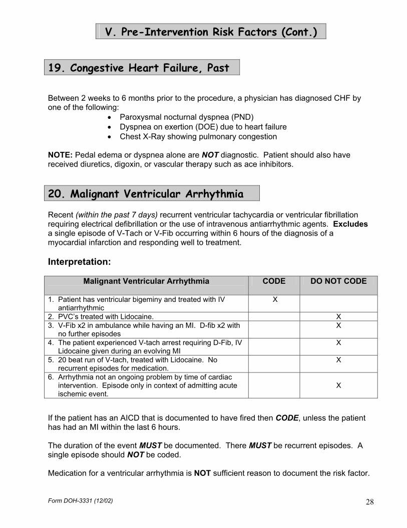
19. Congestive Heart Failure, Past Between 2 weeks to 6 months prior to the procedure, a physician has diagnosed CHF by one of the following:
• Paroxysmal nocturnal dyspnea (PND) • Dyspnea on exertion (DOE) due to heart failure • Chest X-Ray showing pulmonary congestion
NOTE: Pedal edema or dyspnea alone are NOT diagnostic. Patient should also have received diuretics, digoxin, or vascular therapy such as ace inhibitors.
20. Malignant Ventricular Arrhythmia Recent (within the past 7 days) recurrent ventricular tachycardia or ventricular fibrillation requiring electrical defibrillation or the use of intravenous antiarrhythmic agents. Excludes a single episode of V-Tach or V-Fib occurring within 6 hours of the diagnosis of a myocardial infarction and responding well to treatment. Interpretation:
Malignant Ventricular Arrhythmia
CODE DO NOT CODE
1. Patient has ventricular bigeminy and treated with IV antiarrhythmic
X
2. PVC’s treated with Lidocaine. X 3. V-Fib x2 in ambulance while having an MI. D-fib x2 with no further episodes
X
4. The patient experienced V-tach arrest requiring D-Fib, IV Lidocaine given during an evolving MI
X
5. 20 beat run of V-tach, treated with Lidocaine. No recurrent episodes for medication.
X
6. Arrhythmia not an ongoing problem by time of cardiac intervention. Episode only in context of admitting acute ischemic event.
X
If the patient has an AICD that is documented to have fired then CODE, unless the patient has had an MI within the last 6 hours. The duration of the event MUST be documented. There MUST be recurrent episodes. A single episode should NOT be coded. Medication for a ventricular arrhythmia is NOT sufficient reason to document the risk factor.
Form DOH-3331 (12/02) 28

V. Pre-Intervention Risk Factors (Cont.)
21. Chronic Obstructive Pulmonary Disease Patients who require chronic (longer than three months) bronchodilator therapy to avoid disability from obstructive airway disease,
Or Have a forced expiratory volume in one second of less than 75% of the predicted value or less than 1.25 liters,
Or Have a room air pO2 <60 or a pCO2 >50. NOTE: COPD should not be checked unless the patient’s medical record contains documented evidence of the above criteria, regardless of how much the patient may have smoked. Interpretation:
COPD
CODE DO NOT CODE
1. Chest X-Ray as documentation X 2. Patient required bronchodilators prior to surgery X 3. Hyperinflated lungs, wheezing X 4. Fibrotic lungs on chest X-Ray X 5. Hyperinflated lungs at operation X 6. Although admitting notes state no history of COPD, documentation in the chart elsewhere as well as chest X-Ray results and a smoking history are apparent
X
7. Patient has a history of asthma, is on bronchodilator and asthma meds
X
8. Chart states asthma without medications X 9. Sleep Apnea without any of the above criteria X
22. Diabetes Requiring Medication The patient is receiving either oral hypoglycemics or insulin. Interpretation: The following scenario WOULD NOT be coded since the medication was not ongoing:
Patient admitted on 12/28. Nurses note on 12/29: “patient has no hx DM but had insulin (stat) in another hospital.” Glucose level 155 on NO meds.
Form DOH-3331 (12/02) 29

V. Pre-Intervention Risk Factors (Cont.)
23. Renal Failure, Creatinine > 2.5 mg/dl Pre-PCI Creatinine > 2.5 mg/dl. Interpretation: No matter how many renal transplants the patient has had, if the creatinine DOES NOT have at least one value >2.5 mg/dl, prior to the intervention, DO NOT code. If the patient has at least one creatinine > 2.5 mg/dl prior to the intervention then code. If the patient is on dialysis and the creatinine is > 2.5 mg/dl then code both this risk factor and risk factor 24 (Renal Failure, Dialysis)
24. Renal Failure, Dialysis The patient is on chronic peritoneal or hemodialysis. Interpretation: A single dialysis treatment DOES NOT constitute coding this risk factor. If the patient is on dialysis and the creatinine is > 2.5 mg/dl then code both this risk factor and risk factor 23 (Renal Failure, creatinine > 2.5 mg/dl)
26. IABP Required at the Start of Procedure The patient arrives in the cath lab with an intra-aortic balloon pump in place, or requires its insertion before the commencement of the PCI procedure, for ongoing myocardial ischemia, left ventricular failure, or shock, clinical evidence of which should be contained in the patient’s record. Excludes IABP necessitated by a complication of previous PCI. Interpretation: The intervention begins at the first insertion of the guidewire. Can be coded if it is inserted AT THE SAME TIME the intervention is being started. The following scenario WOULD NOT be coded:
IABP inserted at Hospital A. IABP removed before transfer to Hospital B. Intervention was done at Hospital B.
Form DOH-3331 (12/02) 30

V. Pre-Intervention Risk Factors (Cont.)
28. Previous CABG Surgery Previous coronary artery bypass (CABG) surgery. Interpretation: DO NOT code if it occurred during the same admission as the PCI in question. If the patient has an A or V coded in the lesion specific section, then this variable should be coded UNLESS the grafting occurred during this admission.
29. Immune System Deficiency Chronic use, that continues until intervention, of steroids, anti-neoplastic therapy, cyclosporine, or other immunosuppressive therapy or the presence of HIV/AIDS.
30. Smoking History, in past 2 weeks The patient has used any tobacco products within the past two weeks. The use of chewing tobacco would be included here.
31. Smoking History, in past year The patient has used any tobacco products within the past year. The use of chewing tobacco would be included here.
32. Emergency PCI due to DX Cath Complication Catheterization related dissection or obstruction of coronary artery during diagnostic catheterization, requiring immediate, unplanned angioplasty to treat closure or threatened closure of the vessel. 34. Stent Thrombosis Formation of a blood clot/thrombus in the stented segment of the artery and/or adjacent area. This usually results in an acute occlusion, chest pain or development of an acute MI. Stent thrombosis usually occurs up to 30 days following the procedure. Interpretation: An occlusion alone or plaque build-up DOES NOT constitute coding. The thrombus needs to be in or around the area that is stented for the risk factor to be coded. Form DOH-3331 (12/02) 31

V. Pre-Intervention Risk Factors (Cont.)
35. Any Previous Organ Transplant The patient has had any organ transplant prior to the PCI. This includes, but is not limited to, heart, lung, kidney, and liver transplants.
VI. Major Events Following Intervention
Check to be sure that all of the listed major events occurred during or after the intervention. Check at least one box in this section. Please Note: A documented pre-intervention risk factor that persists post-intervention with NO increase in severity is not a major event. Unless otherwise specified, major events are ONLY reported if they occur during or after PCI, but before hospital discharge.
0. None Check if none of the Major Events listed below occurred following the intervention.
1. Stroke (New Neurological Deficit) 24 hrs or less Permanent new focal neurological deficit occurring either during the intervention or within 24 hrs Post-PCI. Interpretation: Exacerbation of a previous CVA with No New Neurological Deficit would NOT be coded. Transient neurological deficits, such as TIA, are no longer reported as a Post-PCI event. If the condition is still present at discharge, then the event should be reported.
Form DOH-3331 (12/02) 32

VI. Major Events Following Intervention (Cont.)
1A. Stroke (New Neurological Deficit) over 24 hours Permanent new focal neurological deficit occurring more than 24 hours Post-PCI. Interpretation: Exacerbation of a previous CVA with No New Neurological Deficit would NOT be coded. Transient neurological deficits, such as TIA, are no longer reported as a Post-PCI event. If the condition is still present at discharge, then the event should be reported. 2. Transmural MI (New Q Waves) New Q waves and a rise in cardiac enzyme (CK) to at least 2.5 times the normal range, occurring within 24 hours after PCI.
3. Non-Transmural MI (No New Q Waves) Utilize your hospital’s clinical quidelines to determine a non-transmural MI, occurring within 24 hours after PCI. 7A. Acute Occlusion in the Targeted Lesion Acute occlusion, complete or partial, in the targeted lesion resulting in reduction of flow through the dilated artery. Usually caused by thrombosis, intimal flap, or dissection. An occlusion which is reopened before the patient leaves the catheterization laboratory and stays open should NOT be reported. An occlusion requiring the patient’s return to the catheterization laboratory SHOULD be reported even if the vessel is then reopened. If the acute occlusion is caused by a stent thrombosis, ONLY code the stent thrombosis.
Form DOH-3331 (12/02) 33

VI. Major Events Following Intervention (Cont.)
7B. Acute Occlusion in a Significant Side Branch Acute occlusion, complete or partial, in a significant side branch resulting in reduction of flow. This should include any occlusion in any location within the significant proximal or distal branches (including the left artery) of the targeted or treated vessel. Usually caused by thrombosis, intimal flap, or dissection. An occlusion which is reopened before the patient leaves the catheterization laboratory and stays open should NOT be reported. An occlusion requiring the patient’s return to the catheterization laboratory SHOULD be reported even if the vessel is then reopened.
8. A/V Injury at Cath Entry Site, requiring intervention Arterial or Venous injury, including, but NOT limited to:
Those requiring femoral or brachial embolectomy Evacuation of a hematoma Repair of false aneurysm, example: ultrasound guided compressions Closure of arterial-venous fistula.
Report occlusions which occur at other coronary arterial sites related to the PCI.
10. Renal Failure Creatinine greater than 2.5 mg/dl for more than 7 days Post-PCI or there is need for temporary or permanent renal dialysis of any type. Do not code this item if Risk Factors 23 (Renal Failure, Creatinine > 2.5 mg/dl) or 24 (Renal Failure, Dialysis) are coded.
Form DOH-3331 (12/02) 34

VI. Major Events Following Intervention (Cont.)
14. Emergency Cardiac Surgery The patient is taken to the operating room for cardiac surgery on an emergency basis due to a complication of PCI.
17. Stent Thrombosis Formation of a blood clot in the stented segment of the artery and/or adjacent area. This usually results in an acute occlusion, chest pain, or development of an acute MI. Stent thrombosis usually occurs within 30 days following the procedure. NOTE: Stent Thrombosis should be reported as a major event even if it does not become apparent until after the patient is discharged from the hospital. It should be reported if apparent up to 6 months post intervention. Interpretation: An occlusion alone or plaque build-up DOES NOT constitute coding. The thrombus needs to be in or around the area that is stented for the major event to be coded. 18. Emergency Return to the Cath Lab for PCI The patient is taken to the Cath Lab for PCI on an emergency basis due to a complication of a previous PCI.
Form DOH-3331 (12/02) 35

VII. Discharge Information
Discharged Alive To Check the appropriate box. Patients discharged to Hospice (including Home with Hospice), code “12”. NOTE: for purposes of analysis a hospice discharge (“12”) is considered an in-hospital mortality. If the patient came from a Prison or Institutional Facility and is being discharged back to the same setting then “11 – Home” would be coded. If the patient is discharged to sub-acute rehab that is in a skilled nursing facility then the discharge status would be “14”, if it is unknown where the sub-acute rehab facility is located then the discharge status would be “19”. If the patient is discharged to an inpatient physical medicine and rehabilitation unit the discharge status should be “15”. “19 – Other (specify)” should NEVER be checked if it is specified as “4 – Died CCU” or “Died”, these cases should be coded in the next section. Any discharge status “19” that does not specify where the patient was discharged to will be sent back to the hospital for verification. Died in Check the appropriate box. If “8 – Elsewhere in Hospital (specify)” is checked, specify where the patient died. Hospital Discharge Date Enter the date the patient was discharged from the hospital. If the patient died in the hospital, the hospital discharge date is the date of death.
VIII. Person Completing Report This section is for hospital use only. It may be helpful to enter the name and telephone number of the person completing the report, and the date the report was completed.
Form DOH-3331 (12/02) 36

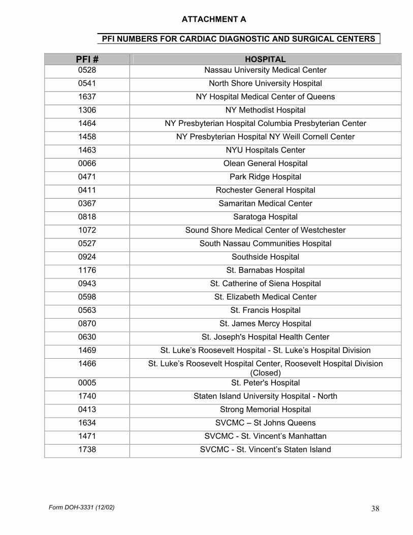
ATTACHMENT A
PFI NUMBERS FOR CARDIAC DIAGNOSTIC AND SURGICAL CENTERS
PFI # HOSPITAL 0001 Albany Medical Center Hospital 0116 Arnot Ogden Medical Center 1438 Bellevue Hospital Center 1439 Beth Israel Medical Center / Petrie Campus 1164 Bronx Lebanon Hospital Center – Fulton Division 1286 Brookdale Hospital Medical Center 0885 Brookhaven Memorial Hospital Medical Center, Inc. 1288 Brooklyn Hospital Center - Downtown 0207 Buffalo General Hospital 0977 Cayuga Medical Center at Ithaca 0135 Champlain Valley Physicians Hospital Medical Center 0208 Children's Hospital of Buffalo 1626 City Hospital Center at Elmhurst 1294 Coney Island Hospital 0636 Crouse Hospital 0829 Ellis Hospital 0210 Erie County Medical Center 0599 Faxton St. Luke’s Healthcare, St. Luke’s Division 0407 Genesee Hospital
(Closed) 1005 Glens Falls Hospital 0925 Good Samaritan Hospital Medical Center (West Islip) 0779 Good Samaritan Hospital of Suffern 1445 Harlem Hospital Center 0913 Huntington Hospital 1300 Interfaith Medical Center, Jewish Hosp. Med Ctr of Brooklyn Division 1629 Jamaica Hospital Medical Center 1450 Lenox Hill Hospital 1302 Long Island College Hospital 1630 Long Island Jewish Medical Center 1304 Lutheran Medical Center 1305 Maimonides Medical Center 0746 Mary Imogene Bassett Hospital 0213 Mercy Hospital of Buffalo 0215 Millard Fillmore Hospital 1169 Montefiore Medical Center – Henry and Lucy Moses Division 3058 Montefiore Medical Center – Jack D. Weiler Hosp. of A. Einstein College Div. 1456 Mount Sinai Hospital
Form DOH-3331 (12/02) 37
ATTACHMENT A
PFI NUMBERS FOR CARDIAC DIAGNOSTIC AND SURGICAL CENTERS
PFI # HOSPITAL 0528 Nassau University Medical Center 0541 North Shore University Hospital 1637 NY Hospital Medical Center of Queens
1306 NY Methodist Hospital 1464 NY Presbyterian Hospital Columbia Presbyterian Center 1458 NY Presbyterian Hospital NY Weill Cornell Center 1463 NYU Hospitals Center 0066 Olean General Hospital 0471 Park Ridge Hospital 0411 Rochester General Hospital 0367 Samaritan Medical Center 0818 Saratoga Hospital 1072 Sound Shore Medical Center of Westchester 0527 South Nassau Communities Hospital 0924 Southside Hospital 1176 St. Barnabas Hospital 0943 St. Catherine of Siena Hospital 0598 St. Elizabeth Medical Center 0563 St. Francis Hospital 0870 St. James Mercy Hospital 0630 St. Joseph's Hospital Health Center 1469 St. Luke’s Roosevelt Hospital - St. Luke’s Hospital Division
1466 St. Luke’s Roosevelt Hospital Center, Roosevelt Hospital Division (Closed)
0005 St. Peter's Hospital 1740 Staten Island University Hospital - North
0413 Strong Memorial Hospital 1634 SVCMC – St Johns Queens 1471 SVCMC - St. Vincent’s Manhattan
1738 SVCMC - St. Vincent’s Staten Island
Form DOH-3331 (12/02) 38

ATTACHMENT A
PFI NUMBERS FOR CARDIAC DIAGNOSTIC AND SURGICAL CENTERS
PFI # HOSPITAL 0058 United Health Services Hospital, Inc – Wilson Hospital Division
1320 University Hospital of Brooklyn
0245 University Hospital at Stony Brook
0635 University Hospital SUNY Health Science Center (Upstate) 0181 Vassar Brothers Hospital 1139 Westchester Medical Center 0511 Winthrop University Hospital 0103 Woman's Christian Association
8888 Catheterization Laboratory at a Veterans Administration Hospital in New York (for use in this reporting system; not an official Permanent Facility Identifier)
9999 Catheterization Laboratory Outside New York State (for use in this reporting system; not an official Permanent Facility Identifier)
Form DOH-3331 (12/02) 39

ATTACHMENT B
Residence Codes The county codes shown below are also used in the SPARCS Discharge Data Abstract: 01 Albany 35 Oswego 02 Allegany 36 Otsego 03 Broome 37 Putnum 04 Cattaraugus 38 Rensselaer 05 Cayuga 39 Rockland 06 Chautauqua 40 St. Lawrence 07 Chemung 41 Saratoga 08 Chenango 42 Schenectady 09 Clinton 43 Schoharie 10 Columbia 44 Schuyler 11 Cortland 45 Seneca 12 Delaware 46 Steuben 13 Dutchess 47 Suffolk 14 Erie 48 Sullivan 15 Essex 49 Tioga 16 Franklin 50 Tompkins 17 Fulton 51 Ulster 18 Genesee 52 Warren 19 Greene 53 Washington 20 Hamilton 54 Wayne 21 Herkimer 55 Westchester 22 Jefferson 56 Wyoming 23 Lewis 57 Yates 24 Livingston 58 Bronx 25 Madison 59 Kings 26 Monroe 60 Manhattan 27 Montgomery 61 Queens 28 Nassau 62 Richmond 29 Niagara
30 Oneida 31 Onondaga 88 Unknown 32 Ontario 33 Orange 99 Outside NYS 34 Orleans
Form DOH-3331 (12/02) 40

ATTACHMENT C
Definitions of CCS Functional Classes Canadian Cardiovascular Society (CCS) Functional Classification: Class I Ordinary physical activity, such as walking or climbing stairs, does not cause angina. Angina may occur with strenuous or rapid or prolonged exertion at work or recreation. Class II There is slight limitation of ordinary activity. Angina may occur with walking or climbing stairs rapidly, walking uphill, walking or stair climbing after meals or in the cold, in the wind, or under emotional stress, or walking more than two blocks on the level, or climbing more than one flight of stairs under normal conditions at a normal pace. Class III There is marked limitation of ordinary physical activity. Angina may occur after walking one or two blocks on the level or climbing one flight of stairs under normal conditions at a normal pace. Class IV There is inability to carry on any physical activity without discomfort; angina may be present at rest.
Form DOH-3331 (12/02) 41

ATTACHMENT D
Codes for Location of Lesion Use the list and diagram below to find the code for location of lesion.
1. Prox RCA 2. Mid RCA 3. Dist RCA 4. R PDA 5. RPLS 6. 1st RPL 7. 2nd RPL 8. 3rd RPL 9. Inf. Septal
10. Ac Marg 11. LMCA 12. Prox LAD * 13. Mid LAD 14. Dist LAD 15. 1st Diag or
Intermediate Branch 16. 2nd Diag 17. 1st Septal 18. Prox CX 19. Dist CX 20. 1st Ob Marginal 21. 2nd Ob Marginal 22. 3rd Ob Marginal 23. L A V 24. 1st LPL 25. 2nd LPL 26. 3rd LPL 27. LPDA
41. Vein Graft to LMCA 42. Artery Graft to LMCA
51. Vein Graft to LAD 52. Artery Graft to LAD
61. Vein Graft to LCX 62. Artery Graft to LCX
71. Vein Graft to RCA 72. Artery Graft to RCA
88. PTMR
* Code 12 refers to the region before the origin of the major septal artery.
Form DOH-3331 (12/02) 42

ATTACHMENT E
Characteristics of Type A, B, and C Lesions If ANY of the characteristics of a Type C lesion are present, use code C. If NO Type C lesion characteristics are present and ANY Type B lesion characteristics are present, use code B. If NO Type B or C lesion characteristics are present, use code A. Type A Lesions: Discrete (< 10 mm length) Concentric Readily accessible Non-angulated segment, < 45 degrees Smooth contour Little or no calcification Less than totally occlusive Not ostial in location No major branch involvement Absence of thrombosis Type B Lesions: 10 to 20 mm length Eccentric Moderate tortuosity of proximal segment Moderately angulated segment, 45 to 90 degrees Irregular contour Moderate to heavy calcification Total occlusion < 3 months old Ostial in location Bi-furcation lesions requiring double guide wires Some thrombosis present Type C Lesions: > 2 cm length Excessive tortuosity of proximal segment Extremely angulated segment, > 90 degrees Total occlusion > 3 months old Inability to protect major side branches Degenerated vein grafts with friable lesions
Form DOH-3331 (12/02) 43

ATTACHMENT F
Procedure/Device List Use the following values to code procedures and/or devices used during the intervention. NOTE: If a Balloon is ONLY used to insert the stent then DO NOT code the Balloon as a device. Brachytherapy should be coded as whatever Primary Device was used to open the vessel (i.e. “1” Balloon, “5” Cutting Balloon), Secondary Device: “10” Brachytherapy Catheter, and “1” Radiation. If the radiation is delivered in a separate Cath Lab visit and no device was used to open the vessel code Primary Device: “10” Brachytherapy Catheter and “1” Radiation. Primary and Secondary Devices: 0 Lesion Not Attempted or No Device Used 1 Balloon 2 Directional Atherectomy 3 Rotational Atherectomy 4 Distal Protective Devices (Including Filter Wires) 5 Cutting Balloon 6 Laser 7 Transluminal Extraction Catheter (TEC) 8 PTMR 10 Brachytherapy Catheter 11 Angiojet 99 Other (Specify) Stents: 0 No Stent Used 1 Un-Coated Stent 2 Covered Stent (membrane coated) 3 Heparin Coated Stent 4 Paclitaxel Coated Stent 5 Tacrolimus Coated Stent 6 Sirolimus Coated Stent 9 Other Coated Stent (Specify)
Form DOH-3331 (12/02) 44