percutaneous fine needle aspirationcytology the … byeither may-grunwald-giemsaor papan-icolaou...
TRANSCRIPT

J Clin Pathol 1989;42:341-347
Percutaneous fine needle aspiration cytology of thepancreas: advantages and pitfallsG KOCJAN,* J RODE,* W R LEES
From the *Department of Histopathology, University College and Middlesex School of Medicine, London, andthe Department ofImaging, Middlesex Hospital, London
SUMMARY Fine needle aspiration of the pancreas was performed in 62 patients with radiologicalsuspicion of malignancy. All fine needle aspirates were taken under computed tomography orultrasound guidance. Fine needle aspirates were positive in 31 of 41 patients with histologically orclinically confirmed pancreatic carcinoma. There were no false positive results. The sensitivity of thismethod for detecting malignant disease was 86%. Cytology was not able to provide conclusive resultsof benign conditions. Difficulties were encountered in diagnosing well differentiated carcinoma andneuroendocrine tumours and distinguishing them from reactive epithelium and islet cell hyperplasia,respectively. This resulted in a 12- 1% false negative rate. There were no complications in our series.Percutaneous fine needle aspiration proved a reliable method of diagnosing pancreatic carcinoma.
The primary objective of the doctor caring for apatient whose symptoms suggest pancreatic carcin-oma should be to establish the diagnosis with aminimum of time, testing, money and suffering.' Themortality of patients with this disease is 99% withinthe first two years.2 Early diagnosis remains the onlyknown method of cure. Fine needle aspirationcytology of the pancreas under radiological guidan-ce,3-13 or during surgery,"" has been widely used as adiagnostic method. It is especially useful in diagnosingsolid neoplasms which account for over 90% ofpancreatic tumours.'6
Material and methods
Fine needle aspirates of the pancreas were taken usinga 21G spinal needle under computed tomographyguidance and local (subcutaneous) anaesthesia. Theneedle was attached to a 20 ml syringe and suctionenhanced by using a syringe holder. Aspiration wasperformed after reaching the desired site by applyingrepeated suction and by releasing it before the needlewas withdrawn from the lesion. This prevented thenegative pressure forcing the blood into the syringe.After removing the needle from the patient the needlewas detached from the holder and aspirated materialwas ejected on to glass slides averaging nine smears ineach case. Cystic fluids and needle washouts weretransported in buffered saline.The fluids were processed in the cytocentrifuge. All
Accepted for publication 3 November 1988
slides were either air dried or fixed in alcohol andstained by either May-Grunwald-Giemsa or Papan-icolaou methods, respectively. Whenever possible,spare slides were kept frozen at -20°C for immun-ocytochemical analysis. Carcinoembrionic antigen(CEA), neuron specific enolase (NSE), andchromogranin were the antibodies used in a limitednumber of cases.
Results
Fine needle aspiration cytology was performed in 62patients with radiological or clinical evidence ofpancreatic mass. Definitive diagnosis of carcinomafrom fine needle aspirate smears was made in 31patients. In 21 patients this was confirmed by thehistological core biopsy specimen taken at the sametime, and in 10 patients clinically by the rapid progressof the disease. There were no false positive results.Cytology was negative in 17 patients; in seven patientsmaterial was inadequate for cytological assessmentdue to poor cell yield or preservation. In seven patientsaspirates were suspicious but not diagnostic. Sub-sequent histology showed carcinoma in five of thenegative and two of the inadequate aspirates. Patientswith findings suspicious but not diagnostic of malig-nancy diagnosed in cytological smears were biopsied.Biopsy specimens showed carcinoma in three cases,were negative in two, inadequate in one and suspiciousof malignancy in one case. The overall sensitivity offine needle aspiration cytology in diagnosing pan-creatic carcinoma was 86%, excluding the cases where
341
on 16 May 2019 by guest. P
rotected by copyright.http://jcp.bm
j.com/
J Clin P
athol: first published as 10.1136/jcp.42.4.341 on 1 April 1989. D
ownloaded from
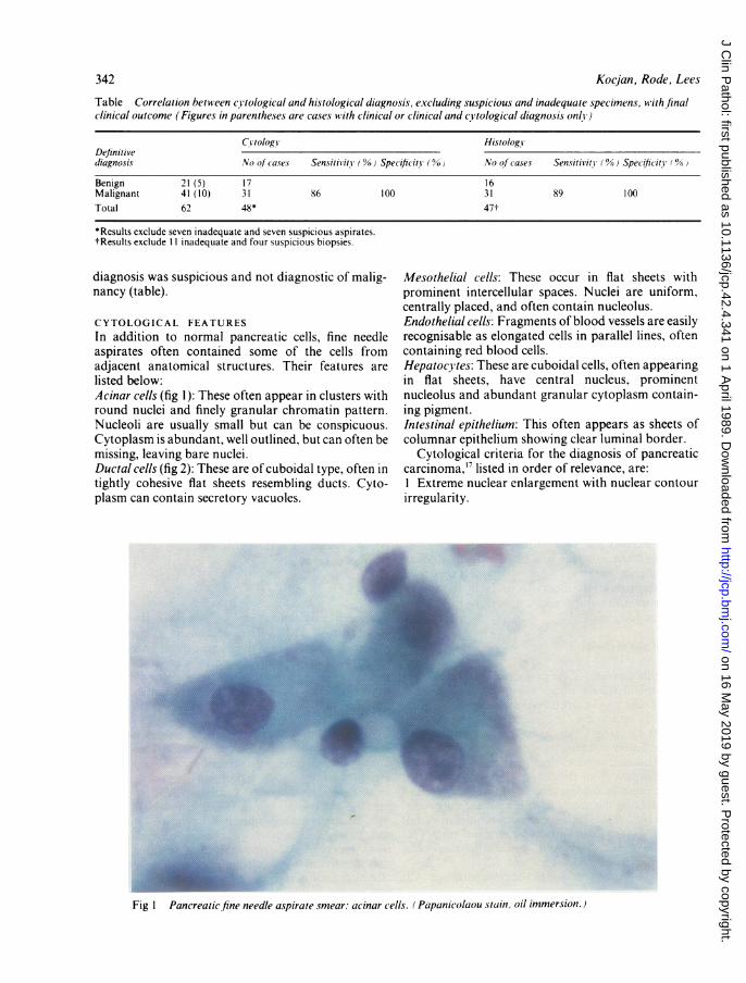
Table Correlation between cytological and histological diagnosis, excluding suspicious and inadequate specimens, with finalclinical outcome (Figures in parentheses are cases with clinical or clinical and cytological diagnosis only)
C}tology HistologsDefinitivediagnosis No of (O.ses Sensitivity 00) Specificitv ( 0) No of cases Sensitivitv (% Specificity 00
Benign 21(5) 1 7 1 6Malignant 41 (10) 31 86 100 31 89 100Total 62 48* 47t
*Results exclude seven inadequate and seven suspicious aspirates.tResults exclude II inadequate and four suspicious biopsies.
diagnosis was suspicious and not diagnostic of malig-nancy (table).
CYTOLOGICAL FEATURESIn addition to normal pancreatic cells, fine needleaspirates often contained some of the cells fromadjacent anatomical structures. Their features arelisted below:Acinar cells (fig 1): These often appear in clusters withround nuclei and finely granular chromatin pattern.Nucleoli are usually small but can be conspicuous.Cytoplasm is abundant, well outlined, but can often bemissing, leaving bare nuclei.Ductal cells (fig 2): These are of cuboidal type, often intightly cohesive flat sheets resembling ducts. Cyto-plasm can contain secretory vacuoles.
Mesothelial cells: These occur in flat sheets withprominent intercellular spaces. Nuclei are uniform,centrally placed, and often contain nucleolus.Endothelial cells: Fragments of blood vessels are easilyrecognisable as elongated cells in parallel lines, oftencontaining red blood cells.Hepatocytes: These are cuboidal cells, often appearingin flat sheets, have central nucleus, prominentnucleolus and abundant granular cytoplasm contain-ing pigment.Intestinal epithelium: This often appears as sheets ofcolumnar epithelium showing clear luminal border.
Cytological criteria for the diagnosis of pancreaticcarcinoma,'7 listed in order of relevance, are:1 Extreme nuclear enlargement with nuclear contourirregularity.
...
Fig I Pancreatic fine needle a.spirate smear: ac inar c ells. ( Papanic olaou stai'n, oil immersion.)
342 Kocjan, Rode, Lees
on 16 May 2019 by guest. P
rotected by copyright.http://jcp.bm
j.com/
J Clin P
athol: first published as 10.1136/jcp.42.4.341 on 1 April 1989. D
ownloaded from

Percutaneous fine needle aspiration cytology of the pancreas
Fi.2Pnet f ei a l . -G s
Fig 2 Pancreaticfine needle aspirate smear: duct cells. (May-Grunwald-Giemsa stain.)
Fig 3 Pancreatic fine needle aspirate smear: well differentiated adenocarcinoma, erroneously interpreted asduct cell atypia. (May-Grinwald-Giemsa.)
343
on 16 May 2019 by guest. P
rotected by copyright.http://jcp.bm
j.com/
J Clin P
athol: first published as 10.1136/jcp.42.4.341 on 1 April 1989. D
ownloaded from

Kocjan, Rode, Lees
l..:~~~~~~~~~~~~~~~~~~~~~~~~~~~~~~~..eVP"._ ;s
F -
Fig 4 Pancreatic fine needle aspirate smear: poor/i' differentiated carcinoma showing classic features ofmalignancv. (Ma -Grinwiald-Giemsa.)
~~~~.*X .. ..
* ~ | t 3 t . 'R
a:' *
Fig 5 Pancreatic fine needle aspirate' islet cells in i.slet cell hYperplasia. (Papanicolaou stain.)
344
on 16 May 2019 by guest. P
rotected by copyright.http://jcp.bm
j.com/
J Clin P
athol: first published as 10.1136/jcp.42.4.341 on 1 April 1989. D
ownloaded from
Percutaneousfine needle aspiration cytology of the pancreas
'.* 0f
C
40
tU sw
! .@;at- P.# <L .* o t
*+"- _ i" > A.. j .V
...
>'4C,
I'
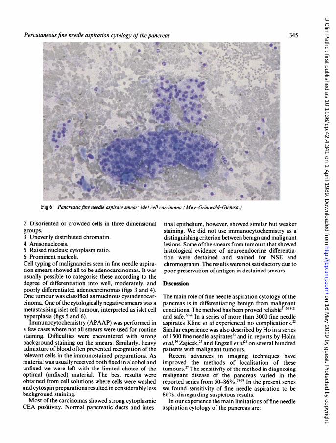
Fig 6 Pancreaticfine needle aspirate smear: islet cell carcinoma (May-Griunwald-Giemsa.)
2 Disoriented or crowded cells in three dimensionalgroups.3 Unevenly distributed chromatin.4 Anisonucleosis.5 Raised nucleus: cytoplasm ratio.6 Prominent nucleoli.Cell typing of malignancies seen in fine needle aspira-tion smears showed all to be adenocarcinomas. It wasusually possible to categorise these according to thedegree of differentiation into well, moderately, andpoorly differentiated adenocarcinomas (figs 3 and 4).One tumour was classified as mucinous cystadenocar-cinoma. One ofthe cytologically negative smears was ametastasising islet cell tumour, interpreted as islet cellhyperplasia (figs 5 and 6).Immunocytochemistry (APAAP) was performed in
a few cases where not all smears were used for routinestaining. Difficulties were encountered with strongbackground staining on the smears. Similarly, heavyadmixture of blood often prevented recognition of therelevant cells in the immunostained preparations. Asmaterial was usually received both fixed in alcohol andunfixed we were left with the limited choice of theoptimal (unfixed) material. The best results wereobtained from cell solutions where cells were washedand cytospin preparations resulted in considerably lessbackground staining.Most of the carcinomas showed strong cytoplasmic
CEA positivity. Normal pancreatic ducts and intes-
tinal epithelium, however, showed similar but weakerstaining. We did not use immunocytochemistry as adistinguishing criterion between benign and malignantlesions. Some ofthe smears from tumours that showedhistological evidence of neuroendocrine differentia-tion were destained and stained for NSE andchromogranin. The results were not satisfactory due topoor preservation of antigen in destained smears.
Discussion
The main role of fine needle aspiration cytology of thepancreas is in differentiating benign from malignantconditions. The method has been proved reliable3 `08-21and safe.2226 In a series of more than 3000 fine needleaspirates Kline et al experienced no complications.22Similar experience was also described by Ho in a seriesof 1500 fine needle aspirates23 and in reports by Holmet al,24 Zajicek,25 and Engzell et afl6 on several hundredpatients with malignant tumours.
Recent advances in imaging techniques haveimproved the methods of localisation of thesetumours." The sensitivity of the method in diagnosingmalignant disease of the pancreas varied in thereported series from 50-86%.2838 In the present serieswe found sensitivity of fine needle aspiration to be86%, disregarding suspicious results.
In our experience the main limitations of fine needleaspiration cytology of the pancreas are:
3450 0
a
on 16 May 2019 by guest. P
rotected by copyright.http://jcp.bm
j.com/
J Clin P
athol: first published as 10.1136/jcp.42.4.341 on 1 April 1989. D
ownloaded from
346 Kocjan, Rode, LeesFalse negative results These are due to poor sampling,technical preparation, and interpretation. Poor sam-pling can be due to scarring and fibrosis associatedwith some tumours. It can also be due to the needlemissing a small neoplasm. Two of the inadequatespecimens in our series were subsequently provedmalignant. Technical preparation of direct smears wasdifficult because of the heavy admixture of blood inmost specimens, causing early clotting and preventingproper spreading of the smear. The cells were trappedin the clot and were difficult to interpret. Problems ofinterpretation can occur when distinguishing epith-elial atypia due to chronic pancreatitis or duct obstruc-tion from well differentiated neoplasms, particularlywhen smears are poorly cellular and contain a highadmixture of blood. Five of the cases reported asbenign or reactive proved malignant (fig 3). Similardifficulties were met in three of the seven cytologicallysuspicious lesions which subsequently proved malig-nant. The false negative rate in our series was 12- 1 %.Diagnosis of neuroendocrine cell tumours One of thecytologically benign cases in our series was a malig-nant neuroendocrine tumour with subsequent matas-tases in the liver. Cytological smears showed remarka-bly uniform islet cells in aggregates (fig 6). As these aresometimes seen in islet cell hyperplasia (fig 5) accom-panying chronic pancreatitis, they were interpreted asbenign. The experience ofsome authors shows that cellmorphology alone is sometimes sufficient for diagnos-ing these tumours.39 '
Differentiation ofcell type Most of the tumours in thisseries were adenocarcinomas, showing various degreesof differentiation. Cytologically we were able toclassify one of the tumours as mucinous cystadenocar-cinoma. It was not possible, however, to detect foci ofneuroendocrine differentiation within individualtumours and report with confidence tumours showinghistologically two components (adenosquamous car-cinoma). Whether the separation of these histologicaltypes will eventually prove relevant to someepidemiological clinical types, clinical feature, or to aresponse to a specific chemotherapeutic regimen is notknown.'The most important question to be answered by fine
needle aspiration cytology is whether a carcinoma ispresent or not. Using the criteria described by Mitchellet al,"7 we feel that extreme nuclear enlargement anddisoriented cells in three dimensional clusters are themost helpful features for distinguishing between ben-ign and malignant lesions, regardless of the quantity ofcells and presence of other features described by thesame authors. In this respect aspiration cytology is areliable method on which to base a decision to proceedwith radical operation in selected cases, as well asprognosis in inoperable cases.
Fine needle aspiration cytology is a quick, safe, and
reliable method of diagnosing pancreatic carcinoma.It is inexpensive and is an easily tolerated procedure.Only positive results are of value and negative resultsdo not exclude malignancy.
Requests for reprints to: Dr G Kocjan, CytologyLaboratory, Department of Histopathology, MiddlesexHospital, Mortimer Street, London WIP 7PN, England.
References
I Malagelada JR. Pancreatic cancer. Mayo Clin Proc 1979;54:459-67.
2 Hovdenak N, Lees WR, Pereira J, Beilby JOW, Cotton PB.Ultrasound guided percutaneous fine needle aspirationcytology in pancreatic cancer. Br Med J 1982;285:284-5.
3 Hastrup J, Thommesen P, Fredriksen P. Pancreatitis and pan-creatic cancer diagnosed by preoperative fine needle aspirationbiopsy. Acda Cytol 1978;21:731-4.
4 Hidvegi D, Nieman HL, James W. Percutaneous transperitonealaspiration of pancreas guided by ultrasound. Acta CYtol1979;23:1814.
5 Ihse I, Isaksson G. Preoperative and operative diagnosis ofpancreatic cancer. World J Surg 1 984;8:846-53.
6 Kolins M, Bernacki EG, Schwab R. Diagnosis of pancreaticlesions by percutaneous aspiration biopsy. Acta C}vtol198 1;25:675-7.
7 Luning M, Kursawa R, Schopke W, ei al. CT guided percutaneousfine needle biopsy of the pancreas. Eur J Radiol 1985;20:22 1-38.
8 Nguyen GK. Percutaneous fine needle aspiration cytology of thepancreas. Pathol Annu 1985;20:221-38.
9 Phillips VM, Hersch T, Erwin BC, et al. Percutaneous biopsy ofpancreatic masses. J Clin Gastroenterol 1985;7:506-10.
10 Tao LC. Ho CS, McLoughlin MJ, McHattie J. Percutaneous fineneedle aspiration biopsy of the pancreas. Acta CYtol1978;22:215-20.
11 Yamamoto R, Tatsuda M, Noguchi S, et al. Histocytologicdiagnosis of pancreatic cancer by percutaneous aspirationbiopsy under ultrasonic guidance. Amn J Clin Pathol1985;83:409-14.
12 Bognel C, Rougier P. Leclere J, et al. Fine needle aspiration of theliver and pancreas with ultrasound guidance. Ac ta CYtol1988;32:22-6.
13 Ihse 1, Toregard B, Akerman M. lntraoperative fine needleaspiration cytology in pancreatic lesions. Ann Surg1979:190:732-4.
14 Keighley MRB, Moore J, Thomson H. The place of fine needleaspiration cytology for the intraoperative diagnosis of pan-creatic malignancy. Ann RoY Coll Surg Engl 1984;66:405-8.
15 Smith S, Vogelszang RL, Donovan J, Atlas SW, Gore RM,Weiman HL. Intraoperative sonography of the pancreas. Am JRadiol 1985;144:557-62.
16 Pinto MM, Avila NA, Criscuolo EM. Fine needle aspiration of thepancreas: a five year experience. Acia CYtol 1988;32:39-42.
17 Mitchell ML, Carney CN. Cytologic criteria for the diagnosis ofpancreatic carcinoma. Am J Surg Pathol 1985;83:171-6.
18 An-Foraker SH, Fong Mui KK. Cytodiagnosis of lesions ofpancreas and related areas. Acia C! tol 1982;26:815-9.
19 Christofferson OP, Poll P. Perioperative pancreas aspirationbiopsies. Acta Pathol Microbiol Scand 1970;212:28-32.
20 Kline TS, Goldstein F, Neal HS. Pancreatic carcinoma, pan-creatitis and needle aspiration biopsy. Arch Surg 1974;109:578-9.
21 Willems JS, Lowhagen T. Aspiration biopsy cytology of thepancreas. Schvei: Med Wochenschr 1980;110:845-58.
on 16 May 2019 by guest. P
rotected by copyright.http://jcp.bm
j.com/
J Clin P
athol: first published as 10.1136/jcp.42.4.341 on 1 April 1989. D
ownloaded from

Percutaneous fine needle aspiration cytology of the pancreas 34722 Kline TS, Neal HS. Needle aspiration biopsy: critical appraisal.
Eight years and 3267 specimens later. JAMA 1978;239:36-9.23 Ho CS, McLoughlin MJ, McHattie JD, et al. Percutaneous. fine
needle aspiration biopsy of the pancreas following endoscopicretrograde cholangiopancreatography. Radiology 1977;125:351-3.
24 Holm HH, Pedersen JF, Kristensen JK, et al. Ultrasonical guidedpercutaneous puncture. Radiol Clin North Am 1975;13:493-503.
25 Zajicek J. Introduction to aspiration biopsy. Clin Cytol 1974;4:1-211.
26 Engzell U, Esposti PL, Rubio C, et al. Investigation on tumourspread in connection with aspiration biopsy. Acta Radiol1971;11:385-98.
27 Hall-Craggs MA, Lees WR. Fine needle aspiration biopsy:pancreatic and biliary tumours. Ann J Radiol 1986;147:399-403.
28 Alpern GA, Dekker A. Fine needle aspiration cytology of thepancreas: An analysis of its use in 52 patients. Acta Cytol1985;29:873-8.
29 Droese M, Altmannsberger M, Kehl A, et al. Ultrasound guidedpercutaneous fine needle aspiration biopsy of abdominal andretroperitoneal masses: accuracy ofcytology in the diagnosis ofmalignancy, cytologic tumour typing and use of antibodies tointermediate filaments in selected cases. Acta Cytol1984;18:368-84.
30 Mitty HA, Efremidis SC, Yeh HC. Impact of fine needle biopsy on
management of patients with carcinoma of the pancreas. Am JRoentgenol 1981;137:1 119-21.
32 Zornoza J. Percutaneous needle biopsy. Baltimore: Williams andWilkins, 1980:102-40.
33 Smith EH, Bartrum RJ, Chang YC, et al. Percutaneous aspirationbiopsy of the pancreas under ultrasonic guidance. N Engl JMed 1975;292:825-8.
34 Hancke S, Holm HH, Koch F. Ultrasonically guided percutan-eous fine needle biopsy of the pancreas. Surg Gynaecol Obstet1975;140:361-4.
35 Tylen U, Arnesjo B, Kindberg LG, Lunderquist A. Percutaneousbiopsy guided by angiography. Surg Gynecol Obstet1976;142:737-9.
36 McLouglin MJ, Ho CS, Langer B, McHattie J, Tao LC. Fineneedle aspiration biopsy of malignant lesions in and aroundpancreas. Cancer 1978;41:2413-9.
37 Goldstein HM, Zornozo J, Wallace S, et al. Percutaneous fineneedle aspiration biopsy of pancreatic and other abdominalmasses. Radiology 1977;123:319-22.
38 Pilotti S, Rilke F, Claren R, Milella M, Lombardi L. Conclusivediagnosis of hepatic and pancreatic malignancies by fine needleaspiration. Acta Cytol 1988;32:27-38.
39 Bell DA. Cytological features of islet cell tumours. Acta Cytol1987;31:485-92.
40 Banner BF, Myrent KL, Memoli VA, Gould VE. Neuroendocrinecarcinoma of the pancreas diagnosed on fine needle aspiration.A case report. Acda Cvtol 1985;29:422-8.
on 16 May 2019 by guest. P
rotected by copyright.http://jcp.bm
j.com/
J Clin P
athol: first published as 10.1136/jcp.42.4.341 on 1 April 1989. D
ownloaded from