perfecting intelligence, transforming medicine
TRANSCRIPT


About UsLunit, abbreviated from “learning unit,” is a medical AI software company
devoted to developing advanced medical image analytics and
novel imaging biomarkers via cutting-edge deep learning technology.
Founded in 2013, Lunit has been internationally acknowledged for its advanced,
state-of-the-art technology and its application in medical images.
Lunit is based in Seoul, South Korea.
Our MissionPerfecting Intelligence, Transforming Medicine.
Through our unprecedented AI technology, we seek to provide AI solutions that open
a new era of diagnostics and therapeutics. We are especially focused on conquering
cancer, one of the leading cause of death worldwide.
Perfecting Intelligence, Transforming Medicine.
2 I Lunit INSIGHT MMG
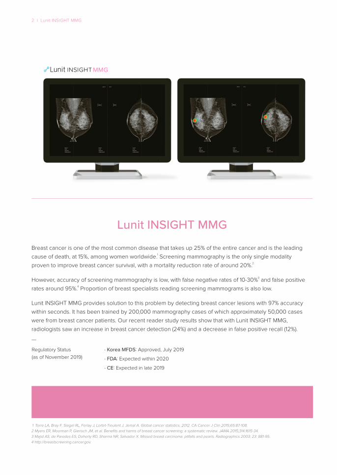
Lunit INSIGHT MMG
Breast cancer is one of the most common disease that takes up 25% of the entire cancer and is the leading
cause of death, at 15%, among women worldwide .1 Screening mammography is the only single modality
proven to improve breast cancer survival, with a mortality reduction rate of around 20% .2
However, accuracy of screening mammography is low, with false negative rates of 10-30%3 and false positive
rates around 95% .4 Proportion of breast specialists reading screening mammograms is also low.
Lunit INSIGHT MMG provides solution to this problem by detecting breast cancer lesions with 97% accuracy
within seconds. It has been trained by 200,000 mammography cases of which approximately 50,000 cases
were from breast cancer patients. Our recent reader study results show that with Lunit INSIGHT MMG,
radiologists saw an increase in breast cancer detection (24%) and a decrease in false positive recall (12%).
—
You can login to https://insight.lunit.io to freely upload images
and get real-time analysis results conducted by Lunit INSIGHT in no time.
Regulatory Status
(as of November 2019)
· Korea MFDS: Approved, July 2019
· FDA: Expected within 2020
· CE: Expected in late 2019
1 Torre LA, Bray F, Siegel RL, Ferlay J, Lortet-Tieulent J, Jemal A. Global cancer statistics, 2012. CA Cancer J Clin 2015;65:87-108.
2 Myers ER, Moorman P, Gierisch JM, et al. Benefits and harms of breast cancer screening: a systematic review. JAMA 2015;314:1615-34.
3 Majid AS, de Paredes ES, Doherty RD, Sharma NR, Salvador X. Missed breast carcinoma: pitfalls and pearls. Radiographics 2003; 23: 881-95.
4 http://breastscreening.cancer.gov.

Internal Validation(Korea, United States, United Kingdom)
Lunit INSIGHT MMG was validated internally throughout various countries with different ethnicity.
Validation dataset consists of approximately 3,200 patients of mammography exams from 3 countries,
of which 1,858 patients from Korea (KR), 750 from United States (US), and 654 from United Kingdom (UK).
Performance Summary: ROC AUC, Sensitivity, Specificity
Density Sub-Group Analysis: ROC AUC
* False-Positive Per Image (FPPI) represents number of FP findings per image; extremely low especially in non-cancer breasts.
INTERNAL VALIDATION I 3
False Positive Analysis: FPPI*
0.965 0.9540.975 0.925
EntirelyFatty
ScatteredFibroglandular Tissue
HeterogeneouslyDense
ExtremelyDense
0.350 0.117 0.031
Cancer Breast Benign Breast Normal Breast
0.970(0.963, 0.978)
0.953(0.938, 0.968)
0.938(0.918, 0.958)
0.903(0.880, 0.926)
0.936(0.906, 0.966)
0.917(0.881, 0.954)
0.917(0.901, 0.932)
0.802(0.767, 0.837)
0.768(0.729, 0.808)
KR
US
UK
Avg. Performance95% C.I (Low, High)
ROC AUC Sensitivity Specificity

Reader Study Results(Korea Ministry of Food and Drug Safety)
Diagnostic Performance: ROC AUC (N=320)
Recall Rate: Cancer (N=160), Non-Cancer (N=160)
Yonsei University Severance Hospital & Soon Chun Hyang University Hospital, Feb. 2019
4 I READER STUDY RESULTS
P - value< 0.00 1
Lunit INSIGHT
P - value< 0.00 1
0.70.6 0.8 0.9 ROC AUC
0.940
0.847
0.893
0.773
0.869
Human Only
Human + Lunit INSIGHT
Breast Specialists(N=7)
General Radiologists(N=7)
Non-cancer Recall Rate
0.20 0.4 0.6 0.8
P - value< 0.00 1
P - value< 0.05
0.277
0.263
0.284
0.245
0.181
P - value< 0.00 1
Lunit INSIGHT
P - value< 0.00 1
Cancer Recall Rate
0.20 0.4 0.6 0.8
0.888
0.800
0.863
0.705
0.832
Human Only
Human + Lunit INSIGHT
Breast Specialists(N=7)
General Radiologists(N=7)

Reader Study Results(Diagnostic Performance and Reading Time)
Subgroup Analysis: Breast Density
Soon Chun Hyang University Hospital, Oct. 2019
A: Entirely Fatty / B: Scattered Fibroglandular Tissue / C: Heterogeneously Dense / D: Extremely Dense
Performance Summary: ROC AUC, Recall Rate, Reading Time
READER STUDY RESULTS I 5
Avg. PerformanceRadiologistOnly (N=14)
Radiologist+ Lunit INSIGHT (N=14)
Lunit INSIGHTOnly
Fatty (A,B)
Dense (C,D)
0.861
0.782
0.905
0.866
0.948
0.932
ROC AUC(N=320)
Fatty (A,B)
Dense (C,D)
0.792
0.738
0.841
0.850
0.864
0.897
CancerRecall Rate(N=160)
Fatty (A,B)
Dense (C,D)
0.205
0.326
0.168
0.305
0.067
0.250
Non-cancerRecall Rate(N=160)
0.751 0.850 0.915ROC AUC (N=200)
0.660
0.348
0.816
0.306
0.870
0.210
71.97 sec
71.00 sec
-
-
60.89 sec
60.88 sec
Cancer (N=100)
Non-cancer(N=100)
Cancer (N=100)
Non-cancer(N=100)
Recall Rate
Reading Time
Avg. PerformanceRadiologistOnly (N=5)
Radiologist+ Lunit INSIGHT (N=5)
Lunit INSIGHTOnly
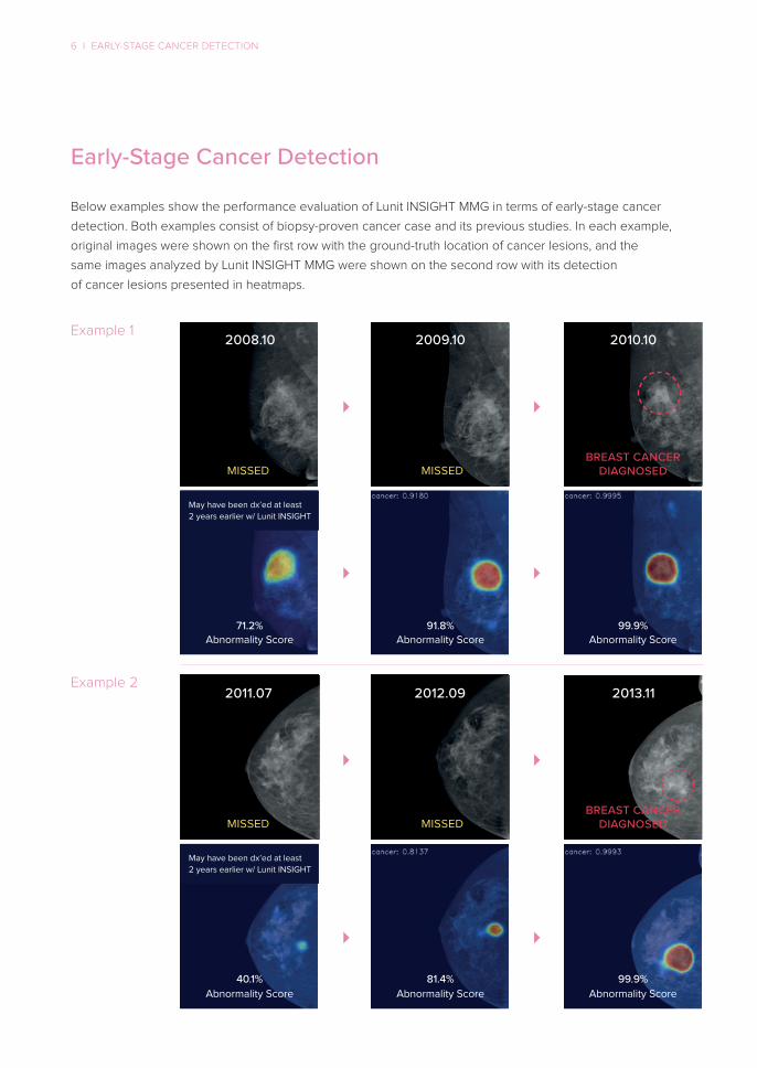
Early-Stage Cancer Detection
Below examples show the performance evaluation of Lunit INSIGHT MMG in terms of early-stage cancer
detection. Both examples consist of biopsy-proven cancer case and its previous studies. In each example,
original images were shown on the first row with the ground-truth location of cancer lesions, and the
same images analyzed by Lunit INSIGHT MMG were shown on the second row with its detection
of cancer lesions presented in heatmaps.
Example 1
Example 2
6 I EARLY-STAGE CANCER DETECTION
MISSED
2008.10
2011.07
2009.10
2012.09
2010.10
2013.11
71.2%Abnormality Score
40.1%Abnormality Score
91.8%Abnormality Score
81.4%Abnormality Score
99.9%Abnormality Score
99.9%Abnormality Score
MISSED
MISSEDBREAST CANCER
DIAGNOSED
BREAST CANCERDIAGNOSEDMISSED
May have been dx’ed at least
2 years earlier w/ Lunit INSIGHT
May have been dx’ed at least
2 years earlier w/ Lunit INSIGHT
CASE 1
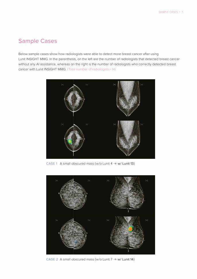
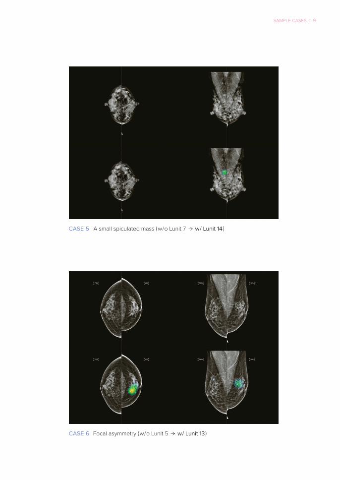
Sample Cases
Below sample cases show how radiologists were able to detect more breast cancer after using
Lunit INSIGHT MMG. In the parenthesis, on the left are the number of radiologists that detected breast cancer
without any AI assistance, whereas on the right is the number of radiologists who correctly detected breast
cancer with Lunit INSIGHT MMG. ( Total number of radiologists = 14 )
SAMPLE CASES I 7
A small obscured mass ( w / o Lunit 4 w / Lunit 13 )
CASE 2 A small obscured mass ( w / o Lunit 7 w / Lunit 14 )

8 I SAMPLE CASES
CASE 3 A small obscured mass with clustered microcalcifications
( w / o Lunit 2 w / Lunit 11 )
CASE 4 A small obscured mass with clustered microcalcifications
( w / o Lunit 5 w / Lunit 12 )
SAMPLE CASES I 9
CASE 5 A small spiculated mass ( w / o Lunit 7 w / Lunit 14 )
CASE 6 Focal asymmetry ( w / o Lunit 5 w / Lunit 13 )

Focal asymmetry ( w / o Lunit 5 w / Lunit 13 )
Focal asymmetry ( w / o Lunit 7 w / Lunit 14 )
10 I SAMPLE CASES
CASE 7
CASE 8
OTHER RESEARCH IN BREAST RADIOLOGY I 11
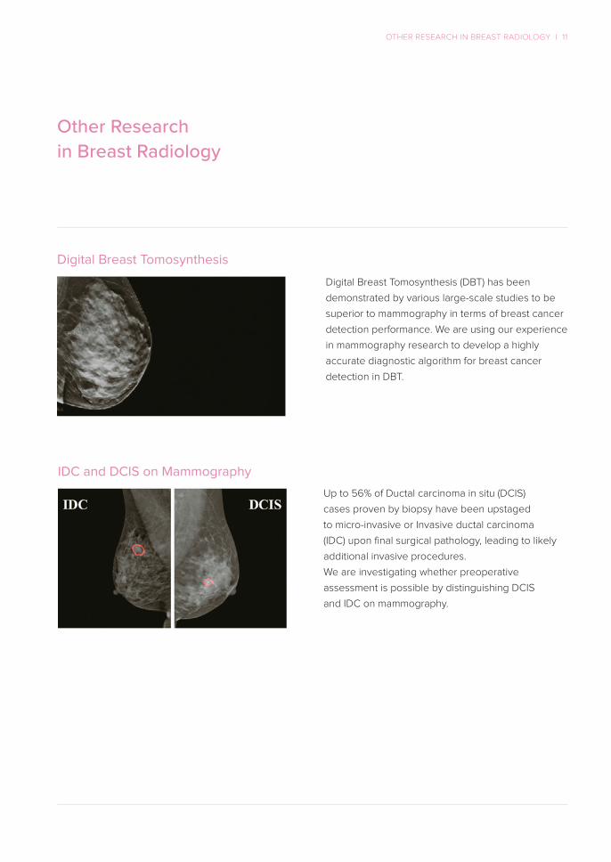
Other Researchin Breast Radiology
Digital Breast Tomosynthesis (DBT) has been
demonstrated by various large-scale studies to be
superior to mammography in terms of breast cancer
detection performance. We are using our experience
in mammography research to develop a highly
accurate diagnostic algorithm for breast cancer
detection in DBT.
Up to 56% of Ductal carcinoma in situ (DCIS)
cases proven by biopsy have been upstaged
to micro-invasive or Invasive ductal carcinoma
(IDC) upon final surgical pathology, leading to likely
additional invasive procedures.
We are investigating whether preoperative
assessment is possible by distinguishing DCIS
and IDC on mammography.
Digital Breast Tomosynthesis
IDC and DCIS on Mammography

12 I RSNA ABSTRACTS
Increase of cancer detection rate and reduction of false-positive recall
in screening mammography using artificial intelligence –
a multi-center reader study
To assess feasibility of artificial intelligence (AI) based
diagnostic-support software whether it can be used to
improve radiologists’ diagnostic performance in terms of
cancer detection and false-positive recall in breast cancer
screening.
This reader study showed a statistically significant improvement
of diagnostic performance (0.071 increase in ROC AUC). Cancer
detection rate was increased by 12.6% and false-positive
recall rate was decreased by 9.6% with assistance of AI-based
diagnostic-support software.
With increase of cancer detection rate and decrease of
false-positive recall rate, AI-based diagnostic-support software
can be practically used in routine breast cancer screening.
A total of 400 exams of screening mammograms were
retrospectively collected from two institutions. For each
institution, 100 cancer, 40 benign, and 60 normal exams
were collected. All cancer exams were proven by biopsy.
Half of the benign exams were proven by biopsy (i.e. recalled
benign) while the remainder were proven by at least 2 years
of follow-up imaging. 80% of the exams were randomly
selected respectively from each category and each institution
(e.g., 16 recalled benign for each institution). All exams were
4-view paired. A blinded multi-reader multi-case study was
performed with a group of 14 radiologists for the selected
320 exams. Each radiologist reads each case without and
then with aid of Lunit INSIGHT for Mammography (Lunit Inc.,
South Korea), a deep learning-based software which shows
per-breast malignancy scores as well as region-ofinterests
(ROIs) for suspicious malignant lesions (Fig.1). The difference
of readers’ decision without and with AI in terms of likelihood-
of-malignancy (LOM; DMIST 7-pt score) and recall-ness (recall
or not) was analyzed.
Significant improvement of diagnostic performance was
shown for all 14 radiologists; average LOM-based ROC
AUC was 0.810 and 0.881 without and with AI, respectively
(p-value=0.0000047, C.I.=95%). Based on readers’ binary
decision whether each exam should be recalled or not,
average cancer detection rate was increased from 75.3%
to 84.8% while false-positive recalls (i.e. non-cancer recalls)
were decreased from 28.0% to 25.4% where 20% of non-
cancer exams were recalled benign cases.
PURPOSE
CONCLUSION
CLINICAL RELEVANCE / APPLICATION
METHOD AND MATERIALS
RESULTS
AI-based diagnosis-support software which shows
per-breast malignancy scores (on the right-side
panel) and ROIs for suspicious malignant lesions
(heatmaps).

RSNA ABSTRACTS I 13
Data-driven Imaging Biomarker
for Breast Cancer Screening in Mammography –
Early Detection of Breast Cancer
To assess feasibility of data-driven imaging biomarker in
mammography (DIB-MMG; an imaging biomarker derived
from large-scale mammography data based on deep learning
technology) whether it can be used for early detection of
breast cancer.
This retrospective study showed feasibility of DIB-MMG for
early detection of breast cancer on mammography, where 32
out of 47 missed cancers, 30 out of 61 interval cancers,
7 out of 17 occult cancers were detected by DIB-MMG. Overall
AUC was 0.738. Further clinical validation with observer
performance study is needed.
With further clinical validation, DIB-MMG can be used as an
effective diagnostic-support tool for early detection of breast
cancer in screening mammography.
A total of 105,592 exams of 4-view digital mammograms
were retrospectively collected from multiple institutions for
developing DIB-MMG, where 22,456 were cancer (confirmed
by biopsy), 36,821 were benign (confirmed by biopsy or at
least 1 year of follow-up imaging), and 46,315 were normal
exams. Based on external validation in a separate institution
with 3,696 exams of mammograms (1,073 were cancer; one for
each patient), DIB-MMG showed 0.963, 94.1%, 80.2% of AUC,
sensitivity, specificity, respectively. Among the 1,073 cancer
patients, 85 patients had 116 exams of prior mammograms
which were diagnosed as non-cancer at that time. A breast
radiologist retrospectively reviewed the 116 exams and
re-classified into three categories – 1) Missed (46 exams;
47 cancer / 45 non-cancer breasts): retrospectively seen in
previous mammogram (mmg-p) and also seen in mammogram
at diagnosis (mmg-d), 2) Interval (55; 61/49): retrospectively not
seen in mmg-p but seen in mmg-d, and 3) Occult (15; 17/13):
not seen both in mmg-p and mmg-d. DIB-MMG was analyzed
for the Missed, Interval, and Occult cancers, respectively.
Per-breast AUC, sensitivity, specificity were used since all
the data is positive in exam-level. Per-breast AUC was 0.841,
0.676, 0.620 for the Missed, Interval, Occult, respectively.
Sensitivity (w/ specificity) at different operating points 0.05, 0.10
were 68.1% (88.9%), 55.3% (91.1%) for Missed, 49.2% (83.7%),
37.7% (91.8%) for Interval, and 41.2% (69.2%), 17.7% (84.6%) for
Occult, respectively. Original operating point of DIB-MMG
for routine screening was 0.10. Fig.1 shows examples of the
Missed and Interval cancers.
PURPOSE
CONCLUSION
CLINICAL RELEVANCE / APPLICATION
METHOD AND MATERIALS
RESULTS
Each patient (left and right) was diagnosed as
cancer (right most column), where the cancer lesion
was seen at diagnosis. Their prior mammograms (first
and second columns) were reviewed retrospectively
by a breast radiologist who already knows location
of the biopsy-confirmed cancer lesions.
1) Missed cancer (left): previously negative but
retrospectively positive, 2) Interval cancer (right):
previously negative and retrospectively negative.
14 I RSNA ABSTRACTS
Data-driven Imaging Biomarker
for Breast Cancer Screening in Mammography –
Prediction of Tumor Invasiveness in Mammography
To assess feasibility of data-driven imaging biomarker in
mammography (DIB-MMG; an imaging biomarker derived
from large-scale mammography data based on deep learning
technology) whether prediction of tumor invasiveness is
applicable on mammography – discrimination of ductal
carcinoma in situ (DCIS), DCIS with microinvasion (DCIS-MI),
and invasive ductal carcinoma (IDC).
This study showed that discrimination of DCIS-MI from
DCIS is more difficult than that from IDC in mammography.
Experimental results showed that DIB-MMG-TI is feasible to
discriminate IDC from the rest. Further clinical validation with
observer performance study is needed.
With further clinical validation, DIB-MMG-TI can be used as a
preoperative diagnostic-support tool for prediction of tumor
invasiveness in mammography.
A total of 151,764 exams of 4-view mammograms were
collected from multiple institutions for developing DIBMMG,
where 31,776 were cancer (confirmed by biopsy), 49,644
were benign (confirmed by biopsy or at least 1 year of follow-
up imaging), and 70,344 were normal exams (confirmed by
at least 1 year of follow-up imaging). Surgical assessment
of tumor invasiveness (459 DCIS, 373 DCIS-MI, and 6,365
IDC) was collected for 7,197 out of 31,776 cancer exams. A
separate set of 777 cancer exams (46 DCIS, 49 DCIS-MI,
682 IDC) were used for evaluation. Previously, we assessed
the feasibility of DIB-MMG as a diagnostic-support tool for
breast cancer screening in mammography. In this study, we
further investigated whether DIB-MMG is applicable to predict
tumor invasiveness in mammography. DIB-MMG-TI (i.e. Tumor
Invasiveness) was developed via two stages of training –
1) training with diagnosis labels (normal, benign, cancer),
followed by 2) fine-tuning with invasiveness labels (DCIS,
DCIS-MI, IDC) on the subset of cancer exams. We exploited
the location of cancer lesions (6,229 among 7,197 exams) for
the purpose of attention (i.e. attention mechanism in AI) in
order to predict the invasiveness in more effective way.
AUC was summarized on two tasks: 1) discrimination of IDC
from DCIS and DCIS-MI, and 2) discrimination of DCIS from
DCIS-MI and IDC. For each task, per-exam AUC of DIB-MMG-
TI on 777 exams of validation dataset was 0.781 and 0.690
respectively, while per-breast AUC for each task was 0.775
and 0.690. Fig.1 shows examples.
PURPOSE
CONCLUSION
CLINICAL RELEVANCE / APPLICATION
METHOD AND MATERIALS
RESULTS
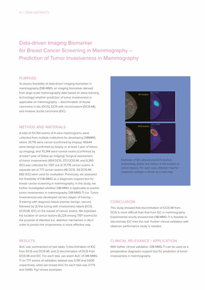
Examples of IDC (above) and DCIS (below)
respectively, where red contour is the location of
cancer lesions. For each case, attention map for
respective subtype is shown as a heat-map.

RSNA ABSTRACTS I 15
Data-driven Imaging Biomarker
for Breast Cancer Screening in Digital Breast Tomosynthesis –
Multidomain Learning with Mammography
To assess feasibility whether mammography data is helpful
for developing data-driven imaging biomarker in digital breast
tomosynthesis (DIB-DBT; an imaging biomarker for detection
of breast cancer, which is derived from DBT data based on
deep learning technology).
This study demonstrated that multi-domain learning with large
-scale MMG is an effective way for developing DIB-DBT
especially with small-scale DBT. Further clinical validation is
needed to utilize DIB-DBT as a reliable diagnostic-support
tool for breast cancer detection.
With further clinical validation, DIB-DBT could be practically
used as an effective diagnostic-support tool for breast cancer
screening in digital breast tomosynthesis.
A total of 1,517 exams of 4-view digital breast tomosynthesis
(DBT) and 49,577 exams of 4-view digital mammograms (MMG)
were retrospectively collected from an institution. We divided
1,517 exams of DBT into 1,187 (970 cancer, 52 benign, 165
normal) and 330 (244 cancer, 34 benign, 52 normal) exams
for training and validation, and 49,577 exams of MMG into
47,719 (5,599 cancer, 17,971 benign, 24,149 normal) and 1,858
(619 cancer, 620 benign, 619 normal) exams for training and
validation, respectively. For external validation, we also
collected 448 exams (148 cancer, 150 benign, and 150 normal)
of 4-view DBT from another institution. Previously, we
demonstrated that using DBT and MMG concurrently is
effective for developing DIB-DBT, where it was first trained
with (large-scale) MMG then fine-tuned with (small-scale) DBT.
We further aimed to enhance the utilization of MMG by
multi-domain learning to boost the performance of DIB-DBT.
Two-stage training was adopted – 1) pre-training with MMG,
followed by 2) multi-domain fine-tuning with both of DBT and
MMG. A total of four different approaches was compared in
order to find the best way to exploit MMG for developing
DIB-DBT – (a) training only with DBT, (b-d) training with MMG
and then fine-tuning with (b) DBT (previous work), (c) DBT and
MMG, (d) DBT and MMG by multi-domain learning.
Per-exam AUC of DIB-DBT on the internal validation dataset
was 0.890, 0.899, 0.901, 0.910 for each method of (a-d)
respectively, while per-exam AUC on the external validation
dataset was 0.871, 0.880, 0.899, 0.901 for (ad) respectively.
Fig.1 shows an example of DIB-DBT (i.e. (d)).
PURPOSE
CONCLUSION
CLINICAL RELEVANCE / APPLICATION
METHOD AND MATERIALS
RESULTS
For visual interpretability of the results, we showed
heat-maps on a set of synthetic 2D images ( just for
visualization). (Left) Heat-maps from DIB-DBT, (Right)
Ground-truth – cancer lesion confirmed by biopsy.

Deepen your INSIGHT
with AI-Powered Breast Radiology
With the help of our AI,
you can make the best decision in less duration of time.
Together, we can save more time, save cost, and save lives.

Partner with Us
We welcome research partnerships and other collaboration with medical institutions,
healthcare providers and companies interested in implementing our software product.
Currently, we have over 20 worldwide research partners throughout USA, UK, China and Korea.
We look forward to hearing from you!
Contact Us
Please feel free to email us about any inquiries or questions.
PARTNER WITH US I 17
Corporate Partners
