perform electrocardiography hltpat407b. objectives 1.check ecg machine against a checklist before...
TRANSCRIPT
Perform Electrocardiography
HLTPAT407B

Objectives1. Check ECG machine against a checklist before each use
2. Correctly identify, measure and inform patient regarding the ECG procedure
3. Record patient information
4. Position patient for attachment of leads
5. Attach leads according to procedure manual
6. Label leads as they are recorded and record if filter is used
7. Record lead two rhythm strip
8. Maintain a straight base line
9. Clean patient’s chest and ECG electrodes
10.Forward trace to cardiologist
11. Identify and manage electrical interference
12. Identify and manage skeletal muscle tremor
13. Identify and manage wandering baseline

definitions
• Cardiac cycle is a single rhythmic repetition of the heartbeat• Artifact is the appearance of a false signal which is obviously not
from the heart• Arrhythmia is an abnormal heart rhythm• Electrocardiogram a record of the electrical activity of the heart• Waves are deflections from the baseline of the ECG• Electrodes are specially prepared tabs with conductive gel placed
on the skin to detect electrical current• Purkinje fibres myocardial cells in the heart that conduct electricity
to the ventricles• Sinoatrial node is the pacemaker of the heart• Atrioventricular node cells found in the right atrium• Bundle of His are nerve fibres within the septum

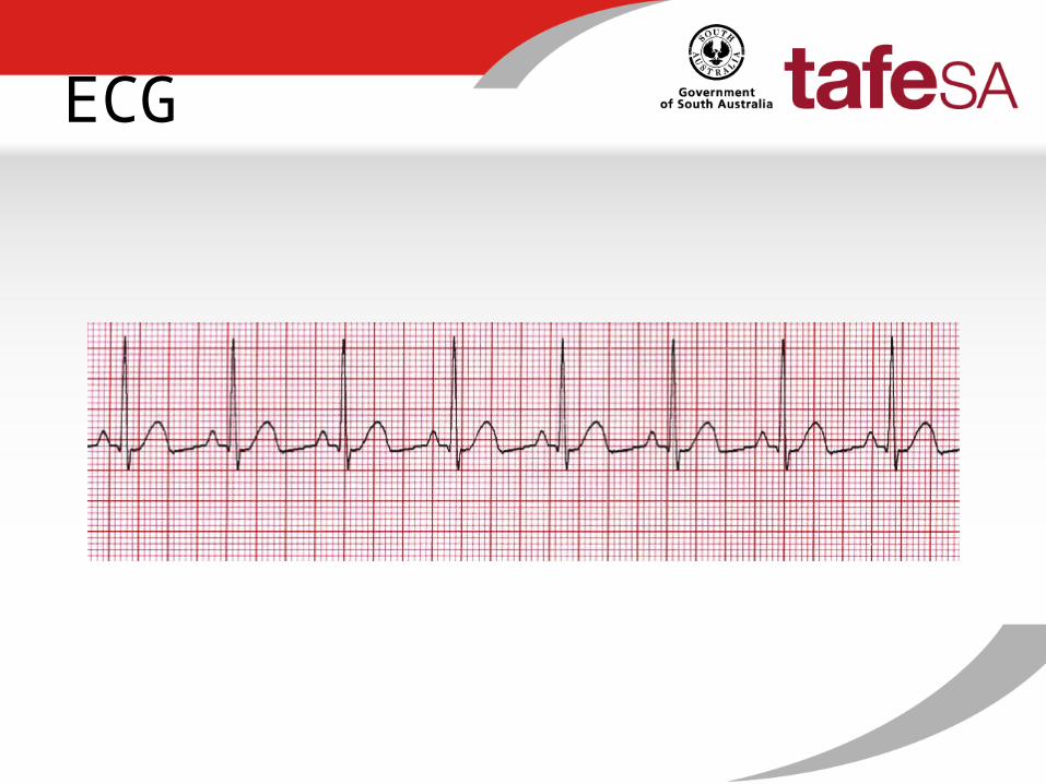
What is an ECG?
• Provides a record or trace of the electrical activity of the heart
• Cardiac muscle tissue generates electrical impulses through the heart
• We measure this activity by placing electrodes on the chest wall, arms and legs
• Electrical activity show as waveforms named P.Q,R,S and Twaves
ECG

Cardiac Cycle
• Electrical impulse begins in the sino-atrial node in the right atrium

Cardiac cycle
• The impulse travels along a network of conductive fibres in the atria to the atrioventricular node

Cardiac cycle
• After a short delay, the impulse passes to the Bundle of His, down the right and left bundle branches, into the purkinje fibres in the ventricles

Cardiac cycle
• When SA node generates impulse, sodium and calcium move into cells making them contract
• This creates a current which flows forward contracting cells next to them
• Contraction of the heart occurs-systole

Cardiac cycle
• potassium leaves the cells and they then relax which returns them to a resting state called diastole
• This current also passes to the nearby tissues to the heart
• This is why electrodes placed on the skin can detect the current

P Wave
• Represents Atrial contraction
• Should be upright, round and symmetrical

P Wave
• High pointed occurs with pulmonary disease
• Mitral valve disease can cause wide notch
• Absence can mean abnormal impulse generation

QRS
• Represents the contraction of the ventricles
• This is a much stronger contraction as it pushes blood out of the ventricles into the arteries
• Known as QRS complex

T wave
• Follows QRS and shows that the ventricles have relaxed
• Usually upright and is taller and less symmetrical than P wave

The rate
• ECG paper has lines to help count time• 3 large squares are equal to 1 sec• To calculate the rate for 1 min, count the number
of cycles in any 6 sec interval and multiply by 10


The rhythm
• Check the atrial rhythm by placing a piece of paper against the P wave and mark with a pencil
• Mark the P wave of the next cycle• Move the paper across the tracing and look to
see if P wave meets the line you have drawn• P wave should be upright, symmetrical and
before the QRS complex

QRS complex
• Measure from beginning of Q or R wave to the point where the S wave returns to the baseline
• Normal time is between 0.5 and 0.10 secs

T wave
• Should be present, upright and look the same

12 lead ECG
• A lead records the electrical activity between 2 points
• Each lead looks at a small part of the heart• A 12 lead ECG gives a complete picture of this
electrical activity• During ECG, some of the electrodes are used twice

Lead Placement
• V1-4th intercostal space at right sternal border• V2-4th intercostal space at left sternal border• V3-midway between 4th and 5th intercostal spaces on left side• V4-5th intercostal space at mid clavicular line•V5- 5th intercostal space anterior axilla•V6-5th intercostal space at midaxillary line

Lead placement

leads
• Leads 1,11 and 111 are attached to arms and legs
• They measure activity in the front of the heart
• Lead 1 measures between LA and RA
• Lead 11 measures between RA and LL
• Lead 111 measures between LA and L chest

Leads
• AVR,AVL and AVF records the centre of the heart to a limb
• AVL measures centre of heart to LA
• AVR measures centre of heart to RA
• AVF measures centre of heart to L foot
• The lead on the R foot is an earth lead

Leads
• Chest leads are V1, V2, V3, V4, V5, V6
• Measures the front to the back of the heart

Limb leads
• Black-left arm
• White-right arm
• Red-left leg
• Green-right leg

Procedure
• Identify patient• Patient needs to be calm and relaxed, no pain,
respect privacy, warmth• Remove top clothes, shoes, socks, jewellery• Patient to lie on back• Prepare skin, shave if necessary, use alcohol
wipe, abrade skin, be aware of perspiration• Apply electrodes• Attach leads

Artifacts
1. Electrical interference• Rapid vibration and spikes that widen the baseline• Look fuzzy and thick
• Correct by moving machine away from bed, disconnect wall socket items, patient not to touch metal on beds

Artifacts
2. Movement• Talking, coughing, crying, muscle tremors• Ask patient to lie with hands under buttocks
• Attach leads accordingly

Artifacts
3. Wandering baseline• Usually due to poor electrode contact with skin• Check that the lead hasn’t come loose• Electrode may be too dry or too wet

Special circumstances• Amputation or plaster- place electrodes
close to body
• Pregnancy- document as may be slightly abnormal due to increased blood volume
• Seizure- remain calm, stay with patient, call for help, do ECG after seizure is over
• Large breasts- place electrodes under breast tissue

Troubleshooting
• Plug into socket, can be used in battery mode but preferable from wall socket
• Good skin contact• Warm, private environment• Simple explanation• Comfort and lying down• Label correctly

Review
• Page 13- check review your tracing