performance dependence of hybrid x-ray computed tomography/fluorescence molecular tomography on the...
TRANSCRIPT

1Ep(isdislssl
adcFttpsqgmp
hpw
Hyde et al. Vol. 26, No. 4 /April 2009 /J. Opt. Soc. Am. A 919
Performance dependence of hybrid x-raycomputed tomography/fluorescence moleculartomography on the optical forward problem
Damon Hyde,1,* Ralf Schulz,2 Dana Brooks,1 Eric Miller,3 and Vasilis Ntziachristos2
1Department of Electrical and Computer Engineering, Northeastern University, 360 Huntington Ave.,Boston, Massachusetts 02115, USA
2Institute for Biological and Medical Imaging (IBMI), Technische Universität München and Helmholtz ZentrumMünchen, Ingolstädter Landstraße 1, D-85764 Neuherberg, Germany
3Department of Electrical and Computer Engineering, Tufts University, 161 College Ave., Medford,Massachusetts 02155, USA
*Corresponding author: [email protected]
Received September 9, 2008; revised January 5, 2009; accepted January 19, 2009;posted February 17, 2009 (Doc. ID 100691); published March 20, 2009
Hybrid imaging systems combining x-ray computed tomography (CT) and fluorescence tomography can im-prove fluorescence imaging performance by incorporating anatomical x-ray CT information into the optical in-version problem. While the use of image priors has been investigated in the past, little is known about theoptimal use of forward photon propagation models in hybrid optical systems. In this paper, we explore theimpact on reconstruction accuracy of the use of propagation models of varying complexity, specifically in thecontext of these hybrid imaging systems where significant structural information is known a priori. Our re-sults demonstrate that the use of generically known parameters provides near optimal performance, evenwhen parameter mismatch remains. © 2009 Optical Society of America
OCIS codes: 110.0113, 110.1758.
lwottgocrrfc
pmgedpgprc
mtodfw
. INTRODUCTIONmerging hybrid imaging systems such as x-ray com-uted tomography fluorescence molecular tomographyXCT-FMT) offer high resolution anatomical informationn conjunction with in vivo quantification of cellular andubcellular tissue biomarkers [1–3]. Recent advances inata collection methodologies, modeling techniques, andmage formation theory have led to small animal FMTystems that employ complete-angle 360°-projection col-ection geometries, use CCD camera detectors for highpatial sampling of photon fields propagating through tis-ue, and offer large field of view to obtain projections overarge volumes.
The development of multimodal imaging methods suchs XCT-FMT has been motivated largely to address fun-amental limitations associated with stand-alone fluores-ence systems. Specifically, the ill-posed nature of theMT image formation problem can be significantly offsethrough the addition of high-resolution anatomic informa-ion from modalities such as XCT and MRI [3–7]. Suchrior information can be used in both modeling and inver-ion, and has been shown to significantly improve imageuality. Different inversion approaches have been sug-ested for the implementation of inverse problems withethodologies that avoid the use of hard priors offering
romising characteristics in order to avoid image bias [8].Here we investigate the forward modeling aspect of the
ybrid tomographic problem. Our imaging system em-loys the normalized Born approximation, or Born ratio,hich divides measurements at the fluorescence wave-
1084-7529/09/040919-5/$15.00 © 2
ength by corresponding measurements at the excitationavelength [9]. This approach allows direct computationf fluorescence parameters without the intermediate de-ermination of tissue optical properties [10]. Additionally,he Born ratio has been shown to grant a significant de-ree of invariance to inhomogeneities in the backgroundptical properties of the medium. That is, the Born ratioorrects for differences between the modeled optical pa-ameters and those present in vivo. Because of this cor-ection, it is unclear whether the use of more elaborateorward models is necessary, or if forward model simplifi-ations will be sufficient to obtain near optimal results.
Additionally, the use of prior information in the inverseroblem also yields significant performance improve-ents in the resulting images. This raises questions re-
arding the interaction between structural prior knowl-dge in the forward and inverse problems, and to whategree inverse structural priors can compensate for sim-lifications of the forward model. To this end, we investi-ated inverse solutions both with and without structuralrior for each potential forward model and examined theelative cost incurred by each subsequent model simplifi-ation.
In stand-alone FMT, where only the air–tissue surfaceay be known using surface extraction techniques, an in-
ernally homogeneous medium can be assumed with eachptical property constant throughout the volume, andata normalization can be employed to correct for the ef-ects of tissue optical heterogeneity [11]. For imaging ofhole animals, the resulting parameter values are com-
009 Optical Society of America
mccpagaiaw
eAoosrttTtqirc
spstptcoh
2TseFolbfgpn
mnm
frdmmmstwlfrtmsw
catmioemtf
tfispT
Fdfl
920 J. Opt. Soc. Am. A/Vol. 26, No. 4 /April 2009 Hyde et al.
only set in the range of �a=0.3 cm−1 and �s�=10 cm−1 toorrespond with the average values of bulk soft tissue. Inontrast, the x-ray CT component of the hybrid approachrovides knowledge of the internal geometry that can en-ble the implementation of more elaborate photon propa-ation models to account for the differences in absorptionnd scattering present within each organ or tissue. Thismproved modeling yields sensitivity functions that moreccurately reflect the physical diffusion taking placeithin the animal.However, the introduction of additional optical param-
ters complicates the implementation of these models.natomic images are frequently segmented into a numberf discrete regions corresponding to individual organs orptically similar tissue types, each of which must be as-igned absorption and scattering values that accuratelyeflect those present in vivo. One option for selectinghese values is to explicitly calculate them through solu-ion of the diffuse optical tomography (DOT) problem [12].his approach is theoretically the most accurate but leadso increased complexity and additional computation re-uirements. Also important is that this calculation maynclude and therefore propagate errors to the fluorescenceeconstruction problem that can bias the final fluores-ence image.
An alternate approach that can be employed when con-tructing forward models for optical tomography is to em-loy average parameter values from published or mea-ured ranges for the different tissue types segmented onhe CT image. While perhaps not as accurate as explicitarameter estimation on a per animal basis, this limita-ion may be offset by the simplicity and lack of additionalomputation. What remains to be established is the trade-ff in terms of reconstruction accuracy that results fromaving a mismatch in modeled issue optical parameters.
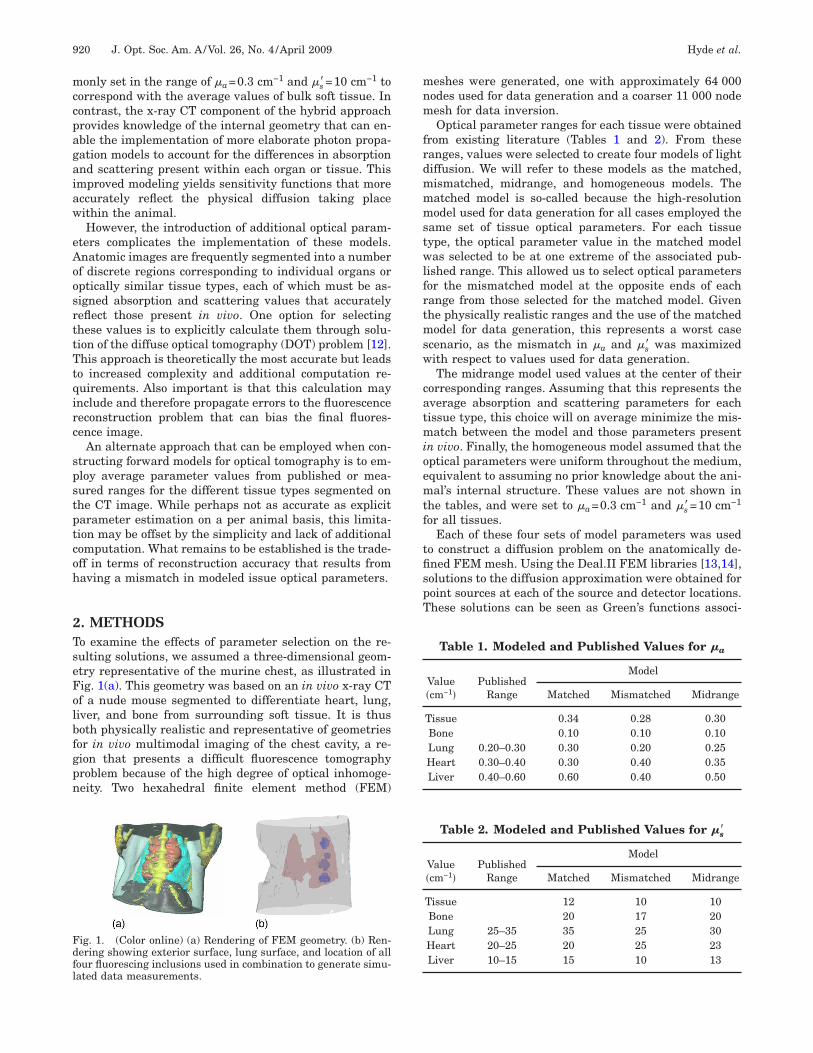
. METHODSo examine the effects of parameter selection on the re-ulting solutions, we assumed a three-dimensional geom-try representative of the murine chest, as illustrated inig. 1(a). This geometry was based on an in vivo x-ray CTf a nude mouse segmented to differentiate heart, lung,iver, and bone from surrounding soft tissue. It is thusoth physically realistic and representative of geometriesor in vivo multimodal imaging of the chest cavity, a re-ion that presents a difficult fluorescence tomographyroblem because of the high degree of optical inhomoge-eity. Two hexahedral finite element method (FEM)
ig. 1. (Color online) (a) Rendering of FEM geometry. (b) Ren-ering showing exterior surface, lung surface, and location of allour fluorescing inclusions used in combination to generate simu-ated data measurements.
eshes were generated, one with approximately 64 000odes used for data generation and a coarser 11 000 nodeesh for data inversion.Optical parameter ranges for each tissue were obtained
rom existing literature (Tables 1 and 2). From theseanges, values were selected to create four models of lightiffusion. We will refer to these models as the matched,ismatched, midrange, and homogeneous models. Theatched model is so-called because the high-resolutionodel used for data generation for all cases employed the
ame set of tissue optical parameters. For each tissueype, the optical parameter value in the matched modelas selected to be at one extreme of the associated pub-
ished range. This allowed us to select optical parametersor the mismatched model at the opposite ends of eachange from those selected for the matched model. Givenhe physically realistic ranges and the use of the matchedodel for data generation, this represents a worst case
cenario, as the mismatch in �a and �s� was maximizedith respect to values used for data generation.The midrange model used values at the center of their
orresponding ranges. Assuming that this represents theverage absorption and scattering parameters for eachissue type, this choice will on average minimize the mis-atch between the model and those parameters present
n vivo. Finally, the homogeneous model assumed that theptical parameters were uniform throughout the medium,quivalent to assuming no prior knowledge about the ani-al’s internal structure. These values are not shown in
he tables, and were set to �a=0.3 cm−1 and �s�=10 cm−1
or all tissues.Each of these four sets of model parameters was used
o construct a diffusion problem on the anatomically de-ned FEM mesh. Using the Deal.II FEM libraries [13,14],olutions to the diffusion approximation were obtained foroint sources at each of the source and detector locations.hese solutions can be seen as Green’s functions associ-
Table 1. Modeled and Published Values for �a
Value�cm−1�
PublishedRange
Model
Matched Mismatched Midrange
Tissue 0.34 0.28 0.30Bone 0.10 0.10 0.10Lung 0.20–0.30 0.30 0.20 0.25Heart 0.30–0.40 0.30 0.40 0.35Liver 0.40–0.60 0.60 0.40 0.50
Table 2. Modeled and Published Values for �s�
Value�cm−1�
PublishedRange
Model
Matched Mismatched Midrange
Tissue 12 10 10Bone 20 17 20Lung 25–35 35 25 30Heart 20–25 20 25 23Liver 10–15 15 10 13
asf
wmgTp
oeactlmstffr
ifrstsc�fipiwrl
3Rws
H(ctdt
usmsTewp
t
TiItysfaasttyAm
gam
Fas
Hyde et al. Vol. 26, No. 4 /April 2009 /J. Opt. Soc. Am. A 921
ted with the diffusing system and can be used to con-truct the appropriate normalized Born models using theormula [9]
w�rs,rd,r� =G�rs,r�G�rd,r�
G�rs,rd�, �1�
here w�rs ,rd ,r� denotes the sensitivity of a measure-ent collected at point rd to fluorescence at a point r
iven a point source of appropriate wavelength at point rs.he function G�r1 ,r2� denotes the solution to the diffusionroblem at point r2 given a point source at r1.Five fluorescing inclusions were constructed on this ge-
metry differing in location and physical dimensions. Inach case, the boundaries of the inclusion were definednd interior voxels given Gaussian distributed fluores-ence intensities. The first simulated a situation wherehe fluorescent probe was spread throughout one entireung, such as might be found when imaging lung inflam-
ation. The remaining four targets were smaller, roughlypherical inclusions, as illustrated in Fig. 1(b). Three ofhese were approximately 3 mm in diameter, while theourth had a diameter of 2 mm. Each was located at a dif-erent nonoverlapping location within the lung to cover aange of possible interference from other organs.
These inclusions were used to construct a total of 16maging scenarios. The first scenario consisted only of theull lung inclusion, while the remaining 15 data sets cor-esponded to all possible combinations of 1–4 of themaller inclusions. For each scenario, multi-angle collec-ion of diffuse data in a transmission geometry was as-umed. At each collection angle, a 3�10 grid of source lo-ations was defined with overall dimensions 0.8 cm1.8 cm. A corresponding set of detector locations was de-
ned using a 10�10 grid of size 1.6 cm�1.0 cm. Using 17rojections spaced evenly every 20° these values resultedn a total of 51 000 source–detector pairs. Simulated dataith 10% added shot noise was generated using the fine-
esolution mesh for Green’s function computation and theinear model presented above.
. RESULTSeconstructions for every data set–model combinationere obtained by solving the Tikhonov regularized least-
quares problem
x̂ = arg minx
�Wx − b�22 + �2�x�2
2. �2�
ere W is the linear system model constructed using Eq.1) for each combination of source, detector, and voxel lo-ation. The vector x contains the fluorescence concentra-ions to be reconstructed, while b is the vector of collectedata points. The regularization parameter � was selectedo minimize 2-norm error with the known true image.
Solution of the above minimization was implementedsing 50 iterations of the LSQR algorithm [15]. Each dataet was reconstructed twice: once using the full weightatrix and once using a structural prior model that con-
trained image values to lie solely within the lung region.he prior model was implemented by eliminating the el-ments of x and corresponding columns of A associatedith voxels lying outside the lung region. For display pur-oses, the values of these voxels were then set to zero.For each solution, 2-norm error with respect to ground
ruth was computed as
ei =�xi − xtrue�2
2
�xtrue�22 . �3�
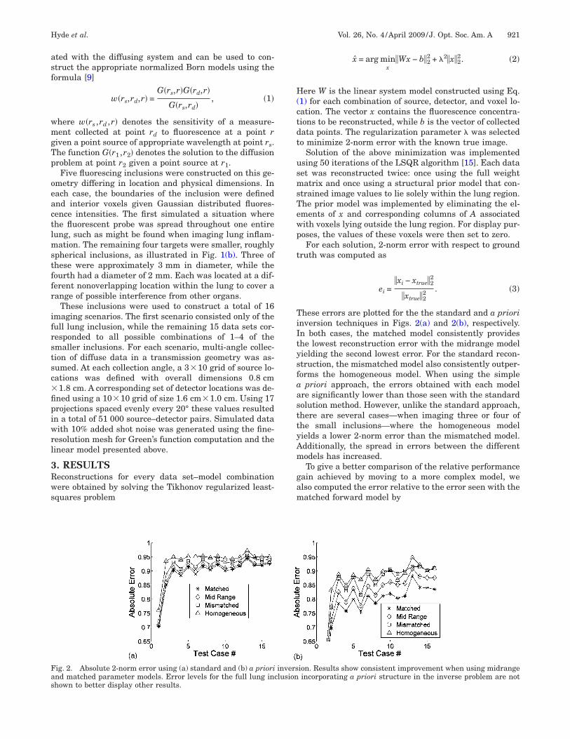
hese errors are plotted for the the standard and a priorinversion techniques in Figs. 2(a) and 2(b), respectively.n both cases, the matched model consistently provideshe lowest reconstruction error with the midrange modelielding the second lowest error. For the standard recon-truction, the mismatched model also consistently outper-orms the homogeneous model. When using the simple
priori approach, the errors obtained with each modelre significantly lower than those seen with the standardolution method. However, unlike the standard approach,here are several cases—when imaging three or four ofhe small inclusions—where the homogeneous modelields a lower 2-norm error than the mismatched model.dditionally, the spread in errors between the differentodels has increased.To give a better comparison of the relative performance
ain achieved by moving to a more complex model, welso computed the error relative to the error seen with theatched forward model by
ig. 2. Absolute 2-norm error using (a) standard and (b) a priori inversion. Results show consistent improvement when using midrangend matched parameter models. Error levels for the full lung inclusion incorporating a priori structure in the inverse problem are nothown to better display other results.

TscsrdimAamm
sfolpg1tw
lrrahufempbe
umattsm
4OcrqmrtfgawtsovmdCrfte
Fs
Fim
922 J. Opt. Soc. Am. A/Vol. 26, No. 4 /April 2009 Hyde et al.
ei,relative =ei
ematched. �4�
hese values are plotted in Figs. 3(a) and 3(b). As alsoeen in the absolute error plots, the homogeneous modelonsistently shows the largest errors when using thetandard inversion approach. As more accurate model pa-ameters are incorporated, solution error correspondinglyecreases in all cases. The mismatched model offers somemprovement over the homogeneous, while the midrange
odel consistently shows less than a 1% increase in error.verage relative error increases of 0.74%, 2.5%, and 4.8%re seen for the midrange, mismatched, and homogeneousodels, respectively, as compared with the matchedodel.The situation changes slightly when incorporating
tructural prior knowledge into the inversion process. Theull fluorescing lung (Test Case #1 on the horizontal axisf Fig. 3) is reconstructed with the lowest overall errorevels, as the structure of the image is provided by therior information. When reconstructing the smaller tar-ets, the homogeneous model yields the highest error in2 out of 16 trials, with the mismatched model yieldinghe highest error for the remaining four cases. Of interest,hile the use of prior information offered consistently
ig. 3. Relative 2-norm error using (a) standard and (b) a priorignificantly higher when using a priori inversion techniques.
ig. 4. (a) True full-lung image. Reconstructions of inclusion us-ng (b) matched model, (c) mismatched model, (d) midrange
odel, (e) homogeneous model.
ower error levels, the relative penalty for using an incor-ect model increased. The average relative increase in er-or was 4.2% for the midrange, 8.9% for the mismatched,nd 10.9% for the fully homogeneous model, significantlyigher than was seen without prior knowledge. These val-es reflect an increase in both the absolute error resultingrom improper model selection and the proportion of totalrror. This indicates that while structural a priori infor-ation in the inverse problem alone can offer improved
erformance, the biggest gains are to be made by a com-ination of prior knowledge and improved diffusion mod-ling.
Sample reconstruction slices from the full lung targetsing a priori knowledge are shown in Fig. 4. While allodels obtain the true structure of the lung, the matched
nd midrange models more accurately resolve the quanti-ative values. When reconstructing smaller inclusions,he more accurate models consistently offered improvedeparation of objects and lower 2-norm errors than the ho-ogeneous and mismatched parameter models.
. CONCLUSIONSur results suggest that for imaging within the murine
hest, the use of established ranges for tissue optical pa-ameters does not significantly degrade reconstructionuality even when mismatch in values remains. Further-ore, by using values from the center of the established
anges, results can be obtained that consistently offer lesshan a 5% increase in error as compared with using per-ectly matched parameters. In contrast, the use of homo-eneous models results in significantly higher error levelsnd reduced image fidelity. These effects are compoundedhen using structural information as prior knowledge in
he inverse problem, which does not appear to compen-ate for simplified diffusion modeling. In fact, the benefitsf improved diffusion modeling are greater when using in-erse priors, suggesting that available structural infor-ation should always be used in the construction of the
iffusion model. We conclude that the use of structuralT data in combination with published optical parameteranges can provide improvements to diffusion modelingor fluorescence molecular tomography without the addi-ional experimental and computational complications ofxplicit parameter estimation.
sion. Note that relative penalty for using the incorrect model is
i inver
R
1
1
1
1
1
1
Hyde et al. Vol. 26, No. 4 /April 2009 /J. Opt. Soc. Am. A 923
EFERENCES1. V. Ntziachristos, A. G. Yodh, M. Schnall, and B. Chance,
“Concurrent MRI and diffuse optical tomography of breastafter indocyanine green enhancement,” Proc. Natl. Acad.Sci. U.S.A. 97, 2767–2772 (2000).
2. A. Joshi, W. Bangerth, and E. M. Sevick-Muraca, “Non-contact fluorescence optical tomography with scanningpatterned illumination,” Opt. Express 14, 6516–6534(2006).
3. S. C. Davis, H. Dehghani, J. Wang, S. Jiang, B. W. Pogue,and K. D. Paulsen, “Image-guided diffuse opticalfluorescence tomography implemented with Laplacian-typeregularization,” Opt. Express 15, 4066–4082 (2007).
4. V. Ntziachristos, E. A. Schellenberger, J. Ripoll, D.Yessayan, E. Graves, J. Alexi Bogdanov, L. Josephson, andR. Weissleder, “Visualization of antitumor treatment bymeans of fluorescence molecular tomography with anannexin V-Cy5.5 conjugate,” Proc. Natl. Acad. Sci. U.S.A.101, 12294–12299 (2004).
5. R. Barbour, S. Barbour, P. Koo, H. L. Graber, R. Aronson,and J. Chang, “MRI-guided optical tomography: Prospectsand computation for a new imaging method,” IEEEComput. Sci. Eng. 2, 63–77 (1995).
6. B. Brooksby, S. Jiang, C. Kogel, M. Doyley, H. Dehghani, J.Weaver, S. Poplack, B. Pogue, and K. Paulsen, “Magneticresonance guided near infrared tomography of the breast,”Rev. Sci. Instrum. 75, 5262–5270 (2004).
7. P. K. Yalavarthy, B. W. Pogue, H. Dehghani, and K. D.Paulsen, “Weight-matrix structured regularizationprovides optimal generalized least-squares estimate in
diffuse optical tomography,” Med. Phys. 34, 2085–2098(2007).
8. M. Guven, B. Yazici, X. Intes, and B. Chance, “Diffuseoptical tomography with a priori anatomical information,”Phys. Med. Biol. 50, 2837–2858 (2005).
9. V. Ntziachristos and R. Weissleder, “Experimental three-dimensional fluorescence reconstruction of diffuse media byuse of the normalized Born approximation,” Opt. Lett. 26,893–895 (2001).
0. A. Soubret, J. Ripoll, and V. Ntziachristos, “Accuracy offluorescent tomography in the presence of heterogeneities:Study of the normalized Born ratio,” IEEE Trans. Med.Imaging 24, 1377–1386 (2005).
1. T. Lasser, A. Soubret, J. Ripoll, and V. Ntziachristos,“Surface reconstruction for free-space 360° fluorescencemolecular tomography and the effects of animal motion,”IEEE Trans. Med. Imaging 27, 188–194 (2008).
2. A. B. Milstein, J. J. Stott, S. Oh, D. A. Boas, R. P. Millane,C. A. Bouman, and K. J. Webb, “Fluorescence opticaldiffusion tomography using multiple-frequency data,” J.Opt. Soc. Am. A 21, 1035–1049 (2004).
3. W. Bangerth, R. Hartmann, and G. Kanschat, “Deal.II—ageneral-purpose object-oriented finite element library,”ACM Trans. Math. Softw. 33, 24:1–24:27 (2007).
4. W. Bangerth, R. Hartmann, and G. Kanschat, Deal.II: AFinite Element Differential Equations Analysis Library,http://www.dealii.org.
5. C. Paige and M. Saunders, “LSQR: An algorithm for sparselinear equations and sparse least squares,” ACM Trans.Math. Softw. 8, 43–71 (1982).