pericardial from blunt chest trauma - thorax.bmj.com · thorax(1974), 29, 329. pericardial...
TRANSCRIPT

Thorax (1974), 29, 329.
Pericardial rupture from blunt chest traumaJ. BORRIE and I. LICHTER
Department of Thoracic Surgery, Otago Medical School, Dunedin, New Zealand
Borme, J. and Lichter, I. (1974). Thorax, 29, 329-337. Pericardial rupture from blunt chesttrauma. Pericardial rupture may occur in two distinct anatomical sites, namely thediaphragmatic pericardium and the pleuropericardium. They may be combined. Theproblems in each type are different.
In ruptured diaphragmatic pericardium the rent may involve the pericardial cavityalone, or may extend into one or both adjoining pleural cavities. Upward herniation ofabdominal viscera can occur, with or without strangulation. The presence of a peri-cardial rent may be suggested by diagnostic pneumoperitoneum, and chest films showingdisplaced abdominal viscera; its extent may be fully revealed only by thoracotomy. If therent involves only the diaphragmatic pericardium without lateral spread into a pleuralcavity, the presence of a rent may be revealed only by exploratory thoracotomy withpericardiotomy.
In ruptured pleuropericardium the rent is usually vertical and may occur on eitherside, more usually on the left. It may be recognized on chest films in its early stages bythe presence of intrapericardial air arising from associated lung trauma. There is seriousrisk of heart dislocation with or without strangulation. The defect must be surgicallyrepaired and, because of pericardial retraction, it may require a fabric patch. Teflonfabric has proved to be a long-term satisfactory pericardial substitute.Two cases of each type of pericardial rupture are described and illustrate these
points.
Rupture of the parietal pericardium is a rare yetimportant sequel to blunt chest trauma. It canoccur in two anatomical sites, namely, the dia-phragmatic pericardium and the pleuroperi-cardium.Rupture in each site brings its own specific
problems.
RUPTURED DIAPHRAGMATIC PERICARDIUM
A tear in this situation results in a free communi-cation between the pericardial and the abdominalcavities. The rupture may involve solely the peri-cardium, or may arise as a medial extension of atorn cupola of the diaphragm. Such extensions aremore common on the left than on the right, as isdemonstrated by the greater frequency of left-sided traumatic diaphragmatic hernia. The line ofrupture may lie in a transverse, anteroposterior,oblique or L-shaped direction.
This type of rent is not usually associated withheart herniation and strangulation unless there isalso a torn pleuropericardium. However, it does
allow upward herniation into the pleural or peri-cardial cavity of stomach, bowel, or omentum.Incarceration or strangulation may occur, withacute restriction of heart and lung function. Thesite and size of the rent determine which serouscavity the herniated bowel will enter. If the tearinvolves solely the diaphragmatic pericardium,bowel may enter the pericardial cavity. If thediaphragmatic pleura is also torn, bowel may enterthat pleural cavity.
RUPTURED PLEUROPERICARDIUM
In patients with rupture of the pleuropericardium,the rent is usually vertical and may communicatewith the right or the left pleural cavity. The rentmay be small and cause no heart herniation, ormoderate in size and allow herniation of part ofthe heart with little or no interference with func-tion. However, if the rent is larger, and placedseveral centimetres above the diaphragm, theheart may escape from the pericardial cavity intothe pleural cavity and become acutely restrictedin its action.
329
E
on 13 March 2019 by guest. P
rotected by copyright.http://thorax.bm
j.com/
Thorax: first published as 10.1136/thx.29.3.329 on 1 M
ay 1974. Dow
nloaded from

J. Borrie and I. Lichter
DIAGNOSIS
In the pleuropericardial group, an unusual heartoutline seen on initial chest films, or appearing inchest films some days after injury, may suggestthe diagnosis. This may be confirmed by cine-angiography showing the heart apex pulsatingbeyond its normal confines. In both groups, di-agnosis of the rent may be confirmed by the radi-ological demonstration of air in the pericardialcavity. This may be gas in bowel herniated intothe pericardial cavity, or may represent a com-municating air leak from contused lung. Whereno air is present diagnostic pneumoperitoneum orpneumothorax may be induced. It is possible forboth types of pericardial rupture to remain un-recognized for years, even when associated withherniation of stomach or bowel, or with partialherniation of the heart.We present in illustration four cases treated in
the Southern Regional Thoracic Surgical Unit,Dunedin, New Zealand during the past 12 years.
RUPTURED DIAPHRAGMATIC PERICARDIUM
CASE 1 Mr. R. M. M., No. 44457, a crane driver aged42, on 12 October 1961 was struck by a falling crane,
sustaining trauma to the chest. He was shocked, withrespiratory distress and severe abdominal pain. Physi-cal examination showed tracheal deviation to theright, a rigid abdomen, dullness in the left lower chest,and diminished air entry.
Chest films showed a traumatic left diaphragmatichernia with a distended stomach, and collapse of thelower lobe of the left lung, with marked deviation ofthe mediastinum to the right and compression of theright lung (Fig. 1).The patient was taken from the admitting room
direct to the operating theatre. A stomach tube waspassed and copious quantities of stomach contentswere removed. Catheterization showed no blood in theurine.Operation (12 Oct. '61): Repair of ruptured diaphrag-matic pericardium and left traumatic diaphragmatichernia, and splenectomy. A left posterolateralthoracotomy was performed. Blood and blood clotwere removed from the left pleural cavity, whichcontained stomach and omentum entering through alarge L-shaped tear in the left diaphragm. The antero-posterior limb of the tear involved the whole of theleft cupola; its transverse limb extended mediallyabove the liver to involve the central tendon anddiaphragmatic pericardium as far as the right cupola.There was an upward extension along the line of theleft phrenic nerve which was torn free. There was thus
FIG. 1. Case 1. Chest film showing mediastinum displaced to theright by left traumatic diaphragmatic hernia. Thoracotomy dis-closed pericardial involvement.
330
on 13 March 2019 by guest. P
rotected by copyright.http://thorax.bm
j.com/
Thorax: first published as 10.1136/thx.29.3.329 on 1 M
ay 1974. Dow
nloaded from
Pericardial rupture from blunt chest trauma
free communication between the pericardial, ab-dominal, and left pleural cavities. Stay sutures wereplaced in the torn edges, and the herniated abdominalcontents were inspected. The spleen was found to beruptured and was excised. Bleeding points in the tailof the pancreas were oversewn. A second small rentin the dome of the diaphragm near the fundus of thestomach was also sutured.The L-shaped pericardial rent and its diaphragmatic
component were closed with interrupted linen sutures,as was its vertical extension. The chest wall was finallyclosed over waterseal drainage.
Postoperatively convalescence was slow, being com-plicated by persistent pyrexia, diarrhoea, and abdom-inal pain. The patient was discharged in good condi-tion on 18 November 1961. Radiological examinationconfirmed left phrenic nerve palsy and demonstratedthe presence of fractured transverse processes of theleft second to fourth lumbar vertebrae. He returnedto work in February 1962 and has remained well since.
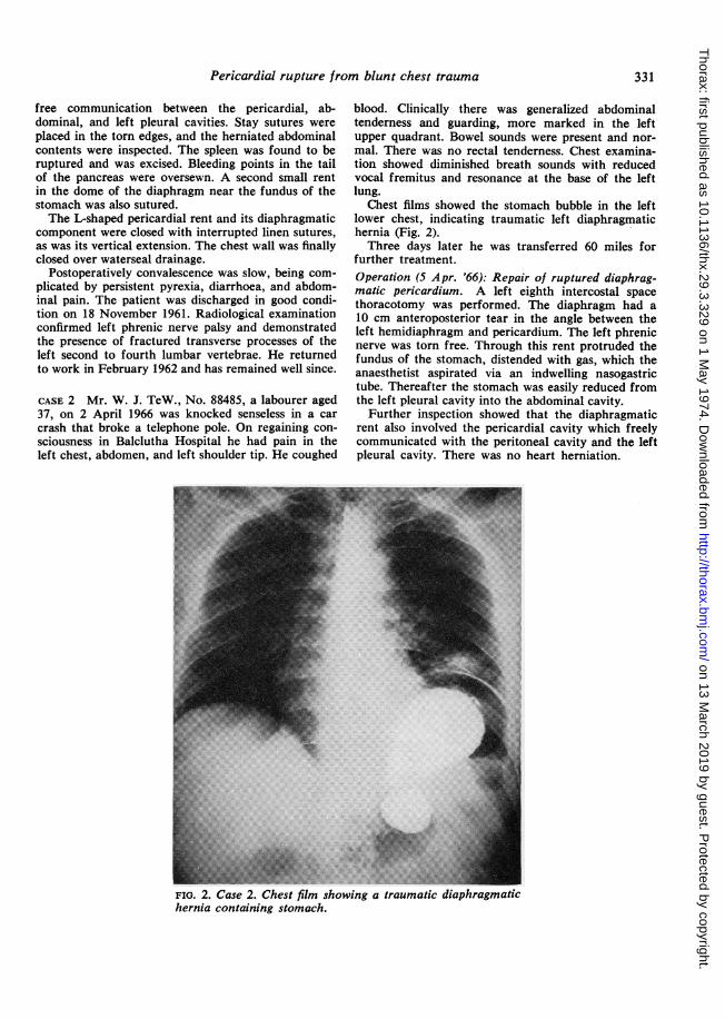
CASE 2 Mr. W. J. TeW., No. 88485, a labourer aged37, on 2 April 1966 was knocked senseless in a carcrash that broke a telephone pole. On regaining con-sciousness in Balclutha Hospital he had pain in theleft chest, abdomen, and left shoulder tip. He coughed
blood. Clinically there was generalized abdominaltenderness and guarding, more marked in the leftupper quadrant. Bowel sounds were present and nor-mal. There was no rectal tenderness. Chest examina-tion showed diminished breath sounds with reducedvocal fremitus and resonance at the base of the leftlung.
Chest films showed the stomach bubble in the leftlower chest, indicating traumatic left diaphragmatichernia (Fig. 2).Three days later he was transferred 60 miles for
further treatment.Operation (5 Apr. '66): Repair of ruptured diaphrag-matic pericardium. A left eighth intercostal spacethoracotomy was performed. The diaphragm had a10 cm anteroposterior tear in the angle between theleft hemidiaphragm and pericardium. The left phrenicnerve was torn free. Through this rent protruded thefundus of the stomach, distended with gas, which theanaesthetist aspirated via an indwelling nasogastrictube. Thereafter the stomach was easily reduced fromthe left pleural cavity into the abdominal cavity.Further inspection showed that the diaphragmatic
rent also involved the pericardial cavity which freelycommunicated with the peritoneal cavity and the leftpleural cavity. There was no heart herniation.
FIG. 2. Case 2. Chest film showing a traumatic diaphragmatichernia containing stomach.
331
on 13 March 2019 by guest. P
rotected by copyright.http://thorax.bm
j.com/
Thorax: first published as 10.1136/thx.29.3.329 on 1 M
ay 1974. Dow
nloaded from

J. Borrie and I. Lichter
The defect was repaired by placing 10 interruptedNo. 35 linen sutures through the lower free edge ofthe torn pericardium. These were continued to en-circle and approximate the torn edges of the dia-phragm. When these sutures were tied the defect wasfirmly closed and the chest wall was repaired. Thepatient made an uneventful recovery and was dis-charged fit 16 days later.
RUPTURED PLEUROPERICARDIUM
CASE 3 Mr. J. J., No. 37698, aged 45, on 26 January1961 fell 60 feet (18 m) from scaffolding, sustainingthe following injuries:
(1) Fracture and depression of the left second toseventh ribs without pueumothorax, haemothorax orparadox but with left lung contusion; anterior fractureof the right sixth to 10th ribs;
(2) fracture of both pubic rami but with intactbladder and urethra;
(3) a double comminuted fracture of the left femur;(4) fracture of the left os calcis.On admission to the orthopaedic ward he was
gravely shocked and 5 litres of blood and 1 litre ofplasma were transfused.Haemopericardium and traumatic left diaphrag-
matic hernia (Fig. 3) were suspected but no immediateaction was taken.
Initially the patient made satisfactory progress,being aided by tracheostomy to assist respirationand the clearing of the tracheobronchial tree. How-ever, the following day chest films showed enlargementof the heart shadow which appeared to be displaced to
the left. The question of haemopericardium was againconsidered but pericardial paracentesis did not supportthis. His condition deteriorated on 28 January.A further chest film showed the heart shadow
displaced to the left (Fig. 4) but the significance of thisdisplacement was not then fully appreciated. Hebecame comatose. His blood pressure continued tofall and he died on 30 January 1961.Necropsy revealed rupture of the left pleuroperi-
cardium with dislocation of the heart into the leftpleural cavity and cardiac 'strangulation'.The initial chest films did not show any air in the
pericardial cavity, which would have been diagnostic,and the cause of the later enlargement of the heartshadow to the left was not appreciated.
CASE 4 Mr. F. S. C., No. 70102, a forestry workeraged 21, on 20 March 1971 was struck by the steeringwheel of his car in a head-on collision. He was admit-ted to Invercargill Hospital with surgical emphysema,flail left chest, left haemopneumothorax, hearttrauma, respiratory failure, and tachypnoea (60/min).He was treated by left tube thoracostomy, tracheos-
tomy, and artificial respiration with a Bird respirator.Two litres of bloodstained fluid drained from the leftpleural cavity. His admission chest film revealedfractures of the first four left ribs and surgicalemphysema over the anterior chest wall, neck, andaxilla. Air was also seen in the pericardial cavity onthe left side (Fig. 5) without evidence of enlargementof the cardiac outline. A chest film on 7 July 1965was normal.
FIG. 3. Case 3. Chest film at time of injury-26 January 1961.
332
on 13 March 2019 by guest. P
rotected by copyright.http://thorax.bm
j.com/
Thorax: first published as 10.1136/thx.29.3.329 on 1 M
ay 1974. Dow
nloaded from

Pericardial rupture from blunt chest trauma
FIG. 4. Case 3. Chest film two days later showing displacement ofthe heart shadow to the left, later shown to be due to dislocationof the heart from the pericardium.
$S,, s w__el° *;,_|K
._......i!,
:'d |_W _.
.. re....::s| ....
.:..,..
....,...:,.RS ._'
*.: |14'' # j
_*,bi&i
..3_F ... |S _Sw. s^_ .. sF _IFIG. 5. Case 4. Chest film of 23 March 1971 showing traumatic 'pneumonitis'in the left lung, surgical emphysema of the left chest wall, and air liftingup the left parietal pericardium, thereby indicating the presence of a rent.
333
on 13 March 2019 by guest. P
rotected by copyright.http://thorax.bm
j.com/
Thorax: first published as 10.1136/thx.29.3.329 on 1 M
ay 1974. Dow
nloaded from

J. Borrie and I. Lichter
I
FIG. 6. Case 4. Film one week after injury, showing the heart displaced tothe left; it is dislocated through the traumatic pleuropericardial rent.
Severe pain was controlled by four days of continu-ous epidural anaesthesia. After six days he was extu-bated. From admission his physician, Dr J. O'Hagan,noted an abnormal pulsation of the left chest wall.When first seen in the thoracic surgical unit on 12
May 1971 the patient was thin, having lost 2 stones(13 kg) in weight. Pulse rate was 88/min and bloodpressure 140/80 mmHg. He had a very obvious pul-sation in the fourth left intercostal space in the leftanterior axillary line. There were no heart murmurs.An electrocardiogram showed inverted T waves inleads III, aVL, and V5 and V6 with tall R waves inV6, all suggesting left ventricular strain. The chestfilm a week after injury showed that the heart wasdisplaced to the left with an unusual upturned andprominent apex (Fig. 6). Though this film suggesteda possible left ventricular aneurysm, when consideredin conjunction with the first film after injury showingair in the pericardial cavity, the film was consideredto represent rupture of the pleuropericardium withpartial dislocation of the heart. This was confirmedby cine-angiography which showed the apex beatherniated into the lefr!pnleural cavity.
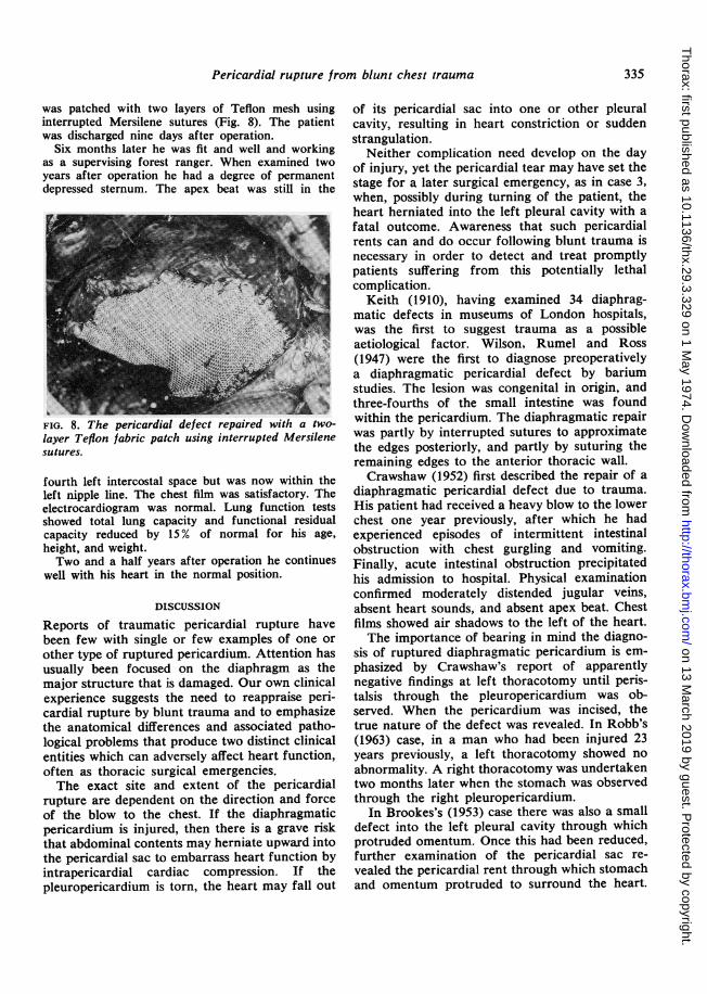
In order to improve feart function and remove therisk of heart strangulation, operative treatment to re-place the heart ai repair the pericardium wasadvised.Operation (24 May '71): Repair of ruptured leftpleuropericardium and Teflon patch to pericardial
defect. A left fifth intercostal thoracotomy revealedfirm adhesions between the heart, pericardium, andleft lung. These were divided. The left ventricle pro-truded through a vertical split in the pericardiumanterior to the left phrenic nerve.Because of retraction of the torn pericardial edges,
there was a large deficiency of parietal pericardiummeasuring 5 X 3 cm (Fig. 7). In order to avoid anyfuture heart herniation and strangulation, this defect
FIG. 7. The rent in the left pleuropericardium afterfreeing the heart. Note the pericardial deficiencycaused by retraction of the pericardial edges.
334
on 13 March 2019 by guest. P
rotected by copyright.http://thorax.bm
j.com/
Thorax: first published as 10.1136/thx.29.3.329 on 1 M
ay 1974. Dow
nloaded from
Pericardial rupture from blunt chest trauma
was patched with two layers of Teflon mesh usinginterrupted Mersilene sutures (Fig. 8). The patientwas discharged nine days after operation.
Six months later he was fit and well and workingas a supervising forest ranger. When examined twoyears after operation he had a degree of permanentdepressed sternum. The apex beat was still in the
_::~~~~~~~~~~~~:FIG. 8. The pericardial defect repaired with a two-layer Teflon fabric patch using interrupted Mersilenesutures.
fourth left intercostal space but was now within theleft nipple line. The chest film was satisfactory. Theelectrocardiogram was normal. Lung function testsshowed total lung capacity and functional residualcapacity reduced by 15% of normal for his age,
height, and weight.Two and a half years after operation he continues
well with his heart in the normal position.
DISCUSSION
Reports of traumatic pericardial rupture havebeen few with single or few examples of one or
other type of ruptured pericardium. Attention hasusually been focused on the diaphragm as themajor structure that is damaged. Our own clinicalexperience suggests the need to reappraise peri-cardial rupture by blunt trauma and to emphasizethe anatomical differences and associated patho-logical problems that produce two distinct clinicalentities which can adversely affect heart function,often as thoracic surgical emergencies.The exact site and extent of the pericardial
rupture are dependent on the direction and forceof the blow to the chest. If the diaphragmaticpericardium is injured, then there is a grave riskthat abdominal contents may herniate upward intothe pericardial sac to embarrass heart function byintrapericardial cardiac compression. If thepleuropericardium is torn, the heart may fall out
of its pericardial sac into one or other pleuralcavity, resulting in heart constriction or suddenstrangulation.
Neither complication need develop on the dayof injury, yet the pericardial tear may have set thestage for a later surgical emergency, as in case 3,when, possibly during turning of the patient, theheart herniated into the left pleural cavity with afatal outcome. Awareness that such pericardialrents can and do occur following blunt trauma isnecessary in order to detect and treat promptlypatients suffering from this potentially lethalcomplication.
Keith (1910), having examined 34 diaphrag-matic defects in museums of London hospitals,was the first to suggest trauma as a possibleaetiological factor. Wilson, Rumel and Ross(1947) were the first to diagnose preoperativelya diaphragmatic pericardial defect by bariumstudies. The lesion was congenital in origin, andthree-fourths of the small intestine was foundwithin the pericardium. The diaphragmatic repairwas partly by interrupted sutures to approximatethe edges posteriorly, and partly by suturing theremaining edges to the anterior thoracic wall.Crawshaw (1952) first described the repair of a
diaphragmatic pericardial defect due to trauma.His patient had received a heavy blow to the lowerchest one year previously, after which he hadexperienced episodes of intermittent intestinalobstruction with chest gurgling and vomiting.Finally, acute intestinal obstruction precipitatedhis admission to hospital. Physical examinationconfirmed moderately distended jugular veins,absent heart sounds, and absent apex beat. Chestfilms showed air shadows to the left of the heart.The importance of bearing in mind the diagno-
sis of ruptured diaphragmatic pericardium is em-phasized by Crawshaw's report of apparentlynegative findings at left thoracotomy until peris-talsis through the pleuropericardium was ob-served. When the pericardium was incised, thetrue nature of the defect was revealed. In Robb's(1963) case, in a man who had been injured 23years previously, a left thoracotomy showed noabnormality. A right thoracotomy was undertakentwo months later when the stomach was observedthrough the right pleuropericardium.
In Brookes's (1953) case there was also a smalldefect into the left pleural cavity through whichprotruded omentum. Once this had been reduced,further examination of the pericardial sac re-vealed the pericardial rent through which stomachand omentum protruded to surround the heart.
335
on 13 March 2019 by guest. P
rotected by copyright.http://thorax.bm
j.com/
Thorax: first published as 10.1136/thx.29.3.329 on 1 M
ay 1974. Dow
nloaded from

J. Borrie and I. Lichter
These defects were closed using interruptedsutures.
Although most reports have described inter-mittent or acute-on-chronic symptoms of intestinalobstruction, this favourable latent period is notalways present. Indeed, Beddingfield (1968) re-ported that, when undertaking early laparotomyfor a chest stab-wound, the patient suddenlydeveloped acute tamponade during induction ofanaesthesia; the blood pressure fell from 140/70to 90/60 mmHg and venous congestion occurred,followed by heart arrest requiring resuscitation bypresternal compression. An immediate leftanterior thoracotomy revealed the pericardial sactightly packed with small bowel loops andomentum that had herniated through a crescenticlaceration in the diaphragmatic pericardium.Ivanov (1965) described a similar immediate
herniation of bowel through a torn diaphragmaticpericardium in a 30-year-old cyclist who hadcrashed. The extent of the lesion was confirmedby laparotomy within an hour of the bluntabdominal trauma.Herman and Goldstein (1965) report that the
diagnosis of diaphragmatic pericardial rupturecan be made preoperatively by inducing a pneu-moperitoneum or by an upper intestinal x-rayseries, especially when a gas-contrast barium mealis performed.Crawshaw emphasizes that, in the presence of a
ruptured diaphragmatic pericardium, chest filmsand barium meal examination may at first sightsimulate a para-oesophageal hiatal hernia; butcareful appraisal of the following points may leadto a correct diagnosis:
(a) history of trauma;(b) symptoms of high intestinal obstruction;(c) absent heart sounds;(d) signs of pericardial tamponade;(e) chest films showing gas shadows in a dis-
tended pericardium.Davis (1968) reported in a 17-year-old boy
traumatic rupture of the diaphragmatic peri-cardium, allowing early herniation of transversecolon through a large transverse rent. Anabdominal approach for repair proved totallyinadequate; he had to perform a left thoracotomy,opening the left pleuropericardium and thenclosing the rent with horizontal mattress sutures.
The effects of rupture of the pleuroperi-cardium closely parallel those of congenital com-plete absence of the pericardium (Borrie, 1969),which are compatible with long life, or those withcongenital partial absence of the pleuroperi-
cardium where, as was described by Boxall as farback as 1886, fatal escape of the heart into theleft pleural cavity may occur. Such partial peri-cardial defects can be surgically created, as firstdescribed by Allison in 1946, during intra-pericardial resection of lung for carcinoma. By1973 some 33 cases variously described as 'acutecardiac herniation, herniation of the heart, incar-ceration of the heart, volvulus of the heart, anddislocation of the heart' had been describedresulting from incomplete pericardial defects leftunclosed after intrapericardial resection of thelung for carcinoma (Patel, Shrivastav, andSabety, 1973). This same danger does not occurafter radical pneumonectomy with excision of allpericardium on that side, as described by Brockand Whytehead (1955).
Blunt trauma may also cause partial pleuro-pericardial defects, as in cases 3 and 4. In case 3there was a fatal outcome when the heartherniated to the left. In case 4, though the pre-sence of a pericardial rent was clearly indicated bydemonstration of intrapericardial air in theearliest post-trauma chest film, the actual heartherniation came later. Only after restoration ofgood lung and chest wall function was the patientreferred for surgical correction of the displacedheart. Fortunately, strangulation had been pre-vented by pericardial and pleuropericardial ad-hesions that had formed round the defect.Reynolds and Davis (1966), in discussing in-
juries of the chest wall, pleura, pericardium, lungs,bronchi, and oesophagus, make the point that airmay enter the pericardial space at the time ofinjury, as in our case 4. Further, they stress thatthe effects of pericardial injury can be delayedand that radiologists must be aware of this fact.
In the repair of pericardial defects (case 4),when freeing of the heart discloses that the defectcannot be directly sutured, a Teflon or Dacronfabric patch may be inserted with excellentresults. One of us (J.B.) has regularly used thisDacron patch technique to close partial peri-cardial defects after intrapericardial resection forlung cancer.Combined lesions of both diaphragmatic peri-
cardium and pleuropericardium may occur.Kuzmich (1965) described a case of traumaticsinistrolateral diaphragmatic hernia and fractureof the pelvis. His patient, a 23-year-old who hadfallen under the wheels of a tractor, was trans-ferred two weeks after injury to a base hospitalwith intestinal noises in the left chest. At electivethoracotomy it was found that the heart had
336
on 13 March 2019 by guest. P
rotected by copyright.http://thorax.bm
j.com/
Thorax: first published as 10.1136/thx.29.3.329 on 1 M
ay 1974. Dow
nloaded from

Pericardial rupture from blunt chest trauma
fallen out of the pericardium through a 15 cmlong rent, and that omentum surrounded theheart and torn pericardium. The defect was 30 cmlong in all, arching over the diaphragm from thecostal margin to the central tendon, and extend-ing up the left pleuropericardium. Because of thesize of the defect, heart strangulation had notoccurred.
In case 1 is an example of the combined typeof tear; a small vertical tear extended from thediaphragmatic rent up the left pleuropericardium.
We wish to acknowledge photographic help fromMr. Gerald Brook, Mr. Brian Connor, and Mr.Donald Weston, of the Photographic Department,Otago Medical School.
REFERENCES
Allison, P. R. (1946). Intrapericardial approach tothe lung root in the treatment of bronchialcarcinoma by dissection pneumonectomy. Journalof Thoracic Surgery, 15, 99.
Beddingfield, G. W. (1968). Cardiac tamponade dueto traumatic hernia of the diaphragm and peri-cardium. Annals of Thoracic Surgery, 6, 178.
Borrie, J. (1969). Congenital complete absence of leftpericardium. Thorax, 24, 756.
Boxall, R. (1886). Incomplete pericardial sac: escapeof heart into left pleural cavity. Transactions ofthe Obstetrical Society, London, 28, 209.
Brock, R. and Whytehead, L. L. (1955). Radicalpneumonectomy for bronchial carcinoma. BritishJournal of Surgery, 43, 8.
Brookes, V. S. (1953). Intrapericardial diaphragmatichernia. British Journal of Surgery, 40, 511.
Crawshaw, G. R. (1952). Herniation of the stomach,transverse colon, and a portion of the jejunuminto the pericardium. British Journal of Surgery,39, 364.
Davis, P. W. (1968). Traumatic herniation into thepericardial sac. Postgraduate Medical Journal, 44,875.
Herman, P. G. and Goldstein, J. E. (1965). Traumaticintrapericardial diaphragmatic hernia. BritishJournal of Radiology, 38, 631.
Ivanov, V. V. (1965). Rupture of the diaphragm andpericardium in closed abdominal injury. VestnikKhirurgii Grekova (Moskva), 94, No. 4, 109.
Keith, A. (1910). Remarks on diaphragmatic herniae.British Medical Journal, 2, 1297.
Kuzmich, V. A. (1965). A case of traumatic ruptureof the pericardium in association with traumaticsinistro-lateral diaphgramatic hernia and fractureof the pelvis. Khirurgiia (Moskva), 41, 136.
Patel, D. R., Shrivastav, R., and Sabety, A. M. (1973).Cardiac torsion following intrapericardial pneu-monectomy. Journal of Thoracic and Cardio-vascular Surgery, 65, 626.
Reynolds, J. and Davis, J. T. (1966). Injuries of thechest wall, pleura, pericardium, lungs, bronchiand esophagus. Radiologic Clinics of NorthAmerica, 4, 383.
Robb, D. (1963). Traumatic diaphragmatic hernia intothe pericardium. British Journal of Surgery, 50,664.
Wilson, A. K., Rumel, W. R., and Ross, 0. L. (1947).Peritoneopericardial diaphragmatic hernia. Reportof a case in a newborn infant successfully cor-rected by surgical operation with recovery of thepatient. American Journal of Roentgenology, 57,42.
Requests for reprints to: J. Borrie, F.R.C.S.,Medical School, Box 913, Dunedin, New Zealand.
337
on 13 March 2019 by guest. P
rotected by copyright.http://thorax.bm
j.com/
Thorax: first published as 10.1136/thx.29.3.329 on 1 M
ay 1974. Dow
nloaded from