perioperative disorders of coagulation and fibrinolysis...
TRANSCRIPT
R. Targoński, P. Cygański, G. Kuciel-Lisiska, M. Drozdowski, M. Kaleczyc, J. Sadowski52
PERIOPERATIVE DISORDERS OF COAGULATION AND FIBRINOLYSIS IN PATIENTS SUBJECTED TO COLORECTAL CANCER RESECTION
Ryszard Targoński1,3, Piotr Cygański1, Grażyna Kuciel-Lisiska2, Marek Drozdowski4, Małgorzata Kaleczyc4, Janusz Sadowski1
1 Department of Cardiology and Internal Medicine, Municipal Hospital in Olsztyn, Poland2 Department of Surgical Oncology, ZOZ MSWiA with the Warmia and Mazury Oncology Center
in Olsztyn, Poland3 Faculty of Medical Sciences, University of Warmia and Mazury in Olsztyn, Poland4 Medical Analytic Laboratory, Municipal Hospital in Olsztyn, Poland
ABSTRACTIntroduction. Venous thromboembolism and disseminated intravascular coagula-tion are frequent complications of malignant neoplasia. Abnormally high coagu-lation activity and fibrinolytic inhibition induced by surgery are suggested to be responsible for frequent occurrences of coagulative disorders. Aim. The aim of this work was to assess the influence of surgery on coagulation and fibrinolitic systems during the early postoperative period in the high risk thrombo-embolism population, receiving heparin prophylaxis. Materials and methods. This study was carried out in a group of 19 patients (12 males and 7 females), ages from 51 to 82 (mean 66.1), all with colorectal adeno-carcinoma, who underwent scheduled elective total tumor resection. Results and Discussion. Following surgical procedures the initially elevated D-dimer plasma level increased significantly. Activated partial thromboplastin time and the prothrombin time were prolonged significantly until the end of the observa-tion period. Substantial reduction of initially normal fibrinogen concentration was revealed 6 hours after surgery, with significant increases at the 24th hour and then after 48 hours. The platelet count decreased linearly between 6 and 48 hours. The same pattern with nadir values after 48 hours was observed for antithrombin, pro-tein C and the plasminogen plasma levels.
Pol. Ann. Med., 2011; 18 (1): 52–65.
ORIGINAL PAPER
Corresponding address: Ryszard Targoński, Miejski Szpital Zespolony, ul. Niepodległości 44, 10-045 Olsztyn, Poland; phone and fax: +48 89 527 22 35, e-mail: [email protected]
Received 15.06.2010, accepted 17.08.2010
Perioperative disorders of coagulation and fibrinolysis in patients subjected to colorectal cancer resection 53
Conclusions. Colorectal cancer and surgery significantly activate the coagulation and fibrinolytic systems, despite prophylaxis with low molecular weight heparin. Elective cancer resection is accompanied by a process resembling consumptive coagulopathy with an impairment of standard coagulation markers as well as sig-nificant reduction in natural plasma anticoagulants. Further studies are required to determine whether substitutional administration of natural anticoagulants added to routine heparin treatment in case of prophylaxis failure should be considered.
Key words: colorectal neoplasms, venous thrombosis, blood coagulation disorders
INTRODUCTIONVenous thromboembolism (VT) and disseminated intravascular coagulation (DIC) are both frequent complications of ongoing neoplasmatic growth and also serve as early indicators of malignant proliferation [3, 15]. The risk of clinically significant VT in patients diagnosed with malignant disease is estimated to be increased up to 4- to 7-fold in comparison to the healthy population [1, 18]. Pathogenesis of developing a hypercoagulative state is multifactorial. Neoplasmatic cells may directly impact on the coagulation system by platelet activation, increased expression of tissue factor (TF) on cell’s surface, release of cancer procoagulant, mucin and factor V receptor, and gen-eration of thrombin [9, 21]. Indirect impact by the stimulation of vascular endothelial growth factor (VEGF) dependent angiogenesis, release of cytokines (e.g., TNF, IL-1) activating monocytes, macrophages and endothelial cells is also considered [9]. This process increases expression of coagulation activators and may suppress fibrinolysis, decreasing the activation of natural anticoagulants. However, their precise roles in this case is unclear. It has been indicated that plasma protein C (PC) levels vary in the course of malignant diseases and do not predict thromboembolic events [9].
Surgery is the next commonly recognized risk factor in developing VT that in-creases the risk of this complication over 20-fold [17, 18]. It has been proved that cancer patients compared to those without malignancy run a 2- to 3-fold higher perioperative risk of VT [2, 34]. The main reason for coagulation activation is related to direct surgical trauma to the vessels, secondary exposure of subendothelial TF, and release of cytokines. Limited postoperative mobility of patients favors the ve-nous stasis and also results in further damage of the endothelium [9, 16]. Addition-ally, surgery is associated with hemodilution, which can be recognized as another cause of hypercoagulability [6, 29, 35].
Furthermore, fibrinolytic activity suppression impacts adversely on the patho-genesis of VT. In cancer patients, fibrinolytic inhibition during surgery has been found to be more enhanced in comparison with those patients having nonmalignant tumors [25].

R. Targoński, P. Cygański, G. Kuciel-Lisiska, M. Drozdowski, M. Kaleczyc, J. Sadowski54
A particularly high activation of coagulation as well as inhibition of the fibrinolytic system have been observed in patients after open colorectal cancer resection [12].
Van Duijnhoven et al. [42] have found that those patients with untreated co-lon carcinoma show increased activation of coagulation and fibrinolysis. The high tendency for thromboembolic complications results from the abnormal balance be-tween both these processes.
It is estimated that despite introducing heparin prophylaxis in the perioperative period, a significant proportion of up to 41% of the colorectal cancer patients in the metastatic stage, suffer from thromboembolism [20]. Therefore, the standards of deep vein thrombosis prophylaxis are not always effective during this vulnerable pe-riod. Recently it has been found that despite prophylaxis with low molecular weight heparin, the frequency rate of pulmonary embolism is the highest during the first 3 postoperative days [37].
However, the question arises as to whether after wide implementation of low mo-lecular weight heparin prophylaxis during the early perioperative period in patients undergoing colon cancer surgery, abnormalities in routine coagulation and fibrino-lytic tests indicative of hypercoagulative stage are still present.
AIMThe aim of this study was to assess the influence of surgery on coagulation and fi-brinolitic systems during the early postoperative period in the high risk thromboem-bolism population, receiving heparin prophylaxis.
MATERIALS AND METHODSThe study was conducted in conformance with the Declaration of Helsinki (Ethical Principles for Medical Research Involving Human Subjects, Edinburgh, 2000). All patients signed a written informed consent and agreed to participate in the study. The study was approved by the local Ethics Committee.
The prospective open study was carried out with a group of 19 patients (7 fe-males and 12 males), aged from 51 to 82 (mean 66.1 years), with histologically con-firmed colorectal adenocarcinoma and scheduled for elective total tumor resection. Till the end of the study none of them received chemotherapy. The average time of surgery was 2.5 (±0.5) hours. Patients with a history of diabetes, coagulopathy, previous thromboembolic disorders, liver or renal dysfunction and those receiv-ing blood transfusions within 48 hours were excluded from the study. For throm-boprophylaxis all patients received Enoxaparin in doses of 40 mg subcutaneously 2 hours before surgery and 40 mg daily thereafter for 4 weeks [19]. Thromboprophy-laxis was the only indication for administering low molecular weight heparin and it was not given before the perioperative period. Each patient was premedicated with 7.5–15 mg of Midazolam. The induction of anesthesia included intravenous (IV) bo-
Perioperative disorders of coagulation and fibrinolysis in patients subjected to colorectal cancer resection 55
lus consisting of 100 µg of fentanyl, 0.1 mg kg-1 of norcuron, 1.5 mg kg-1 of propofol, and 0.5– 1 mg kg-1 of chlorsuccillin. During general anesthesia norcuron 60 μg kg-1 per hour, fentanyl 20–60 μg kg-1 per hour, propofol 3–6 mg kg-1 per hour were ad-ministered together with ventilation with oxygen and nitrous oxide mixture in a ra-tio of 1 : 2. The blood samples were taken immediately before surgery (T1) – these served as control values for assessing surgery’s impact on the coagulation system. Next, samples were taken 6 (T2), 24 (T3) and 48 (T4) hours after surgery comple-tion. Three different peripheral blood samples were obtained. One was collected into 3.8% trisodium citrate in 9 : 1 volume, centrifuged at 2 500 g for 15 minutes at 4°C. Separated plasma samples were stored at –70°C until used for the analytical tests. These included antithrombin (AT), PC and plasminogen (PG), which were deter-mined by chromogenic substrate tests (Diagnostica Stago). From the second sample plasma fibrinogen (FG) levels were assessed by the Clauss method. D-dimer (DD) plasma levels were determined by STA Liatest DDI optical density method, activated partial thromboplastin time (APTT), and prothrombin time (PT) estimated as inter-national normalized ratio (INR) were determined using standard kits manufactured by Diagnostica Stago. Additionally, platelet count, hemoglobin concentration, and hematocrit values were measured from the third sample.
The hypothesis of normal distribution of analyzed variables was verified and con-firmed by the Kolmogorov–Smirnov test. Statistical analysis was performed using a SPSS software (V6.0 SPSS PC, Inc. Chicago, USA). Statistical significances of differ-ences between means for continuous parameters of normal distribution were tested with Student’s t-test.
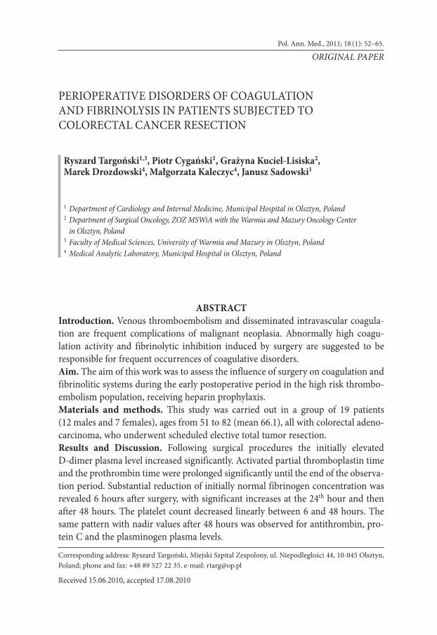
RESULTSBefore surgery the plasma DD level exceeded the upper limit of reference value (0.5 mg/dL) more than 2-fold and increased significantly 6 hours following surgery. Maximal values were found after 24 hours with an insignificant decrease on the next day (Fig. 1).
R. Targoński, P. Cygański, G. Kuciel-Lisiska, M. Drozdowski, M. Kaleczyc, J. Sadowski56
1.10
2.06
2.412.29
0
1
2
3
4
T1 T2 T3 T4Time
DD [m
g/dL
]
p<0.001
p=0.01
Fig. 1. DD plasma levels after colorectal cancer resection. Comments: p < 0.01 for T2 value compared to T1 (control sample). Values expressed as mean ±SD
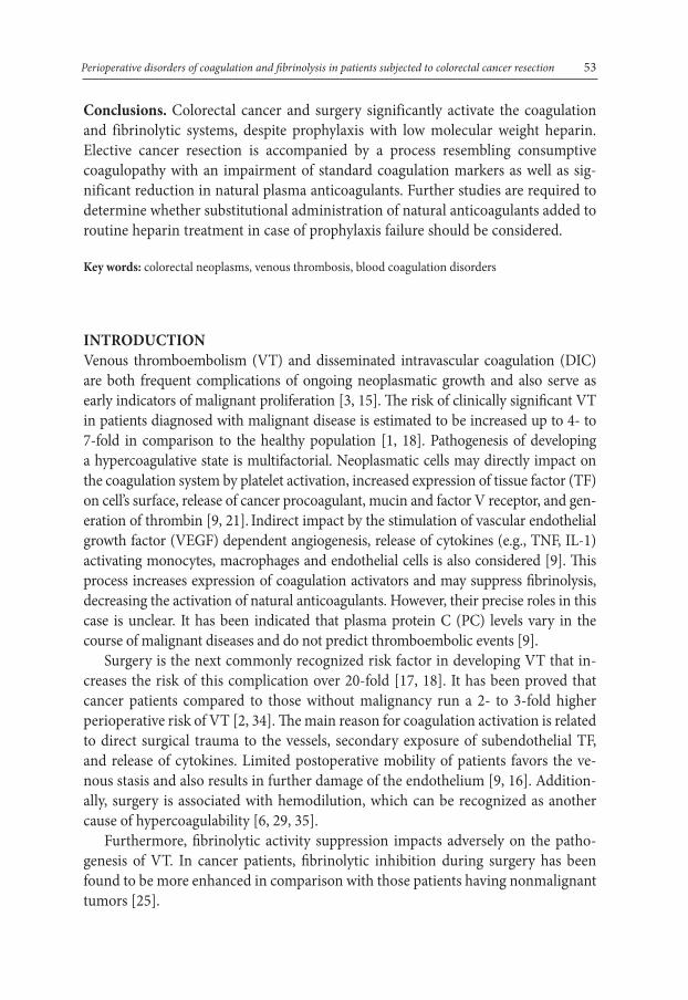
APTT results, presented in Fig. 2, did not change during the initial 6 hours after surgery, but then lengthened significantly after 24 and 48 hours.
35.9
44.3
48.0
36.4
25
30
35
40
45
50
55
60
T1 T2 T3 T4Time
APTT
[s]
p=0.11
p<0.001
Fig. 2. APTT after colorectal cancer resection. Comments: p < 0.001 for T3 and T4 values compared to T1 and for T2 compared to T3 and T4 values. Values expressed as mean ±SD

Perioperative disorders of coagulation and fibrinolysis in patients subjected to colorectal cancer resection 57
PT defined as INR increased significantly 6 hours after surgery, reaching maximal value after 24 hours, with subsequent insignificant diminishing after 48 hours (Fig. 3).
INR
1.12
1.27
1.481.44
1.0
1.1
1.2
1.3
1.4
1.5
1.6
1.7
1.8
T1 T2 T3 T4
Time
p<0.001
p=0.009
Fig. 3. INR levels after colorectal cancer resection. Comments: p < 0.001 for T2, T3, T4 values compared to T1; p < 0.001 for T2 compared to T3 value and p = 0.009 for T2 compared to T4 value. Values expres-sed as mean ±SD
FG plasma levels in perioperative period are summarized in Fig. 4.
390.7
308.9
431.7
552.5
0
100
200
300
400
500
600
700
T1 T2 T3 T4
Time
FG [m
g/dL
]
p<0.001
p=0.003 p<0.001
Fig. 4. FG levels after colorectal cancer resection. Comments: p < 0.001 for T2 value compared to T1. Line-ar increase of T3 and T4 values (p < 0.001) compared to T2 value. Values expressed as mean ±SD

R. Targoński, P. Cygański, G. Kuciel-Lisiska, M. Drozdowski, M. Kaleczyc, J. Sadowski58
A substantial reduction of initially normal FG concentration was revealed 6 hours after surgery. However, FG plasma level significantly increased after 24 hours com-pared to the lowest detected value and exceeded the upper reference limit. On the next day, the FG concentration plasma level showed a further significant increase.
Platelet count diminished significantly after 6 hours, showing a further linear de-cline up to 48 hours. At that time the mean value was the lowest one. Results are presented in Fig. 5.
244.79
201.79
190.05
173.00
150
170
190
210
230
250
270
290
310
T1 T2 T3 T4
Time
Plat
elets
[103 /u
L]
p=0.06
p=0.04
Fig. 5. Platelets count after colorectal cancer resection. Comments: p = 0.04 for T2 value compared to T1. Linear decrease of all subsequent values (T3, T4); p = 0.009 for T1 compared to T3 value and p < 0.001 for T1 compared to T4 value. Values expressed as mean ±SD
The mean AT plasma level before surgery was within normal limits. At 6 hours after surgery a significant drop was noted and lasted up to 48 hours (Fig. 6).
PC plasma level before surgery reached a level of 110.79%. All the subsequent mean postoperative values were significantly and progressively decreased. The lowest value was observed 48 hours after surgery. PG plasma level before surgery amounted to 97.67% and a substantial decrease was detected up to 48 hours (Fig. 6).
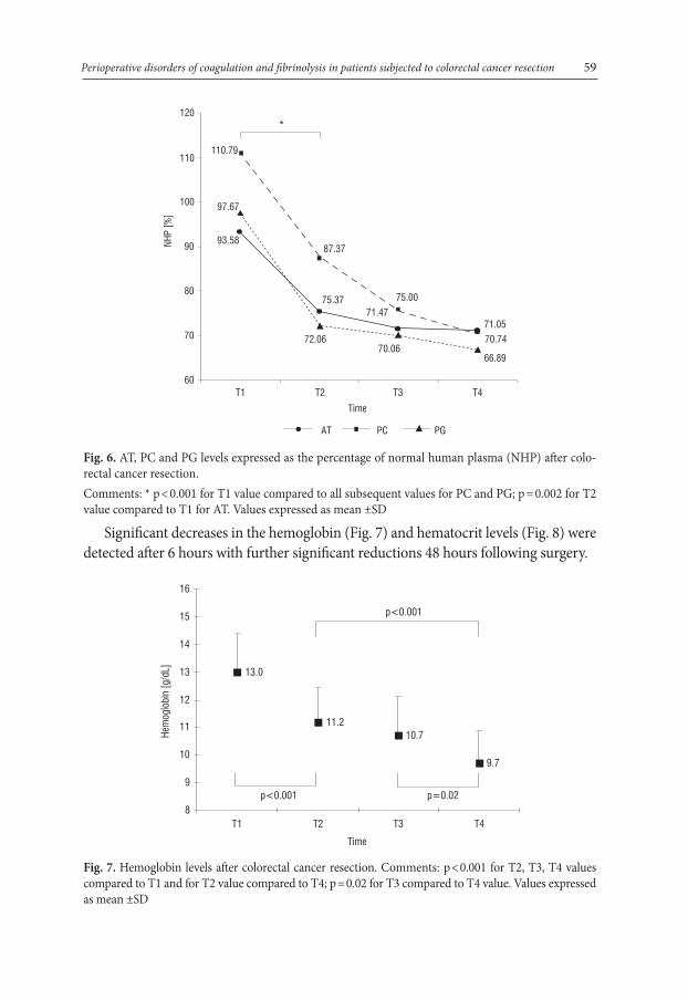
Perioperative disorders of coagulation and fibrinolysis in patients subjected to colorectal cancer resection 59
66.89
93.58
71.05
75.3771.47
70.74
75.00
87.37
110.79
72.0670.06
97.67
60
70
80
90
100
110
120
T1 T2 T3 T4Time
NHP
[%]
AT PC PG
*
Fig. 6. AT, PC and PG levels expressed as the percentage of normal human plasma (NHP) after colo-rectal cancer resection. Comments: * p < 0.001 for T1 value compared to all subsequent values for PC and PG; p = 0.002 for T2 value compared to T1 for AT. Values expressed as mean ±SD
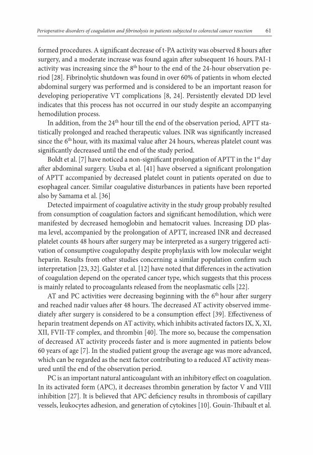
Significant decreases in the hemoglobin (Fig. 7) and hematocrit levels (Fig. 8) were detected after 6 hours with further significant reductions 48 hours following surgery.
13.0
11.210.7
9.7
8
9
10
11
12
13
14
15
16
T1 T2 T3 T4
Time
Hem
oglo
bin
[g/d
L]
p<0.001
p<0.001
p=0.02
Fig. 7. Hemoglobin levels after colorectal cancer resection. Comments: p < 0.001 for T2, T3, T4 values compared to T1 and for T2 value compared to T4; p = 0.02 for T3 compared to T4 value. Values expressed as mean ±SD

R. Targoński, P. Cygański, G. Kuciel-Lisiska, M. Drozdowski, M. Kaleczyc, J. Sadowski60
38.8
33.332.0
29.0
25
27
29
31
33
35
37
39
41
43
45
T1 T2 T3 T4
Time
Hem
atoc
rit [%
]
p<0.001
p<0.001
p=0.02
Fig. 8. Hematocrit levels after colorectal cancer resection. Comments: p < 0.001 for T2, T3, T4 values compared to T1 and for T2 value compared to T4; p = 0.02 for T3 compared to T4 value. Values expressed as mean ±SD
DISCUSSIONIn the studied group of patients, DD plasma levels before surgery increased over 2-fold compared to the upper reference value. DD value is considered to be an indicator of coagulation activation, secondary clot proteolysis and correlates with colorectal cancer progression [5, 12, 19, 20, 38]. A high plasma DD level before surgery confirms numer-ous, already published, conclusions concerning preoperative activation of coagulation and fibrinolytic systems in patients with colorectal cancer [12, 13, 19, 42].
It seems that increased DD plasma concentration in the studied group of patients with the highest level at 24 hours after surgery was caused mostly by the compensa-tory fibrinolysis process. Galster et al. [12] have found that in the patients who un-derwent gastrointestinal cancer resection in comparison to these operated on due to non-malignant disease, fibrinolysis assessed by DD plasma level was more intense, and lasted longer, until the 14th day of the postoperative follow-up. The mean values of the DD increased up to 48 hours, and were similar to those in the study group. However, these authors observed a decrease of fibrinolytic processes known as fi-brinolytic shutdown assessed after 24 hours, which was not observed in our study.
Postoperative fibrinolysis is crucial for fibrin degradation rate and protects against the new thrombotic incidents. Its activity depends on t-PA activity and PAI-1 activity. Neudecker et al. [28] have found that patients who have undergone colorec-tal cancer resections regardless of the operation type – conventional or laparoscopic, have had increased t-PA activity without an increase in PAI-1 activity during per-
Perioperative disorders of coagulation and fibrinolysis in patients subjected to colorectal cancer resection 61
formed procedures. A significant decrease of t-PA activity was observed 8 hours after surgery, and a moderate increase was found again after subsequent 16 hours. PAI-1 activity was increasing since the 8th hour to the end of the 24-hour observation pe-riod [28]. Fibrinolytic shutdown was found in over 60% of patients in whom elected abdominal surgery was performed and is considered to be an important reason for developing perioperative VT complications [8, 24]. Persistently elevated DD level indicates that this process has not occurred in our study despite an accompanying hemodilution process.
In addition, from the 24th hour till the end of the observation period, APTT sta-tistically prolonged and reached therapeutic values. INR was significantly increased since the 6th hour, with its maximal value after 24 hours, whereas platelet count was significantly decreased until the end of the study period.
Boldt et al. [7] have noticed a non-significant prolongation of APTT in the 1st day after abdominal surgery. Usuba et al. [41] have observed a significant prolongation of APTT accompanied by decreased platelet count in patients operated on due to esophageal cancer. Similar coagulative disturbances in patients have been reported also by Samama et al. [36]
Detected impairment of coagulative activity in the study group probably resulted from consumption of coagulation factors and significant hemodilution, which were manifested by decreased hemoglobin and hematocrit values. Increasing DD plas-ma level, accompanied by the prolongation of APTT, increased INR and decreased platelet counts 48 hours after surgery may be interpreted as a surgery triggered acti-vation of consumptive coagulopathy despite prophylaxis with low molecular weight heparin. Results from other studies concerning a similar population confirm such interpretation [23, 32]. Galster et al. [12] have noted that differences in the activation of coagulation depend on the operated cancer type, which suggests that this process is mainly related to procoagulants released from the neoplasmatic cells [22].
AT and PC activities were decreasing beginning with the 6th hour after surgery and reached nadir values after 48 hours. The decreased AT activity observed imme-diately after surgery is considered to be a consumption effect [39]. Effectiveness of heparin treatment depends on AT activity, which inhibits activated factors IX, X, XI, XII, FVII-TF complex, and thrombin [40]. The more so, because the compensation of decreased AT activity proceeds faster and is more augmented in patients below 60 years of age [7]. In the studied patient group the average age was more advanced, which can be regarded as the next factor contributing to a reduced AT activity meas-ured until the end of the observation period.
PC is an important natural anticoagulant with an inhibitory effect on coagulation. In its activated form (APC), it decreases thrombin generation by factor V and VIII inhibition [27]. It is believed that APC deficiency results in thrombosis of capillary vessels, leukocytes adhesion, and generation of cytokines [10]. Gouin-Thibault et al.

R. Targoński, P. Cygański, G. Kuciel-Lisiska, M. Drozdowski, M. Kaleczyc, J. Sadowski62
[15] explain that decreased AT, PC and protein S (PS) activities in cancer patients result from diminished hepatic synthesis and overconsumption. Nguyen et al. [30] found laparoscopic and open gastric bypass surgery to result in a gradual postopera-tive decrease of initially normal PC and AT plasma levels as we detected in our group of patients [38]. Garcia-Avello et al. [13] found a decreased PC plasma level only in samples taken from the tumor draining vein after colorectal cancer resection. In the study group both the consumption of coagulation factors and postoperative hemodi-lution can be regarded as contributing to decreased plasma levels of natural antico-agulants. In a large prospective study Folsom et al. [11] concluded that in a popula-tion without cancer, a low PC level rather than AT deficiency is responsible for new onsets of thrombotic complications. When comparing patients with thrombosis and cancer to those without malignancy, PC concentration is found to be significantly decreased in the former group [14]. These observations suggest a clinical significance of low PC plasma levels in the development of thromboembolic complications.
Decreased PG values 6 hours after surgery in the study group was accompanied by a rapid increase in FG plasma level. Low PG level may lead to deterioration in plasma fibrinolytic potential. However, isolated congenital PG deficiency was not recognized as a risk factor for VT [31].
Nguyen et al. [30] suggested that a decrease in PG level and a concomitant in-creased FG concentration may facilitate fibrin generation. This process shifts the coagulation-fibrinolysis balance towards a hypercoagulable state [42].
Before heparin prophylaxis era, it was found that general anesthesia was associ-ated with a statistically significant increase in postoperative thromboembolic com-plications [33]. Despite the introduction of low molecular weight heparin prophy-laxis, it was proved that general anesthetics may still produce more VT complica-tions [26]. There is no doubt that heparin prophylaxis by impairing the coagulation system reduces the risk of thromboembolic complications in patients subjected to open surgery. Nevertheless, the activation of the coagulation system occurs as a re-sult of processes described above, and leads to impairment of intrinsic and extrinsic coagulation pathways, decreased platelet count as well as the lowering of natural an-ticoagulants. Thus, a question arises: What is the significance of both these processes in pathogenesis of thromboembolic complications? It seems that decreased levels of anticoagulants may be of the greatest importance.
The limitation of this study is the fact that the prothrombotic effect of applied general anesthetics in this research cannot be excluded. Due to the safety reasons, we did not compare our patients with a control group without heparin prophylaxis. It is therefore impossible to establish whether the observed hemostatic disarrangements are related to the applied standard general anesthesia or to what extent they were prevented by the use of prophylaxis with low molecular weight heparin.
Perioperative disorders of coagulation and fibrinolysis in patients subjected to colorectal cancer resection 63
CONCLUSIONSColorectal cancer induces a significant activation of coagulation and fibrinolytic sys-tems. Despite prophylaxis with low molecular weight heparin, colorectal cancer re-section interferes with hemostasis by induction of a process resembling consumptive coagulopathy with an impairment of standard coagulation laboratory tests and trig-gers reactions leading to a decreased level of natural anticoagulants. Further studies are required to determine whether substitutional administration of natural antico-agulants added to routine heparin treatment in case of prophylaxis failure should be considered.
REFERENCES1. Adess M., Eisner R., Nand S., Godwin J., Messmore H. L., Wehrmacher W. H.: Thromboembolism in
cancer patients: pathogenesis and treatment. Clin. App. Thromb. Hemost., 2006; 12 (3): 254–266.2. Anderson F. A., Spencer F. A.: Risk factors for venous thromboembolism. Circulation, 2003; 107: I9–
I16.3. Baron J. A., Gridley G., Weiderpass E., Nyrén O., Linet M.: Venous thromboembolism and cancer.
Lancet, 1998; 351 (9109): 1077–1080.4. Bergqvist D., Agnelli G., Cohen A. T., Eldor A., Nilsson P. E., Le Moigne-Amrani A., Dietrich-Neto F.:
Duration of prophylaxis against venous thromboembolism with enoxaparin after surgery for cancer. N. Engl. J. Med., 2002; 346 (13): 975–980.
5. Blackwell K., Hurwitz H., Lieberman G., Novotny W., Snyder S., Dewhirst M., Greenberg C.: Circu-lating D-dimer levels are better predictors of overall survival and disease progression than carcinoem-bryonic antigen levels in patients with metastatic colorectal carcinoma. Cancer, 2004; 101: 77–82.
6. Boldt J., Haisch G., Suttner S., Kumle B., Schellhase F.: Are lactated Ringer’s solution and normal saline solution equal with regard to coagulation? Anesth. Analg., 2002; 94 (2): 378–384.
7. Boldt J., Hüttner I., Suttner S., Kumle B., Piper S. N., Berchthold G.: Changes of haemostasis in patients undergoing major abdominal surgery – is there a difference between elderly and younger patients? Br. J. Anaesth., 2001; 87 (3): 435–440.
8. D’Angelo A., Kluft C., Verheijen J. H., Rijken D. C., Mozzi E., Mannucci P. M.: Fibrinolytic shut-down after surgery: impairment of the balance between tissue-type plasminogen activator and its specific in-hibitor. Eur. J. Clin. Invest., 1985; 15 (6): 308–312.
9. De Cicco M.: The prothrombotic state in cancer: pathogenic mechanisms. Crit. Rev. Oncol. Hematol., 2004; 50 (3): 187–196.
10. Esmon C. T.: The protein C pathway. Chest, 2003; 124 (Suppl. 3): 26–32.11. Folsom A. R., Aleksic N., Wang L., Cushman M., Wu K. K., White R. H.: Protein C, antithrombin,
and venous thromboembolism incidence: a prospective population-based study. Arterioscler. Thromb. Vasc. Biol., 2002; 22 (6): 1018–1022.
12. Galster H., Kolb G., Kohsytorz A., Seidlmayer C., Paal V.: The pre-, peri-, and postsurgical activa-tion of coagulation and the thromboembolic risk for different risk groups. Thromb. Res., 2000; 100 (5): 381–388.
13. Garcia-Avello A., Galindo-Alvarez J., Martinez-Molina E., Cesar-Perez J., Navarro J. L.: Coagulative system activation and fibrinolytic system inhibition activities arise from tumoral draining vein in colon carcinoma. Thromb. Res., 2001; 104 (6): 421–425.
14. Goldenberg N., Kahn S. R., Solymoss S.: Markers of coagulation and angiogenesis in cancer-associated venous thromboembolism. J. Clin. Oncol., 2003; 21 (22): 4194–4199.
15. Gouin-Thibault I., Achkar A., Samama M. M.: The thrombophilic state in cancer patients. Acta Hae-matol., 2001; 106 (1–2): 33–42.

R. Targoński, P. Cygański, G. Kuciel-Lisiska, M. Drozdowski, M. Kaleczyc, J. Sadowski64
16. Gupta P. K., Charan V. D., Kumar H.: Cancer related thrombophilia: clinical importance and manage-ment strategies. J. Assoc. Physicians India, 2005; 53: 877–882.
17. Heit J. A., O’Fallon W. M., Petterson T. M., Lohse C. M., Silverstein M. D., Mohr D. N., Melton L. J.: Relative impact of risk factors for deep vein thrombosis and pulmonary embolism: a population-based study. Arch. Intern. Med., 2002; 162 (11): 1245–1248.
18. Heit J. A., Silverstein M. D., Mohr D. N., Petterson T. M., O’Fallon W. M., Melton L. J.: Risk factors for deep vein thrombosis and pulmonary embolism: a population-based case-control study. Arch. Intern. Med., 2000; 160 (6): 809–815.
19. Iversen L. H., Okholm M., Thorlacius-Ussing O.: Pre- and postoperative state of coagulation and fibrinolysis in plasma of patients with benign and malignant colorectal disease – a preliminary study. Thromb. Haemost., 1996; 76 (4): 523–528.
20. Iversen L. H., Thorlacius-Ussing O.: Relationship of coagulation test abnormalities to tumor burden and postoperative DVT in resected colorectal cancer. Thromb. Haemost., 2002; 87 (3): 402–408.
21. Lee A. Y.: Thrombosis and cancer: the role of screening for occult cancer and recognizing the underlying biological mechanisms. Hematol. Am. Soc. Hematol. Educ. Program, 2006: 438–443.
22. Lee A. Y., Levine M. N.: Venous thromboembolism and cancer: risk and outcomes. Circulation, 2003; 107: 17–21.
23. Mal’kov O. A., Dolgikh V. T., Lukach V. N.: [Complex prevention of thrombotic complications in pa-tients with colorectal cancer during different stages of surgical treatment][Article in Russian]. Anest. Reanimatol., 2001; (5): 52–54.
24. Mellbring G., Dahlgren S., Wiman B.: Plasma fibrinolytic activity in patients undergoing major ab-dominal surgery. Acta Chir. Scand., 1985; 151 (2):109–114.
25. Modrau I. S., Iversen L. H., Thorlacius-Ussing O. O.: Hemostatic alterations in patients with benign and malignant colorectal disease during major abdominal surgery. Thromb. Res., 2001; 104 (5): 309– –315.
26. Nathan S. S., Simmons K. A., Lin P. P., Hann L. E., Morris C. D., Athanasian E. A., Boland P. J., Hea- S., Simmons K. A., Lin P. P., Hann L. E., Morris C. D., Athanasian E. A., Boland P. J., Hea-S., Simmons K. A., Lin P. P., Hann L. E., Morris C. D., Athanasian E. A., Boland P. J., Hea- A., Lin P. P., Hann L. E., Morris C. D., Athanasian E. A., Boland P. J., Hea-A., Lin P. P., Hann L. E., Morris C. D., Athanasian E. A., Boland P. J., Hea- P., Hann L. E., Morris C. D., Athanasian E. A., Boland P. J., Hea-P., Hann L. E., Morris C. D., Athanasian E. A., Boland P. J., Hea- E., Morris C. D., Athanasian E. A., Boland P. J., Hea-E., Morris C. D., Athanasian E. A., Boland P. J., Hea- D., Athanasian E. A., Boland P. J., Hea-D., Athanasian E. A., Boland P. J., Hea- A., Boland P. J., Hea-A., Boland P. J., Hea- J., Hea-J., Hea-ley J. H.: Proximal deep vein thrombosis after hip replacement for oncologic indications. J. Bone Joint Surg. Am., 2006; 88 (5): 1066–1070.
27. Nesheim M.: Thrombin and fibrinolysis. Chest, 2003; 124 (Suppl. 3): 33–39.28. Neudecker J., Junghans T., Ziemer S., Raue W., Schwenk W.: Prospective randomized trial to de-
termine the influence of laparoscopic and conventional colorectal resection on intravasal fibrinolytic capacity. Surg. Endosc., 2003; 17 (1): 73–77.
29. Ng K. F., Lam C. C., Chan L. C.: In vivo effect of haemodilution with saline on coagulation: a random-ized controlled trial. Br. J. Anaesth., 2002; 88 (4): 475–480.
30. Nguyen N. T., Owings J. T., Gosselin R., Pevec W. C., Lee S. J., Goldman C., Wolfe B. M.: Systemic co-agulation and fibrinolysis after laparoscopic and open gastric bypass. Arch. Surg., 2001; 136: 909–916.
31. Okamoto A., Sakata T., Mannami T., Baba S., Katazama Z., Matsuo H., Yasaka M., Minematusu K., Tomoike K., Miyata T.: Population-based distribution of plasminogen activity and estimated preva-lence and relevance to thrombotic diseases of plasminogen deficiency in the Japanese: the Suita Study. J. Thromb. Haemost., 2003; 1 (11): 2397–2403.
32. Ozturk M., Sengul N., Dagli M., Kosar A., Bavbek N.: Global fibrinolytic capacity in colorectal cancer: a new clue to occult fibrinolysis. Clin. Appl. Thromb. Hemost., 2003; 9 (2): 151–154.
33. Prins M. H., Hirsh. A comparison of general anesthesia and regional anesthesia as a risk factor for deep vein thrombosis following hip surgery: a critical review. Thromb. Haemost., 1990; 64 (4): 497– –500.
34. Rickles F. R., Levine M. N.: Epidemiology of thrombosis in cancer. Acta Haematol., 2001; 106 (1–2): 6–12.
35. Ruttmann T. G., James M. F., Viljoen J. F.: Haemodilution induces a hypercoagulable state. Br. J. An-Br. J. An-aesth., 1996; 76 (3): 412–414.
36. Samama C. M., Thiry D., Elalamy I., Diaby M., Guillosson J. J., Kieffer E., Coriat P.: Perioperative activation of hemostasis in vascular surgery patients. Anesthesiology, 2001; 94 (1): 74–78.
Perioperative disorders of coagulation and fibrinolysis in patients subjected to colorectal cancer resection 65
37. Schizas C., Neumayer F., Kosmopoulos V.: Incidence and management of pulmonary embolism fol-lowing spinal surgery occurring while under chemical thromboprophylaxis. Eur. Spine J., 2008; 17 (7): 970–974.
38. Subramanian S., Rameshkumar K., Damodar P.: Do plasma D-dimer levels correlate with disease stage in colonic carcinoma? Indian J. Surg., 2006; 68 (6): 306–309.
39. Szczepański M., Szostek P., Pypno W., Borówka A.: The lack of effect of a prophylactic dose of enox-aparin on thrombin generation in patients subjected to nephrectomy because of kidney cancer. Thromb. Res., 2001; 104 (6): 427–432.
40. Urbach D., Matzen K. A., Heitmann D., Neumann H. W.: Relation between perioperative antithrombin activity and deep vein thrombosis after elective hip replacement surgery. Vasa, 2003; 32 (l): 14–17.
41. Usuba A., Motoki R., Watanabe M., Koizumi Y., Kanno R., Matayoshi K., Ohishi A., Endho Y., Konno O., Inoue H.: [Hypercoagulability after surgery of esophageal carcinoma] [Article in Japa-nese]. Nippon Kyobu Geka Gakkai Zasshi, 1990; 38 (3): 401–411.
42. van Duijnhoven E. M., Lustermans F. A., van Wersch J. W.: Evaluation of the coagulation/fibrinolysis balance in patients with colorectal cancer. Haemostasis, 1993; 23 (3): 168–172.