peritoneal dialysis fundamentals - msic
TRANSCRIPT

Peritoneal Dialysis Fundamentals
Dr.Jebril S. Elabidi •Consultant nephrologist
•Lecturer in Faculty of medicine
Benghazi university
•Head of dialysis unit in BCH
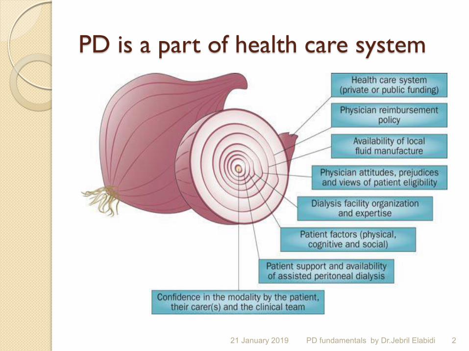
PD is a part of health care system
21 January 2019 PD fundamentals by Dr.Jebril Elabidi 2
21 January 2019 PD fundamentals by Dr.Jebril Elabidi 3

PD Introduction
Basics of peritoneal dialysis
anatomy and physiology
Principles of PD
Indication and contraindication
Advantages and disadvanges
Complications
Adequacy of PD dialysis
21 January 2019 4 PD fundamentals by Dr.Jebril Elabidi

Automated PD
CAPD
ESRD Treatment Modalities
Transplantation
Peritoneal
Dialysis
Renal
Replacement
Therapy
?
Hemofiltration Hemodialysis
Hemodiafiltration 21 January 2019 5 PD fundamentals by Dr.Jebril Elabidi

21 January 2019 6 PD fundamentals by Dr.Jebril Elabidi

WHY PD
21 January 2019 7 PD fundamentals by Dr.Jebril Elabidi

Why PD
No vascular access
No extracorporeal circuit
CAPD less expensive than HD
Home treatment
Better preservation of residual renal function
No rapid change in blood volume and body fluid compostion
21 January 2019 8 PD fundamentals by Dr.Jebril Elabidi

Indications Hemdynamically unstable
Uremic syndrome
Fluid overload
Pediatric patient
21 January 2019 9 PD fundamentals by Dr.Jebril Elabidi

21 January 2019 10 PD fundamentals by Dr.Jebril Elabidi

21 January 2019 11 PD fundamentals by Dr.Jebril Elabidi

Absolute of contraindication Peritoneal fibrosis
More than 50 % intestinal resection
Pleural preitoneal leak
Active inflammatory bowel disease
21 January 2019 12 PD fundamentals by Dr.Jebril Elabidi
Peritoneal dialysis Peritoneal dialysis is the method of removing waste and excess water from the body using the peritoneal membrane as a filter . Peritoneal dialysis cleans the blood without it
being removed ,
Patients need not to have a schedule dialysis sessions at a center but can have treatment at home, at work or on trips; in the same time he should have close contact with his health care team.
21 January 2019 13 PD fundamentals by Dr.Jebril Elabidi
21 January 2019 14 PD fundamentals by Dr.Jebril Elabidi

Introduction PD
Intracorporeal dialysis used peritoneum as dialyzer and heart as blood pump without vascular access
PD using peritoneum as semipermeable membrane
21 January 2019 15 PD fundamentals by Dr.Jebril Elabidi
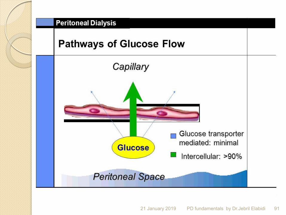
Solutes are transported across the membrane by diffusion.
The driving force is the concentration gradient between the PD fluid and the blood.
Waste products present in the blood per fusing the peritoneum will diffuse from the blood vessels into the “cleaner” dialysis fluid.
21 January 2019 16 PD fundamentals by Dr.Jebril Elabidi
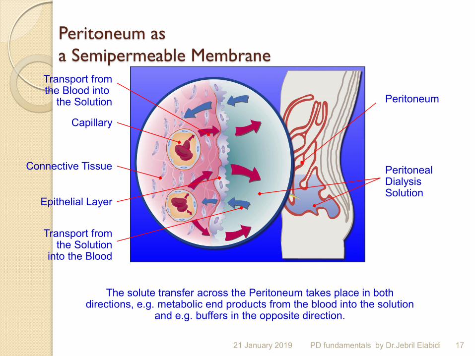
Peritoneum as a Semipermeable Membrane
Peritoneum
Peritoneal Dialysis Solution
Transport from the Solution
into the Blood
Epithelial Layer
Connective Tissue
Capillary
Transport from the Blood into
the Solution
The solute transfer across the Peritoneum takes place in both directions, e.g. metabolic end products from the blood into the solution
and e.g. buffers in the opposite direction.
21 January 2019 17 PD fundamentals by Dr.Jebril Elabidi
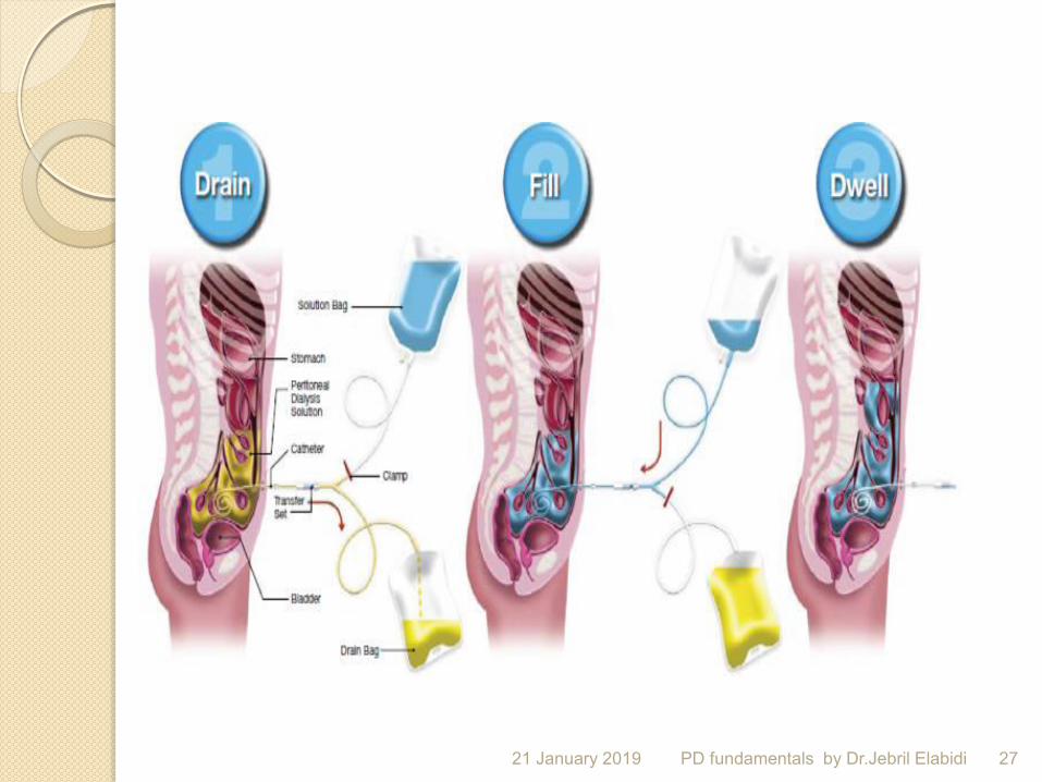
The dialysis fluid should be instilled for
4 to 6 hours.
When the dialysis fluid is drained from the abdominal cavity, it contains waste products and excess fluid extracted from the blood.
PD is most often applied and effective as a continuous therapy
21 January 2019 18 PD fundamentals by Dr.Jebril Elabidi
21 January 2019 19 PD fundamentals by Dr.Jebril Elabidi

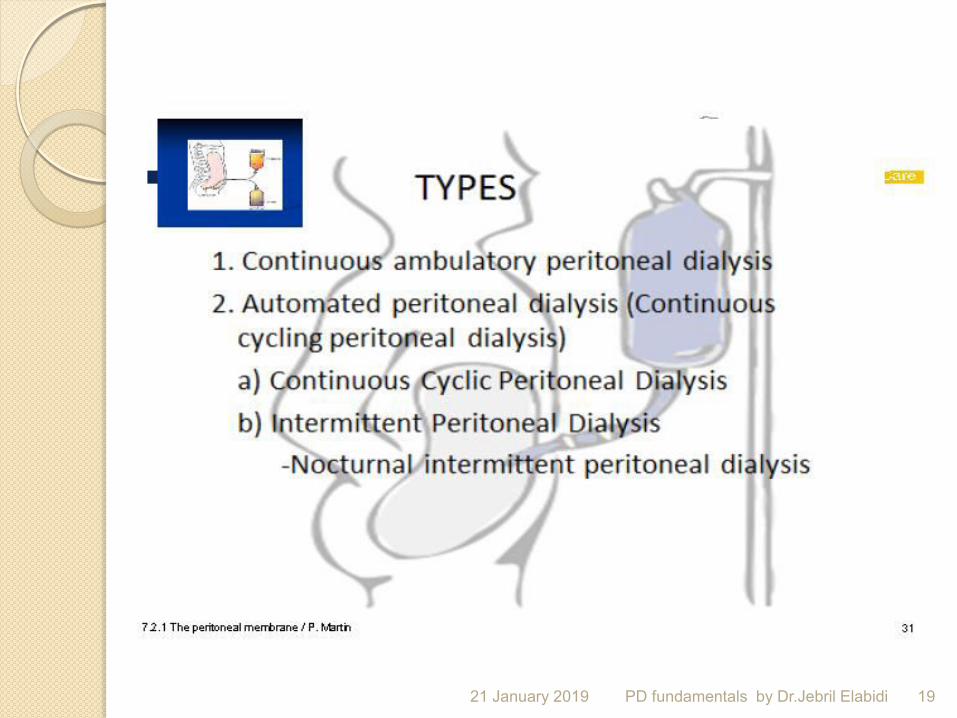
PD: Major Techniques
CAPD APD
21 January 2019 20 PD fundamentals by Dr.Jebril Elabidi

CAPD
PD
fun
da
menta
ls b
y D
r.Je
bril E
lab
idi
21 January 2019 21

APD
PD fundamentals by Dr.Jebril Elabidi 21 January 2019 22
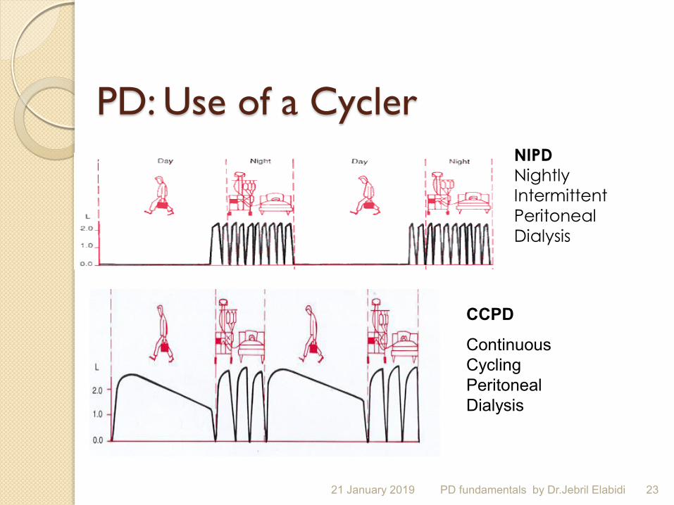
PD: Use of a Cycler NIPD Nightly
Intermittent
Peritoneal
Dialysis
CCPD
Continuous
Cycling
Peritoneal
Dialysis
21 January 2019 23 PD fundamentals by Dr.Jebril Elabidi

21 January 2019 24 PD fundamentals by Dr.Jebril Elabidi

21 January 2019 25 PD fundamentals by Dr.Jebril Elabidi
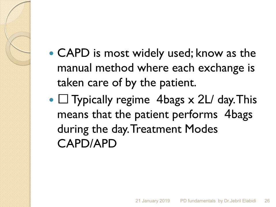
CAPD is most widely used; know as the manual method where each exchange is taken care of by the patient.
Typically regime 4 bags x 2L/ day. This means that the patient performs 4 bags during the day. Treatment Modes CAPD/APD
21 January 2019 26 PD fundamentals by Dr.Jebril Elabidi
21 January 2019 27 PD fundamentals by Dr.Jebril Elabidi

21 January 2019 28 PD fundamentals by Dr.Jebril Elabidi

21 January 2019 29 PD fundamentals by Dr.Jebril Elabidi

Fill process About 10 min and dependent on
Amount of solution
Height of solution bag
Diameter of tubing
Inta abdominal pressure
21 January 2019 30 PD fundamentals by Dr.Jebril Elabidi

Dwell Time required for transport of solutes
across the peritoneum
In CAPD usually 4-5 hours per exchange during the day and 9 hours at night
In APD 8-10 hours at night , 14 hours during day if required
21 January 2019 31 PD fundamentals by Dr.Jebril Elabidi

Drain About 15-25 min depends on
Amount of solution
Gravity
Diameter of tubing
Position
21 January 2019 32 PD fundamentals by Dr.Jebril Elabidi

21 January 2019 33 PD fundamentals by Dr.Jebril Elabidi

Basics of peritoneal dialysis Anatomy of the peritoneum
Peritoneum is a serous membrane, derived from the mesenchyma.
It is composed of the parietal and visceral peritoneum that lines the peritoneal space
21 January 2019 34 PD fundamentals by Dr.Jebril Elabidi

21 January 2019 35 PD fundamentals by Dr.Jebril Elabidi

21 January 2019 36 PD fundamentals by Dr.Jebril Elabidi

21 January 2019 37 PD fundamentals by Dr.Jebril Elabidi
21 January 2019 38 PD fundamentals by Dr.Jebril Elabidi
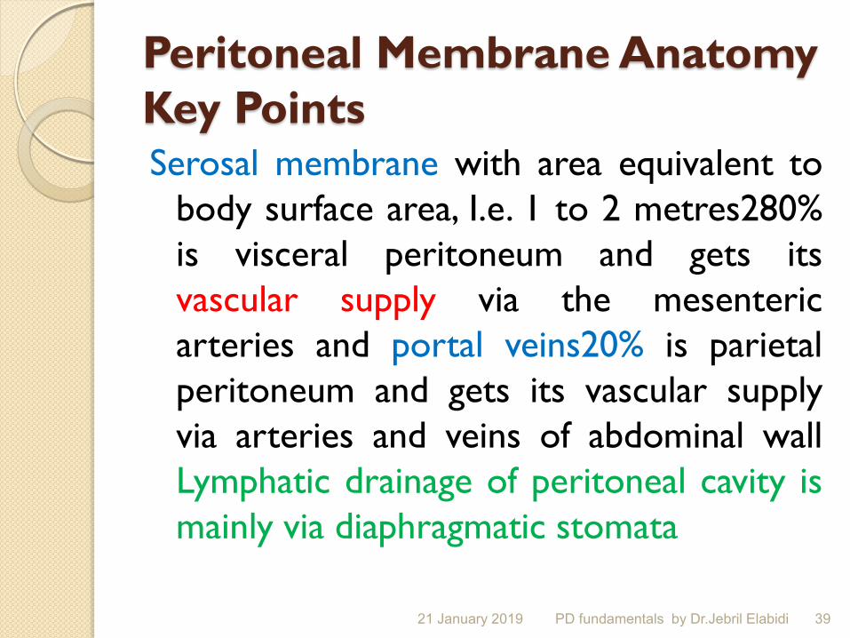
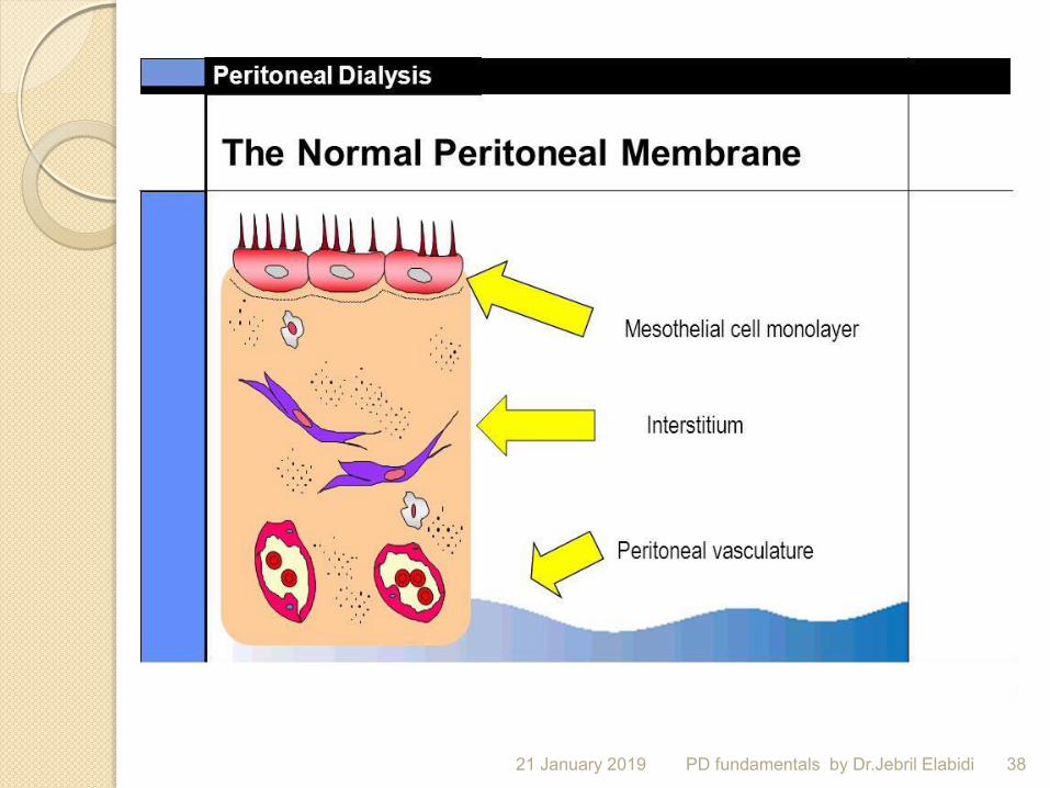
Peritoneal Membrane Anatomy Key Points Serosal membrane with area equivalent to
body surface area, I.e. 1 to 2 metres280% is visceral peritoneum and gets its vascular supply via the mesenteric arteries and portal veins20% is parietal peritoneum and gets its vascular supply via arteries and veins of abdominal wall Lymphatic drainage of peritoneal cavity is mainly via diaphragmatic stomata
21 January 2019 39 PD fundamentals by Dr.Jebril Elabidi

Peritoneal cavity is lined by a mesothelial monolayer which produces a lubricating fluid under the mesothelium is a gel-like interstitium containing connective tissue fibres, capillaries and lymphatics
The effective surface area is critical for dialysis and depends on the vascularity of the peritoneum as well as its surface area
21 January 2019 40 PD fundamentals by Dr.Jebril Elabidi
PD access
21 January 2019 41 PD fundamentals by Dr.Jebril Elabidi

Up to 20 % of the patients are transferred HD , related to catheter problems .
21 January 2019 42 PD fundamentals by Dr.Jebril Elabidi

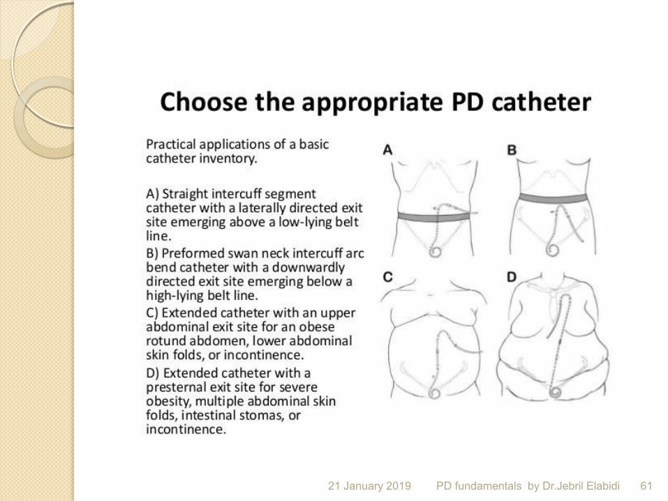
PD Catheters specification Intended for long term use
Soft and flexible
Double cuff ( most common )
Radio opaque stripe
Different sizes
Larges practical internal diameter
Kink prevention
Prevents obstruction and improves the flow
Biocompatible material to reduce inflammation
Ease of implantation and removal
21 January 2019 43 PD fundamentals by Dr.Jebril Elabidi

Common materials Silicone
Polyurethane
Silver impregnated
21 January 2019 44 PD fundamentals by Dr.Jebril Elabidi

Catheters type A. Tenckhoff catheter
B. T.W.H . Catheter with perpendicular discs
C.Swan neck Missouri catheter ( subcutaneous curved 170 degrees )
21 January 2019 45 PD fundamentals by Dr.Jebril Elabidi

21 January 2019 46 PD fundamentals by Dr.Jebril Elabidi

PD
fun
da
menta
ls b
y D
r.Je
bril E
lab
idi
21 January 2019 47

A tenckhoff catheter provides a permanent long term access to the peritoneal cavity .
it’s thin non irritating flexible tube , one end of this tube rests in the peritoneal cavity while the other extends from the body by about four inches
21 January 2019 48 PD fundamentals by Dr.Jebril Elabidi

21 January 2019 49 PD fundamentals by Dr.Jebril Elabidi
Catheter implantation 3 types of placement
1.blind placement
2.surgical placement by lapraotomy
3.peritoneoscopic visualisation
21 January 2019 50 PD fundamentals by Dr.Jebril Elabidi

21 January 2019 51 PD fundamentals by Dr.Jebril Elabidi

21 January 2019 52 PD fundamentals by Dr.Jebril Elabidi

21 January 2019 53 PD fundamentals by Dr.Jebril Elabidi

21 January 2019 54 PD fundamentals by Dr.Jebril Elabidi

Good catheter Fluid transit occurs rapidly more than
200 ml /min
No pain with fluid transit
Drain completely ( 100 ml residual vol is considered
Discourages infection and biocompatible
21 January 2019 55 PD fundamentals by Dr.Jebril Elabidi

Types of catheters Acute
Chronic
21 January 2019 56 PD fundamentals by Dr.Jebril Elabidi

Acute PD: THE PROCEDURE: Indications: Any patient who has acute
kidney injury needing RRT
Contraindications:
Deranged coagulation profile,
presence of hernia, distended bowel loops, features of peritoneal adhesions due to repeated intrabdominal surgery
or intrabdominal infections like tuberculous peritonitis.
21 January 2019 57 PD fundamentals by Dr.Jebril Elabidi
The procedure: Acute PD procedure can be divided into
the following four sequential steps:
Step 1 -Preparation of the abdomen
Step 2- Priming of the abdomen with fluid,
Step 3- PD catheter insertion and
Step 4- Conducting PD fluid exchanges
21 January 2019 58 PD fundamentals by Dr.Jebril Elabidi

STEP 1. Preparation of the abdomen: The rectum and urinary bladder are to be
emptied before the procedure for which rectal enema and evacuation of urinary bladder may be needed.
The abdominal wall hair is to be clipped shaved in case of adult males.
The skin of all patients should be cleaned initially with chlorehxidine/isoprolyl alcohol or if chlorhexidine unavailable, povidone iodine scrub and later povidone iodine lotion to ensure sterility.
The operator should use sterile gloves, gown and mask.
21 January 2019 59 PD fundamentals by Dr.Jebril Elabidi

STEP 2. Priming of the peritoneal cavity with fluid:
Placement of the priming needle in the correct place is a crucial step in the procedure.
Instruments required for priming include long wide bore needle or wide bore lumbar puncture needle, two intravenous (IV) fluid of blood administration sets, surgical blade and PD fluid bottles(or bags), 2 liters in case of adults and 50 ml/kg body weight fluid in case of children
21 January 2019 60 PD fundamentals by Dr.Jebril Elabidi
21 January 2019 61 PD fundamentals by Dr.Jebril Elabidi

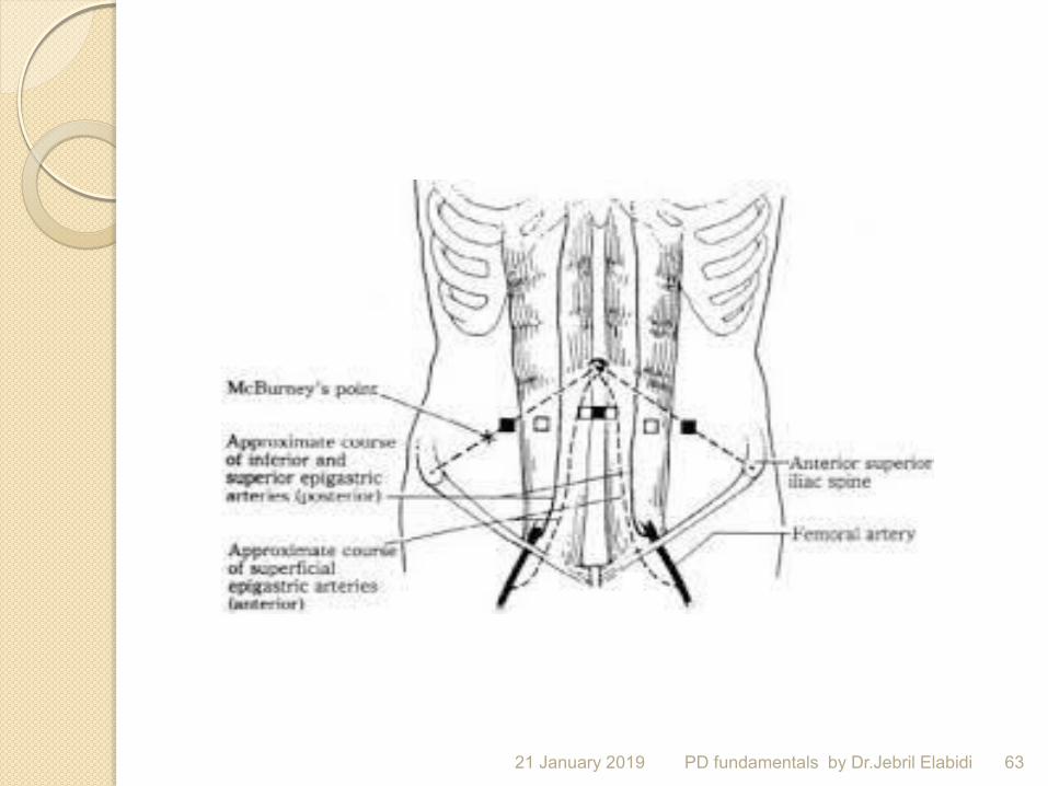
Most preferred site of PD catheter insertion is in the midline one inch below the umbilicus.
The other sites are McBurney’s point on right side or similar position in left iliac fossa.
21 January 2019 62 PD fundamentals by Dr.Jebril Elabidi
21 January 2019 63 PD fundamentals by Dr.Jebril Elabidi

21 January 2019 64 PD fundamentals by Dr.Jebril Elabidi

21 January 2019 65 PD fundamentals by Dr.Jebril Elabidi
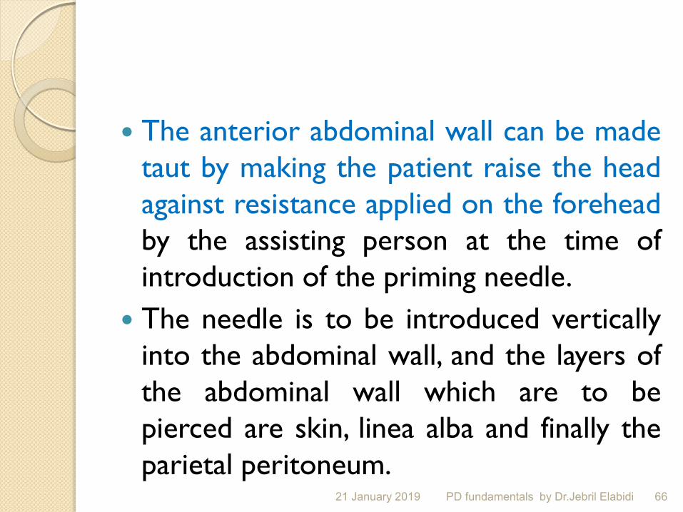
The anterior abdominal wall can be made taut by making the patient raise the head against resistance applied on the forehead by the assisting person at the time of introduction of the priming needle.
The needle is to be introduced vertically into the abdominal wall, and the layers of the abdominal wall which are to be pierced are skin, linea alba and finally the parietal peritoneum.
21 January 2019 66 PD fundamentals by Dr.Jebril Elabidi

The most important phenomenon to be experienced at the time of piercing each layer of abdominal wall is initial resistance followed by a feeling of ‘giving in’.
The final ‘giving in’ feeling is felt when the parietal peritoneum is pierced after which one should be careful not to advance the needle further as the intestine might get punctured.
21 January 2019 67 PD fundamentals by Dr.Jebril Elabidi

Correct position of the priming needle is ascertained by visualisation of good flow of fluid in the air chamber of the IV set. Wrong placement of the needle tip in the intestinal lumen will result in immediate diarrhea and, urinary bladder puncture will cause sudden increase in ‘urine’ flow which is actually the PD fluid draining out
21 January 2019 68 PD fundamentals by Dr.Jebril Elabidi

STEP 3. PD catheter insertion: instruments needed are,
PD catheter with stylet , two IV sets ,
a 3 way connector,
PD fluid bottles or bags and drain bag After priming of the required quantity of fluid the priming needle is removed .
The PD catheter is kept ready with the stylet well inside the catheter and its sharp tip protruding out
21 January 2019 69 PD fundamentals by Dr.Jebril Elabidi

STEP 4. PD exchanges: The three way connector is used to
regulate the direction of PD fluid.
Each fluid cycle has three components a) PD fluid inflow ( 10 minutes) b) 35 minutes dwell c) drain time ( 10 to 15 minutes)
21 January 2019 70 PD fundamentals by Dr.Jebril Elabidi
21 January 2019 71 PD fundamentals by Dr.Jebril Elabidi

Acute catheter Intended for short term use
Are stiff
Are placed via blind / Trocar method
NOT in obese
21 January 2019 72 PD fundamentals by Dr.Jebril Elabidi
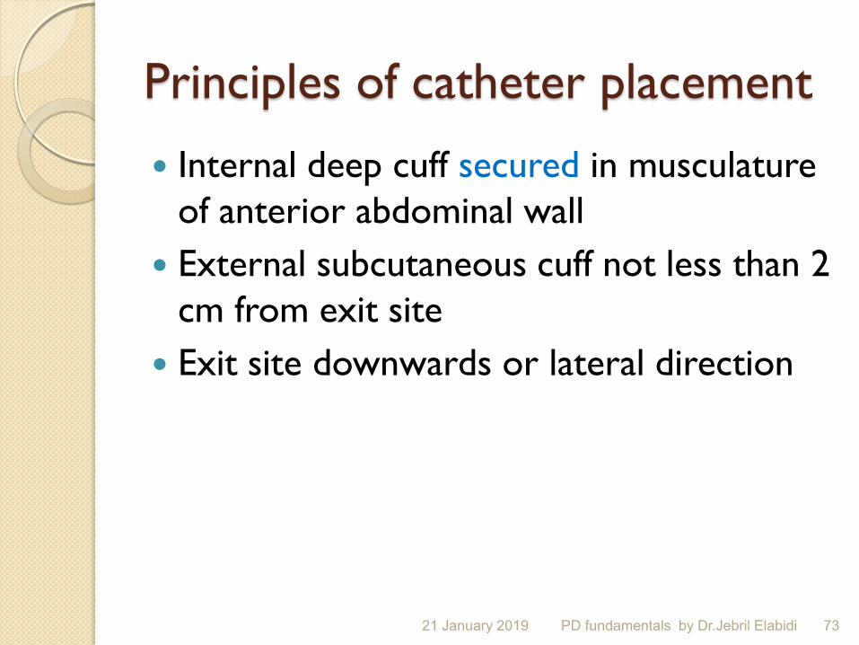
Principles of catheter placement Internal deep cuff secured in musculature
of anterior abdominal wall
External subcutaneous cuff not less than 2 cm from exit site
Exit site downwards or lateral direction
21 January 2019 73 PD fundamentals by Dr.Jebril Elabidi

21 January 2019 74 PD fundamentals by Dr.Jebril Elabidi

Laproscopic procedure
21 January 2019 75 PD fundamentals by Dr.Jebril Elabidi

Post implantation dialysis
Postpone flushing for 1-3 days to permit good tissue healing , then twice weekly how fill volume
Heparin 500-1000/l to prevent fibrin or blood clot formation
Does not start before 2 weeks of catheter insertion
21 January 2019 76 PD fundamentals by Dr.Jebril Elabidi

peritoneal Dialysis Catheter Choice and Outcomes
21 January 2019 77 PD fundamentals by Dr.Jebril Elabidi

Catheter survival rates Optimal 80 % three years of use
Minimal 50 % one year of
21 January 2019 78 PD fundamentals by Dr.Jebril Elabidi

RECOMMENDATIONS Catheter survival of >80% at one year desirable Double cuffed catheter preferred to single cuff Downward directed exit-site decreases the risk of catheter related infections (advantage being its- preformed arcuate bend)
No catheter appears to be superior to the 2 cuff standard Tenckhoff catheter - experience with swan- neck catheters is promising.
21 January 2019 79 PD fundamentals by Dr.Jebril Elabidi
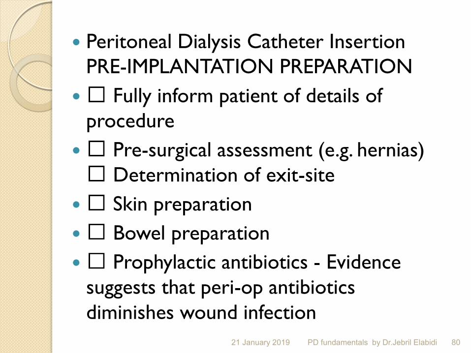
Peritoneal Dialysis Catheter Insertion PRE-IMPLANTATION PREPARATION
Fully inform patient of details of procedure
Pre-surgical assessment (e.g. hernias) Determination of exit-site
Skin preparation
Bowel preparation
Prophylactic antibiotics - Evidence suggests that peri-op antibiotics diminishes wound infection
21 January 2019 80 PD fundamentals by Dr.Jebril Elabidi
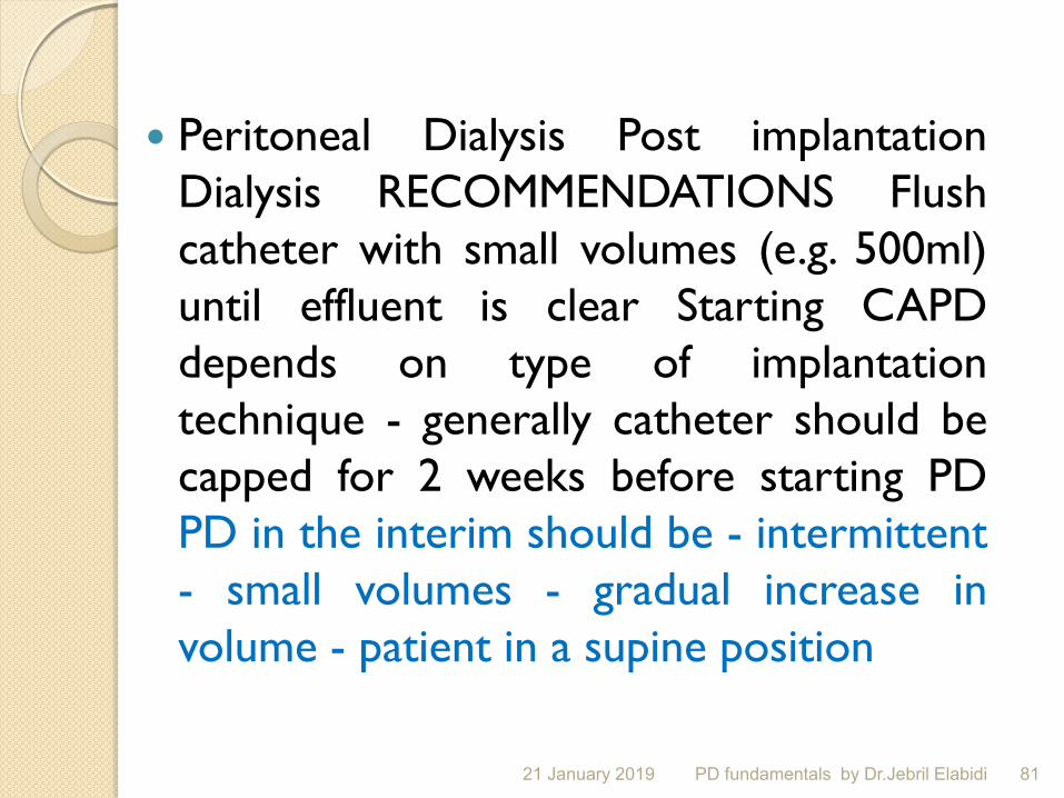
Peritoneal Dialysis Post implantation Dialysis RECOMMENDATIONS Flush catheter with small volumes (e.g. 500ml) until effluent is clear Starting CAPD depends on type of implantation technique - generally catheter should be capped for 2 weeks before starting PD PD in the interim should be - intermittent - small volumes - gradual increase in volume - patient in a supine position
21 January 2019 81 PD fundamentals by Dr.Jebril Elabidi

Physiology of Peritoneal Dialysis Barriers of peritoneal transport There are
three main barriers to peritoneal transport of solutes and fluid:
1. Mesothelium. 2. Interstitial tissue. 3. Blood vessels (endothelium & basement
membrane) The parietal peritoneum is more important
intransport than the visceral as only 25-30% of the VP it is in contact with the peritoneal fluid
21 January 2019 82 PD fundamentals by Dr.Jebril Elabidi

Interstitium Blood vessels Mesothelium Mesothelium It consists of a single layer of
flattened cuboidal cells (30,000cells/cm²) lying on a basement membrane.
Microvilli present on the luminal side increases the effective surface area of the peritoneal cavity up to 40m².
Tight junctions and desmosomes are present between the mesothelial cells.
Transport through the mesothelium occurs via endocytosis, transcytosis
21 January 2019 83 PD fundamentals by Dr.Jebril Elabidi

Interstitium Consists of cells (pred. fibroblast) & fibers (pred. collagen) embedded in an amorphous substance.
Thickness of the interstitium varies from
1-2µm to ≥30µm.
This thickness influences transport characteristics as this also defines the distance between the mesothelium and bv’s. Movement of solute is determined by the difference in concentration per unit distance (Fick’s law of diffusion).
21 January 2019 84 PD fundamentals by Dr.Jebril Elabidi

21 January 2019 85 PD fundamentals by Dr.Jebril Elabidi

Peritoneal blood flow 50-100mL/min. Peritoneal clearance is not blood flow limited as long as blood flow is >30% of normal.
Similarly UF also does not appear to be blood flow limited.
Vasoactive agents can affect peritoneal clearance by means of capillary recruitment & increasing the micropore diameters.
21 January 2019 86 PD fundamentals by Dr.Jebril Elabidi
21 January 2019 87 PD fundamentals by Dr.Jebril Elabidi

88 PD fundamentals by Dr.Jebril Elabidi 21 January 2019

89 PD fundamentals by Dr.Jebril Elabidi 21 January 2019

90 PD fundamentals by Dr.Jebril Elabidi 21 January 2019
21 January 2019 91 PD fundamentals by Dr.Jebril Elabidi

21 January 2019 92 PD fundamentals by Dr.Jebril Elabidi

21 January 2019 93 PD fundamentals by Dr.Jebril Elabidi

Factors that influence solute diffusion
Concentration gradient.
Effective peritoneal surface area.
Intrinsic membrane permeability characteristics.
Solute characteristics.
Blood flow (no significant role).
Dwell time and total volume of the dialysate.
21 January 2019 94 PD fundamentals by Dr.Jebril Elabidi
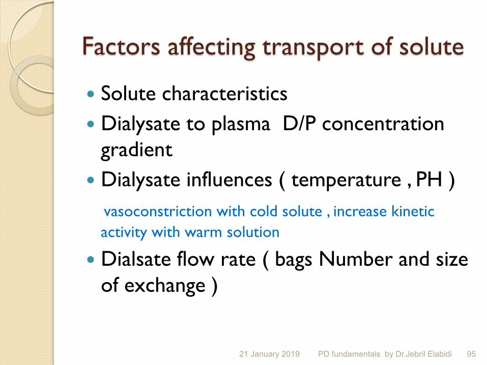
Factors affecting transport of solute
Solute characteristics
Dialysate to plasma D/P concentration gradient
Dialysate influences ( temperature , PH )
vasoconstriction with cold solute , increase kinetic
activity with warm solution
Dialsate flow rate ( bags Number and size of exchange )
21 January 2019 95 PD fundamentals by Dr.Jebril Elabidi

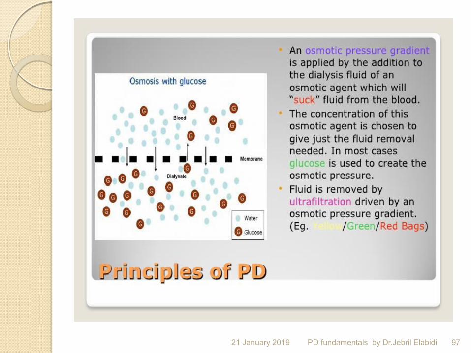
Principles of PD Osmosis is the process in which water moves
through a semi permeable membrane from an area of high water concentration (ie; low solute concentration) to an area of low water concentration (ie; higher solute concentration
21 January 2019 96 PD fundamentals by Dr.Jebril Elabidi
21 January 2019 97 PD fundamentals by Dr.Jebril Elabidi

21 January 2019 98 PD fundamentals by Dr.Jebril Elabidi

21 January 2019 99 PD fundamentals by Dr.Jebril Elabidi

Factors affecting osmosis Peritoneal surface area
Peritoneal permeability
Oncotic pressure
Hydrostatic pressure gradient
Osmotic pressure gradient
Hyperglycemia or hyperosmolar
21 January 2019 100 PD fundamentals by Dr.Jebril Elabidi

Factors affecting osmosis Prolonged dell time will decrease fluid removal
- osmotic pressure
dialysate is diluted
dextrose is absorbed
- lymphatic absorbtion continous
-transcapillary absorption occurs
Shorter dwell enhance fluid removal
21 January 2019 101 PD fundamentals by Dr.Jebril Elabidi

Solutes are transported across the membrane by diffusion.
The driving force is the concentration gradient between the PD fluid and the blood.
Waste products present in the blood
per fusing the peritoneum will diffuse from the blood vessels into the “cleaner” dialysis fluid.
21 January 2019 102 PD fundamentals by Dr.Jebril Elabidi
The dialysis fluid should be instilled for
4 to 6 hours.
When the dialysis fluid is drained from the abdominal cavity, it contains waste products and excess fluid extracted from the blood.
PD is most often applied and effective as a continuous therapy. In this way it is a more physiological treatment then Haemodialysis (HD)
21 January 2019 103 PD fundamentals by Dr.Jebril Elabidi

The osmotic agent normally used in PD fluid is glucose. Not an ideal osmotic agent, as it is readily transported
across the peritoneum.
Large concentration glucose creates a temporary osmotic gradient before being adsorbed into the blood.
The higher the glucose concentration, the larger the osmotic pressure, resulting in a larger fluid removal.
If PD exchanges are missed or dwell more than 6-8 hours, fluid may be gained by the patient rather then lost.
The Volume of dialysis solution administered is also important for the total fluid removal, as it will take longer for the concentration gradient to decline in a large volume of fluid.
21 January 2019 104 PD fundamentals by Dr.Jebril Elabidi
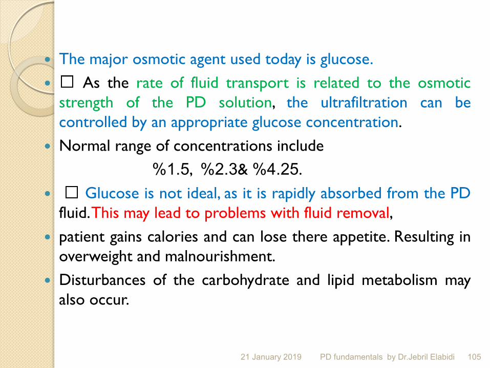
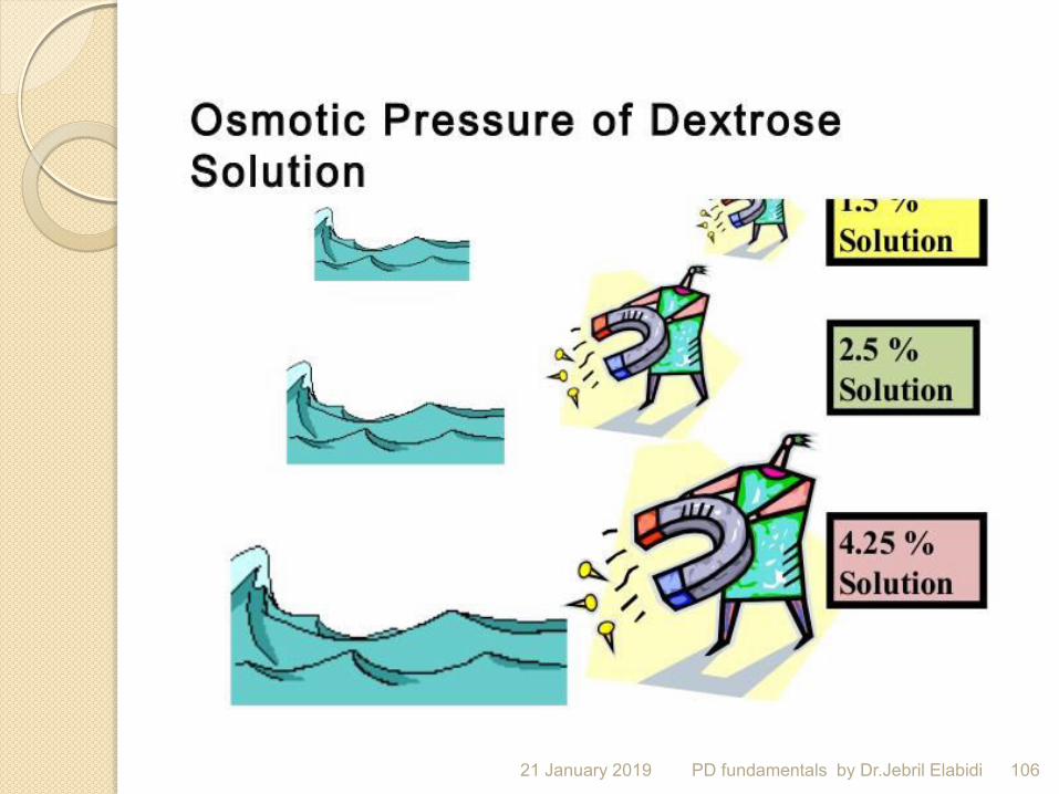
The major osmotic agent used today is glucose.
As the rate of fluid transport is related to the osmotic strength of the PD solution, the ultrafiltration can be controlled by an appropriate glucose concentration.
Normal range of concentrations include
1.5% , 2.3% & 4.25% .
Glucose is not ideal, as it is rapidly absorbed from the PD fluid. This may lead to problems with fluid removal,
patient gains calories and can lose there appetite. Resulting in overweight and malnourishment.
Disturbances of the carbohydrate and lipid metabolism may also occur.
21 January 2019 105 PD fundamentals by Dr.Jebril Elabidi
21 January 2019 106 PD fundamentals by Dr.Jebril Elabidi

peritoneal transport Two clinical end-points Clearance of solutes (by diffusion and convection)
Fluid removal (transcapillary UF – fluid absorption)
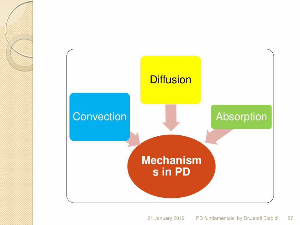
Peritoneal Transport Three Distinct Processes Diffusion Ultrafiltration Fluid Absorption
21 January 2019 107 PD fundamentals by Dr.Jebril Elabidi
What Happens with Solute Removal During a CAPD Dwell?
Diffusion is at a maximum, and urea and creatinine equilibration are fastest, in the first hour but become slower as the gradient lessons with timeBy 4 hours, urea is >90% and creatine > 65% equilibrated in most patients
Dialysate to plasma (D/P) ratios measure degree of equilibration at a given dwell time (e.g. D/P Urea, D/P Creatine)
21 January 2019 108 PD fundamentals by Dr.Jebril Elabidi

Natural functions of the peritoneum
Facilitate motion
Minimize friction
To conduct vessels and nerves to the viscera.
Solute transfer and exchange.
Regulation of fluid dynamics and UF.
21 January 2019 109 PD fundamentals by Dr.Jebril Elabidi
21 January 2019 110 PD fundamentals by Dr.Jebril Elabidi

21 January 2019 111 PD fundamentals by Dr.Jebril Elabidi

21 January 2019 112 PD fundamentals by Dr.Jebril Elabidi

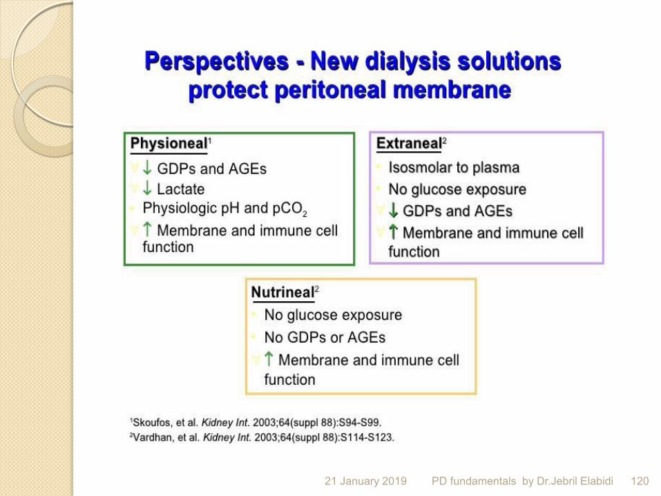
Research to find alternative osmotic agents has resulted in new products which are still not widely used. Amino acids are an interesting alternative as they provide nutritional supplement.
High molecular weight glucose polymer (extraneal/icodextrin) provide sustained ultrafiltration for long overnight dwells
21 January 2019 113 PD fundamentals by Dr.Jebril Elabidi

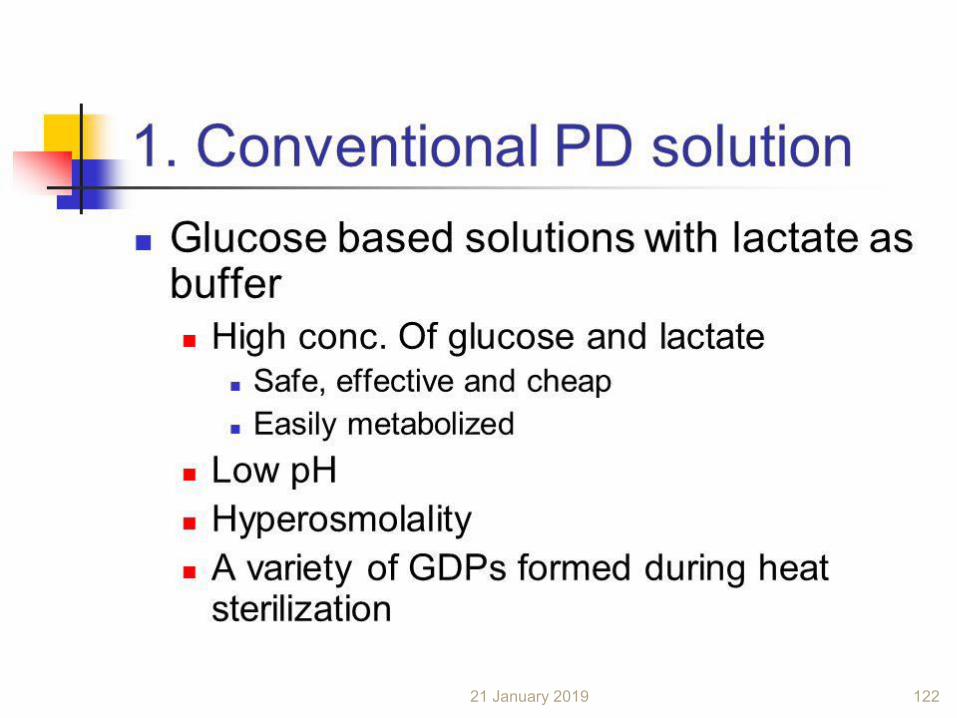
Gulcose degradation products GDPs
Effects of GDPs
1.inhibation of viability and function in different peritoneal cells
2. inflow pain during initial peritoneal contact
3.long term PD leads to morphological changes which leads to functional change
( UF failure )
21 January 2019 114 PD fundamentals by Dr.Jebril Elabidi
Biocompatibility Measurement of biocompatibility
parameters like CA 125
21 January 2019 115 PD fundamentals by Dr.Jebril Elabidi

Pure bicarbonate Buffer ( bicavera ) Less inflow pain
Improved biocompatibilty
More effective in correcting metabolic acidosis and prevents further bicarbonate loss
Pure bicarbonate obligatry in hepatic dysfunction
Preserves the homeostasis of the body
21 January 2019 116 PD fundamentals by Dr.Jebril Elabidi

The solutions The sugar solutions can be a problem for
diabetic patients and changes in therapy may be needed,
New solutions are being developed protein or starch .
Isodextrin (glucose polymer) produced by hydrolysis of starch, It’s effective in patients with poor UF even when using high concentration dextrose, but Very expensive and metabolized to maltose resulting in high maltose levels
21 January 2019 117 PD fundamentals by Dr.Jebril Elabidi

21 January 2019 118 PD fundamentals by Dr.Jebril Elabidi
Amino acids, it’s very expensive and only used if diffusion of amino acids from the dialysate into the blood compartment would he beneficial for nutrition, it’s use is limited to one bag per day as amino acids are metabolized to urea causing increased uremic symptoms.
21 January 2019 119 PD fundamentals by Dr.Jebril Elabidi
21 January 2019 120 PD fundamentals by Dr.Jebril Elabidi
21 January 2019 121 PD fundamentals by Dr.Jebril Elabidi
Absorbtion of glucose from peritoneal solutions
Solutions containing glucose (green) lead to significant glucose absorbtion
Solutions based on another osmotic agent (blue, violet) do not lead to glucose absorbtion, so decrease total daily glucose load).
PD
fun
da
menta
ls b
y D
r.Je
bril E
lab
idi
2.5 L
Physioneal
1.36%
2.5 L
Physioneal
1.36%
2.5 L
Physioneal
1.36%
2.5 L
Physioneal
3.86%
Glucose absorbed = 159 g/day
2.5 L
Physioneal
1.36%
2.5 L
Nutrineal
2.5 L
Physioneal
1.36%
2.5 L
Extraneal
Glucose absorbed = 50 g/day
21 January 2019 122
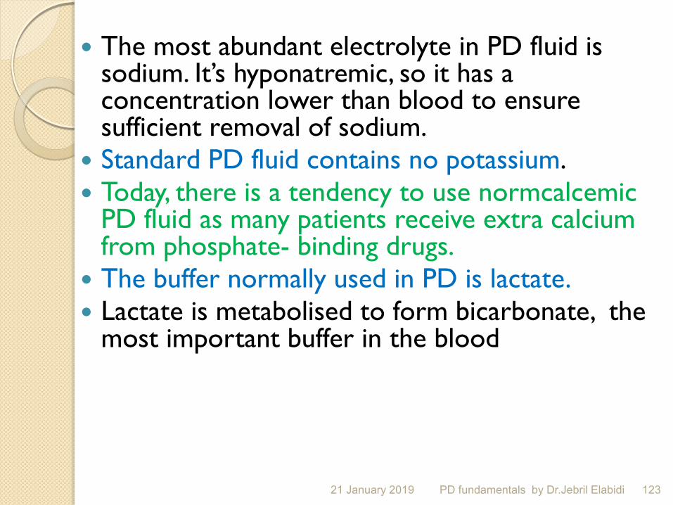
The most abundant electrolyte in PD fluid is sodium. It’s hyponatremic, so it has a concentration lower than blood to ensure sufficient removal of sodium.
Standard PD fluid contains no potassium. Today, there is a tendency to use normcalcemic
PD fluid as many patients receive extra calcium from phosphate- binding drugs.
The buffer normally used in PD is lactate. Lactate is metabolised to form bicarbonate, the
most important buffer in the blood
21 January 2019 123 PD fundamentals by Dr.Jebril Elabidi

If hypercatabolic state or hyperkalemia the dwell time is shortened to 10 minutes for faster correction.
If extra ultrafiltration is necessary 25 to 100ml of high concentration dextrose (25% or50%) is added into each PD fluid bottle/bag before instilling the fluid into the abdomen.
Blood glucose needs to be monitored every 4 hours in diabetics and insulin is to be administered .
21 January 2019 124 PD fundamentals by Dr.Jebril Elabidi

To increase the efficiency of PD and help the patient with the exchanges, a machine can be used, known as Automated Peritoneal Dialysis of APD.
Advantages of APD v CAPD are
1) higher clearance of solutes, as higher volumes can be used
2) better fluid removal, as shorter dwell time can be used
3) more freedom during the daytime as no exchanges need to be made.
Drawbacks of APD are that of a higher cost and
portability. Treatment Modes CAPD/APD
21 January 2019 125 PD fundamentals by Dr.Jebril Elabidi

Complications
Infectious
Non-infectious
21 January 2019 126 PD fundamentals by Dr.Jebril Elabidi

Peritonitis Peritonitis can occur without cloudy effluent and can
present with other symptoms, such as abdominal pain, fever, constipation, and diarrhea.
Likewise, cloudy effluent does not necessarily indicate infectious peritonitis.
Nevertheless, patients presenting with cloudy effluent should be presumed to have peritonitis, which is confirmed by a cell count, blood differential test, and blood culture of the peritoneal fluid.
An effluent white blood cell count of 100/μL after a 2-hour dwell with at least 50% neutrophilic cells indicates inflammation, with peritonitis as the most likely cause.
21 January 2019 127 PD fundamentals by Dr.Jebril Elabidi
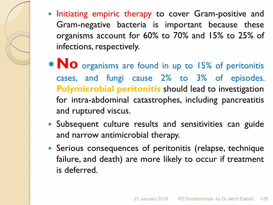
Initiating empiric therapy to cover Gram-positive and Gram-negative bacteria is important because these organisms account for 60% to 70% and 15% to 25% of infections, respectively.
No organisms are found in up to 15% of peritonitis
cases, and fungi cause 2% to 3% of episodes. Polymicrobial peritonitis should lead to investigation for intra-abdominal catastrophes, including pancreatitis and ruptured viscus.
Subsequent culture results and sensitivities can guide and narrow antimicrobial therapy.
Serious consequences of peritonitis (relapse, technique failure, and death) are more likely to occur if treatment is deferred.
21 January 2019 128 PD fundamentals by Dr.Jebril Elabidi

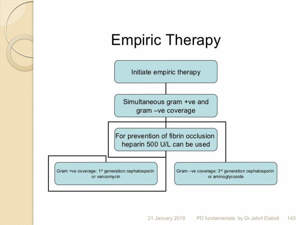
Guidelines from the ISPD offer a thoughtful approach to therapy.
First- and third-generation cephalosporins are typically used for empiric coverage
unless the patient had prior infections with resistance to first-generation cephalosporins or the institution has high rates of resistance to them.
21 January 2019 129 PD fundamentals by Dr.Jebril Elabidi

In either case, vancomycin can be used with a third-generation cephalosporin. Patients with minimal RKF (defined as urine output < 100 mL/d) can use aminoglycosides.
Intraperitoneal administration is recommended unless the patient is hospitalized and acutely ill.
Then, intravenous administration should be considered.
21 January 2019 130 PD fundamentals by Dr.Jebril Elabidi

The treatment course should be continued for 2 weeks, except in the case of Saureus, Enterococcus species, Psuedomonas/Stenotrophomonas species, or multiorganism peritonitis, which require
3 weeks of therapy.
If No organisms are found, Gram-negative coverage can be discontinued at 96 hours if the patient is clinically improving, and Gram-positive coverage can continue for a total of 2 weeks.
21 January 2019 131 PD fundamentals by Dr.Jebril Elabidi

Treatment of fungal infections usually fails, and early catheter removal is a prudent way to proceed.
21 January 2019 132 PD fundamentals by Dr.Jebril Elabidi
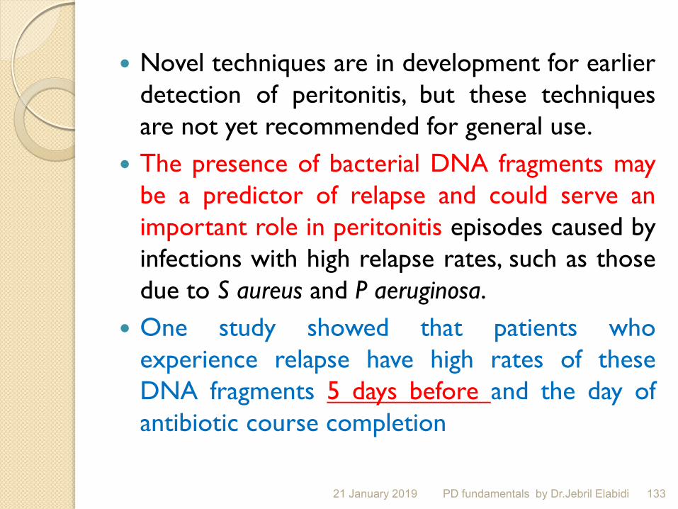
Novel techniques are in development for earlier detection of peritonitis, but these techniques are not yet recommended for general use.
The presence of bacterial DNA fragments may be a predictor of relapse and could serve an important role in peritonitis episodes caused by infections with high relapse rates, such as those due to S aureus and P aeruginosa.
One study showed that patients who experience relapse have high rates of these DNA fragments 5 days before and the day of antibiotic course completion
21 January 2019 133 PD fundamentals by Dr.Jebril Elabidi
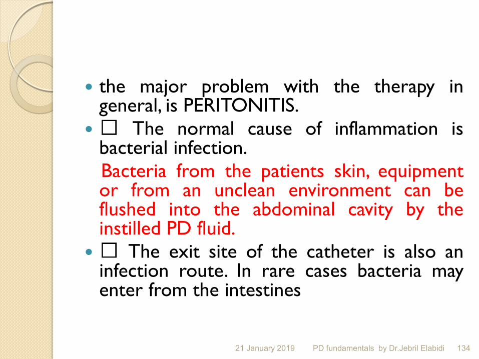
the major problem with the therapy in general, is PERITONITIS.
The normal cause of inflammation is bacterial infection.
Bacteria from the patients skin, equipment or from an unclean environment can be flushed into the abdominal cavity by the instilled PD fluid.
The exit site of the catheter is also an infection route. In rare cases bacteria may enter from the intestines
21 January 2019 134 PD fundamentals by Dr.Jebril Elabidi

PERITONITIS MANAGEMENT
Initial symptoms may include;
diarrhoea, vomiting, nausea, abdominal pain,
mental confusion or feeling unwell
21 January 2019 135 PD fundamentals by Dr.Jebril Elabidi

COLLECT DRAINED BAG
*See additional resources (pink section) for drainage instructions
* Send entire bag for urgent MC&S (including WCC differential) and Fungal elements
21 January 2019 136 PD fundamentals by Dr.Jebril Elabidi

CLEAR BAG CLOUDY BAG Intraperitoneal (IP) Antibiotics
21 January 2019 137 PD fundamentals by Dr.Jebril Elabidi

How does bacteria gain entry into peritoneal cavity? During catheter connection
Tracking around the catheter around the exit site Across the bowel wall; diverticulosis li
Transvaginal
Rarely hematogenous
Bacteremia can cause peritoneal seeding and peritonitis Peritonitis rarely causes bacteremia
Use antibiotic prophylaxis for anticipated bacteremia during procedures like dental work, colonoscopy, GU instrumentation
Drain effluent before colonoscopy or colposcopy
21 January 2019 138 PD fundamentals by Dr.Jebril Elabidi

Pathophysiology Multiple connection and disconnections from the
transfer set
Presence of non-physiologic fluid in the peritoneal cavity may impair host defenses
High glucose concentration, low pH and hyperosmolality dilute resident peritoneal macrophage and cytokine levels
Constant removal of macrophage and cytokines during each exchange
Alteration of mesothelial cell defense properties over time
21 January 2019 139 PD fundamentals by Dr.Jebril Elabidi

Diagnosis
At least two of the following three features
1. Peritoneal fluid leucocytosis; >100 cells/mm 3 and at least 50% PMNs
2. Abdominal pain
3. Positive culture of the dialysis effluent
21 January 2019 140 PD fundamentals by Dr.Jebril Elabidi

Specimen collection and processing Effluent fluid sent for cell count with differential,
culture and gram stain
Collection of effluent: 50ml of effluent is centrifuged for 15 min followed by re-suspension of sediment in 3-5 ml of sterile saline and inoculation to media
Dwell time of at least 2 – 4 hours before effluent collection
If peritoneal cavity is dry, 1L of dialysate infused to dwell for at least 1 – 2 hours
Peripheral blood cultures usually not necessary
21 January 2019 141 PD fundamentals by Dr.Jebril Elabidi

Treatment Non-antimicrobial measures
1. Heparin 500 units/L can be used to lyse or prevent fibrin clots when dialysate remains cloudy
2. Pain control
Dwell time
Long dwell exchanges (4-6 hrs) when compared with short dwells are associated with higher number of functional macrophages
Membrane properties: changes during peritonitis
Patients may transiently become rapid transporters, thereby requiring the use of hypertonic glucose or shorter dwells
Alternatively, icodextrin may be helpful 21 January 2019 142 PD fundamentals by Dr.Jebril Elabidi
21 January 2019 143 PD fundamentals by Dr.Jebril Elabidi

Empiric therapy 1 st generation cephalosporin: cefazolin or cephalothin
Vancomycin used at centers with high rate of MRSA 3 rd generation cephalosporin: ceftazidime or cefepime Short term use of aminoglycoside is safe and does not diminish residual renal function
Aztreonam can be used in cephalosporin allergic patients
Monotherapy with imipenem/cilastatin is possible.
One study with 102 patients randomly assigned to either imipenem/cilastatin or cefazolin + ceftazidime showed similar outcomes in both groups
21 January 2019 144 PD fundamentals by Dr.Jebril Elabidi
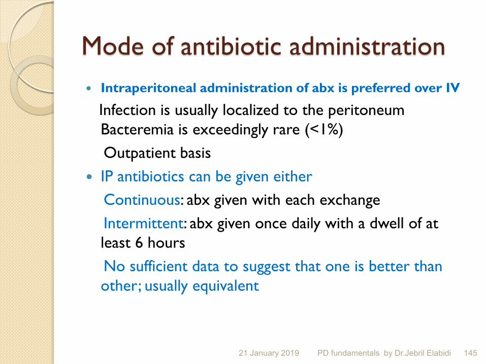
Mode of antibiotic administration Intraperitoneal administration of abx is preferred over IV
Infection is usually localized to the peritoneum Bacteremia is exceedingly rare (<1%)
Outpatient basis
IP antibiotics can be given either
Continuous: abx given with each exchange
Intermittent: abx given once daily with a dwell of at least 6 hours
No sufficient data to suggest that one is better than other; usually equivalent
21 January 2019 145 PD fundamentals by Dr.Jebril Elabidi

21 January 2019 146 PD fundamentals by Dr.Jebril Elabidi
21 January 2019 147 PD fundamentals by Dr.Jebril Elabidi

21 January 2019 148 PD fundamentals by Dr.Jebril Elabidi

21 January 2019 149 PD fundamentals by Dr.Jebril Elabidi

Other causes of peritonitis Fungal peritonitis
1. Catheter removal
2. Flucytosine 1gm/day + Fluconazole 200 mg/day PO for 10 days after catheter removal
Mycobacterial peritonitis
M. tuberculosis: Rifampin + INH (for 12 months) + pyrazinamide + ofloxacin (3 months) Consider catheter removal
21 January 2019 150 PD fundamentals by Dr.Jebril Elabidi

21 January 2019 151 PD fundamentals by Dr.Jebril Elabidi

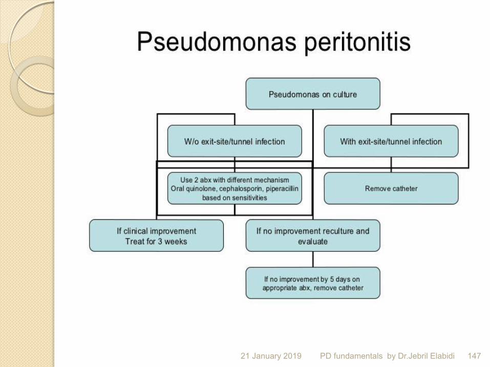
Catheter removal in peritonitis patients ISPD guidelines recommend catheter removal in
following
Relapsing peritonitis: another episode with same species that caused the preceding episode within 4 weeks of completing abx
Refractory peritonitis: failure to respond to abx in 5 days
Refractory catheter infection
Fungal peritonitis , Fecal peritonitis
Peritonitis associated with intra-abdominal pathology Consideration to catheter removal in mycobacterial and multiple enteric organisms peritonitis
21 January 2019 152 PD fundamentals by Dr.Jebril Elabidi

21 January 2019 153 PD fundamentals by Dr.Jebril Elabidi
Eosinophilic peritonitis Relatively new PD catheter
Effluent is cloudy w/o abdominal pain
PD differential count: eosinophils ++
Effluent culture: no growth
Cause: ?immune reaction to catheter
Treatment
Usually self limited, goes away in few days
Some reports of benefit with IP steroids
21 January 2019 154 PD fundamentals by Dr.Jebril Elabidi

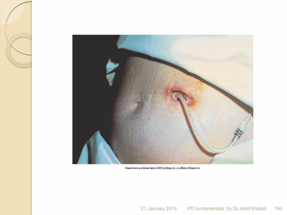
Exit-site/Tunnel infection Exit-site infection: presence of purulent
discharge with or w/o erythema of the skin at catheter-epidermal interface.
Tunnel infection: usually occult but may be present with erythema, edema or tenderness over subcutaneous path.
Rarely occurs alone.
Staph aureus and pseudomonas exit site infections are often associated with concomitant tunnel infection
21 January 2019 155 PD fundamentals by Dr.Jebril Elabidi
21 January 2019 156 PD fundamentals by Dr.Jebril Elabidi

21 January 2019 157 PD fundamentals by Dr.Jebril Elabidi

Non infectious complications
21 January 2019 158 PD fundamentals by Dr.Jebril Elabidi

Outflow failure Incomplete recovery of instilled dialysate Unable to
remove dialysate from peritoneal cavity
Fluid is no longer in peritoneal cavity
Incidence: 5-20%
Etiologies
Constipation (anytime)
Catheter malposition (days)
Intraluminal catheter occlusion by thrombus.
Extraluminal catheter occlusion by omentum or adhesions (weeks)
Kinking (soon after placement, positional)
Loss of dialysate from peritoneal cavity 21 January 2019 159 PD fundamentals by Dr.Jebril Elabidi
Treatment Constipation
More than half of the cases are cured with releif of constipation
Laxatives, stool softeners, suppositories or enema
Fibrin clot Heparin 500 units/L of dialysate for lysis
Urokinase – instilled in catheter for 1 hour and then removed
21 January 2019 160 PD fundamentals by Dr.Jebril Elabidi

Treatment Malpositioned catheter
Fluoroscopy with stiff wire manipulation Redirection either laproscopically or surgically Replace catheter if not successful
Catheter kinking Usually requires catheter replacement
Superficial cuff removal if kinking is due to placement of the catheter cuffs too close to each other
Abdominal exploration may be necessary for catheter redirection, omentectomy or adhesiolysis or catheter replacement
21 January 2019 161 PD fundamentals by Dr.Jebril Elabidi
Pericatheter leakage Early after placement
Increased intra-abdominal pressure on CAPD 2ry to increased activity
Weak abdominal wall (pervious surgeries, pregnancies)
High dialysate volumes
Catheter placement techniques: poor evidence of technique with incidence Peritoneoscopically placed catheters may be better
Double cuff catheters are considered less likely to leak
21 January 2019 162 PD fundamentals by Dr.Jebril Elabidi

Pericatheter leakage Treatment Reduce physical activity
Reduce dialysate volumes
Conversion to cycler
Temporary conversion to HD
If conservative measures fails then surgical repair of deep cuff or catheter replacement
21 January 2019 163 PD fundamentals by Dr.Jebril Elabidi
Abdominal X- ray/Peritoneography Plain abdominal X-rays are performed to evaluate for
malposition of the peritoneal catheter.
The normal position of catheter is in the pelvic gutter.
In patients with normal inflow of dialysate but problems with outflow, the most common underlying cause is constipation. However, if symptoms do not improve after resumption of normal bowel movements, abdominal X-ray should be done to assess catheter position.
For peritoneography, the initial X ray is taken, then 100-200 ml non-ionic contrast is mixed into a 2L dialysate bag and instilled in the patient. The patient changes positions to mix dialysate and a repeat X ray is taken.
Can be used to diagnose an entrapped catheter or a peritoneal leak.
21 January 2019 164 PD fundamentals by Dr.Jebril Elabidi

Monitoring patients on peritoneal dialysis
21 January 2019 PD fundamentals by Dr.Jebril Elabidi 165

PD adequacy
21 January 2019 PD fundamentals by Dr.Jebril Elabidi 166

21 January 2019 167 PD fundamentals by Dr.Jebril Elabidi

British Renal Association: A peritoneal equilibration test (PET)
should be performed after 4–8 weeks on dialysis, and when clinically indicated,
e.g. when biochemical indices or loss of ultrafiltration raise suspicion of changes in peritoneal transport characteristics, or when therapy is changed to APD.
21 January 2019 PD fundamentals by Dr.Jebril Elabidi 168

The peritoneal function test (PFT), The test allows assessment of total delivered
therapy for urea and creatinine, protein and calorie nutrition, fluid balance, and peritoneal transport.
The results are expressed as Pt50 or the time required for a solute to achieve 50% equilibration between dialysate and plasma.
The PFT has been extensively used as part of a kinetic modeling program and the data can be displayed for individual patients, clinics, or
regional groups of dialysis centers. 21 January 2019 PD fundamentals by Dr.Jebril Elabidi 169

The peritoneal dialysis capacity (PDC) program was designed to measure
transperitoneal passage of fluid and solutes under normal conditions with a non-invasive test.
The PDC is based on the three-pore-model of Rippe et al13,14.
It describes the peritoneal membrane characteristics
21 January 2019 PD fundamentals by Dr.Jebril Elabidi 170

The dialysis adequacy and transport test (DATT) was introduced by Rocco et al. in an
attempt to develop
an easier test for classifying peritoneal transport type
21 January 2019 PD fundamentals by Dr.Jebril Elabidi 171
The accelerated peritoneal examination (APEX) test was designed by Verger et al.
using a similar protocol as their initial equilibration test with 3.86% glucose solution, but summarizes in a single number the peritoneal permeability for both glucose and urea.
It represents the time at which the glucose and urea equilibration curves cross.
21 January 2019 PD fundamentals by Dr.Jebril Elabidi 172

Monitoring patients on peritoneal dialysis Total (peritoneal plus residual renal)
weekly Kt/Vurea and
CCr measurement and a peritoneal equilibration test (PET) should be performed approximately 4 weeks after dialysis commencement, but no sooner than 2 weeks after dialysis commencement because of unstable peritoneal permeability at this stage (Level III evidence).
21 January 2019 173 PD fundamentals by Dr.Jebril Elabidi

The standard peritoneal permeability analysis (SPA)
is a more sophisticated way to assess
peritoneal function
It uses intraperitoneally administered dextran 70 to study fluid kinetics during a 4-hour dwell using an infusion volume consistent with the patient’s usual prescription
21 January 2019 PD fundamentals by Dr.Jebril Elabidi 174
Residual renal Kt/V and CCr measurements should be repeated at the following times:
i. every 2 months in automated peritoneal dialysis (APD) patients and every 4–6 months in continuous ambulatory peritoneal dialysis (CAPD) patients who are dependent on residual renal function to achieve small solute clearance targets, particularly those with a small ‘safety margin’ (e.g. patients treated with ‘incremental’ rather than ‘full-dose’ peritoneal dialysis), 21 January 2019 175 PD fundamentals by Dr.Jebril Elabidi

ii. following a history of a substantial decline in urine output ,
iii. following unexplained fluid overload, and
iv. with clinical or biochemical evidence of worsening uraemia.
21 January 2019 176 PD fundamentals by Dr.Jebril Elabidi
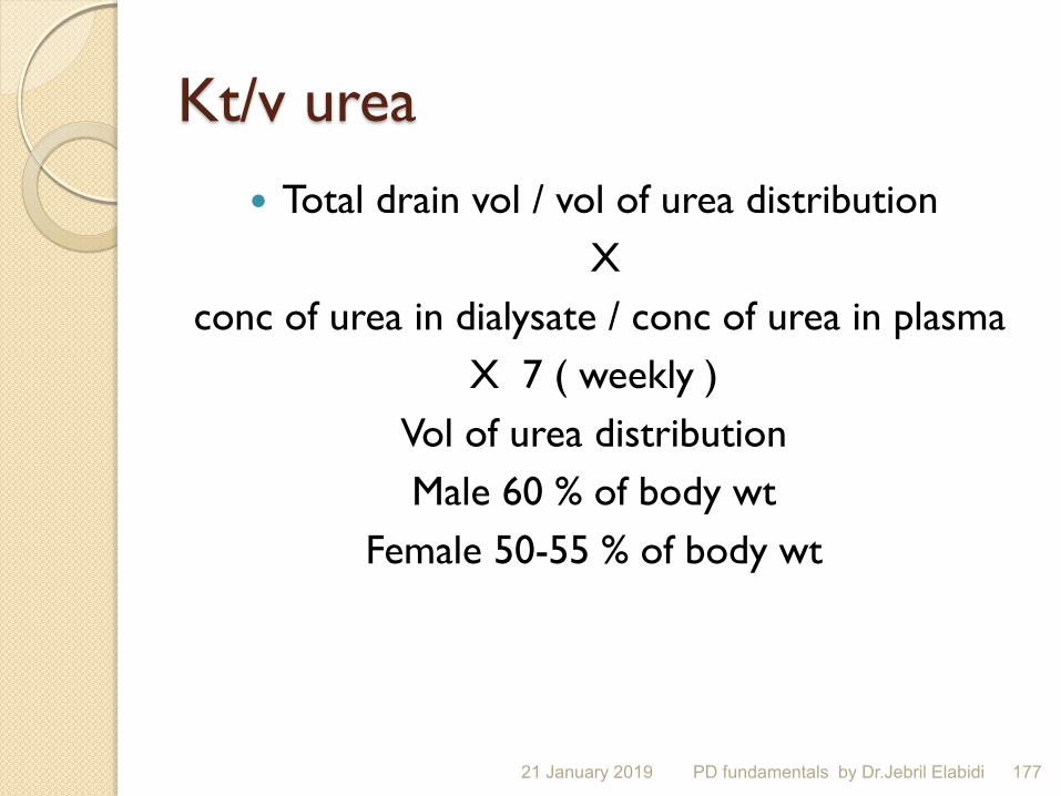
Kt/v urea Total drain vol / vol of urea distribution
X
conc of urea in dialysate / conc of urea in plasma
X 7 ( weekly )
Vol of urea distribution
Male 60 % of body wt
Female 50-55 % of body wt
21 January 2019 PD fundamentals by Dr.Jebril Elabidi 177

21 January 2019 PD fundamentals by Dr.Jebril Elabidi 178

21 January 2019 179 PD fundamentals by Dr.Jebril Elabidi

21 January 2019 180 PD fundamentals by Dr.Jebril Elabidi
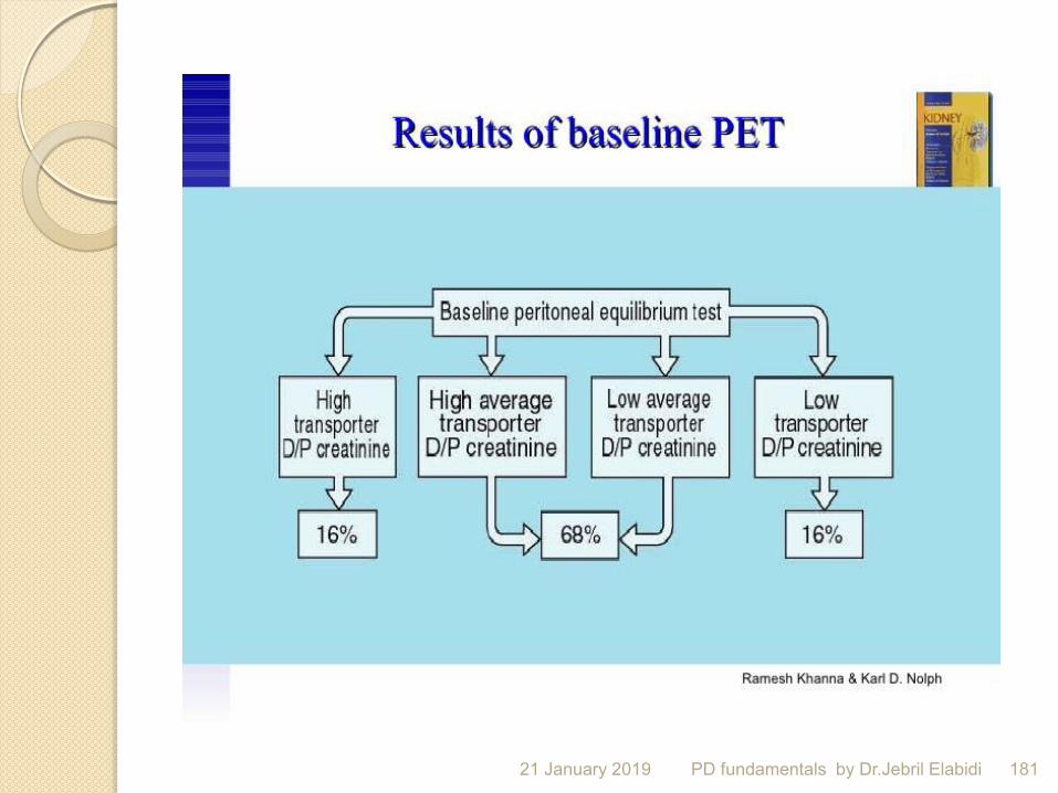
21 January 2019 181 PD fundamentals by Dr.Jebril Elabidi
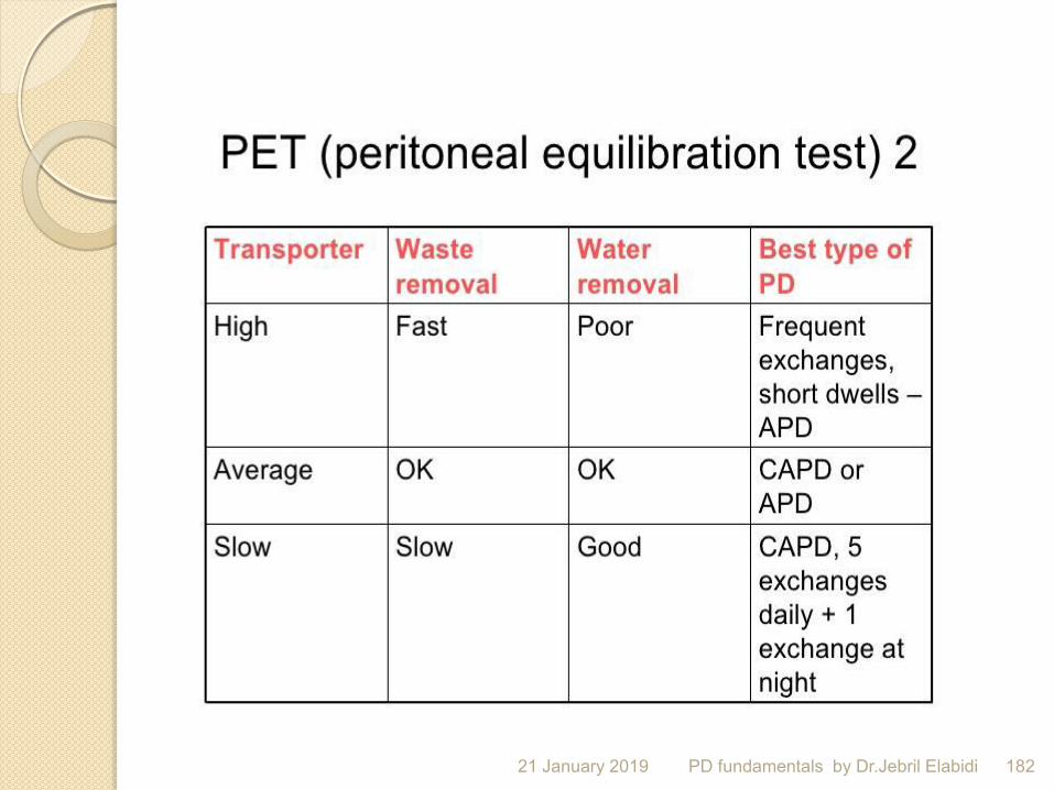
21 January 2019 182 PD fundamentals by Dr.Jebril Elabidi

21 January 2019 183 PD fundamentals by Dr.Jebril Elabidi

21 January 2019 PD fundamentals by Dr.Jebril Elabidi 184
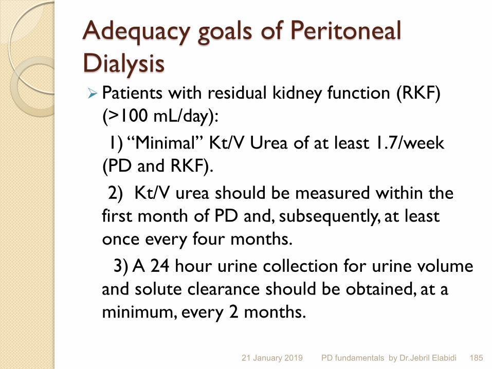
Adequacy goals of Peritoneal Dialysis Patients with residual kidney function (RKF)
(>100 mL/day):
1) “Minimal” Kt/V Urea of at least 1.7/week (PD and RKF).
2) Kt/V urea should be measured within the first month of PD and, subsequently, at least once every four months.
3) A 24 hour urine collection for urine volume and solute clearance should be obtained, at a minimum, every 2 months.
21 January 2019 185 PD fundamentals by Dr.Jebril Elabidi
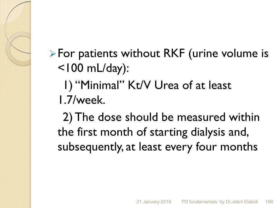
For patients without RKF (urine volume is <100 mL/day):
1) “Minimal” Kt/V Urea of at least 1.7/week.
2) The dose should be measured within the first month of starting dialysis and, subsequently, at least every four months
21 January 2019 186 PD fundamentals by Dr.Jebril Elabidi
Why different Kt/V in PD vs HD BUN may not be a good index uremic
toxin and middle molecule clearance may be different between PD and HD
Peak BUN concentration may be related to uremic symptoms and maybe to survival?
21 January 2019 187 PD fundamentals by Dr.Jebril Elabidi

21 January 2019 188 PD fundamentals by Dr.Jebril Elabidi

21 January 2019 PD fundamentals by Dr.Jebril Elabidi 189

21 January 2019 PD fundamentals by Dr.Jebril Elabidi 190

21 January 2019 PD fundamentals by Dr.Jebril Elabidi 191

Initial prescription Fill volume related to body size
21 January 2019 PD fundamentals by Dr.Jebril Elabidi 192

DOQI guidelines for initial prescription GFR More than 2 ml/min
21 January 2019 PD fundamentals by Dr.Jebril Elabidi 193
Cycler CAPD Patient size
4 C X 2L 4 cycles x 2 L BSA < 1.7 m2
4 X 2.5 L 4 x 2.5 L BSA 1.7 – 2 m2
4 X 3 L 4 X 3 L BSA > 2.0m2

East , west and south , LIBYAN nephrologists are together .
21 January 2019 PD fundamentals by Dr.Jebril Elabidi 194