peritrophic matrix of phlebotomus duboscqi and its ... · regular article peritrophic matrix of...
TRANSCRIPT

REGULAR ARTICLE
Peritrophic matrix of Phlebotomus duboscqi and its kineticsduring Leishmania major development
Jovana Sádlová & Petr Volf
Received: 5 December 2008 /Accepted: 31 March 2009 /Published online: 27 May 2009# The Author(s) 2009. This article is published with open access at Springerlink.com
Abstract Light microscopy of native preparations, histolo-gy, and electron microscopy have revealed that Phlebotomusduboscqi belongs to a class of sand fly species with promptdevelopment of the peritrophic matrix (PM). Secretion ofelectron-lucent fibrils, presumably chitin, starts immediatelyafter the ingestion of a blood meal and, about 6 h later, isfollowed by secretion of amorphous electron-dense compo-nents, presumably proteins and glycoproteins. The PMmatures in less than 12 h and consists of a thin laminarouter layer and a thick amorphous inner layer. No differenceshave been found in the timing of the disintegration of the PMin females infected with Leishmania major. In both groups offemales (infected and uninfected), the disintegration of thePM is initiated at the posterior end. Although parasites arepresent at high densities in the anterior part of the blood mealbolus, they escape from the PM at the posterior end only.These results suggest that L. major chitinase does not havean important role in parasite escape from the PM. Promas-tigotes remain in the intraperitrophic space until the PM isbroken down by sand-fly-derived chitinases and only thenmigrate anteriorly. Disintegration of the PM occurs simulta-neously with the morphological transformation of parasitesfrom procyclic forms to long nectomonads. A novel role isascribed to the anterior plug, a component of the PMsecreted by the thoracic midgut; this plug functions as atemporary barrier to stop the forward migration of nectomo-nads to the thoracic midgut.
Keywords Chitinase .Midgut . Digestion . Peritrophicmatrix .Phlebotomus duboscqi (Insecta) . Leishmania major
Introduction
The peritrophic matrix (PM, previously referred to as theperitrophic membrane) is an acellular chitin-containingenvelope, which, in most insects separates the gut lumenfrom the midgut epithelium; it is composed of chitin,proteins, and proteoglycans (Peters 1992; Lehane 1997). Innematoceran Diptera, including sand flies, females producea type 1 PM, which is secreted by the entire midgut indirect response to the distension of the midgut caused byblood feeding (Jacobs-Lorena and Oo 1996).
The role of the PM is associated with the protection ofthe midgut epithelium against pathogens and abrasion byfood particles and with the compartmentalization ofdigestion between the ecto- and endoperitrophic spaces(Peters 1992; Lehane 1997). In mosquitoes and otherhematophagous insects, the PM also performs a centralrole in heme detoxification (Pascoa et al. 2002). Althoughelectron-microscopy studies on the PM of sand flies havebeen published (Gemetchu 1974; Blackburn et al. 1988;Walters et al. 1993, 1995; Andrade-Coelho et al. 2001;Secundino et al. 2005), the data presented by the differentauthors are often fragmentary. Moreover, descriptions ofPM formation vary, even in studies when two groups haveworked on the same sand fly species (Reznik andKuznetsova 1983; Blackburn et al. 1988).
Detailed knowledge of the formation and morphology ofthe sand fly PM is important as sand flies are vectors ofLeishmania parasites, causative agents of serious humandiseases. In the early phase of infection, when Leishmaniaare vulnerable to proteolytic damage, the PM is supposed to
Cell Tissue Res (2009) 337:313–325DOI 10.1007/s00441-009-0802-1
This work was supported by the Ministry of Education of the CzechRepublic (projects MSM0021620828 and LC06009).
J. Sádlová : P. Volf (*)Department of Parasitology, Charles University,Vinicna 7,Prague 2, Czech Republice-mail: [email protected]

protect the parasites against the rapid diffusion of digestiveenzymes (Pimenta et al. 1997). On the other hand,following digestion of the blood meal, the PM canadversely influence the development of Leishmania pro-mastigotes by creating a potential physical barrier thatprevents their escape from the endoperitrophic space (for areview, see Bates and Rogers 2004). In unnatural parasite-vector combinations, Feng (1951) and Walters et al. (1992)have reported that the PM does not break down duringblood digestion, and that parasites are excreted.
Leishmania promastigotes produce chitinase (Shakarianand Dwyer 1998, 2000), and experiments with chitinase-overexpressing mutants of L. mexicana have confirmed thedamaging effects of the enzyme on the stomodeal valve ofthe sand fly (Rogers et al. 2008). The role of chitinase inparasite interactions with the sand fly PM is still not clear.Schlein et al. (1991) have reported that L. major promas-tigotes cause the disintegration of the anterior end of thePM of P. papatasi by means of their own chitinase.However, in most vector-parasite combinations, parasiteescape from the PM seems to coincide with the breakdownof the PM by sand-fly-derived chitinases (Ramalho-Ortigaoand Traub-Cseko 2003; Ramalho-Ortigao et al. 2005).
In this study, we describe the PM in the natural vector-parasite pair of Phlebotomus duboscqi Neveu-Lemaireinfected with Leishmania major Yakimoff et Schokhor.We have studied the morphology, ultrastructure, and timingof assembly and degradation of the PM. In contrast toprevious descriptions, we have found that Leishmaniaparasites do not cause the disintegration of the PM at theanterior end but migrate through posterior openings thatexist in the PM even in non-infected females. In addition,we suggest a novel role for the anterior plug (AP), a PMprojection separating the abdominal (AMG) and thoracic(TMG) regions of the midgut. This structure seems totemporarily limit the anterior migration of Leishmania intothe TMG.
Materials and methods
Sand flies
The colony of P. duboscqi was maintained on 50% sucroseat 26°C under a 14-h light/10-h dark photoperiod (for moredetails, see Benková and Volf 2007).
Parasites and sand fly infections
The Leishmania major strain LV561 (LRC-L137; MHOM/IL/1967/Jericho-II), which is highly virulent in BALB/cmice (Sádlová et al. 1999), and the green-fluorescentprotein (GFP) line from the same strain were kindly
provided by Dr. J. Votýpka and Dr. D. Folková (CharlesUniversity, Prague). BALB/c mice were inoculated with107 promastigotes in the rump to generate amastigotes forsand fly infections. The lesions from infected animals weredissected and homogenized in Schneider’s Drosophilamedium with gentamicin (40 mg/100 ml). Sand fly femaleswere fed through a chick-skin membrane on a suspensionof amastigotes mixed 1:1 with rabbit erythrocytes. The finalconcentration of parasites was 106 (wild strain, GFP line) or107 (GFP line) amastigotes/ml. Engorged sand flies weremaintained under the same conditions as those describedabove.
Light microscopy
A total of 186 blood-fed females were dissected at varioustime intervals after feeding on anesthetized mice; 6 within10 min (T0) post-blood-meal (PBM) and 20 for each of thefollowing times PBM: 1, 3, 6, 12, 24, 48, 72, 96, 120 h(T1–T120). In addition, 257 females fed through a chick-skin membrane were dissected for Leishmania infections atT48, T72, and T96. Dissections were carried out in isotonicsaline solution, and then the gut from each fly was brieflywashed in distilled water for better isolation of the PM(Gemetchu 1974). Slides were observed under an OlympusBX51 microscope with Nomarski contrast and photo-graphed with an Olympus D70 camera and software (DPController). Parasite loads were graded according toMyskova et al. (2008) as light (<100 parasites/gut),moderate (>100 and <1000 parasites/gut), or heavy(>1000 parasites/gut).
Morphometry of parasites
Gut smears of females infected with L. major weredissected at T72 and T96, fixed with methanol, stainedwith Giemsa, and examined under the light microscopewith an oil-immersion objective. Three smears were madefor each of four categories of PM (intact, slightlydisintegrated, opened/heavily disintegrated, and defecated),and 40 randomly selected promastigotes were measured ineach smear. The body length, flagellar length, and bodywidth were measured, and the position of the kinetoplast inrelation to the nucleus was noted. Five morphologicalforms were distinguished based on Čiháková and Volf(1997) and Rogers et al. (2002): (1) procyclic promastigotes(body length <14 µm and flagellar length ≤ body length),(2) elongated nectomonads (body length ≥14 µm), (3) shortnectomonads (leptomonads; body length <14 µm andflagellar length > body length but ≤ twice body length),(4) metacyclic promastigotes (body length <14 µm andflagellar length > twice body length), (5) paramastigotes(kinetoplast lateral to the nucleus).
314 Cell Tissue Res (2009) 337:313–325

Histology
Unfed females and females at T0–T96 time intervals PBMwere fixed at 4°C in AFA solution (formaldehyde:ethanol:acetic acid:distilled water, 1.5:12.5:1:10). After beingwashed in phosphate-buffered saline (pH 7.6) and 70%ethanol, the samples were embedded in JB-4 following themanufacturer’s protocol (Polysciences). Histological sec-tions (4–6 µm thick) were stained with Ehrlich’s acidhematoxylin and 0.2% eosin, mounted on glass slides withPlastic UV Mount (Polysciences), and observed under anOlympus BX51 microscope. Three to five females wereused for each of the time intervals.
Electron microscopy
Unfed females or gut samples and blood-fed females or gutsamples were dissected at T0–T72, fixed in modifiedKarnovsky fixative (Karnovsky 1965), and post-fixed witha 2% osmium tetroxide solution (both at 4°C). The sampleswere dehydrated in ascending concentrations of ethanol andembedded in Poly/Bed 812/Araldite 502 (Polysciences).Semithin sections (400 nm) and ultrathin sections (70 nm)were obtained by using a Reichert-Jung Ultracut Eultramicrotome. Semithin sections were stained with tolu-idine blue for light microscopy. Ultrathin sections stainedwith uranyl acetate and lead citrate (Reynolds 1963) wereobserved and photographed with a JEOL 1011 transmissionelectron microscope. Three to seven females were used foreach of the time intervals.
Statistical analysis
PM development in the groups of infected and uninfectedfemales was compared by using the Chi-squared test.Measurements of parasites and the representation ofmorphological forms were compared by using an analysisof variance and the Chi-squared test, respectively. Allstatistical evaluations were performed with statisticalsoftware (SPSS version 12).
Results
Development of the PM: light microscopy
Immediately after blood feeding (T0), the blood meal wasspread throughout the TMG and AMG with no apparentagglutination of erythrocytes (data not shown). Within 1 h,the TMG was free of blood, and the whole blood meal wasconcentrated in the AMG. Erythrocytes were agglutinatedto a compact dark central bolus surrounded by a lightperiphery formed from extruded blood plasma. In some of
the flies, a sharp line separating the blood bolus from boththe plasma in the periphery and the midgut epitheliumindicated the first trace of the future PM (Fig. 1a).
At T3, a thin transparent PM had formed in half of thedissected females (Figs. 1b, 2). In some cases, it did notcover the whole surface of the blood bolus but only theanterior part. At T6, the PM was strong enough to enabledissection of the entire peritrophic sac in all studied females(Fig. 2). By T24, the PM darkened as it became encrustedwith heme, a product of erythrocyte degradation (data notshown). This process affected both the anterior and posteriorends of the PM in most of the flies (85% of females) and lessfrequently affected only the anterior (5%) or posterior ends(10%). At T48, further encrustation with heme led to adenser PM with a paler median region (Fig. 1c).
By T72, the erythrocytes had been disintegrated, and thePM showed a reduced volume and a folded surface and beganlosing its dark color in some flies. In most females, the PMwas only slightly disintegrated or intact, but in some cases, theposterior end had widely opened (Fig. 2). At 4 days PBM(T96), most females had defecated (Fig. 2); their midgut wasfree of the blood meal and often contained air bubbles.Rarely, slightly disintegrated PM, posteriorly opened PM(Fig. 1d), or heavily disintegrated PM was found. By day 5PBM (T120), all but one female had defecated.
The mature PM of P. duboscqi took the form of a closedsac in all females. The anterior and posterior regions of thePM protruded into structures termed here the AP andposterior tail (PT), respectively. The AP represented thepart of matrix secreted by the TMG and, because ofperistaltic contractions of the midgut, regressed to theanterior end of the peritrophic sac (Richardson andRomoser 1972). Both the AP and PT were visible by T6(in 50% and 5% of females, respectively) and, at T24, werewell formed in all females studied. These structures werenot in a direct contact with blood and therefore remainedtransparent, without a heme encrustation (Fig. 1e, f)
Development of the PM: histological sections
During the first hour PBM, the irregular marginal line ofthe blood meal mass changed to a clear line separating thered blood cells from the extruded plasma and the midgutepithelium. This line, the future PM, was often found inspecific regions of the midgut from T1 to T3 (data notshown). Heme deposits were visible in histological sectionsfrom earlier time points when compared with nativeobservations; both the anterior and posterior ends becamecolored with heme at T6 (Fig. 3a). At T24, the PM wasintensely brown, and several distinct layers were observed(Fig. 3b). By T48, the heme encrustations became evenmore pronounced (Fig. 3c) and affected all parts of the PM,except for the AP and PT. The AP region, with heme absent
Cell Tissue Res (2009) 337:313–325 315

or only scarcely incorporated, showed a strikingly differentappearance from other parts of the PM (Fig. 3d, e). As theblood meal decreased in volume, the PM creased (T48,Fig. 3f) and shrank (T72).
Development of the PM: electron microscopy
The epithelium of the AMG consisted of a single layer ofdigestive cells surrounded by the basal lamina. The cells were
characterized with centrally located large nuclei, denselypacked apical microvilli, and intricate basal labyrinth (i.e.,invaginated basal cell membrane). The cytoplasm was filledwith numerous mitochondria, free ribosomes, and roughendoplasmic reticulum (RER). Cells were connected togetherwith a belt of intercellular junctions (zonula continua) in theapical region. Invaginations of the basal and baso-lateralcellular membranes together formed an extensive network ofchannels, viz., the basal labyrinth (Figs. 4, 5).
Fig. 1 Development of the peritrophic matrix (PM) in P. duboscqifemales observed by light microscopy. a The sharp line (arrow)separating the blood bolus from the midgut epithelium is the first signof the future PM at T1 (BB blood bolus, ME midgut epithelium). ×400.b Detail of the peritrophic sac dissected from the gut at T3; a thin PM
surrounds the blood bolus. ×400. c Heme incrustation at T48 reachesboth the anterior and posterior ends of the PM; the anterior plug (AP)remains transparent. ×40. d The PM opens widely at the posterior end atT96. ×200. e Anterior plug in a female dissected at T72. ×200. fPosterior tail in a female dissected at T24. ×400
Fig. 2 Presence of the peritro-phic matrix in P. duboscqifemales at various time intervalspost-blood-meal (PBM)
316 Cell Tissue Res (2009) 337:313–325

In unfed females, the cells in this region of the gut werecolumnar in shape; the cytoplasm was either vacuolatedwith vesicular RER or filled with tubular RER that oftenformed whorls (Fig. 4a). Distension of the AMG caused byengorgement (T0) resulted in extreme flattening of diges-tive cells to a squamous form with remote microvilli.Additionally, nuclei were flattened, the basal labyrinth waswell formed, and the cytoplasm was densely filled withRER with the occasional appearance of whorls. Abundantsecondary lysosomes emerging at the apical cytoplasmindicated the rapid initiation of blood digestion (Fig. 4b).From T1 onward, large lipoid spheres appeared within thecytoplasm, and the RER was observed as tubular orvesicular cisternae distributed throughout the cytoplasm(Fig. 4c, d). The epithelium of the AMG returned to itsprevious height from T12 (Fig. 4d), and the cells regainedtheir original columnar shape by T72 (Fig. 4e). Blood mealremnants among the microvilli and the abundance of largesecondary lysosomes in the cytoplasm demonstrated thatT72 was the most active period of digestion (Fig. 4f).
Immediately after blood feeding (T0), all the spacesbetween the epithelium and compact blood cells mass werefilled with an electron-dense fine granular material. Simul-taneously, the secretion of an electron-lucent fibrillar materialwas observed along the entire cell surface (data not shown).This secretion continued at T1, when the electron-lucentmicrofibrils started to form a distinct zone near the microvilli(Fig. 5a). The loosely organized fibrillar network had a
random texture. The electron-lucent secretion continued atT3 when additional electron-dense secretion started. Theelectron-dense amorphous material fused with the electron-lucent microfibrils by 3 h PBM (Fig. 5b). From T6, theelectron-lucent secretion was no longer detectable, whereasthe electron-dense secretion was observed until the end ofthe experiment (T72).
At T6, the first signs of heme encrustation (electron-denseaggregates) were apparent in the immature PM (Fig. 5c).Partially lysed blood cells were often present in the peripheryof the alimentary bolus. From T12 to T48, particles fromlysed blood cells filled the space between the bolus and PM.The mature but thin (less than 2 µm) PM was present in allstudied females at T12 (Fig. 5d) and consisted of a laminarouter layer and a thick amorphous inner layer. At 1 day PBM(T24), the PM had become thicker (more than 2 µm) andmultilayered with several electron-dense laminae at themidgut site (Fig. 5e). From T48 to T72, the PM becameeven thicker (about 5 µm) and was wrinkled (Fig. 5f).
The epithelial cells of the TMG were coated with longdense microvilli that filled almost the whole luminal space.After a blood meal, the TMG epithelial cells were lessflattened than epithelial cells in AMG. Regardless of theirdifferent morphology, TMG epithelial cells appeared to beactive during the processes of secretion (both electron-lucentand electron-dense secretions were observed) and digestion(primary and secondary lysosomes were present), althoughthese processes were not as intensive as those in AMG cells.
Fig. 3 Development of the peritrophic matrix (PM) in P. duboscqifemales observed in histological sections. a Heme (arrow) bound tothe PM at T6 (ME midgut epithelium, ES endoperitrophic space).×400. b Distinct layers of the PM observed at T24. ×400. c
Multilayered PM with intensive heme incrustation at T48. ×400. dTransparent anterior plug (AP) without heme incrustation at T48.×200. e PM with heme deposits, and AP without heme deposits atT48. ×400. f Folded PM at T48 ×400
Cell Tissue Res (2009) 337:313–325 317

Effect of Leishmania infections on PM
The intensity of L. major infection (number of parasites)increased from T48 to T72 and T96. Then, on T120, itdecreased because of defecation. At the latter stage (days8 and 9 PBM), heavy infections with colonization of thestomodeal valve prevailed (Fig. 6).
The development of the PM was compared in femalesinfected with L. major amastigotes and those fed anuninfected blood meal. Groups of 10 females weredissected at T48 and groups of 30 females at T72, T96,and T120. No significant differences were found in thetiming of disintegration of the PM between these twogroups (Table 1). Two days after blood feeding (T48), thePM was intact in both groups. One day later (T72), the PMremained mostly intact or was only slightly disintegrated.Parasites were still located inside the PM, but the intensityof the infection rapidly increased (Fig. 6). In some femaleswith an opened PM and disintegrated AP, long nectomo-
nads were found in ectoperitrophic space and even in theTMG (not shown).
By T96, the pattern of infection was highly diverse. Halfof the females had defecated, and parasites colonized themidgut and, in some cases, reached the stomodeal valve.One group of females (21%) showed a heavily disintegratedPM still present in AMG, and parasites had invaded theAMG and TMG; however, when the AP was still fullyformed, parasites were restricted to the AMG. In anothergroup of females (25%), parasites were found only insidethe slightly disintegrated PM. By T120, most infectedfemales had defecated, and parasites were spread through-out both the AMG and TMG, with colonization of thestomodeal valve being observed in 48% of the flies.
A GFP-expressing line of L. major produced a similarpattern of infection of P. duboscqi, with an infective dose of106amastigotes/ml, although an infective dose of 107
amastigotes/ml resulted in greater parasite abundance. By2 days PBM (T48), promastigotes remained inside the PM,
Fig. 4 Electron micrographs ofcross sections of the abdominalmidgut of P. duboscqi femalesshowing the process of diges-tion. a Columnar midgut cells ofan unfed female (BL basallamina, BLb basal labyrinth, GGolgi apparatus, mv microvilli,M mitochondria, N nucleus,RER rough endoplasmic reticu-lum). Bar 2 µm. b Flattenedmidgut cells immediately afterblood feeding at T0 (RBC redblood cells, SL secondary lyso-somes). Bar 2 µm. Insert: Whorlof RER. Bar 0.5 µm. c Midgutepithelium at T6; still flattenedand separated from the bloodbolus by the peritrophic matrix(PM). Bar 2 µm. d Digestion ofred blood cells (RBC) at T12(IJ intercellular junctions,L lipidic inclusions). Bar 2 µm.e Columnar midgut cells at T72.Bar 2 µm. f Intense digestion atT72. Note the small food par-ticles (arrow) penetrating amongthe microvilli (mv) and thecytoplasm filled with secondarylysosomes (SL). Bar 2 µm
318 Cell Tissue Res (2009) 337:313–325

and most of the population consisted of short and roundedprocyclic forms. At the higher infection dose, the maximaldensity of parasites was clearly seen in the center and theanterior part of the blood bolus (Fig. 7a, b).
Parasites escaping from the posterior end of the PM wereoften observed by T72, and the posterior PM was open inmost (70%) females by T96 (Fig. 7c). Even in sand flieswith high infective doses (107 amastigotes/ml), parasitesescaped only from the posterior end of the PM, despite theextremely high concentration of parasites at the anterior endof the blood meal bolus (Fig. 7d). No escape ofpromastigotes through the anterior end of the PM wasobserved. Promastigotes within the ectoperitrophic spacemigrated anteriorly to the region blocked with the AP(Fig. 7c, d) and, after disintegration of the AP, migrated tothe TMG (Fig. 7e).
Histological sections and electron microscopy showedthat all promastigotes were located inside the PM by T48,and that a few free-swimming parasites occurred in theectoperitrophic space by T72. The disintegration of the PMby T96 allowed a massive migration of parasites into theectoperitrophic space through the posterior opening. Longnectomonads migrated along the disintegrated AP andcolonized the TMG (data not shown). In some P. duboscqi
Fig. 5 Electron micrographs ofcross sections of the abdominalmidgut of P. duboscqi femalesshowing formation of the peri-trophic matrix (PM). a Electron-lucid zone (arrow) formed nearmicrovilli (mv) at T1; theelectron-dense background orig-inates from blood digestion. Bar1 µm. b Electron-dense areabetween microvilli and theblood bolus at T3 (RBC redblood cells). Bar 2 µm. cElectron-dense secretion frommicrovilli and heme incrustation(arrow) of the immature PM atT6 (IJ intercellular junctions).Bar 1 µm. d Thin PM with solidelectron-dense outer layer (blackarrow) and diffuse inner layer atT12 (white arrows membranesfrom digested RBC). Bar 2 µm.Insert: Higher magnification ofPM (white arrows membranesfrom digested RBC in theendoperitrophic space, blackarrow solid electron-dense outerlayer). Bar 1 µm. e MultilayeredPM at T24. Bar 2 µm. f Thickand wrinkled PM at T72. Bar5 µm
Fig. 6 Rates and intensities of L. major LV561 infections in P.duboscqi
Cell Tissue Res (2009) 337:313–325 319
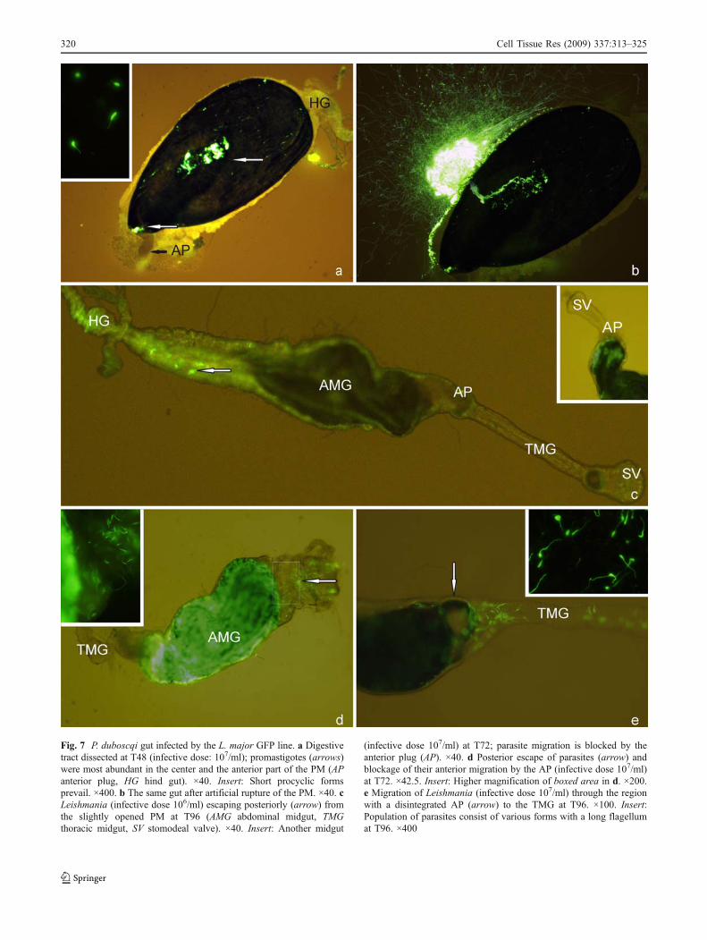
Fig. 7 P. duboscqi gut infected by the L. major GFP line. a Digestivetract dissected at T48 (infective dose: 107/ml); promastigotes (arrows)were most abundant in the center and the anterior part of the PM (APanterior plug, HG hind gut). ×40. Insert: Short procyclic formsprevail. ×400. b The same gut after artificial rupture of the PM. ×40. cLeishmania (infective dose 106/ml) escaping posteriorly (arrow) fromthe slightly opened PM at T96 (AMG abdominal midgut, TMGthoracic midgut, SV stomodeal valve). ×40. Insert: Another midgut
(infective dose 107/ml) at T72; parasite migration is blocked by theanterior plug (AP). ×40. d Posterior escape of parasites (arrow) andblockage of their anterior migration by the AP (infective dose 107/ml)at T72. ×42.5. Insert: Higher magnification of boxed area in d. ×200.e Migration of Leishmania (infective dose 107/ml) through the regionwith a disintegrated AP (arrow) to the TMG at T96. ×100. Insert:Population of parasites consist of various forms with a long flagellumat T96. ×400
320 Cell Tissue Res (2009) 337:313–325

females, an unidentified contaminating yeast was observedin the infected blood meal. Where Leishmania promasti-gotes were never seen burrowing inside the PM, yeast wereable to disrupt and penetrate through the PM layers(Fig. 8a, b).
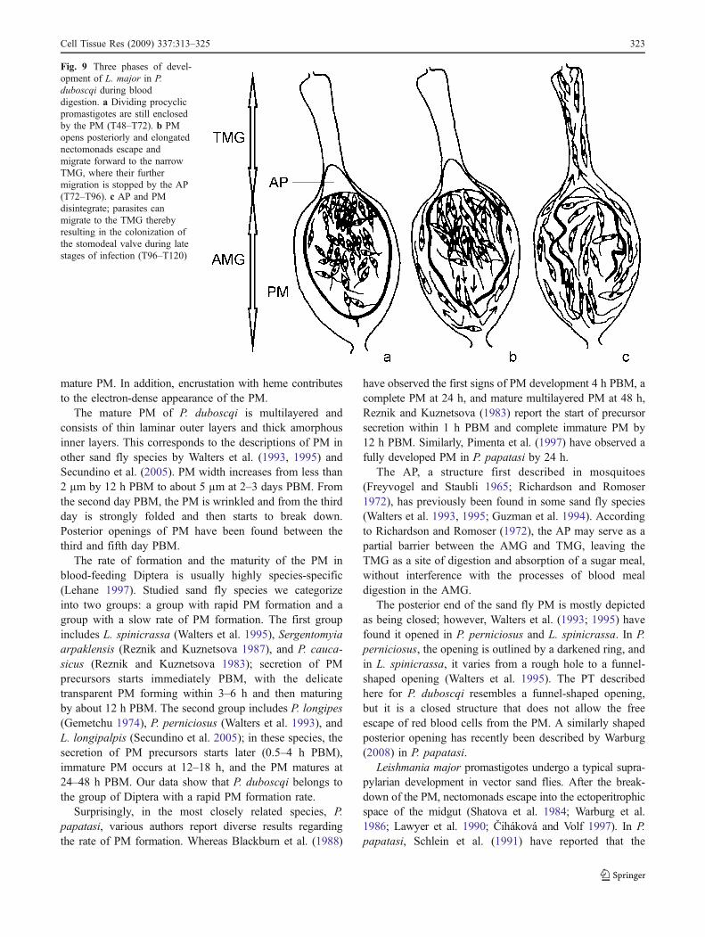
A proposed model of the course of L. major earlydevelopment in P. duboscqi is depicted in Fig. 9.
Morphological transformations of Leishmania in relationto disintegration of the PM
According to the state of the PM, four categories of femaleswere distinguished: females with an intact PM, females witha slightly disintegrated PM, females with a PM that wasopened/heavily disintegrated, and females that had defecated.The effect of the disintegration of the PM on the morpho-logical transformation of promastigotes was more pro-nounced than the effect of the time PBM (Table 2, 3). By
T72, promastigotes from females with a slightly disinte-grated PM had a significantly longer (P=0.012) and moreslender (P=0.009) body and longer flagella (P=0.002) thanthe promastigotes from females with an intact PM (Table 2).Parasites from flies with an opened or heavily disintegratedPM had a much longer body and flagellum than those fromboth the above-mentioned groups (P<0.0001 for all traits).Additionally, the width of the parasite body was smaller thanin flies with an intact PM (P=0.005). Similarly, by T96,promastigotes from females with a slightly disintegrated PMhad significantly shorter flagella and shorter and widerbodies than did parasites from an opened/heavily disinte-grated PM and from defecated flies (P<0.0001 in all cases).These morphological changes reflected the transformationfrom short procyclic promastigotes to elongated nectomo-nads in correlation with the stages of PM deformation, viz.,from an intact PM, to a slightly disintegrated PM, and to anopened/heavily disintegrated PM (Table 3). On the other
Table 1 Disintegration of the peritrophic matrix (PM) in females infected with Leishmania major amastigotes and in controls fed an uninfectedblood meal (PBM post-blood-meal)
DaysPBM
Number of infected (INF) andcontrol (C) flies
State of PM Significance of between-group differences
Intact Slightlydisintegrated
Opened Heavily disintegrated/blood remnants
Defecated
2 INF 8 8 0 0 0 0 P=1, χ2=0.0000C 10 10 0 0 0 0
3 INF 26 11 13 2 0 0 P=0.9881, χ2=0.0239C 30 13 15 2 0 0
4 INF 24 0 6 1 5 12 P=0.8911, χ2=1.1201C 30 1 8 2 5 14
5 INF 25 0 0 1 1 23 P=0.3243, χ2=2.2520C 30 0 0 1 5 24
Table 2 Measurements of Leishmania major promastigotes: effect of disintegration of PM on Leishmania morphological transformation
DaysPBM*
State of PM** Number ofmeasurements
Body length Body width Flagellar length
Mean (SD)in µm
Rangein µm
Mean (SD)in µm
Rangein µm
Mean (SD)in µm
Rangein µm
3 Intact 120 10.4 (2.0) 6.8–15.3 2.2 (0.8) 0.9–5.1 10.5 (2.9) 3.4–20.4
Slightly disintegrated 120 11.3 (2.6) 5.1–17.0 1.9 (0.7) 0.9–3.4 11.7 (2.8) 6.8–17.0
Opened / heavily disintegrated 80 15.6 (2.9) 8.5–23.8 1.8 (0.7) 0.9–3.4 15.8 (2.5) 10.2–20.4
Total 320 12.0 (3.2) 5.1–23.8 2.0 (0.7) 0.9–5.1 12.3 (3.5) 3.4–20.4
4 Slightly disintegrated 120 12.6 (4.5) 3.4–25.5 1.8 (1.0) 0.9–5.1 13.6 (3.7) 5.1–20.4
Opened / heavily disintegrated 120 16.4 (3.0) 6.8–25.5 1.4 (0.4) 0.9–1.7 16.4 (2.7) 10.2–25.5
Defecated 120 17.1 (3.5) 8.5–27.2 1.3 (0.4) 0.9–1.7 16.7 (2.7) 10.2–23.8
Total 360 15.4 (4.2) 3.4–27.2 1.5 (0.7) 0.9–5.1 15.6 (3.4) 5.1–25.5
*General effect of the time PBM on morphological transformation of promastigotes: F=17.917, d.f.=1, P<0.0001
** General effect of the state of the PM on morphological transformation of promastigotes: F=80.154, d.f.=3, P<0.0001
Cell Tissue Res (2009) 337:313–325 321

hand, parasites from an opened/heavily disintegrated PM andfrom defecated flies were morphologically indistinguishable(Tables 2, 3).
Discussion
The combination of light microscopy, histology, andelectron microscopy has revealed new details concerningPM formation in P. duboscqi females. Within 1 h PBM,erythrocytes are concentrated in the AMG forming acompact central bolus surrounded with extruded bloodplasma. The volume of blood plasma is apparently muchlower than that of blood cells, presumably because ofprediuresis of excess liquid by females during feeding(Sádlová et al. 1998).
The formation of the PM has several distinct stages. Itstarts with the secretion of electron-lucent componentshaving a fibrillar appearance (T0–T3), and at later timeintervals, the secretion of electron-dense components pre-vails. Both electron-dense and electron-lucent componentsare released into the gut lumen directly from the plasmamembrane on the microvillar surface, without the participa-tion of any secretory vesicles. This is in agreement withobservations in P. perniciosus (Walters et al. 1993),Lutzomyia spinicrassa (Walters et al. 1995), or Culicoidespunctatus and C. grisescens (Filimonova 2005). We attributethe electron-lucent fibrils to chitin, whereas the electron-dense amorphous secretion presumably represents proteinsand glycoproteins. The later type of secretion continues for3 days; proteins and glycoproteins coat the chitin frameworkand are responsible for the electron-dense appearance of the
Fig. 8 Electron micrographs ofthe gut of P. duboscqi femalesshowing yeast (Y) surroundedby a lysed space within theperitrophic matrix (PM) layers(a) and L. major promastigotes(Le) filling the endoperitrophicspace (a, b). mv Microvilli Bars3 µm (a), 2 µm (b)
Table 3 Representation of L. major morphological forms in distinct stages of PM deformation
Days PBM* Morphological forms State of PM**
Intact Slightly disintegrated Opened and heavily disintegrated Defecated
3 Procyclic promastigotes 88 (73%) 77 (64%) 16 (20%) -
Elongated nectomonads 6 (5%) 13 (11%) 53 (66%) -
Short nectomonads/leptomonads 26 (22%) 30 (25%) 11 (14%) -
Total 120 (100%) 120 (100%) 80 (100%) -
4 Procyclic promastigotes - 34 (28%) 12 (10%) 15 (12%)
Elongated nectomonads - 40 (33%) 90 (75%) 92 (77%)
Short nectomonads/leptomonads - 41 (34%) 18 (15%) 13 (11%)
Metacyclic promastigotes - 1 (1%) 0 0
Paramastigotes - 4 (4%) 0 0
Total - 120 (100%) 120 (100%) 120 (100%)
*General effect of the time PBM on morphological transformation of promastigotes: Pearson χ2 =139.3, d.f.=4, P<0.0001
**General effect of the state of the PM on morphological transformation of promastigotes: Pearson χ2 =261.2, d.f.=12, P<0.0001
322 Cell Tissue Res (2009) 337:313–325
mature PM. In addition, encrustation with heme contributesto the electron-dense appearance of the PM.
The mature PM of P. duboscqi is multilayered andconsists of thin laminar outer layers and thick amorphousinner layers. This corresponds to the descriptions of PM inother sand fly species by Walters et al. (1993, 1995) andSecundino et al. (2005). PM width increases from less than2 µm by 12 h PBM to about 5 µm at 2–3 days PBM. Fromthe second day PBM, the PM is wrinkled and from the thirdday is strongly folded and then starts to break down.Posterior openings of PM have been found between thethird and fifth day PBM.
The rate of formation and the maturity of the PM inblood-feeding Diptera is usually highly species-specific(Lehane 1997). Studied sand fly species we categorizeinto two groups: a group with rapid PM formation and agroup with a slow rate of PM formation. The first groupincludes L. spinicrassa (Walters et al. 1995), Sergentomyiaarpaklensis (Reznik and Kuznetsova 1987), and P. cauca-sicus (Reznik and Kuznetsova 1983); secretion of PMprecursors starts immediately PBM, with the delicatetransparent PM forming within 3–6 h and then maturingby about 12 h PBM. The second group includes P. longipes(Gemetchu 1974), P. perniciosus (Walters et al. 1993), andL. longipalpis (Secundino et al. 2005); in these species, thesecretion of PM precursors starts later (0.5–4 h PBM),immature PM occurs at 12–18 h, and the PM matures at24–48 h PBM. Our data show that P. duboscqi belongs tothe group of Diptera with a rapid PM formation rate.
Surprisingly, in the most closely related species, P.papatasi, various authors report diverse results regardingthe rate of PM formation. Whereas Blackburn et al. (1988)
have observed the first signs of PM development 4 h PBM, acomplete PM at 24 h, and mature multilayered PM at 48 h,Reznik and Kuznetsova (1983) report the start of precursorsecretion within 1 h PBM and complete immature PM by12 h PBM. Similarly, Pimenta et al. (1997) have observed afully developed PM in P. papatasi by 24 h.
The AP, a structure first described in mosquitoes(Freyvogel and Staubli 1965; Richardson and Romoser1972), has previously been found in some sand fly species(Walters et al. 1993, 1995; Guzman et al. 1994). Accordingto Richardson and Romoser (1972), the AP may serve as apartial barrier between the AMG and TMG, leaving theTMG as a site of digestion and absorption of a sugar meal,without interference with the processes of blood mealdigestion in the AMG.
The posterior end of the sand fly PM is mostly depictedas being closed; however, Walters et al. (1993; 1995) havefound it opened in P. perniciosus and L. spinicrassa. In P.perniciosus, the opening is outlined by a darkened ring, andin L. spinicrassa, it varies from a rough hole to a funnel-shaped opening (Walters et al. 1995). The PT describedhere for P. duboscqi resembles a funnel-shaped opening,but it is a closed structure that does not allow the freeescape of red blood cells from the PM. A similarly shapedposterior opening has recently been described by Warburg(2008) in P. papatasi.
Leishmania major promastigotes undergo a typical supra-pylarian development in vector sand flies. After the break-down of the PM, nectomonads escape into the ectoperitrophicspace of the midgut (Shatova et al. 1984; Warburg et al.1986; Lawyer et al. 1990; Čiháková and Volf 1997). In P.papatasi, Schlein et al. (1991) have reported that the
Fig. 9 Three phases of devel-opment of L. major in P.duboscqi during blooddigestion. a Dividing procyclicpromastigotes are still enclosedby the PM (T48–T72). b PMopens posteriorly and elongatednectomonads escape andmigrate forward to the narrowTMG, where their furthermigration is stopped by the AP(T72–T96). c AP and PMdisintegrate; parasites canmigrate to the TMG therebyresulting in the colonization ofthe stomodeal valve during latestages of infection (T96–T120)
Cell Tissue Res (2009) 337:313–325 323

breakdown of the PM differs between uninfected females andthose infected by L. major; the disintegration of the anteriorregion has been observed in infected sand flies. In this studyof a closely related species, P. duboscqi, we have not foundany differences in the timing of the degeneration of the PMbetween infected and uninfected females. Disintegration ofthe PM starts in both groups by day 3 PBM. Although themaximal density of parasites is present in the central andanterior parts of the blood bolus, Leishmania escape onlyfrom the posterior opening of the PM, with a similaropenning being present in uninfected sand flies.
Schlein et al. (1991) have postulated that the lysis of thechitin layer of the PM is caused by the chitinase of parasiteorigin and have demonstrated chitinase activity in L. major.More recently, the chitinase gene and enzyme activity havebeen characterized in several Leishmania species (Shakarianand Dwyer 1998, 2000). Phlebotomus papatasi treated witha chitinase inhibitor allosamidin forms a thicker PM thatprevents the escape of L. major parasites from the bloodmeal (Pimenta et al. 1997), whereas over-expression of theparasite chitinase causes the earlier arrival of Leishmaniamexicana at the stomodeal valve of Lutzomyia longipalpis(Rogers et al. 2008). However, the role of Leishmaniachitinase during promastigote escape to the ectoperitrophicsac remains elusive, as its activity is inhibited by hemoglobinpresent in the blood meal (Schlein and Jacobson 1994). In P.duboscqi females, the PM opens similarly in uninfected andinfected females, most likely as a result of the sand-fly-derived chitinase activity described in P. papatasi byRamalho-Ortigao and Traub-Cseko (2003) and Ramalho-Ortigao et al. (2005). Our transmission electron-microscopestudy has not revealed any signs of PM lysis caused byLeishmania, whereas contaminating yeast produces clearlytic plaques (Fig. 8). The above-mentioned findingssuggest that L. major chitinase does not have an importantrole in the disintegration of sand fly PM. Parasites “wait”until the PM is broken down by sand fly chitinase (seemodel in Fig. 9). Rogers et al. (2008) have also suggestedthat at least some Leishmania species rely upon thechitinases of the sand fly.
Nevertheless, we cannot exclude that the process ofPM disintegration is species-specific. In a previousstudy aimed at the development of different L. majorstrains within various sand fly species, Čiháková and Volf(1997) have found a correlation between the timing ofescape of a particular strain from the endoperitrophicspace and its ability to produce successful late-stageinfections in P. papatasi. In contrast, in P. duboscqi, theescape of various strains from the endoperitrophic space ismore uniform and does not correlate with late stagedevelopment in the vector.
Although mycoses in populations of sand flies have beenreported to reduce the incidence of Leishmania in endemic
areas and negatively influence the survival of vectors(Killick-Kendrick 1979; Schlein et al. 1985), we have notobserved any impact of yeast contaminating P. duboscqimidgut on sand fly mortality or Leishmania development.
The disintegration of PM coincides with the transforma-tion of procyclic forms to long nectomonads. Procyclicpromastigotes are transformed after the first signs of PMbreakdown, perhaps because of contact with salivarycomponents. A similar differentiation of procyclic tonectomonad forms has been demonstrated in vitro byCharlab and Ribeiro (1993) and Charlab et al. (1995) usingsalivary gland homogenates. In vivo, saliva-derived pro-teins (Volf et al. 2002) and salivary enzymes such asamylase (Ribeiro et al. 2000) have been demonstrated in themidgut. Accordingly, saliva ingested into the midgut, eitherwith a sugar meal (Bates and Rogers 2004) or alone, cantrigger the transformation of parasites.
Finally, our study has revealed an interesting and novelrole of the AP during Leishmania infection. After initialestablishment in the ectoperitrophic space of the AMG,promastigotes move anteriorly. However, this forwardmigration to the narrowed TMG can be stopped by theAP until the disintegration of the PM. In P. duboscqifemales with an infective dose of 106 parasites/ml,Leishmania occurrence in the TMG is always connectedwith the disintegration of the AP. In female sand flies withan opened PM, but complete AP, all the parasites arerestricted to the AMG. Thus, the AP can temporarilyfunction as a barrier that prevents the forward migration ofnectomonads, as depicted in our model of parasite infectionin the sand fly midgut (Fig. 9).
Acknowledgements We are grateful to Dr J. Votýpka and Dr. D.Folková (Charles University, Prague) for providing us with GFP-transfected parasites and to Dr. R. Jochim (NIH, Rockville) for criticalreading of the manuscript.
Open Access This article is distributed under the terms of theCreative Commons Attribution Noncommercial License which per-mits any noncommercial use, distribution, and reproduction in anymedium, provided the original author(s) and source are credited.
References
Andrade-Coelho CA, Santos-Mallet J, Souza NA, Lins U, MeirellesMNL, Rangel EF (2001) Ultrastructural features of the midgutepithelium of females Lutzomyia intermedia (Lutz & Neiva,1912) (Diptera: Psychodidae: Phlebotominae). Mem InstOswaldo Cruz 96:1141–1151
Bates PA, Rogers ME (2004) New insights into the developmentalbiology and transmission mechanisms of Leishmania. Curr MolMed 4:601–609
Benková I, Volf P (2007) Effect of temperature on metabolism ofPhlebotomus papatasi (Diptera: Psychodidae). J Med Entomol44:150–154
324 Cell Tissue Res (2009) 337:313–325

Blackburn K, Wallbanks KR, Molyneux DH, Lavin DR, WinstanleySL (1988) The peritrophic membrane of the female sandflyPhlebotomus papatasi. Ann Trop Med Parasit 82:613–619
Charlab R, Ribeiro JMC (1993) Cytostatic effect of Lutzomyialongipalpis salivary gland homogenates on Leishmania parasites.Am J Trop Med Hyg 48:831–838
Charlab R, Tesh RB, Rowton ED, Ribeiro JMC (1995) Leishmaniaamazonensis: sensitivity of different promastigote morphotypesto salivary gland homogenates of the sand fly Lutzomyialongipalpis. Exp Parasitol 80:167–175
Čiháková J, Volf P (1997) Development of different Leishmaniamajor strains in the vector sandflies Phlebotomus papatasi and P.duboscqi. Ann Trop Med Parasit 91:267–279
Feng LC (1951) The role of the peritrophic membrane in Leishmaniaand trypanosome infections of sandflies. Peking Nat Hist Bull19:327–334
Filimonova SA (2005) Morphological study of digestive cycle inbloodsucking biting midges of genus Culicoides. J Evol BiochemPhysiol 41:221–232
Freyvogel TA, Staubli W (1965) The formation of the peritrophicmembrane in Culicidae. Acta Trop 22:118–147
Gemetchu T (1974) The morphology and fine structure of the midgutand peritrophic membrane of the adult female, Phlebotomuslongipes Parrot and Martin (Diptera: Psychodidae). Ann TropMed Parasit 68:111–124
Guzman H, Walters LL, Tesh RB (1994) Histologic detection ofmultiple blood meals in Phlebotomus duboscqi (Diptera: Psy-chodidae). J Med Entomol 31:890–897
Jacobs-Lorena M, Oo MM (1996) The peritrophic matrix of insects.In: Beaty J, Marquardt WC (eds) The biology of disease vectors.University Press of Colorado, Boulder, pp 318–332
Karnovsky MJ (1965) Formaldehyde-glutaraldehyde fixative of highosmolarity for use in electron microscopy. J Cell Biol 27:137
Killick-Kendrick R (1979) Biology of Leishmania in phlebotominesandflies. In: Lumsden WH, Evans DA (eds) Biology of theKintoplastida, vol 2. Academic Press, London New York, pp396–460
Lawyer PG, Ngumbi PM, ChO A, Odongo SO, Mebrahtu YB, GithureJI, Koech DK, Roberts CR (1990) Development of Leishmaniamajor in Phlebotomus duboscqi and Sergentomyia schwetzi(Diptera: Psychodidae). Am J Trop Med Hyg 43:31–43
Lehane MJ (1997) Peritrophic matrix structure and function. AnnuRev Entomol 42:525–550
Myskova J, Votypka J, Volf P (2008) Leishmania in sand flies: comparisonof quantitative polymerase chain reaction with other techniques todetermine the intensity of infection. J Med Entomol 45:133–138
Pascoa V, Oliveira PL, Dansa-Petretski M, Silva JR, Alvarenga PH,Jacobs-Lorena M, Lemos FJA (2002) Aedes aegypti peritrophicmatrix and its interaction with heme during blood digestion.Insect Biochem Mol 32:517–523
Peters W (1992) Peritrophic membranes. Springer, BerlinPimenta PFP, Modi GB, Pereira ST, Shahabuddin M, Sacks DL (1997)
A novel role for the peritrophic matrix in protecting Leishmaniafrom the hydrolytic activities of the sand fly midgut. Parasitology115:359–369
Ramalho-Ortigao JM, Traub-Cseko ZM (2003) Molecular charac-terization of Llchit1, a midgut chitinase cDNA from theleishmaniasis vector Lutzomyia longipalpis. Insect BiochemMol 33:279–287
Ramalho-Ortigao JM, Kamhawi S, Joshi MB, Reynoso D, LawyerPG, Dwyer DM, Sacks DL, Valenzuela JG (2005) Characteriza-tion of a blood activated chitinolytic system in the midgut of thesand fly vectors Lutzomyia longipalpis and Phlebotomus papa-tasi. Insect Mol Biol 14:703–712
Reynolds ES (1963) The use of lead citrate at high pH as an electron-opaque stain in electron microscopy. J Cell Biol 17:208
Reznik EP, Kuznetsova LA (1983) The migration of microfilariansThamugadia ivaschkini from the intestine to haemocoel of sandflies of the genus Phlebotomus. Parazitologiya 17:36–41
Reznik EP, Kuznetsova LA (1987) Some peculiarities of digestion andperitrophic membrane formation in sand flies Sergentomyiaarpaklensis. Parazitologiya 21:16–21
Ribeiro JMC, Rowton ED, Charlab R (2000) Salivary amylase activityof the phlebotomine sand fly, Lutzomyia longipalpis. InsectBiochem Mol 30:271–277
Richardson MW, Romoser WS (1972) The formation of theperitrophic membrane in adult Aedes triseriatus (Say) (Diptera:Culicidae). J Med Entomol 9:495–500
Rogers ME, Chance ML, Bates PA (2002) The role of promastigotesecretory gel in the origin and transmission of the infective stageof Leishmania mexicana by the sandfly Lutzomyia longipalpis.Parasitology 124:495–507
Rogers ME, Hajmová M, Joshi MB, Sádlová J, Dwyer DM, Volf P,Bates P (2008) Leishmania chitinase facilitates colonization ofsand fly vectors and enhances transmission to mice. CellMicrobiol 10:1363–1372
Sádlová J, Reishig J, Volf P (1998) Prediuresis in female Phlebotomussandflies (Diptera: Psychodidae). Eur J Entomol 95:643–647
Sádlová J, Svobodová M, Volf P (1999) Leishmania major: effect ofrepeated passages through sandfly vectors or murine host. AnnTrop Med Parasitol 93:599–611
Schlein Y, Jacobson RL (1994) Haemoglobin inhibits the developmentof infective promastigotes and chitinase secretion in Leishmaniamajor cultures. Parasitology 109:23–28
Schlein Y, Polacheck I, Yuval B (1985) Mycoses, bacterial infections andantibacterial actvity in sandflies (Psychodidae) and their possiblerole in the transmision of leishmaniasis. Parasitology 90:57–66
Schlein Y, Jacobson RL, Shlomai J (1991) Chitinase secreted byLeishmania functions in the sandfly vector. Proc R Soc Lond[Biol] 245:121–126
Secundino NFC, Eger-Mangrich I, Braga EM, Santoro MM, PimentaPFP (2005) Lutzomyia longipalpis peritrophic matrix: formation,structure, chemical composition. J Med Entomol 42:928–938
Shakarian AM, Dwyer DM (1998) The LdCht1 gene encodesthe secretory chitinase from Leishmania donovani. Gene208:315–322
Shakarian AM, Dwyer DM (2000) Pathogenic Leishmania secreteantigenically related chitinases which are encoded by a highlyconserved gene locus. Exp Parasitol 94:238–242
Shatova SM, Shulga MA, Safianova VM, Avakian AA (1984)Sravnitelnoe elektronmikroskopicheskoe izuchenie Leishmaniamajor i L. tropica pri eksperimentalnem zarazhenii moskitaPhlebotomus papatasi. Parazitologiya 18:353–355
Volf P, Skarupova S, Man P (2002) Characterization of the lectin fromfemales of Phlebotomus duboscqi sand flies. Eur J Biochem269:6294–6301
Walters LL, Irons KP, Modi GB, Tesh RB (1992) Refractory barriersin the sand fly Phlebotomus papatasi (Diptera: Psychodidae) toinfection with Leishmania panamensis. Am J Trop Med Hyg46:211–228
Walters LL, Irons KP, Guzman H, Tesh RB (1993) Formation andcomposition of the peritrophic membrane in the sand fly Phlebotomusperniciosus (Diptera: Psychodidae). J Med Entomol 30:179–198
Walters LL, Irons KP, Guzman H, Tesh RB (1995) Peritrophicenvelopes of Lutzomyia spinicrassa (Diptera: Psychodidae). JMed Entomol 32:711–725
Warburg A (2008) The structure of the female sand fly (Phlebotomuspapatasi) alimentary canal. Trans R Soc Trop Med Hyg102:161–166
Warburg A, Hamada GS, Schlein Y, Shire D (1986) Scanning electronmicroscopy of Leishmania major in Phlebotomus papatasi. ZParasitenkd 72:423–431
Cell Tissue Res (2009) 337:313–325 325