periungual abscess caused by citrobacter braakii in a ... species, aerobic, gram-negative bacilli in...
TRANSCRIPT
Brief Report
528 Ann Dermatol
Received January 25, 2015, Revised August 20, 2015, Accepted for publication August 26, 2015
Corresponding author: Seong-Jin Kim, Department of Dermatology,Chonnam National University Hospital, 42 Jebong-ro, Dong-gu, Gwangju 61469, Korea. Tel: 82-62-220-6683, Fax: 82-62-222-4058, E-mail: seongkim@ chonnam.ac.kr
This is an Open Access article distributed under the terms of the Creative Commons Attribution Non-Commercial License (http://creativecommons.org/licenses/by-nc/4.0) which permits unrestricted non-commercial use, distribution, and reproduction in any medium, provided the original work is properly cited.
Copyright © The Korean Dermatological Association and The Korean Society for Investigative Dermatology
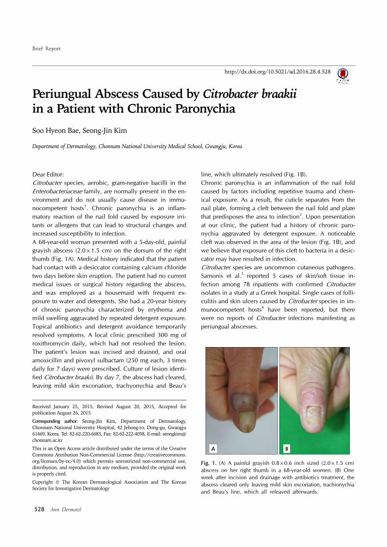
Fig. 1. (A) A painful grayish 0.8×0.6 inch sized (2.0×1.5 cm) abscess on her right thumb in a 68-year-old women. (B) One week after incision and drainage with antibiotics treatment, the abscess cleared only leaving mild skin excoriation, trachionychia and Beau’s line, which all releaved afterwards.
http://dx.doi.org/10.5021/ad.2016.28.4.528
Periungual Abscess Caused by Citrobacter braakii in a Patient with Chronic Paronychia
Soo Hyeon Bae, Seong-Jin Kim
Department of Dermatology, Chonnam National University Medical School, Gwangju, Korea
Dear Editor:Citrobacter species, aerobic, gram-negative bacilli in the Enterobacteriaceae family, are normally present in the en-vironment and do not usually cause disease in immu-nocompetent hosts1. Chronic paronychia is an inflam-matory reaction of the nail fold caused by exposure irri-tants or allergens that can lead to structural changes and increased susceptibility to infection.A 68-year-old woman presented with a 5-day-old, painful grayish abscess (2.0×1.5 cm) on the dorsum of the right thumb (Fig. 1A). Medical history indicated that the patient had contact with a desiccator containing calcium chloride two days before skin eruption. The patient had no current medical issues or surgical history regarding the abscess, and was employed as a housemaid with frequent ex-posure to water and detergents. She had a 20-year history of chronic paronychia characterized by erythema and mild swelling aggravated by repeated detergent exposure. Topical antibiotics and detergent avoidance temporarily resolved symptoms. A local clinic prescribed 300 mg of roxithromycin daily, which had not resolved the lesion. The patient’s lesion was incised and drained, and oral amoxicillin and pivoxyl sulbactam (250 mg each, 3 times daily for 7 days) were prescribed. Culture of lesion identi-fied Citrobacter braakii. By day 7, the abscess had cleared, leaving mild skin excoriation, trachyonychia and Beau’s
line, which ultimately resolved (Fig. 1B).Chronic paronychia is an inflammation of the nail fold caused by factors including repetitive trauma and chem-ical exposure. As a result, the cuticle separates from the nail plate, forming a cleft between the nail fold and plate that predisposes the area to infection2. Upon presentation at our clinic, the patient had a history of chronic paro-nychia aggravated by detergent exposure. A noticeable cleft was observed in the area of the lesion (Fig. 1B), and we believe that exposure of this cleft to bacteria in a desic-cator may have resulted in infection. Citrobacter species are uncommon cutaneous pathogens. Samonis et al.3 reported 5 cases of skin/soft tissue in-fection among 78 inpatients with confirmed Citrobacter isolates in a study at a Greek hospital. Single cases of folli-culitis and skin ulcers caused by Citrobacter species in im-munocompetent hosts4 have been reported, but there were no reports of Citrobacter infections manifesting as periungual abscesses.
Brief Report
Vol. 28, No. 4, 2016 529
Received September 24, 2015, Revised January 14, 2016, Accepted for publication January 18, 2016
Corresponding author: Sung Ku Ahn, Department of Dermatology, Yonsei University Wonju College of Medicine, 20 Ilsan-ro, Wonju 26426, Korea. Tel: 82-33-741-0621, Fax: 82-33-748-2650, E-mail: [email protected]
This is an Open Access article distributed under the terms of the Creative Commons Attribution Non-Commercial License (http://creativecommons.org/ licenses/by-nc/4.0) which permits unrestricted non-commercial use, distribution, and reproduction in any medium, provided the original work is properly cited.
Copyright © The Korean Dermatological Association and The Korean Society for Investigative Dermatology
Antibiotic susceptibility differs among Citrobacter sub-types3 and treatment has been based on individual anti-microbial profiles. One study of 22 cases of C. freundii showed 100% resistance to erythromycin, ampicillin, and methicillin5. As was demonstrated in our case study, mac-rolides including erythromycin and roxithyromycin (often indicated for gram positive/negative cocci infections) were ineffective treatments for C. braakii, a gram-negative bacillus. While oral amoxicillin and pivoxyl sulbactam were pre-scribed after incision and drainage (I&D) in our study, the Citrobacter isolate was resistant to amoxicillin. Therefore, the I&D procedure was primarily responsible for patient improvement. To our knowledge, this is the first reported case of C. braa-kii-induced periungual abscess in a patient with chronic paronychia. Additional data on Citrobacter infections are needed to improve comprehension of risk factors, etiol-ogy, clinical features, and management of these infections.
REFERENCES
1. Nayar R, Shukla I, Sultan A. Epidemiology, prevalence and
identification of Citrobacter species in clinical specimens in a tertiary care hospital in India. Int J Sci Res Publ 2014;4:1-6.
2. Tosti A, Piraccini BM, Ghetti E, Colombo MD. Topical
steroids versus systemic antifungals in the treatment of chronic paronychia: an open, randomized double-blind and
double dummy study. J Am Acad Dermatol 2002;47:73-76.
3. Samonis G, Karageorgopoulos DE, Kofteridis DP, Matthaiou DK, Sidiropoulou V, Maraki S, et al. Citrobacter infections in
a general hospital: characteristics and outcomes. Eur J Clin
Microbiol Infect Dis 2009;28:61-68.4. Whalen JG, Mully TW, English JC 3rd. Spontaneous
Citrobacter freundii infection in an immunocompetent
patient. Arch Dermatol 2007;143:124-125.5. Abo-State MA, Mahdy HM, Ezzat SM, El Shakour EHA,
El-Bahnasawy MA. Antimicrobial resistance profiles of
Enterobacteriaceae isolated from rosetta branch of river Nile, Egypt. World Appl Sci J 2012;19:1234-1243.
http://dx.doi.org/10.5021/ad.2016.28.4.529
A Rare Cutaneous Adverse Reaction after Intra-Articular Injection of Hyaluronic Acid
Hee-Chul Chung, Hana Bak, Jin Wook Lee, Sung Ku Ahn
Department of Dermatology, Yonsei University Wonju College of Medicine, Wonju, Korea
Dear Editor:Hyaluronic acid (HA) is a high-molecular-weight poly-saccharide found throughout the extracellular matrix and is one of the major components of articular joints. Intra-ar-ticular injection of HA is often performed for the treatment of symptomatic osteoarthritis. The efficacy of HA injection
in relieving pain and improving function in patients with osteoarthritis has been reported previously. Adverse re-actions are usually limited to local reactions at the in-oculation site, including transient local tenderness1. Although HA has been acknowledged as safe, there have been many reports related to a variety of adverse effects to