personalized cancer immunotherapy
TRANSCRIPT
Towards personalized cancer treatments using
Immunoinformatics
Bjoern PetersZeynep Kosaloglu-YalcinLa Jolla Institute for Immunology

La Jolla Institute for Immunology (LJI)

Overview
§ Background Biology: The immune system can detect and eliminate mutated cancer cells
§ Background Immunoinformatics: Mutations that can be recognized by T cells can be predicted
§ Hands on: Design a personalized cancer vaccine

Cancer immune surveillance and escape
§ Mutations in cells occur frequently
§ The immune system has the capacity to detect and eliminate such
mutated cells, and will do so on a regular basis
§ Only when mutated cells find ways to hide from- or suppress an attack
by the immune system, they can grow and spread unhindered leading
to clinically apparent tumors
4 Van der Burg et al, Nature Reviews Cancer 16, 219–233 (2016)

CD8+ T cell epitope recognition
MHC-I
Antigen
Antigen Presenting Cell (APC)
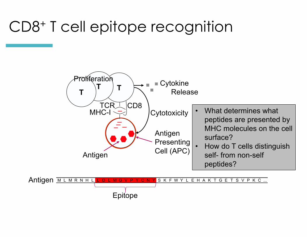
MHC-I
T Cytokine Release
Cytotoxicity
TT
Proliferation
TCR CD8
ORF 1 M G Q I V T M F E A L P H I I D E V I N I V I I V L I V I T G I K A V Y N ...ORF 2 M G L K G P D I Y K G V Y Q F K S V E F D M S H L N L T M P N A C S A N N ...ORF 3 M H N F C N L T S A F N K K T F D H T L M S I V S S L H L S I D G N S N Y ...ORF 4 M S A Q S Q C R T F R G R V L D M F R T A F G G K Y M R S G W G W T G S D ...ORF 5 M H C T Y A G P F G M S R I L L S Q E K T K F F T R R L A G T F T W T L S ...ORF 6 M K C F G N T A V A K C N V N H D A E F C D M L R L I D Y N K A A L S K F ...ORF 7 M L M R N H L L D L M G V P Y C N Y S K F W Y L E H A K T G E T S V P K C ...ORF 8 M N M I T E M L R K D Y I K R Q G S T P L A L M D L L M F S T S A Y L V S ...ORF 9 M S L S K E V K S F Q W T Q A L R R E L Q S F T S D V K A A V I K D A T N ...
T cell epitope mapping
CD8+ T cell epitope recognition
Antigen
Antigen Presenting Cell (APC)
Antigen
Epitope
• What determines what peptides are presented by MHC molecules on the cell surface?
• How do T cells distinguish self- from non-self peptides?

MHC I - Antigen processing and presentation pathway
ER
TAP
ProteasomeProtein
Golgi
T Cell Receptor
MHC-I
Peters et al, J Mol Biol 2002, Bioinformatics 2003, J Immunol.2003; CMLS 2005 ; Assarson, J Immunol 2007

MHC:peptide binding mode
X-Ray Structure: Madden, Cell 1993. Viewer: Beaver and Ponomarenko, Immunome Research, 2007
• Each human expresses up to 6 different MHC-I molecules in every cell
• >3000 MHC variants are known
• Distinct binding specificities à individual epitope repertoire

Self –reactive T cells are deleted during maturation

Cancer immune surveillance and escape
§ Mutations in cells occur frequently
§ The immune system has the capacity to detect and eliminate such
mutated cells, and will do so on a regular basis
§ Only when mutated cells find ways to hide from- or suppress an attack
by the immune system, they can grow and spread unhindered leading
to clinically apparent tumors
10 Van der Burg et al, Nature Reviews Cancer 16, 219–233 (2016)
Can the immune response against an ‘escaped’ cancer be re-activated?

Nobel Prize 2018 in Medicine
§ Identification of the molecules PD-1 and CTLA-4 that function as ‘T cell brakes’ (immune checkpoints)
§ Blockade of PD-1 and CTLA-4 results in activation of T cells which has ”fundamentally changed the outcome for certain groups of patients with advanced cancer”
§ “Similar to other cancer therapies, adverse side effects are seen, which can be serious and even life threatening. They are caused by an overactive immune response leading to autoimmune reactions […]”
11
“for their discovery of cancer therapy by inhibition
of negative immune regulation”
https://www.nobelprize.org/uploads/2018/10/press-medicine2018.pdf

Rationale for Personalized Cancer Immunotherapy
§ Vaccination: Introduce or boost an immune response against a specific target (antigen)
§ Cancer cells contain non-self antigens that could be recognized by T cells, but presence of cancer means this mechanism has failed, typically by the tumor suppressing immune responses
§ Checkpoint blockade treatments: Block immune suppressive mechanisms to boost T cell immune responses against cancer cells.
§ Problem: Checkpoint blockade is unspecific, and will also boost unwanted autoimmune responses
§ Personalized Cancer Immunotherapy: Boost anti-tumor response with vaccine containing peptides corresponding to cancer mutations that can be recognized by T cells.

Several trials for personalized cancer vaccines are currently ongoing
13

14
Personalized Cancer Immunotherapy

15
Personalized Cancer Immunotherapy

Overview
§ Background Biology: The immune system can detect and eliminate mutated cancer cells
§ Background Immunoinformatics: Mutations that can be recognized by T cells can be predicted
§ Hands on: Design a personalized cancer vaccine

Neoepitopes (Neoantigens)
§ Cancers genomes accumulate mutations§ Mutations in coding regions are translated in
mutated protein sequences§ Mutated peptides can be presented as epitopes on
MHC to T cells
Neoepitopes are presumably recognized by tumor-infiltratinglymphocytes (TILs)
Neoepitopes are highly tumor-specific!
Coulie et al, Nat Rev Cancer. 2014 Feb;14(2):135-46Schumacher & Schreiber, Science. 2015 Apr 3;348(6230):69-74

Which mutations can be recognized by the patient’s T cells? à Resulting peptides have to bind HLA molecules of the patient
DNA and RNA sequencing identifies tumor specific somatic mutations

DNA Isolation
PCR Primary Amplification(exons 1-5)
PCR Primary Amplification
Product Purification
Sequencing Reactions(forward & reverse orientations)
Sequencing Reaction
Precipitation
Utilization of 96 samplesequencing instrument
Sequencing Analysis
Sequence-BasedTyping
•http://www.ashi-hla.org/publicationfiles/ASHI_Quarterly/25_2_2001/highthrusbt3.htm
HLA Typing: Targeted sequencing of HLA locus

Experimental Basis: MHC Binding Assay
Sequence IC50QIVTMFEAL 3.6LKGPDIYKG 308NFCNLTSAF 50,000AQSQCRTFR 38,000CTYAGPFGM 143CFGNTAVAK 50,000
...
List of peptides with allele specific binding affinity
Impossible to measure all peptides
à Predict binding peptides using machine learning
Find function Fi inFi (Sequence) ≈ Affinity
Many different approaches (ANN, SVM, HMM, LP, ... )
F1, F2, F3, ...
Measuring and predicting MHC:peptidebinding
log(IC50) ~ Binding free Energy
low IC50 à high affinityORF 1 M G Q I V T M F E A L P H I I D E V I N I V I I V L I V I T G I K A V Y N ...ORF 2 M G L K G P D I Y K G V Y Q F K S V E F D M S H L N L T M P N A C S A N N ...ORF 3 M H N F C N L T S A F N K K T F D H T L M S I V S S L H L S I D G N S N Y ...ORF 4 M S A Q S Q C R T F R G R V L D M F R T A F G G K Y M R S G W G W T G S D ...ORF 5 M H C T Y A G P F G M S R I L L S Q E K T K F F T R R L A G T F T W T L S ...ORF 6 M K C F G N T A V A K C N V N H D A E F C D M L R L I D Y N K A A L S K F ...ORF 7 M L M R N H L L D L M G V P Y C N Y S K F W Y L E H A K T G E T S V P K C ...ORF 8 M N M I T E M L R K D Y I K R Q G S T P L A L M D L L M F S T S A Y L V S ...ORF 9 M S L S K E V K S F Q W T Q A L R R E L Q S F T S D V K A A V I K D A T N ...
T cell epitope mapping
ORF 1 M G Q I V T M F E A L P H I I D E V I N I V I I V L I V I T G I K A V Y N ...ORF 2 M G L K G P D I Y K G V Y Q F K S V E F D M S H L N L T M P N A C S A N N ...ORF 3 M H N F C N L T S A F N K K T F D H T L M S I V S S L H L S I D G N S N Y ...ORF 4 M S A Q S Q C R T F R G R V L D M F R T A F G G K Y M R S G W G W T G S D ...ORF 5 M H C T Y A G P F G M S R I L L S Q E K T K F F T R R L A G T F T W T L S ...ORF 6 M K C F G N T A V A K C N V N H D A E F C D M L R L I D Y N K A A L S K F ...ORF 7 M L M R N H L L D L M G V P Y C N Y S K F W Y L E H A K T G E T S V P K C ...ORF 8 M N M I T E M L R K D Y I K R Q G S T P L A L M D L L M F S T S A Y L V S ...ORF 9 M S L S K E V K S F Q W T Q A L R R E L Q S F T S D V K A A V I K D A T N ...
T cell epitope mapping
1 2 3 4 5 6 7 8 9 1 2 3 4 5 6 7 8 9A -0.3 0.8 -0.3 -0.3 -0.2 -0.3 0.0 0.0 -0.9 -0.3 -0.2 0.1 -0.1 0.2 -0.3 -0.1 -0.4 0.5C 0.2 0.9 0.0 0.3 -0.5 -0.1 0.1 0.2 0.4 0.3 0.4 0.0 0.4 0.2 0.3 0.2 0.5 0.3D 0.8 0.9 -0.4 -0.3 0.3 0.2 0.4 0.3 0.6 0.8 0.4 0.6 0.3 0.7 0.4 0.8 0.6 1.1E 0.6 -0.4 0.7 -0.2 0.1 -0.4 -0.2 -0.2 -0.5 0.3 0.3 0.4 -0.1 0.5 0.1 0.3 0.1 0.4F -1.3 0.5 -0.5 0.1 -0.1 0.0 -0.3 -0.4 -0.8 0.4 0.7 -0.5 -0.5 -0.6 -0.3 -0.4 0.2 -1.5G -0.2 0.1 0.3 -0.1 0.0 0.4 0.3 -0.1 0.2 0.2 0.4 0.2 -0.1 -0.1 0.3 0.5 -0.2 -0.1H 1.1 0.9 -0.1 0.4 0.1 0.2 0.0 0.2 0.8 -0.3 0.1 0.1 -0.1 0.2 -0.1 -0.3 -0.1 0.0I -0.4 -0.7 -0.4 0.1 -0.1 -0.4 -0.5 0.5 -1.4 -0.4 -0.7 -0.3 0.3 -0.4 -0.3 0.3 -0.2 -1.4K -0.3 0.0 1.1 0.1 0.1 0.6 0.9 0.2 0.9 -0.7 0.9 0.5 -0.1 0.1 0.1 0.8 0.2 0.3L 0.0 -1.9 -0.4 -0.2 0.0 -0.2 0.0 -0.1 -1.1 -0.4 -0.7 -0.3 0.5 -0.4 0.1 -0.7 0.1 -0.9M -0.7 -1.2 -0.7 0.2 -0.6 0.0 0.0 0.0 -0.8 -0.6 -1.0 -0.5 -0.3 -0.1 0.0 0.2 -0.2 -0.4N -0.1 0.3 0.1 -0.3 -0.1 -0.3 0.0 0.2 0.7 0.2 0.4 -0.4 -0.3 0.2 -0.2 -0.4 -0.1 0.4P 1.2 0.5 0.6 -0.3 0.4 0.0 -0.4 -0.5 0.7 0.6 0.5 0.4 0.5 0.2 0.2 0.3 -0.4 0.9Q 0.4 -1.1 0.0 -0.1 0.4 -0.2 -0.3 0.2 0.7 0.0 -0.7 -0.1 0.0 -0.2 0.4 -0.3 0.0 0.4R -0.2 0.9 1.0 0.3 0.1 0.4 0.7 0.0 0.9 -0.7 0.9 0.2 0.1 -0.1 0.1 0.2 0.1 0.1S -0.3 0.1 0.1 -0.4 0.1 0.3 -0.2 -0.1 0.2 -0.3 -0.5 0.0 0.0 0.0 -0.3 0.1 -0.4 0.7T -0.2 -0.5 0.1 0.4 0.1 -0.5 0.2 0.0 -0.1 0.3 -1.2 0.3 0.0 0.6 -0.3 0.1 -0.4 0.9V -0.1 -0.9 -0.1 0.2 0.0 -0.3 0.1 0.1 -1.9 -0.1 -0.5 0.0 0.0 -0.3 -0.4 -0.8 -0.2 -0.2W 0.0 0.7 -0.5 -0.2 -0.1 0.2 -0.3 -0.1 0.4 0.5 0.3 -0.5 0.0 -0.5 0.2 -0.4 0.6 -0.8Y -0.3 0.2 -0.6 0.2 0.0 0.4 -0.4 -0.3 0.8 0.2 0.3 -0.5 -0.4 -0.1 0.0 -0.5 0.2 -0.9
HLA A*3201HLA A*0201
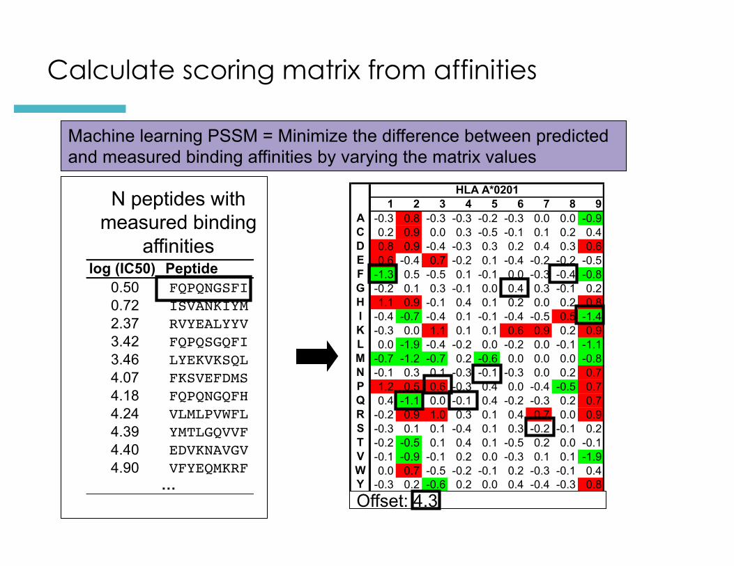
Calculate scoring matrix from affinities
N peptides with measured binding
affinitieslog (IC50) Peptide
0.50 FQPQNGSFI0.72 ISVANKIYM2.37 RVYEALYYV3.42 FQPQSGQFI3.46 LYEKVKSQL4.07 FKSVEFDMS4.18 FQPQNGQFH4.24 VLMLPVWFL4.39 YMTLGQVVF4.40 EDVKNAVGV4.90 VFYEQMKRF
… Offset: 4.3
Machine learning PSSM = Minimize the difference between predicted and measured binding affinities by varying the matrix values

Predictions available as webserver
§ Immune Epitope Database (IEDB) Analysis resource§ http://tools.iedb.org/mhci/

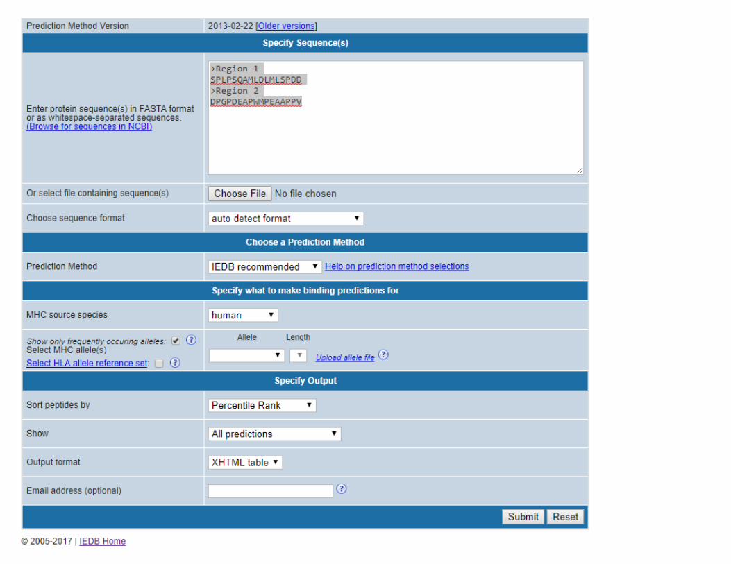


Evaluating binding predictions
§ Percentile rank < 0.5% = high affinity binder§ Percentile rank 0.5%-1% = intermediate binder§ Percentile rank 1% - 2% = low affinity binder§ Percentile rank 2% - 5% = borderline§ Percentile rank >5% is a non-binder

Overview
§ Background Biology: The immune system can detect and eliminate mutated cancer cells
§ Background Immunoinformatics: Mutations that can be recognized by T cells can be predicted
§ Hands on: Design a personalized cancer vaccine

Input data from patient:
>P53_HUMAN Cellular tumor antigen p53 - Healthy TissueMEEPQSDPSVEPPLSQETFSDLWKLLPENNVLSPLPSQAMDDLMLSPDDIEQWFTEDPGPDEAPRMPEAAPPVAPAPAAPTPAAPAPAPSWPLSSSVPSQKTYQGSYGFRLGFLHSGTAKSVTCTYSPALNKMFCQLAKTCPVQLWVDSTPPPGTRVRAMAIYKQSQHMTEVVRRCPHHERCSDSDGLAPPQHLIRVEGNLRVEYLDDRNTFRHSVVVPYEPPEVGSDCTTIHYNYMCNSSCMGGMNRRPILTIITLEDSSGNLLGRNSFEVRVCACPGRDRRTEEENLRKKGEPHHELPPGSTKRALPNNTSSSPQPKKKPLDGEYFTLQIRGRERFEMFRELNEALELKDAQAGKEPGGSRAHSSHLKSKKGQSTSRHKKLMFKTEGPDSD
>P53_HUMAN Cellular tumor antigen p53 - Tumor TissueMEEPQSDPSVEPPLSQETFSDLWKLLPENNVLSPLPSQAMLDLMLSPDDIEQWFTEDPGPDEAPWMPEAAPPVAPAPAAPTPAAPAPAPSWPLSSSVPSQKTYQGSYGFRLGFLHSGTAKSVTCTYSPALNKMFCQLAKTCPVQLWVDSTPPPGTRVRAMAIYKQSQHMTEVVRRCPHHERCSDSDGLAPPQHLIRVEGNLRVEYLDDRNTFVHSVVVPYEPPEVGSDCTTIHYNYMCNSSCMGGMNRRPILTIITLEV
HLA typing results:HLA-A*02:01, HLA-A*68:01HLA-B*07:02 , HLA-B*35:01
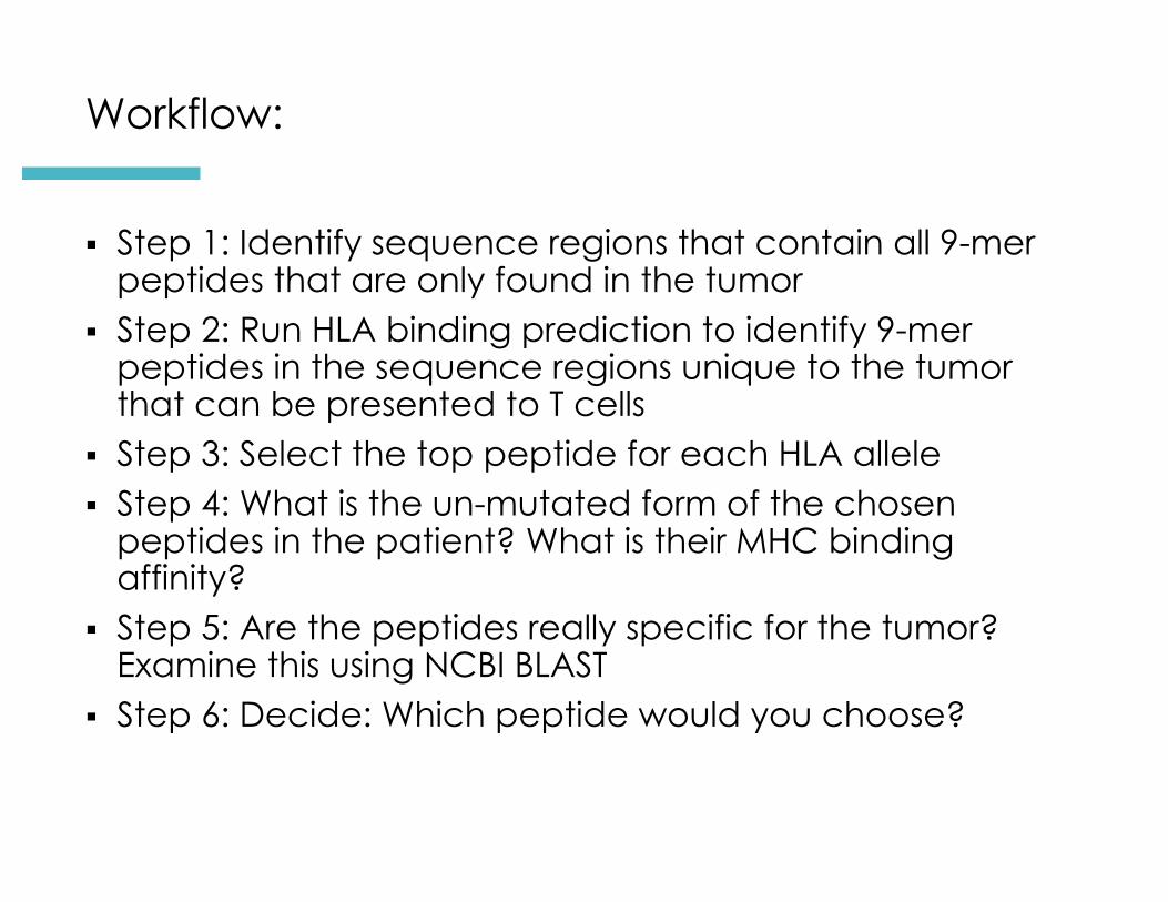
Workflow:
§ Step 1: Identify sequence regions that contain all 9-mer peptides that are only found in the tumor
§ Step 2: Run HLA binding prediction to identify 9-mer peptides in the sequence regions unique to the tumor that can be presented to T cells
§ Step 3: Select the top peptide for each HLA allele § Step 4: What is the un-mutated form of the chosen
peptides in the patient? What is their MHC binding affinity?
§ Step 5: Are the peptides really specific for the tumor? Examine this using NCBI BLAST
§ Step 6: Decide: Which peptide would you choose?

Workflow:
§ Step 1: Identify sequence regions that contain all 9-mer peptides that are only found in the tumor
§ Step 2: Run HLA binding prediction to identify 9-mer peptides in the sequence regions unique to the tumor to select peptides that can be presented to T cells in this patient
§ Step 3: Determine if the identified peptides are specific for the tumor
§ Final question: Which peptide would you choose?