pet and radiotherapy for head and neck
TRANSCRIPT

MAESTROJan. 2007
PET and Radiotherapy for Head andNeck Cancer: today and tomorrow…
Vincent GREGOIRE, M.D., Ph.D.
Head and Neck Oncology Program, RadiationOncology Dept., & Center for Molecular Imaging
and Experimental Radiotherapy, UniversitéCatholique de Louvain, St-Luc University
Hospital, Brussels, Belgium

MAESTROJan. 2007
S / RxTh / CH
Work-up-stagingprognostic evaluation
GTV/CTVSelection/delineation
Final responseevaluation
Early responseevaluation
Functional Image-guidedIMRT
Early detectionof recurrence
FDGC-methionineEF3 - F-miso - CuATSMBFU - FLT…
Potential added-value of PET in oncology

MAESTROJan. 2007
Imaging in radiotherapy:… today and tomorrow …
•Selection of Target Volumes with FDG-PET
•Delineation of GTV with FDG-PET
•Selection / delineation of CTV

MAESTROJan. 2007
The use of FDG-PET for the selection ofTarget Volume: setting the scene
Q: unilateral vs bilateralneck irradiation?
A: highly sensitiveexamination
Laryngeal SCC: T2-N1-M0

MAESTROJan. 2007
Detection of metastatic disease in the neck
• N=106 patients
• oral cavity tumors
• Neck dissection for all patients (2196 lymph nodes)
Stuckensen et al., 2000
Sensitivity Specificity Accuracy NPV PPV
PET 70% 82% 75% 71% 81%
CT 66% 69% 70% 66% 74%
MRI 64% 69% 66% 62% 71%
US 84% 68% 76% 79% 75%

MAESTROJan. 2007
Potential added-value of PET in oncology
Grégoire, 2004
Comparison between CT and FDG-PET for nodal staging.
Site Sensitivity Specificity
CT FDG-PET CT FDG-PET
Head and neck cancer 36-86% 50-96% 56-100% 88-100%
NSC lung cancer 45% 80-90% 85% 85-100%
Cervix carcinoma 57-73%1 75-91% 83-100%1 92-100%
Esophageal cancer 11-87% 30-78% 28-99% 86-98%1CT or MRI

MAESTROJan. 2007
Potential added-value of PET in oncology
Antoch et al., 2004
Sensitivity (%) Specificity (%) PPV NPV
PET/CT 92 93 88 94
PET+CT 88 89 83 92
PET 85 88 82 90
CT 64 83 70 79
Comparison between PET and PET/CT for N staging (n=260)

MAESTROJan. 2007
Target
J. John, 1974
MAESTROJan. 2007
The Gross Tumor volume (GTV)
Daisne et al., Radiology, 233: 93-100, 2004

MAESTROJan. 2007 Daisne et al, 2004
The Gross Tumor volume (GTV)
MAESTROJan. 2007
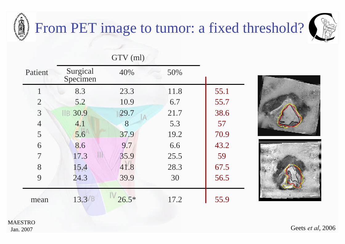
From PET image to tumor: a fixed threshold?
55.917.226.5*13.3mean
56.53039.924.3967.528.341.815.485925.535.917.37
43.26.69.78.6670.919.237.95.65575.384.14
38.621.729.730.9355.76.710.95.2255.111.823.38.31
50%40%SurgicalSpecimen
Patient
GTV (ml)
Geets et al, 2006

MAESTROJan. 2007
Volume delineation based on automatic thresholding with 18F-FDG
Daisne et al, 2003Geets et al, 2004
Functional imaging and automaticsegmentation

MAESTROJan. 2007
Imaging modalities, TVs and dose distribution

MAESTROJan. 2007
Image-Guided Radiation Therapy in HNSCC
0
50
100
150
200
GTV CTV PTV
Ave
rage
(±se
m)v
olum
e(c
c) CT-scan
FDG-PET
Larynx/hypopharynx (n=9)
ANOVA: p<0.05
0
50
100
150
200
250
GTV CTV PTV
CT-scan
FDG-PET
ANOVA: p<0.01
Oropharynx (n=10)
Ave
rage
(±se
m)
volu
me
(cc)
Impact of imaging modality on CTV/PTV delineation
Geets et al, 2003

MAESTROJan. 2007
Impact of imaging modality on dose distribution
Image-Guided Radiation Therapy in HNSCC
Geets et al, 2006

MAESTROJan. 2007
DAHANCA: http://www.dshho.suite.dk/dahanca/guidelines.htmlEORTC: http://www.eortc.be/home/ Radio/EDUCATION.htmRTOG: http://www.rtog.org/hnatlas/main.htm
Conformal radiotherapy and IMRTin Head and Neck Tumors

MAESTROJan. 2007
Ant. submandibular glandpost. belly of digastric m.
Post. sternocleidomastoid m.Lat. sternocleidomastoid m.Med. paraspinal m.
int. carotid arteryCra. lateral process of C1Cau. hyoid bone
Level II
LV
LII
CT-based delineation of lymph node levels inthe neck: Brussels- Rotterdam consensus guidelines

MAESTROJan. 2007
Imaging in radiotherapy:… today and tomorrow …
•Atlases
•Adaptive Target Volume delineation
•Non-rigid registration
•Adaptive dose distribution
•Other PET tracers

MAESTROJan. 2007
H&N IMRT practice heterogeneity
Harari et al., 2005
Atlases?

MAESTROJan. 2007
CT MRI (T2) FDG-PET
PRE-R/
WEEK 3
WEEK 5
(Week 2)
(Week 4)

MAESTROJan. 2007
•• 10 patients with stage III10 patients with stage III--IVIV pharyngopharyngo--laryngeallaryngeal SCC treated by CTSCC treated by CT--RTRT•• Images acquired before R/ and during RT after means doses of 14,Images acquired before R/ and during RT after means doses of 14, 25, 3525, 35
and 45and 45 GyGy..
Week 1 Week 2 Week 3 Week 4 Week 5 Week 7Week 6Before R/
R/ start
Images acquisitions
Dynamic FDG-PETAnatomic imaging
CT MR T2 FS MR T2 FDG-PET
Biological adaptive IMRT
Geets, 2006

MAESTROJan. 2007
Raw
imag
e
Image segmentation
SBR
Image processing
UG 4mm
PET image segmentation during RxTh
Lee & Geets 2006

MAESTROJan. 2007
Raw
imag
e
Image processing Image segmentation
SBR
W&C
UG 4mm
BG 6mm + deconvolution
PET image segmentation during RxTh
Lee & Geets 2006

MAESTROJan. 2007
PhantomPhantom imagesimages Synthetic phantomSynthetic phantom imagesimages Jaszczak phantomJaszczak phantom (6(6 spheresspheres))
Patient imagesPatient images TotalTotal laryngectomylaryngectomy
((surgical specimen is frozensurgical specimen is frozen,,slicedsliced,,digitizeddigitized,, delineateddelineated,, and registredand registred)) 16.6**16.6**24.7*24.7*14.714.7meanmean
27.827.833.433.430.930.977
252534.134.124.324.366
25.325.335.435.417.317.355
19.719.737.237.215.415.444
8.28.216.316.35.65.633
5.55.57.47.45.25.222
4.74.78.78.74.14.111
GradientGradient--basedbasedmethodmethod
ThresholdThreshold--based methodbased method
SurgicalSurgicalspecimenspecimen
PatientPatientNoNo
* in comparison with surgical specimens, p = 0.014 (Student’s t-test)** in comparison with surgical specimens, p = 0.19 (Student’s t-test)
Lee & Geets 2006
PET image segmentation

MAESTROJan. 2007
Impact on TV delineation
P<0.001
Geets, 2006

MAESTROJan. 2007
Classic CT-based planning Adaptive PET-based planning
58%58%67%67%73%73%98%98%100%100%99%99%Adaptive PETAdaptive PET--basedbased81%81%82%82%83%83%98%98%99%99%99%99%Classic PETClassic PET--basedbased66%66%80%80%85%85%100%100%100%100%99%99%Adaptive CTAdaptive CT--basedbased100%100%100%100%100%100%100%100%100%100%100%100%Classic CTClassic CT--basedbasedVV100100VV9595VV9090VV8080VV5050VV1010PlanningPlanning
P<0.001
Impact on TV delineation
Geets, 2006

MAESTROJan. 2007
Referenceimage
Rigidregistration
Non-rigidregistration
Image registration…
Loeckx & MaesESAT, 2004

MAESTROJan. 2007
Week 0
30 Gy SIB
Week 3
Image registration…
Parraga, Castadot & Lee, 2006

MAESTROJan. 2007
Differential dysplay
Image registration…
Parraga, Castadot & Lee, 2006

MAESTROJan. 2007
Week 3 Week 3 on week 0
Non-rigidRegistration
(Samba)
Image registration…
Parraga, Castadot & Lee, 2006

MAESTROJan. 2007
Time 1 (t1)
Deformed checkerboardshowing the non- rigid
transformation from t1 to t2 Deformed contourDeformed dose at t1
on CT at t2
Time 2 (t2)
Lee, 2006
Dose registration…

MAESTROJan. 2007
Metabolism: 18F-FDG11C-Met
Proliferation: 76Br-BFU
Hypoxia: 18F-EF3
Other PET tracers…

MAESTROJan. 2007
From microscopy to PET images…
N. Christian, 2006
CT-scan MRI
µPET-scan Autoradiography

MAESTROJan. 2007
• Target Volume selection and delineation
• Adaptive IMRT: geometrical, biological & dosimetrical
•which imaging modalities??
•which biological pathways??
•which volume/dose registration algorithms??
•how frequently??
• Clinical validation
Conclusions: future challenges …

MAESTROJan. 2007
Acknowledgements• Communication and Remote Sensing Lab. Alois du BOIS d’AISCHE, Eng.
Pierre-François d’HAESE, Eng.Benoit MACQ,Eng., Ph.D.
• ENT and Head & Neck surgery Marc HAMOIR, M.D.Philippe ROMBAUX, M.D.
• Imaging Emmanuel COCHE, M.D.Thierry DUPREZ, M.D.Max LONNEUX, M.D.
• Oral & Maxillo-Facial surgery Pierre MAHY, M.D.Hervé REYCHLER, M.D., D.M.D.
• Pathology Birgit WEYNAND, M.D.
• PET laboratory Anne BOL, Ph.D.Daniel LABARE, Ph.D.
• Radiation Oncology Nicholas CHRISTIAN, M.D.Jean-François DAISNE, M.D., Ph.D.Xavier GEETS, M.D.John LEE, eng., Ph.D.Pierre SCALLIET, M.D., Ph.D.Milan TOMSEJ, M. Sc.

MAESTROJan. 2007
Acknowledgements
• KULeuven, Radiation Oncology Karin HAUSTERMANS, M.D., Ph.D.Sarah ROELS, M.D.Tom DEPUYDT, Ph.D.Pieter SLAGMOLEN, Eng.
• KULeuven, ESAT Frederic MAES, Eng., Ph.DFrederic LOECKX, Eng.

MAESTROJan. 2007
Betrayal of images
This is not anapple…
R. Magritte
Target selection and delineation

MAESTROJan. 2007 R. Wouters, 1915

MAESTROJan. 2007
Impact of imaging modality on dose distribution
Image-Guided Radiation Therapy in HNSCC
CT-based target volume FDG PET-based target volume

MAESTROJan. 2007
IMRT in Head and Neck TumorsIMRT in Head and Neck Tumors
PTVs
Spinal cord
Left parotid
Right parotid
Hypopharyngeal SCCT4-N0-M0Dose: 25 x 1.8 Gy bid

MAESTROJan. 2007
T
XY
Z
XYZ T
c/s c/s
D
D
XYZ
T
c/s
T
c/s
D
T
XY
Z
9.Segmented images
1.Raw images 2.Image denoising 4.TAC distances
8.Cluster Analysis 7.TAC distances
5.Watersheds
6.Wat.sh. mean TACs
XYZ
c/s
3.Image deblurring
Bil. PSF
Image improvement
Watershed & Clustering segmentation
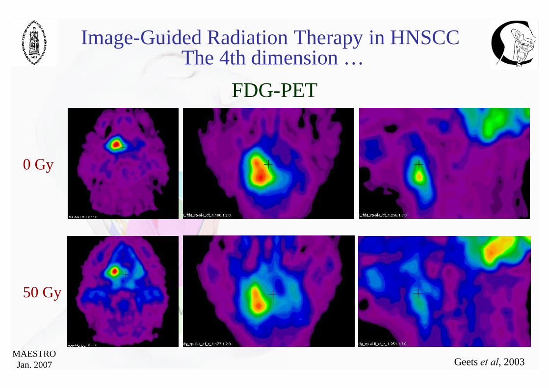
MAESTROJan. 2007 Geets et al, 2003
0 Gy
50 Gy
FDG-PET
Image-Guided Radiation Therapy in HNSCCThe 4th dimension …

MAESTROJan. 2007
Bayesian terminology for diagnostic imaging studiesDisease status
“+”“-”
Tes
t
“+”
“-” TN
FP
FN
TP
Sensitivity = TP/TP+FNSpecificity = TN/TN+FP
PPV = TP/TP+FP
NPV = TN/TN+FN
“-” “+”
“-”
“+”
48 2
20 30
NPV = 96%
PPV = 60%Test
Disease
Sensitivity = 94%
Specificity = 70%
“-” “+”
“-”
“+”
25 25
4 46
NPV = 50%
PPV = 92%Test
Disease
Sensitivity = 65%Specificity = 86%

MAESTROJan. 2007
From microscopy to PET images…
N. Christian, 2006
MR-CT: 0.21 0.06 mm 2.23 0.31
PET-CT: 0.23 0.11 mm 2.52 0.74
PET-MR: 0.28 0.07 mm 2.47 0.46
PET-AR: 0.18 0.04 mm 3.53 0.39
Mean minimal distance Mean angle (°)between two skew lines

MAESTROJan. 2007
PET-FLT
FLT
FDG
?
Haustermans et al, 2006
Work in progress: other tracers…
0 Gy 20 Gy
Lee & Geets 2006
Before surgery

MAESTROJan. 2007
Gradient Intensity Image
Image saturation (window level)
Raw
imag
eG
rad.
Int.
imag
e
Boundaries do not move!Lee & Geets 2006