pet/mri: technical design challenges and...
TRANSCRIPT

Restricted © Siemens AG 2013 All rights reserved. Answers for life.
PET/MRI: Technical Design
Challenges and Innovations Niraj K. Doshi, PhD, PMP, ExecJD – August 2013
Restricted © Siemens AG 2013 All rights reserved.
• Historical development of MR and
PET technology
• Current hybrid technology
• Potential future outlook
PET/MRI: Technical Design Challenges and
Innovations Agenda
Restricted © Siemens AG 2013 All rights reserved.
Historical Development of MR
and PET Technology

Restricted © Siemens AG 2013 All rights reserved.
MR Compatible PET System (McPET)
Insert concept by Cherry, et al. – Circa 1997
Courtesy of Simon R. Cherry, PhD – UCLA School of Medicine. Crump Institute for Biological Imaging.

Restricted © Siemens AG 2013 All rights reserved.
McPET
System design
Courtesy of Simon R. Cherry, PhD – UCLA School of Medicine. Crump Institute for Biological Imaging.
Restricted © Siemens AG 2013 All rights reserved.
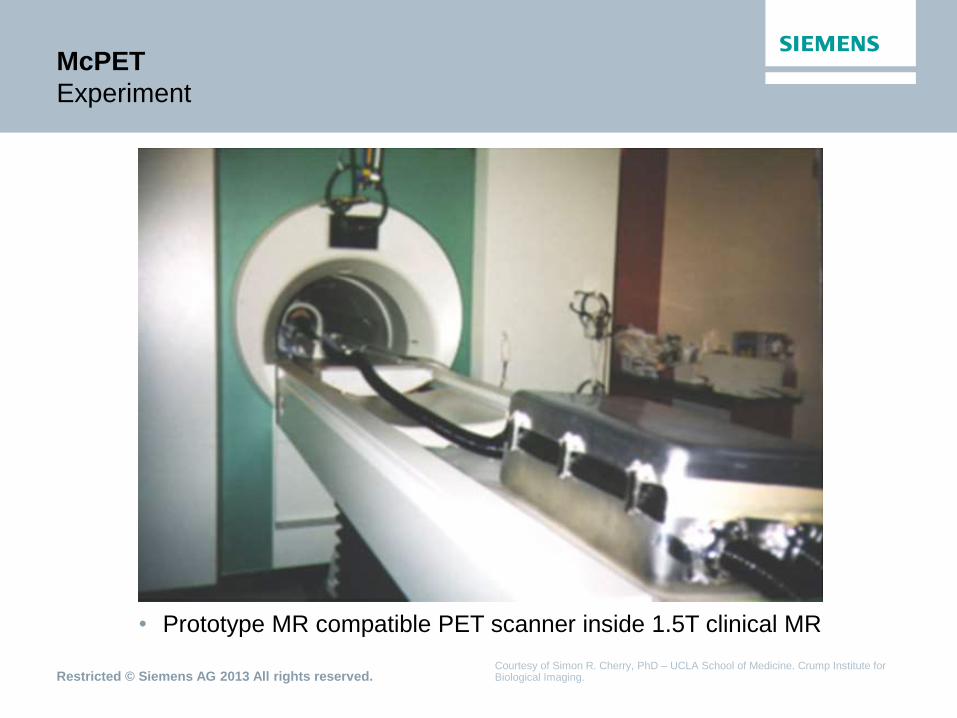
McPET
Experiment
Courtesy of Simon R. Cherry, PhD – UCLA School of Medicine. Crump Institute for Biological Imaging.
• Prototype MR compatible PET scanner inside 1.5T clinical MR

Restricted © Siemens AG 2013 All rights reserved.
McPET
Initial images ~1997-98
Courtesy of Simon R. Cherry, PhD – UCLA School of Medicine. Crump Institute for Biological Imaging.
PET Brain MR Brain
Simultaneous In Vivo Imaging
• 200 g rat injected with 1.3 mCi 18F
FDG
• Imaging time = 30 mins
• Slice thickness ~ 2 mm
• TE = 12 msec, TR = 280 msec
• Continuous 75 sec acquisition during
PET study
• Slice thickness = 4 mm

Restricted © Siemens AG 2013 All rights reserved.
BrainPET
Siemens’ 1st Clinical PET/MRI – Head Insert – Circa 2006
Technical Hurdles:
• Physical size constraints
• Cooling requirements
• RF interference
• Perturbation of MR image quality
• Perturbation of PET image quality
• Serviceability

Restricted © Siemens AG 2013 All rights reserved.
Data courtesy of Townsend and Nahmias University of Tennessee, USA and Schlemmer, Pichler, Claussen University of Tuebingen, Germany
BrainPET
Clinical results ~2006
T2 TSE PET MR-PET ADC
• Diffusion EPI sequence applied during PET acquisition.
Restricted © Siemens AG 2013 All rights reserved.
Current Hybrid Technology
Restricted © Siemens AG 2013 All rights reserved.
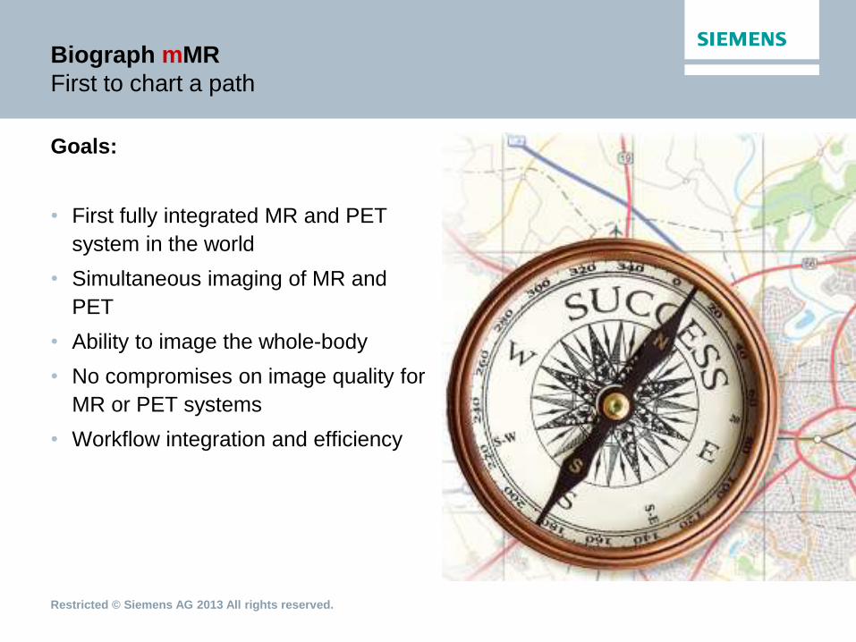
Biograph mMR
First to chart a path
Goals:
• First fully integrated MR and PET
system in the world
• Simultaneous imaging of MR and
PET
• Ability to image the whole-body
• No compromises on image quality for
MR or PET systems
• Workflow integration and efficiency
Restricted © Siemens AG 2013 All rights reserved.
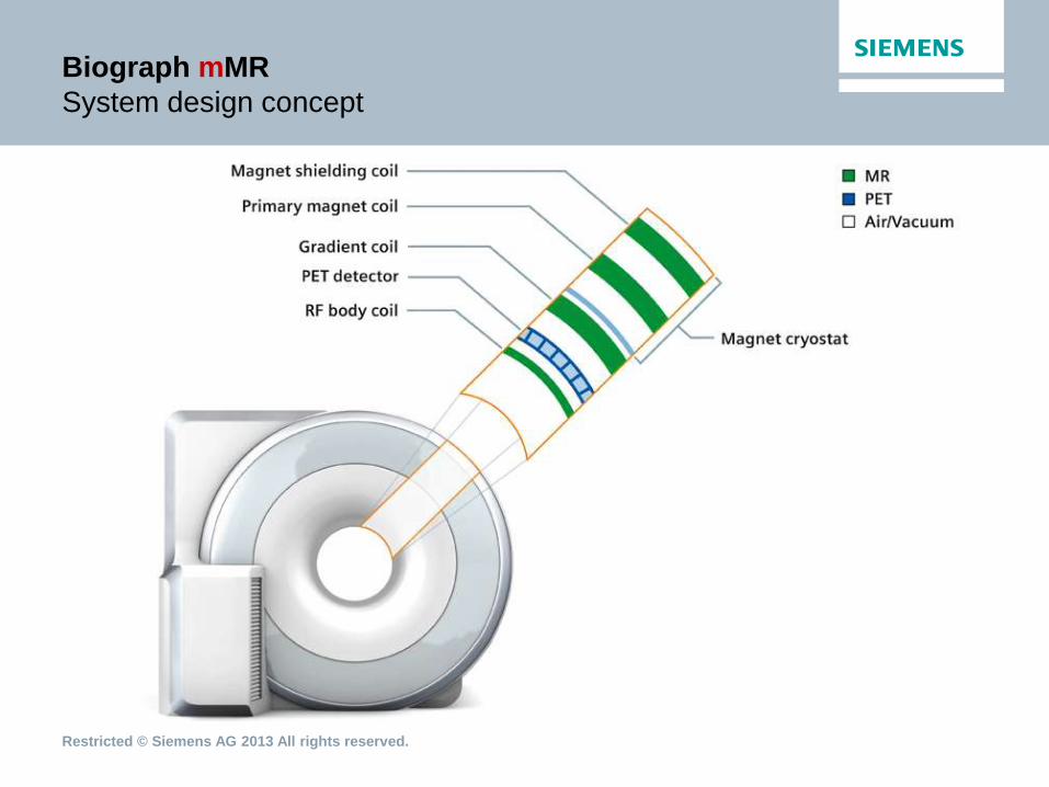
Biograph mMR
System design concept

Restricted © Siemens AG 2013 All rights reserved.
Biograph mMR
System architecture
Technical Innovations:
• Solid state detector design
• Common cooling and feedback system
• Component redesign for PET
optimization (reduce attenuation)
• RF interference optimization using
filtering
• Heat dissipation through design and
component selection
• Baseline correction to avoid gradient
amplifier noise effects

Restricted © Siemens AG 2013 All rights reserved.
PET Detector Technology
Photomultiplier tube (PMT) based
Detector block
PMT
Lightguide
A B
D

Restricted © Siemens AG 2013 All rights reserved.
Biograph mMR
Solid state detector design concept

Restricted © Siemens AG 2013 All rights reserved.
Corrected
PET
Fused
Data courtesy of the Institute of Medical Physics and University of Erlangen, Erlangen, Germany
Attenuation Correction (AC)
MRI-based AC of a patient body
Fat
μ-Map
Water
Opp Phase In Phase
Dixon VIBE Sequence
Attenuation
correction
Uncorrected
PET

Restricted © Siemens AG 2013 All rights reserved.
Attenuation Correction
Rigid stationary hardware
• CT-based AC for table, head/neck
and spine coils
• µ-map is part of PET reconstruction
process
• Relevant part of µ-map is
automatically selected for each table
position

Restricted © Siemens AG 2013 All rights reserved.
Data courtesy of Hirsch et al.: PET/MR in children. Initial clinical experience in pediatric oncology using an integrated PET/MR scanner. Pediatr Radiol (2013)
Biograph mMR
Pediatric images
11-year-old, male with T-cell
lymphoma; DWI and PET provide
the full picture
Restricted © Siemens AG 2013 All rights reserved.
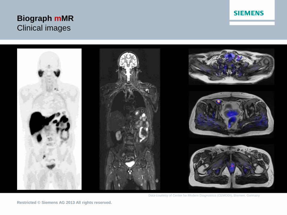
Data courtesy of Center for Modern Diagnostics (CEMODI), Bremen, Germany
Biograph mMR
Clinical images

Restricted © Siemens AG 2013 All rights reserved.
Data courtesy of Mallinckrodt Institute of Radiology, Washington University, St. Louis, USA
Biograph mMR
Temporal co-registration
Simultaneously gated PET and cardiac MR images
• PET data acquired in list mode and binned retrospectively
• MRI acquired in diastole, fused with diastolic PET data
Cardiac molecular MR
Restricted © Siemens AG 2013 All rights reserved.
Future Outlook

Restricted © Siemens AG 2013 All rights reserved.
Biograph mMR
Road forward
Facts:
• Biograph mMR is a clinical reality
• Numerous sites are performing
clinical and research studies
Future Path:
• Incorporate further customer
feedback
• Focus on clinical outcomes

Restricted © Siemens AG 2013 All rights reserved.
Let the
clinicians
lead the
way!

Restricted © Siemens AG 2013 All rights reserved.
Biograph mMR
Clinical perspective
“Dose reduction is a major concern of
ours -- and this can be quite
considerable.
This is particularly critical in the
examination of children and young
adults, especially in cases of frequent
follow-up examinations in therapy
monitoring.”
Markus Lentschig, MD
Radiologist and partner at the CEMODI
practice
Bremen, Germany
First purely private Biograph mMR user
The statements by Siemens’ customers described herein are based on results that were achieved in the customer's unique setting. Since there is no "typical" setting and many variables exist there can be no guarantee that other customers will achieve the same results.

Restricted © Siemens AG 2013 All rights reserved.
Biograph mMR
Clinical perspective
“From our own experience in cervical
cancer patients, we see that
simultaneous MR and PET is the ideal
imaging tool covering the whole range
of clinical care from diagnosis, therapy
planning, monitoring and follow-up.
The benefits over other modalities are
improved spatial registration, the high
soft tissue contrast and functional
capabilities of MRI, and minimizing the
patient’s scan time or dose burden.”
Perry W. Grigsby, MD
Professor, Radiation Oncology
Director, Brachytherapy Center
Washington University, St. Louis, MO, USA
The statements by Siemens’ customers described herein are based on results that were achieved in the customer's unique setting. Since there is no "typical" setting and many variables exist there can be no guarantee that other customers will achieve the same results.

Restricted © Siemens AG 2013 All rights reserved.
Biograph mMR
Early adopters > 35
Europe
• IMP Erlangen, Germany
• Klinikum r. d. Isar, Munich, Germany
• Univ. Hospital Tübingen, Germany
• Univ. Hospital Leipzig, Germany
• CEMODI Bremen, Germany
• Univ. Hospital Essen, Germany
• Univ. College London Hospitals, UK
• SDN, Naples, Italy
• DKFZ, Heidelberg, Germany
• Rigshospitalet, Copenhagen, Denmark
• St. Olav Hospital, Trondheim, Norway
• DLRZ, Bonn, Germany
• University of Padua, Italy
• CERMEP/Univ. of Lyon, France
• AKH/MUW, Vienna, Austria
• OAO RZD, Moscow, Russia
• King‘s College London, UK
• Centr. Onkologii, Bydgoszcz, Poland
North America
• MGH, Boston, USA
• NIH, Bethesda, USA
• Washington Univ., St. Louis, USA
• Univ. of N. Carolina, Chapel Hill, USA
• Mt. Sinai Medical Center, New York, USA
• Indiana University, Indianapolis, USA
• Lawson Health Research Institute,
London, Canada
• UPMC, Pittsburgh, USA
• NYU, New York, USA
• Cleveland Clinic Foundation, Cleveland, USA
• Stony Brook University, Stony Brook, USA
• Weill Cornell Imaging, New York Presbyterian,
NY, USA (2x)
• Cedar-Sinai Medical Center, Los Angeles, USA
• UHN Toronto, Canada
Asia
• PLA 301 Hospital, Beijing, China
• Parkway Mt. Elizabeth Novena Hospital,
Singapore
• CIRC/NUS/A*Star, Singapore
• Yeungnam Univ. Hospital, Daegu, Korea
• SNUH, Seoul, Korea
• Apollo Hospitals, Delhi, India
• Fukushima Medical Univ, Fukushima,
Japan
• TNUH, Taipei, Taiwan
• NIMHANS, Bangalore, India
• HKSH, Hong Kong, China

Restricted © Siemens AG 2013 All rights reserved.
Future innovations*...
• Workflow improvements
• Extended MR FoV
• Motion correction
• Whole-body bone segmentation
*These features are not commercially available in all countries. Due to regulatory reasons their future availability cannot be guaranteed. Please contact your local Siemens organization for further details.

Restricted © Siemens AG 2013 All rights reserved.
Global Business Unit
Siemens Medical Solutions USA, Inc.
Molecular Imaging
810 Innovation Drive
Knoxville, TN
USA
www.siemens.com/mi
Answers for life.
Thank you!