pex22p of pichia pastoris , essential for peroxisomal...
TRANSCRIPT
The Rockefeller University Press, 0021-9525/99/07/99/14 $5.00The Journal of Cell Biology, Volume 146, Number 1, July 12, 1999 99–112http://www.jcb.org 99
Pex22p of
Pichia pastoris
, Essential for Peroxisomal Matrix Protein Import, Anchors the Ubiquitin-conjugating Enzyme, Pex4p, on thePeroxisomal Membrane
Antonius Koller,* William B. Snyder,* Klaas Nico Faber,* Thibaut J. Wenzel,* Linda Rangell,
‡
Gilbert A. Keller,
‡
and Suresh Subramani*
*Department of Biology, University of California San Diego, La Jolla, California 92093-0322; and
‡
Pharmacological Science, Genentech, South San Francisco, California 94080
Abstract.
We isolated a
Pichia pastoris
mutant that was unable to grow on the peroxisome-requiring media, methanol and oleate. Cloning the gene by complemen-tation revealed that the encoded protein, Pex22p, is a
new peroxin. A
D
pex22
strain does not grow on metha-nol or oleate and is unable to import peroxisomal ma-trix proteins. However, this strain targets peroxisomal membrane proteins to membranes, most likely peroxi-somal remnants, detectable by fluorescence and elec-tron microscopy. Pex22p, composed of 187 amino acids, is an integral peroxisomal membrane protein with its NH
2
terminus in the matrix and its COOH terminus in the cytosol. It contains a 25–amino acid peroxisome membrane-targeting signal at its NH
2
terminus. Pex22p interacts with the ubiquitin-conjugating enzyme Pex4p,
a peripheral peroxisomal membrane protein, in vivo, and in a yeast two-hybrid experiment. Pex22p is re-quired for the peroxisomal localization of Pex4p and in strains lacking Pex22p, the Pex4p is cytosolic and unsta-ble. Therefore, Pex22p anchors Pex4p at the peroxiso-mal membrane. Strains that do not express Pex4p or Pex22p have similar phenotypes and lack Pex5p, sug-gesting that Pex4p and Pex22p act at the same step in peroxisome biogenesis. The
Saccharomyces cerevisiae
hypothetical protein, Yaf5p, is the functional homo-logue of
P
.
pastoris
Pex22p.
Key words: organelle • peroxin • peroxisome • pro-tein transport • yeast
P
EROXISOMES
are single-membrane–bound organellespresent in all eukaryotic cells. They contain en-zymes that are responsible for such metabolic path-
ways as hydrogen peroxide metabolism,
b
-oxidation oflong-chain fatty acids, synthesis of plasmalogens, choles-terol, and bile acids, and degradation of purines and aminoacids (for review see Van den Bosch et al., 1992). To en-sure that all enzymes for these metabolic pathways areproperly targeted to the peroxisomes, cells have evolvedseveral mechanisms to direct these enzymes to their cor-rect locations after they have been translated.
Matrix-localized enzymes contain either one of two per-
oxisome-targeting signals (PTSs)
1
. PTS1 is located at theextreme COOH terminus of peroxisomal proteins. It con-sists of three amino acids and has the sequence SKL orsome variants of it. PTS2 is present at the NH
2
terminusand has a consensus sequence of R/K-L/V/I-X
5
-H/Q-L/A(for review see Subramani, 1998). Each of these PTSs isrecognized by a specific receptor, peroxin (Pex)5p orPex7p, respectively. Mutants lacking functional Pex5p arestill able to import PTS2-containing proteins, whereas cellslacking Pex7p are only able to import PTS1-containingproteins (for review see Subramani, 1998). These resultssuggested the existence of two distinct import pathwaysfor peroxisomal matrix proteins. The localization of thesetwo receptors is still controversial. It seems, however, thatboth receptors are localized to the cytosol and peroxi-
Address correspondence to Professor S. Subramani, Department of Biol-ogy, University of California San Diego, Bonner Hall, Room 3230, 9500Gilman Drive, La Jolla, CA 92093-0322. Tel.: (619) 534-2327. Fax: (619)534-0053. E-mail: [email protected]
Klaas Nico Faber’s present address is University of Groningen, Gron-ingen Biomolecular Sciences and Biotechnology Institute, Eukaryotic Mi-crobiology, Kerklaan 30, 9751 NN Haren, The Netherlands.
Thibaut J. Wenzel’s present address is Gist-Brocades, Food SpecialitiesDivision, Wateringseweg 1, 2600 MA Delft, The Netherlands.
1.
Abbreviations used in this paper:
AD, activation domain; AOX, alcoholoxidase; DB, DNA-binding domain; DSP, dithiobis(succinimidylpropi-onate); G6PDH, glucose-6-phosphate dehydrogenase; GFP, green fluo-
rescent protein; IPTG, isopropyl
b
-
D
-thiogalactopyranoside; mPTS, mem-brane peroxisome targeting signal; Pex, peroxin; PNS, post nuclearsupernatant; PTS, peroxisome-targeting signal; UBC, ubiquitin-conjugat-ing enzyme.
on October 17, 2007
ww
w.jcb.org
Dow
nloaded from

The Journal of Cell Biology, Volume 146, 1999 100
somes, suggesting that the receptors shuttle from the cyto-plasm to the peroxisomes, where they bind to the tightlyassociated peroxisomal membrane protein Pex13p (Gir-zalsky et al., 1999) and Pex14p (Albertini et al., 1997; Bro-card et al., 1997; Fransen et al., 1998). Pex5p and Pex7p, aswell as Pex14p, are in a complex with another peripheralperoxisomal membrane protein, Pex17p (Huhse et al.,1998). The binding of Pex5p and Pex7p to Pex17p, how-ever, was dependent on the presence of Pex14p (Huhse etal., 1998). Deleting the genes encoding Pex14p or Pex17pinhibited both the PTS1- and PTS2-dependent importpathways, suggesting that these proteins function at apoint of convergence for the two import pathways. Pex5p,Pex7p, and Pex14p were also shown to interact with theSH3 domain–containing, peroxisomal integral membraneprotein Pex13p (Elgersma et al., 1996; Erdmann and Blo-bel, 1996; Gould et al., 1996; Albertini et al., 1997; Girzal-sky et al., 1999). Pex7p is only targeted to the peroxisomeswith the help of the interacting proteins Pex18p andPex21p (Purdue et al., 1998). Several other proteins havebeen implicated in the import of peroxisomal matrix pro-teins. Antibodies against cytosolic HSP70 inhibit the im-port of SKL-containing proteins into peroxisomes (Wal-ton et al., 1994; Fransen et al., 1998). Deleting the geneencoding Djp1p, a cytosolic DnaJ-like protein, had a dras-tic effect on peroxisomal import of certain PTS-containingproteins (Hettema et al., 1998). These data suggest that af-ter being translated, PTS1- and PTS2-containing proteinsare recognized by their respective receptors. This interac-tion could be facilitated by the action of chaperones andtheir cofactors. The complex of PTS-containing proteinand receptor is then transferred to the peroxisomeswhere the receptor is recognized by the complex com-prised of Pex13p, Pex14p, and Pex17p. Then the receptorreleases the cargo which is then transported into the per-oxisome.
There is little known of the mechanism for targetingperoxisomal membrane proteins. Different consensus se-quences for peroxisomal membrane targeting have beenproposed (Dyer et al., 1996; Elgersma et al., 1997). This se-quence (called mPTS [membrane peroxisomal targetingsignal]) seems to be on the lumenal side of membrane pro-teins, close to a transmembrane sequence, and consists ofseveral positively charged amino acids (Dyer et al., 1996;Elgersma et al., 1997). How these proteins reach the per-oxisomal membrane is still a matter of debate. A few per-oxisomal membrane proteins (Pmp70 and Pmp22) are in-serted directly from the cytoplasm into the membrane(Fujiki et al., 1984; Bodnar and Rachubinski, 1991; Dis-telkötter and Just, 1993; Imanaka et al., 1996), whereas itwas shown that other membrane proteins (Pex3p andPex15p) are also targeted to the ER, at least when they areoverexpressed, suggesting that these proteins transit viathe ER to the peroxisome (Baerends et al., 1996; Elgersmaet al., 1997).
Another interesting protein involved in peroxisomalprotein transport is Pex4p. Pex4p was shown to be highlyhomologous to ubiquitin-conjugating enzymes (UBCs)(Wiebel and Kunau, 1992; Crane et al., 1994; van der Kleiet al., 1998) and is located on the peroxisomal membraneas a peripheral protein facing the cytosol. The proteinbinds ubiquitin and the active-site cysteine residue is im-
portant for Pex4p function (Crane et al., 1994). In
S
.
cere-visiae
and
P
.
pastoris
, deletion of
PEX4
disrupts target-ing via the PTS1 and PTS2 pathway (Wiebel and Kunau,1992; Gould et al., 1992; our unpublished observation). In
Hansenula polymorpha
, only the PTS1 pathway was im-paired and this defect could be rescued by overexpressionof the PTS1 receptor, Pex5p
(van der Klei et al., 1998), im-plying an important role for Pex4p in the import of PTS1-containing proteins in this yeast.
In this study we have characterized a novel peroxin,Pex22p, from
P
.
pastoris
.
We analyzed the subcellular lo-calization, targeting signal, topology, interacting partners,and the functions of this protein. Our data shed light onthe roles of Pex4p and Pex22p in peroxisomal matrix pro-tein import and show that these proteins are also con-served in
S
.
cerevisiae
.
Materials and Methods
Strains and Media
P
.
pastoris
strains used in this study were as follows: parental wild-typestrains PPY1, PPY12 (PPY1
arg4
,
his4
), and SMD1163 (
his4
,
pep4
,
prb1
);STK10 (PPY12,
pex22
.
1
,
ARG4
::pTW84 (PTS2-GFP [green fluorescentprotein] (S65T)/
GAPDH
, PTS2-BLE/
GAPDH
); STK11 (PPY12,
D
pex22
::Zeocin); STK12 (SMD1163,
D
pex22
::Zeocin); STK13 (PPY12,
D
pex4
::Zeocin); STK14 (SMD1163,
D
pex4
::Zeocin); STK15 (PPY12, pTK51[
PEX4p::NH-PEX4
, Zeocin
r
]).
S
.
cerevisiae
strains used: BJ1991
(
MAT
a
,
leu2
,
trp1
,
ura3-251
,
prb1-1122
,
pep4-3
); STK16 (BJ1991,
D
yaf5
); L40(
MATa
,
his3
D
200,
trp1-901
,
leu2-3,112
,
ade2
,
LYS2
::(lexAop)
4
-HIS3
,
URA3
::(lexAop)
8
-lacZ). The
Escherichia coli
strain used for cloning pro-cedures was JM109 and for protein expression, SG13009. Yeast mediawere as described in Faber et al. (1998).
Cloning Procedures
Standard cloning procedures were used (Sambrook et al., 1989). DNA se-quencing was performed according to the Sanger method (Sanger et al.,1977). Restriction site ends were made blunt with Klenow polymerasefrom Boehringer Mannheim. PCR was performed using Vent DNA poly-merase from New England Biolabs. The resulting PCR products werecloned into pCR2.1 by adding a 3
9
A-overhang with Taq polymerase orinto pCR-Blunt (Invitrogen). Usually, a restriction site was introducedwithin the primers to facilitate further cloning of the products, otherwiserestriction sites from the pCR vectors were used. PCR fragments were cutout with specified restriction enzymes, purified with Qiaex (Qiagen) andcloned into the specified vectors according to standard protocols.
Isolation of pex Mutants and Cloning of PEX22
The isolation of
pex
mutants was performed according to Elgersma et al.(1998). A genomic library was transformed into the
pex22
.
1
strain(STK10). Five different plasmids (p82.2, p82.3, p82.9, p82.13, and p82.15)restored growth on methanol and oleate medium. Restriction analysis ofthe inserts revealed that the five inserts contained an overlapping frag-ment of 1.1 kb. This fragment was excised from plasmid p82.13 as aBamHI fragment, sequenced on both strands, and shown to include the
PEX22
gene.
Construction of Disruptions
To disrupt
PEX22
, the 5
9
and 3
9
regions of the gene were amplified withPCR (TK45 and TK46 for the 5
9
region and TK47 and TK48 for the 3
9
re-gion). The 5
9
fragment was cloned as a BamHI-SmaI fragment into pBlue-scriptSKII (Stratagene). The 3
9
fragment was then ligated as an EcoRI-SmaI fragment into this vector. The resulting plasmid was cut with SmaIand a blunt-ended HaeII-BamHI Zeocin fragment (cut out from plasmidpPICZ A; Invitrogen) was inserted. The resulting plasmid, pTK29, was cutwith BamHI and EcoRI and transformed into PPY12 and SMD1163. Thedisruptions were confirmed by PCR.
The 5
9
and 3
9
regions of the
PEX4
gene were amplified with PCR
on October 17, 2007
ww
w.jcb.org
Dow
nloaded from

Koller et al.
Role of PpPex22p in Peroxisome Biogenesis
101
(primers TK41 and TK42 for the 5
9
region and TK43 and TK44 for the 3
9
region). The 5
9
fragment was cloned as a BamHI-SmaI fragment intopBluescriptSKII. The 3
9
fragment was then ligated as an HindIII-SmaIfragment into this vector (cut with HindIII-SmaI). The resulting fragmentwas then cut with SmaI and a blunt-ended HaeII-BamHI Zeocin fragmentwas inserted. The resulting plasmid, pTK35, was cut with BamHI andHindIII and transformed into PPY12 and SMD1163. The disruptions wereconfirmed by PCR.
The
ScYAF5
gene was disrupted according to Wach et al. (1994). Prim-ers TK53, TK62, TK63, and TK64 were used to isolate a fragment usingPCR that contains the 5
9
region of
ScYAF5
, followed by kanMX2, fol-lowed by the 3
9
region of
ScYAF5
. This construct was transformed intothe
S
.
cerevisiae
strain BJ1991 and G418-resistant colonies were checkedfor correct disruption of the
ScYAF5
gene by PCR.
Construction of Plasmids
Plasmids used are in Table I and DNA primers are in Table II. Plasmidp82.20 contains the 1.1-kb BamHI fragment of p82.13 in vector pSG560(Gould et al., 1992). p82.21 contains the 0.7-kb BglII-BamHI fragment,p82.22 the 0.8-kb BamHI-HincII fragment, p82.23 the 0.3-kb HincII frag-ment, p82.24 the 0.5-kb NarI-BamHI fragment, and p82.25 the 0.9-kbEcoRV-BamHI fragment. All these fragments were cloned into pSG560either as a blunt-ended fragment or as a blunt-ended BamHI fragmentcontaining one blunt end and one BamHI end.
Plasmid pTK10, which expresses the
PEX22
gene from the alcohol oxi-dase (AOX) promoter, was cloned as follows: the gene was amplified byPCR using primer TK31 and TK40, thereby introducing a BamHI site im-mediately upstream of the ATG. The
PEX22
gene was excised withBamHI and EcoRI and cloned into pPIC3K (Invitrogen) cut with BamHIand EcoRI.
Yeast two-hybrid plasmids were made by fusing appropriate gene frag-ments downstream of the DNA binding (DB) domain of LexA or the acti-vation domain (AD) of VP16.
PEX22
fusions were generated by cloning,in-frame, parts of or the full-length
PEX22
fused to either domains in
plasmids pKNSD55 (pBTM116 based) or pKNSD52 (pVP16 based)(Faber et al., 1998). Plasmids pTK12 and pTK13 were constructed by fus-ing a BamHI-EcoRI PCR fragment of
PEX22
(primer TK34 and TK40)with BamHI-EcoRI cut pKSND55 or pKNSD52, respectively. PlasmidpTK14, expressing a
Pex22p
lacking the first 25 NH
2
-terminal amino ac-ids, was constructed by cloning the BamHI-EcoRI PCR fragment ob-tained with primers TK35 and TK40 into BamHI-EcoRI cut pKNSD55.Plasmid pTK16, expressing the COOH-terminal part of Pex22p, was con-structed by cloning the BamHI-EcoRI PCR fragment obtained with prim-ers TK36 and TK40 into BamHI-EcoRI cut pKNSD55. Plasmid pTK18,expressing the NH
2
-terminal part of Pex22p, was constructed by cloningthe BamHI-EcoRI PCR fragment obtained with primers TK34 and TK37into BamHI-EcoRI cut pKNSD55.
Plasmids containing
PEX4
(as a BamHI-EcoRI fragment made byPCR with primers KNF13 and KNF14) in pKNSD55 or pKNSD52 (Faberet al., 1998) for two-hybrid analysis were named pKNF119 and pKNF118,respectively. Plasmid pTK21 was constructed as follows: a BamHI-EcoRVfragment of
PEX4
(cut out of pKNF118) was cloned into pKNSD55,which had been cut with BamHI and EcoRI (blunt ended). PlasmidpTK23 contains a BamHI-SspI fragment of
PEX4
cloned into pKNSD55.Plasmid pTK25 contains an EcoRV-EcoRI fragment of
PEX4
cloned intopKNSD53, cut with BamHI (blunt ended) and EcoRI. Plasmid pTK27contains an SspI-EcoRI fragment of
PEX4
in pKNSD53, cut with BamHI(blunt ended) and EcoRI. Plasmid pTK36, expressing a 6HIS-taggedPex4p from the GAPDH promoter was made as follows:
PEX4
was am-plified with primers TK51 and KNF14 (Faber et al., 1998) and cloned as aBamHI-HindIII fragment into the BamHI-HindIII cut pQE30 (Qiagen).This plasmid was cut with EcoRI-HindIII, blunt ended using Klenow en-zyme and cloned into the EcoRI site of pTW71, which was blunt ended us-ing Klenow enzyme.
Plasmids expressing GFP-SKL (pTW51) and PTS2-GFP (pTW66) wereas described (Wiemer et al., 1996). Plasmid pTK30, containing full-length
Table I. Plasmids Used in This Study
Plasmid Relevant features Source
pTW5 GFP-SKL Wiemer et al., 1996pTW66 PTS2-GFP Wiemer et al., 1996pTK10 pPIC3K
PEX22
This studypTK12 pKNSD55
PEX22
This studypTK13 pKNSD52
PEX22
This studypTK14 pKNSD55
PEX22
(26-187) This studypTK16 pKNSD55
PEX22
(88-187) This studypTK18 pKNSD55
PEX22
(1-89 This studypTK20 pQE30
PEX22
(26-187) This studypTK21 pKNSD52
PEX4
(1-87) This studypTK23 pKNSD52
PEX4
(1-124) This studypTK25 pKNSD53
PEX4
(88-204) This studypTK27 pKNSD53
PEX4
(125-204) This studypTK29 pBluescript 5
9
PEX22-Zeocin-39 PEX22
This study
pTK30 pPIC3K PEX22-GFP This studypTK32 pTW71 PEX22(1-25)-GFP This studypTK34 pTW71 PEX22(1-7)-GFP This studypTK35 pBluescript 59PEX4-Zeocin-39PEX4 This studypTK36 pTW71 6HIS-PEX4 This studypTK37 pQE30 PEX4 (1-124) This studypTK45 pRS306 ScYAF5 This studypTK46 pKNSD52 ScYAF5 This studypTK47 pKNSD55 ScYAF5 This studypTK48 pKNSD52 ScPEX4 This studypTK49 pKNSD55 ScPEX4 This studypTK50 pTW71 NH-PEX4 This studypTK51 pBluescript 59PEX4-NH-PEX4-
Zeocin-39PEX4This study
pKNSD119 pKNSD55 PEX4 This studypKNSD118 pKNSD52 PEX4 This study
Table II. Primers Used in This Study
Primer 59-Sequence-39
TK31 59-GGATCCATGAAATCCACAAAGAGAAACTK34 59-GGATCCAAATCCACAAAGAGAAACACTK35 59-GGATCCAAGAGTTTTATAACGTCCGACTK36 59-GGATCCGCCAAGGACGAATTTCTACGAGTK37 59-CTACTTGGCTATTGTCGGCGCCAGCACTK40 59-GGCAAGAGAGACATCACCGGACGCCGTK41 59-GGATCCTGAGGAAATGCTTGGGCCTTAACAGGCTK42 59-CCCGGGTTCTTTGGATTATCAAGTGGGGAAAGAGAGTK43 59-CCCGGGTTAATGCTATTTGAATCCGAAAAATTCCTCAAT-
GTCTK44 59-AAGCTTGATCTAACCCCTCCAAAGAAGACCCCGTK45 59-GGATCCACCCCGTGAAATGGACCCCGTGAGTK46 59-CCCGGGGTTGAAGAAGGGAGAGTATTGAGGTK47 59-CCCGGGCGGCGTCCGGTGATGTCTCTCTK48 59-CGACTTTCTAGACAGTTGACTK51 59-GGATCCTCAGCTGAAAAGCGTTTGTK52 59-GGAGAAAGTGACCAACTCGGCGACGTK53 59-GGATCCATGCCACCACCATCAAGAAGTAGTK59 59-AAGATCTCATTGTATATATATTGATTCACTK61 59-AAGATCTTCTTGTAAACAGAGTACCCCAGTK62 59-GTAGTTGGTGTCGGTGGCACTK63 59-GGGGATCCGTCGACCTGCAGCGTACCATGTTTTATATCT-
TCTTCTTCTTTATTCTK64 59-AACGAGCTCGAATTCATCGATGATATAATGTAGAAAAGA-
GTTTCTTGTAACAGTATGTK67 59-GGATCCATGCCAAACTTCTGGATTCTTGAGTK68 59-CCTCAACGATCTACTGGGCTGCCTK95 59-CGGATCCATGACTTTTTTTGGCTTGGCAGCCCTTGGGGCTK96 59-GGGATCCATGAAATCCACAAAGAGAAACAGTGCAGGAGA-
AGAACTTTTCACTGGAGTTGTW6 59-AGAGAATTCTTATTATTTGTATAGTTCKNF13 59-GGCGGATCCATGTCAGCTGAAAAGCGTTTGKNF14 59-GGAGATGAATTCTATCGGGpPAS8-1 59-AGATCTACCATGTCGCTTATTGGCGG
on October 17, 2007
ww
w.jcb.org
Dow
nloaded from

The Journal of Cell Biology, Volume 146, 1999 102
PEX22 fused to GFP, was made by amplifying PEX22 with primers TK31and TK59, cutting the fragment with BglII and BamHI and cloning it intothe BglII-BamHI cut plasmid pTW113, which contains a GFP gene with-out the ATG in plasmid pTW71. The plasmid containing the first 25 aminoacids of Pex22p fused to GFP was made as follows: the fragment encodingthe first 25 amino acids was amplified by PCR with primers TK31 andTK61. This HindIII-BglII fragment was cloned into the HindIII-BglII cutplasmid pTW103, which contains a full-length GFP fragment missing theATG in pCR2.1 (Invitrogen). The resulting plasmid was cut with BamHIand EcoRI to excise the fragment containing Pex22(1–25)-GFP andcloned into BamHI-EcoRI cut pTW71 resulting in plasmid pTK32. Theplasmid expressing the first 7 amino acids of Pex22p fused to GFP(Pex22(1–7)-GFP) was made as follows: a BamHI-EcoRI cut PCR frag-ment with primer TK96 and TW6 was ligated into BamHI-EcoRI cutpTW71 resulting in plasmid pTK34. Plasmid pTK44, expressing a GFPfused to the amino acids 8–25 of Pex22p (Pex22(8–25)-GFP) was cloned asfollows: PCR was performed with primer TK95 and Pichia primer 39AOX(Invitrogen) with plasmid pTK32 as template. The resulting fragment wascut with BamHI and EcoRI and cloned into plasmid pTW71, cut withBglII and EcoRI.
Plasmid pTK50 expressing a NH-tagged Pex4p from the acyl-CoA oxi-dase (ACO) promoter was cloned as follows: a BamHI-EcoRI fragmentcontaining the full-length PEX4 was cloned into plasmid pM22 cut withBamHI-EcoRI (Elgersma et al., 1998). Plasmid pTK51, expressing NH-Pex4p from its own promoter was cloned as follows: the fragment express-ing NH-Pex4p was cut out of pTK51 with BglII-EcoRI, treated withKlenow enzyme and cloned into the SmaI site of the pBluescriptSKII con-taining the 59 end of PEX4 and the 39 end of PEX4 (see above). Ablunt-ended HaeII-BamHI Zeocin fragment was then cloned into theblunted EcoRI site in the 39 end of PEX4. This whole fragment (59-PEX4-NH-PEX4-Zeocin-39PEX4) was cut out of the plasmid with XbaI-HindIIIand transformed into a Dpex4 strain (PPY12, Dpex4::ARG4). Arginineminus and Zeocin resistant colonies were checked for their expression ofNH-Pex4p.
A fragment of ScYAF5 containing the full-length gene was amplifiedwith primers TK52 and TK62 on genomic S. cerevisiae DNA. The result-ing EcoRI fragment was cloned into pRS306, cut with EcoRI to yield plas-mid pTK45. The two-hybrid vectors with ScYAF5 were made as follows:ScYAF5 was amplified with PCR with primers TK52 and TK53. The re-sulting BamHI-EcoRI fragment was cloned into either pKNSD55 (toyield plasmid pTK46) or pKNSD52 (pTK47). Plasmids for the two-hybridexperiment expressing ScPEX4 were made by amplifying ScPEX4 withprimers TK67 and TK68. The resulting fragment was cloned as a BamHI-EcoRI fragment into pKNSD55 (to yield pTK48) and pKNSD52 (to yieldpTK49).
Production of AntibodiesFor the construction of a 6HIS-Pex22p, a BamHI-EcoRI fragment ofPEX22 produced by PCR with primers TK35 and TK40 was cloned into aBamHI-EcoRI cut plasmid pQE30 (Qiagen) creating plasmid pTK20.This plasmid, expressing Pex22p missing the first 25 amino acids, wastransformed into E. coli SG13009 and the gene induced with isopropylb-D-thiogalactopyranoside (IPTG). The protein was purified under nativeand denaturing conditions on Ni21-NTA beads according to the manufac-turer’s manual (Qiagen). The purified proteins were used to immunizerabbits. The antibodies were preabsorbed against an acetone powder ex-tract from a Dpex22 strain. Basically, the deletion strain was grown in oneliter of methanol medium to an OD of 1. The cells were pelleted and re-suspended in PBS at a cell density of 20 OD. Zymolyase was added (1 mg)and the cells were incubated with gentle shaking for 20 min. The cellswere placed on ice for 5 min. Cold acetone (four volumes) was added, fol-lowed by another incubation on ice for 30 min. The cells were rewashedwith cold acetone and placed on ice for another 30 min. The cells werepelleted, put into a mortar and dried. The cells were subsequently groundto a fine powder using a pestle. This powder was incubated with undilutedsera at a concentration of 1% (wt/vol) at 4°C. After an overnight incuba-tion, the tube was centrifuged for 10 min, the supernatant collected andused for further studies.
Plasmid pTK37, expressing a 6HIS-tagged NH2 terminus of Pex4p, wasmade by cloning the BamHI-SspI fragment of pTK23 into pQE30, whichwas cut with BamHI-SmaI. This plasmid was transformed into strainSG13009 and the protein induced with IPTG. Expressed protein was puri-fied under denaturing conditions according to the manufacturers proce-dure (Qiagen). The pure protein was then injected into rabbits for anti-
body production. The resulting antibody was then further purified usingan affinity-purification protocol according to Harlow and Lane (1988).
Differential Centrifugation, Nycodenz Gradient, Floatation Gradient, Membrane Extraction, and Protease ProtectionDifferential centrifugation and Nycodenz gradients were done as de-scribed (Faber et al., 1998). Floatation gradient was done as described(Faber et al., 1998) with the difference that the 27,000-g pellets weretaken. Membrane extraction and protease protection experiments weredone as described (Wiemer et al., 1996).
Binding of Pex22p to Pex4pA Dpex4 strain (STK14) was transformed with plasmid pTK36, expressinga 6HIS-Pex4p. This strain and SMD1163 as a control were grown in meth-anol and spheroplasts were prepared. Cross-linking of cell extracts wasperformed as previously described (Rieder and Emr, 1997). 50 ml of a50% slurry of Ni21-NTA agarose (Qiagen) was added with 10 mM imida-zole to the supernatant to precipitate protein complexes. This mixture wasincubated at 48C for 1 h. After this incubation period the beads werewashed five times with buffer containing 20 mM imidazole. The pelletswere resuspended in sample buffer and part of it loaded onto an SDS gel.
Fluorescence and Electron MicroscopyFluorescence microscopy for the detection of GFP-tagged proteins wasdone as described by Monosov et al. (1996). Fluorescence images were ac-quired using a CCD camera (model 4995; Cohu Inc.) and a CG-7 FrameGrabber (Scion Corp.). Samples for immunofluorescence were induced inmethanol, spheroplasted, fixed, and prepared as described (Babst et al.,1998). a-Pex3p and a-AOX were used at a dilution of 1:10,000. Micros-copy for immunofluorescence was as described (Odorizzi et al., 1998).
MiscellaneousTCA lysates were made as follows: 2 OD of cells were collected by centrif-ugation, resuspended in 10% TCA and incubated on ice for .30 min. Thesuspension was centrifuged and the pellet washed three times with ace-tone. The pellet was resuspended in sample buffer and glass beads added.The tube was vortexed for 1 min and heated at 1008C for 1 min. This pro-cedure was repeated four times. The sample was separated from the glassbeads and loaded on gels.
Digitonin permeabilization was done according to Elgersma et al.(1998). Western blotting was performed according to standard proce-dures. Antibodies were used at the following dilutions: a-Sccatalase,1:10,000; a-Scthiolase, 1:10,000; a-ScG6PDH (glucose-6-phosphate dehy-drogenase), 13,000; a-F1b subunit of mitochondrial ATPase, 1:10,000;a-PpPex3p, 1:10,000; a-PpPex4p, 1:1,000; a-PpPex5p, 10,000; a-PpPex7p,1:10,000; a-PpPex22p, 1:2,000; a-GFP, 1:2,000.
Results
Isolation of Peroxisomal Protein Import Mutants
The screen employed for the isolation of import mutantswas based on a positive screening procedure (Elgersma etal., 1993, 1998). It used the bleomycin-resistance protein,which binds the toxic drug phleomycin, thereby preventingthe drug from intercalating into DNA. The bleomycingene (BLE) was fused to 51 basepairs, encoding the NH2-terminal 17 amino acids (containing the PTS2 signal), of S.cerevisiae thiolase (FOX3). The fusion protein was tar-geted to the peroxisomes in P. pastoris wild-type cells,thereby rendering the cells sensitive to phleomycin. In pexmutants, however, this fusion protein would not be tar-geted into peroxisomes, therefore rendering the cells resis-tant to the drug. A wild-type yeast strain (PPY12 1pTW84; Elgersma et al., 1998) was mutagenized, grown inoleate and treated with phleomycin. Two phleomycin-
on October 17, 2007
ww
w.jcb.org
Dow
nloaded from

Koller et al. Role of PpPex22p in Peroxisome Biogenesis 103
resistant mutants (Pppex7.1 and Ppfox3.1) did not growon oleate, but grew on methanol (Elgersma et al., 1998;Koller, A., and S. Subramani, unpublished results). Oneother mutant did not grow on methanol and oleate, al-though it grew on glucose and glycerol, and was namedpex22.1. This mutant was backcrossed twice against wild-type and the resulting strain (STK10) was used for furtherexperiments.
Cloning of PEX22
The pex22.1 mutant (STK10) was transformed with a wild-type genomic library and plasmids (p82.2, p82.3, p82.9,p82.13, and p82.15) from colonies that grew on methanolmedium were isolated and rechecked for their ability torestore growth on methanol and oleate. The five insertscontained an overlapping fragment of 1.1 kb which wasisolated from p82.13 as a BamHI fragment and subclonedinto the pSG560 vector (Gould et al., 1992) to check forcomplementation (p82.20; Fig. 1 A). The smallest, comple-menting fragment was the 0.9-kb EcoRV-BamHI frag-ment (p82.25). The whole 1.1-kb fragment was sequencedto obtain the PEX22 gene which is 564 bp long, encoding aprotein of 187 amino acid (calculated molecular mass of20,984 D and pI of 5.76; Fig. 2). The protein contains a pu-tative membrane-spanning region between amino acids 7or 8 and 24 or 25. Otherwise the protein does not containany known motifs. The whole PEX22 gene was replaced inwild-type cells with the Zeocin-resistance gene (see Mate-
rials and Methods). The resulting Dpex22 strain grew nor-mally on glucose, but not on methanol and oleate, forwhich growth was complemented upon reintroduction ofPEX22 (pTK10; Fig. 1 B).
The Dpex22 Strain Does Not Import PTS1- andPTS2-Containing Proteins
The Dpex22 (STK11) strain was transformed with GFPconstructs to determine the ability of this strain to importperoxisomal matrix proteins. The GFP constructs usedwere shown to be properly localized to peroxisomes inwild-type cells (Wiemer et al., 1996; Fig. 3). A PTS1-GFP(pTW51) introduced into the Dpex22 strain was not tar-geted into peroxisomes when grown in methanol mediumbut was localized in the cytosol (Fig. 3). A PTS2-GFP (ex-pressing the first 17 amino acids of S. cerevisiae thiolasefused to GFP; pTW61) was also not targeted to peroxi-somes when grown in oleate but was localized in the cyto-sol (Fig. 3). However, immunofluorescence with Pex3p an-tibody showed that this peroxisomal membrane proteinlocalized to punctate structures in the cytosol in the mu-tant strain, suggesting that the Dpex22 strain retains theability to target peroxisomal membrane proteins to someperoxisome-like structures, so called remnants (Fig. 3).Electron microscopy revealed that in wild-type cells, the
Figure 2. Nucleotide sequence of PEX22 and deduced aminoacid sequence. Complete nucleotide sequence of the PEX22open reading frame and flanking sequences are shown. The de-duced amino acid sequence is denoted by the one-letter code.The putative transmembrane region is underlined. These se-quence data are available from GenBank under accession num-ber AF133103.
Figure 1. Map of PEX22, PEX22 disruption construct and com-plementation assay of PEX22 constructs. (A) Plasmid p82.09 andderivatives (p82-20–p82-25 and pTK10) containing the DNA in-dicated by a line were tested for ability to complement thepex22.1 mutant (STK20). Plus (1) and minus (2) signs indicateability and inability, respectively, to complement the mutant. Forthe Dpex22 disruption, the whole open reading frame of PEX22was replaced by the Zeocin-resistance gene (see Materials andMethods). (B) Dpex22::Zeocin (STK11) was transformed with anempty plasmid (pPIC3K) or plasmid pTK10 and streaked onminimal methanol medium (SMethanol). Wt is PPY12.
on October 17, 2007
ww
w.jcb.org
Dow
nloaded from

The Journal of Cell Biology, Volume 146, 1999 104
peroxisomes were clearly present in both methanol (Fig. 4A) and oleate (Fig. 4 B) grown cells. In Dpex22 cells, nonormal peroxisomes could be observed (Figs. 4, C and D).However, in both growth media, small single-membraneorganelles could be observed, suggesting that Dpex22 cellscontain peroxisome remnants.
Differential centrifugation experiments confirmed theresults obtained with the GFP fusions. Wild-type cells,SMD1163 (for control), and the Dpex22 strain (STK12)were grown in oleate to induce peroxisomes. Post-nuclearsupernatants (PNS) from these strains were centrifuged at27,000 g (27 k). The supernatant was spun further at100,000 g (100 k). Equal portions of these fractions (PNS,27-k pellet, 100-k pellet, and 100-k supernatant) wereanalyzed by immunoblotting. Both catalase and thiolase,which are PTS1- and PTS2-containing proteins, respec-tively, in yeasts and mammals, were localized in the 27-kpellet in the wild-type strain, whereas in the Dpex22 strainthese proteins were cytosolic (100-k supernatant) (Fig. 5A). Pex3p, however, was localized in the 27-k pellet inboth strains. To check if the pelletable Pex3p is membranebound, the 27-k pellet was resuspended in 65% sucroseand overlaid with layers of 50% and 30% sucrose, respec-tively. After centrifugation, fractions were collected fromthe top and analyzed. Immunoblots showed that in bothstrains, Pex3p floated to the middle or top of the gradient,as did a mitochondrial marker (F1b-ATPase), suggestingthat Pex3p is membrane-bound in the Dpex22 strain (Fig. 5B). Together, these data suggest that both PTS1- andPTS2-containing proteins are not properly targeted in aDpex22 strain, whereas peroxisomal membrane proteins(Pex3p) are targeted to membrane structures, most likelythe peroxisome remnants seen by immunofluorescenceand electron microscopy.
Pex22p Is Localized to Peroxisomes
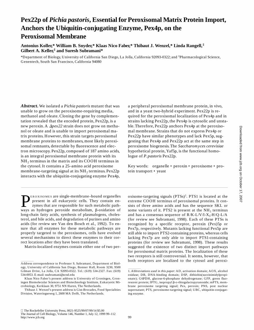
Antibodies raised against Pex22p (see Materials and Meth-ods) specifically detected a protein of z23 kD in cellsgrown on oleate and methanol (Fig. 5 A). Cells grown inglucose only showed a faint band corresponding to Pex22p(data not shown). No band was apparent in Dpex22 strainsas expected (Fig. 5 A). The same fractions as above (PNS,27-k pellet, 100-k pellet, and 100-k supernatant) takenfrom the wild-type strain were checked for the presence ofPex22p by immunoblotting. Pex22p was localized to the27-k pellet, suggesting an organellar localization for thisprotein (Fig. 5 A). The PNS of the wild-type strain wasfractionated on a linear Nycodenz gradient and analyzedby immunoblotting. Catalase and thiolase migrated, al-though with some trailing most likely due to rupture ofsome peroxisomes, near the bottom of the gradient, asdid Pex3p (Fig. 5 C). Pex22p colocalized with the perox-isomal markers catalase, thiolase, and Pex3p. Furtherevidence that Pex22p is a peroxisomal protein wasobtained by immunoelectron microscopy. Sections ofmethanol- and oleate-grown cells were decorated withPex22p antibodies followed by incubation with gold-con-jugated protein A. The gold particles almost exclusivelydecorated the peroxisomal membrane in the wild-type(Fig. 6, B and D), but not the Dpex22 strain (Fig. 6 A).Sometimes, Pex22p was localized to patches on peroxi-somes (Fig. 6 C).
Pex22p Is a Peroxisomal Membrane Protein with Its COOH Terminus Facing the Cytosol
The topology of Pex22p within the peroxisomal membranewas analyzed by organelle subfractionation. The wild-typestrain, SMD1163, was grown in oleate and the 27-k pelletwas fractionated into soluble and insoluble fractions aftertreatment with 0.1 M Na2CO3, pH 11.5, 10 mM Tris, pH8.5 (no salt), 1 M NaCl in 10 mM Tris, pH 8.5 (high salt),and 0.1% Triton X-100. Pex22p behaved like Pex3p, a per-oxisomal membrane protein (Wiemer et al., 1996), in allthe experiments, whereas catalase, a soluble matrix pro-tein, was found in the supernatant under all the conditionstested (Fig. 5 D). The 27-k pellet was further incubatedwith increasing amounts of trypsin in the presence or ab-sence of Triton X-100 to assess the availability of Pex22pfor the protease. Fig 5 E shows that Pex22p, as well asPex3p, were degraded even in the absence of detergent.Thiolase was well protected upon protease treatmentin the absence of Triton X-100, but degraded in the pres-ence of detergent. The immunocytochemistry experimentshowed that several gold particles are actually localized onthe cytosolic side of the peroxisomes (Fig. 6, B and D). Se-quence analysis of Pex22p showed that it contains one pu-tative membrane span near the NH2 terminus. The factsthat the bulk of the protein is protease accessible even inthe absence of Triton X-100 and that the antibody that de-tects Pex22p was raised against a protein lacking the first25 amino acids, suggest that the NH2 terminus faces theperoxisomal lumen whereas the COOH terminus is cyto-solic.
The First 25 Amino Acids of Pex22p Contain an mPTS
Sequence analysis of Pex22p did not reveal an obvious
Figure 3. Detection of GFP-PTS1, PTS2-GFP, and Pex3p inwild-type and Dpex22 cells. Wild-type (PPY12) and Dpex22(STK11) were transformed with constructs expressing GFP-PTS1and PTS2-GFP. Cells expressing GFP-PTS1 were induced inmethanol medium for 16 h, and those producing PTS2-GFP onoleate medium for 16 h. Pex3p was detected by immunofluores-cence (IF) in methanol-induced cells. Pictures in the left columnshow the subcellular localization of the GFP constructs andPex3p examined by fluorescence microscopy. Pictures in the rightcolumn were obtained by using Nomarski optics.
on October 17, 2007
ww
w.jcb.org
Dow
nloaded from

Koller et al. Role of PpPex22p in Peroxisome Biogenesis 105
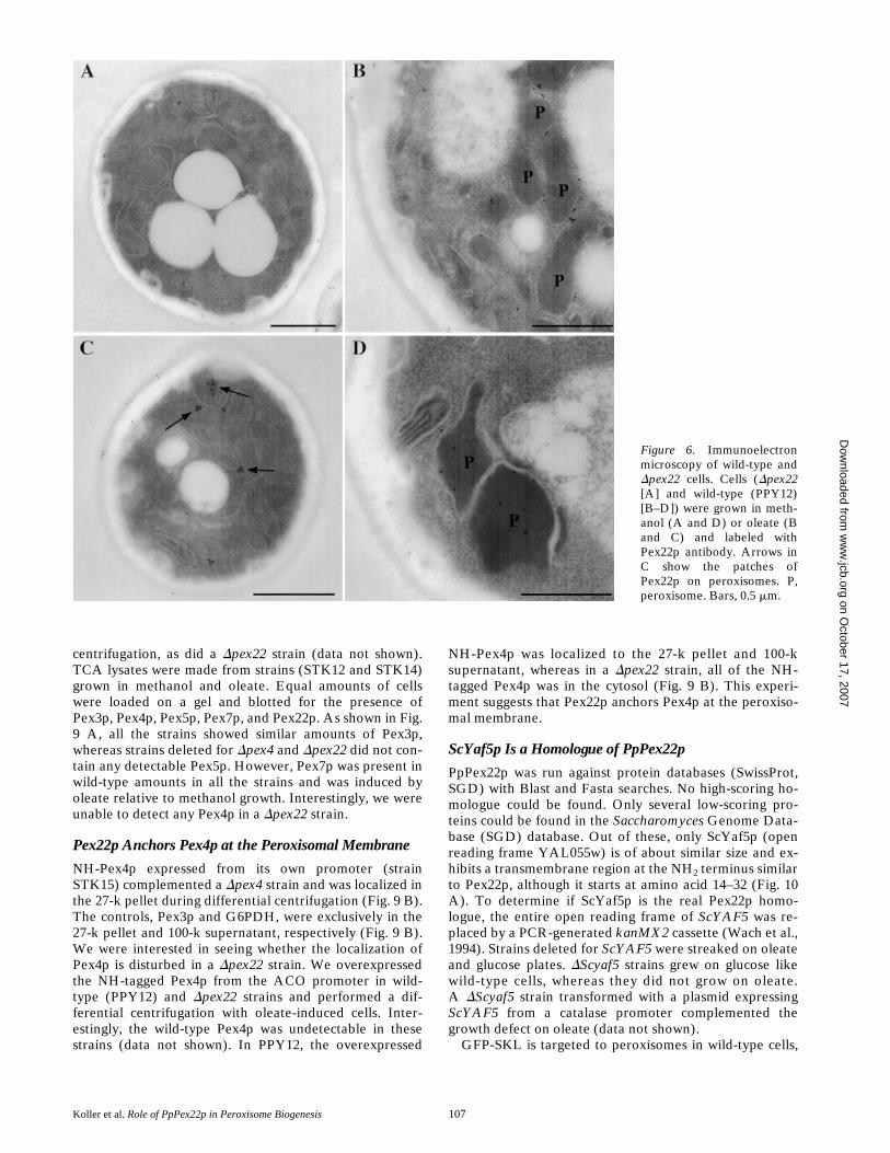
more, this fusion was organelle associated since the fusionprotein (Pex22(1–25)-GFP) only leaked from cells at digi-tonin concentrations that released membrane proteins(Fig. 7 B). The cytosolic protein, G6PDH, was releasedinto the supernatant at low concentrations (25 mg/ml),whereas the peroxisomal matrix protein GFP-SKL startedto leak at digitonin concentrations of 50–100 mg/ml, andrelease was not complete until the concentration of digito-nin was 500 mg/ml. Pex3p, a peroxisomal membraneprotein, was only fully released into the supernatantat digitonin concentrations exceeding 1,000 mg/ml. ThePex22(1–25)-GFP fusion protein was released into themedium at very high concentrations (1,000–1,500 mg/ml),or when the cells were treated with 0.2% Triton X-100(Fig. 7 B). These results show that the Pex22(1–25)-GFPconstruct is targeted to peroxisomal membranes.
Pex22p Interacts with Pex4p
To determine interactions of Pex22p with other Pex pro-teins, the yeast two-hybrid system was employed. PEX22was fused to the DB domain of LexA, or the AD of VP16.All published P. pastoris PEX genes (PEX1, PEX2,PEX3, PEX4, PEX5, PEX6, PEX7, PEX8, PEX10,PEX12, and PEX13) were also fused to these domains.
Figure 4. Electron micros-copy of wild-type and Dpex22cells. Morphological analysisof wild-type cells (PPY12)(A and B) and Dpex22(STK11) (C and D). Cellswere grown in methanol (Aand C) or oleate medium (Band D), and prepared forelectron microscopy. Ar-rows point to the peroxisomeremnants. N, nucleus; P, per-oxisome. Bars, 0.5 mm.
mPTS. There is, however, a stretch of positively chargedamino acids near the extreme NH2 terminus of Pex22pwhich does not completely fit the consensus sequence foran mPTS (Elgersma et al., 1997). This stretch is at thesame location as the putative mPTS of Pex3p (Hp, Sc, Pp).Therefore, we constructed GFP fusions with full-lengthPex22 (Pex22-GFP, pTK30, this construct is able to com-plement a Dpex22 mutant for growth on oleate and metha-nol), a second fusion with the first 25 amino acids of Pex22(Pex22(1–25)-GFP; pTK32), containing the transmem-brane domain, a third fusion with only the transmembranedomain (Pex22(8–25)-GFP; pTK44), and a fourth fusionwith the first seven amino acids of Pex22 (Pex22(1–7)-GFP; pTK34), not containing the transmembrane domain.These constructs were transformed into PPY12 and theresulting strains induced on methanol. The constructsexpressing full-length Pex22-GFP and Pex22(1–25)-GFPshowed colocalization with alcohol oxidase, a bona fideperoxisomal matrix protein (Fig. 7 A), proving that theseconstructs get targeted to peroxisomes, whereas the othertwo constructs (Pex22(1–7)-GFP, Pex22(8–25)-GFP) werelocalized in the cytosol (data not shown). The Pex22(1–25)-GFP fusion protein could also be shown to colocalizewith peroxisomes when an organelle fraction was sepa-rated on Nycodenz gradients (data not shown). Further-
on October 17, 2007
ww
w.jcb.org
Dow
nloaded from

The Journal of Cell Biology, Volume 146, 1999 106
These plasmids were then transformed in combinationinto the S. cerevisiae strain L40 and interaction of theseproteins was assessed by the production of b-galactosidaseactivity. Only the combination of Pex22p with Pex4p, aubiquitin-conjugating enzyme, produced any detectableenzyme activity. Almost the whole Pex22p protein (con-struct Pex22.1) was needed for interaction with Pex4p,whereas the COOH-terminal 39% of Pex4p (constructPex4.2) interacted with Pex22p (Fig. 8 A). Control experi-ments performed by exchanging the backbone vectorsconfirmed our findings (data not shown). We were alsoable to show that these two fragments of Pex22p (Pex22.1)and Pex4p (Pex4.2) interacted with each other (data notshown).
To show that Pex22p and Pex4p interact in vivo, 6HIS-Pex4p was expressed from the GAPDH promoter (plas-mid pTK36). This plasmid was then transformed into theDpex4 strain (STK14). The 6HIS-Pex4p complementedthe disrupted strain as assessed by growth on methanoland oleate (data not shown). This strain was grownin methanol, and spheroplasts were prepared. Thecross-linker dithiobix(succinimidylpropionate) (DSP) wasadded to the lysates to cross-link neighboring proteins.6HIS-Pex4p and associated proteins were precipitatedwith Ni21-NTA beads. Bound proteins were run on anSDS gel, blotted onto nitrocellulose and checked for thepresence of Pex4p, Pex22p, and Pex3p. The 6HIS-Pex4pspecifically bound Pex22p in the presence of the cross-linker DSP (Fig. 8 B), whereas no Pex22p could be de-tected in the sample without DSP. Pex3p, another peroxi-somal membrane protein, did not bind to the beads or to6HIS-Pex4p. Pex22p and Pex4p did also not bind to thebeads, as seen in the wild-type strain, not expressing any6HIS-tagged protein. These experiments confirm the spe-cific interaction between Pex4p and Pex22p by two differ-ent methods.
Dpex4 and Dpex22 Strains Share Similar Phenotypes
PpPex4p was previously characterized as a ubiquitin-con-jugating enzyme, similar to ScPex4p (Crane et al., 1994). ADpex4 strain (STK14) behaved similarly in differential
Figure 5. Subcellular localization, floatation gradient, Nycodenzgradient, membrane extraction and protease protection assaysfor Pex22p. (A) Postnuclear supernatant (PNS) was producedfrom wild-type (SMD1163) and Dpex22 (STK12) cells grown inoleate and subfractionated into a 27,000-g pellet (27 k p), a100,000-g pellet (100 k p) and a 100,000-g supernatant (100 k s).
Equivalent volumes were loaded on gels, transferred to nitrocel-lulose and blotted for the specified proteins. (B) The 27-k pelletof wild-type and Dpex22 strains grown in oleate were overlaidwith sucrose and centrifuged. Fractions were taken from the topand checked for the localization of Pex3p and the b-subunit ofthe mitochondrial F1-ATPase (F1). (C) PNS from wild-type cells(SMD1163) grown on oleate was loaded on top of Nycodenz gra-dients. Equal volumes of fractions from the gradient were ana-lyzed by immunoblotting. (D) The 27-k pellet of oleate-grownwild-type cells was subfractionated into an insoluble pellet frac-tion (p) and a soluble fraction (s) after treatment with 0.1 M car-bonate (pH 11.5), 10 mM Tris (pH 8), 10 mM Tris (pH 8), 1 MNaCl, and 0.1% Triton X-100. The distributions of the specifiedproteins between supernatant and membranous pellet fractionswere examined by immunoblotting. (E) A 27-k pellet of oleate-grown, wild-type cells was treated with the specified amount oftrypsin in the presence (1) or absence (2) of 0.1% Triton X-100.The disappearance of the specified proteins was examined by im-munoblotting.
on October 17, 2007
ww
w.jcb.org
Dow
nloaded from
Koller et al. Role of PpPex22p in Peroxisome Biogenesis 107
centrifugation, as did a Dpex22 strain (data not shown).TCA lysates were made from strains (STK12 and STK14)grown in methanol and oleate. Equal amounts of cellswere loaded on a gel and blotted for the presence ofPex3p, Pex4p, Pex5p, Pex7p, and Pex22p. As shown in Fig.9 A, all the strains showed similar amounts of Pex3p,whereas strains deleted for Dpex4 and Dpex22 did not con-tain any detectable Pex5p. However, Pex7p was present inwild-type amounts in all the strains and was induced byoleate relative to methanol growth. Interestingly, we wereunable to detect any Pex4p in a Dpex22 strain.
Pex22p Anchors Pex4p at the Peroxisomal Membrane
NH-Pex4p expressed from its own promoter (strainSTK15) complemented a Dpex4 strain and was localized inthe 27-k pellet during differential centrifugation (Fig. 9 B).The controls, Pex3p and G6PDH, were exclusively in the27-k pellet and 100-k supernatant, respectively (Fig. 9 B).We were interested in seeing whether the localization ofPex4p is disturbed in a Dpex22 strain. We overexpressedthe NH-tagged Pex4p from the ACO promoter in wild-type (PPY12) and Dpex22 strains and performed a dif-ferential centrifugation with oleate-induced cells. Inter-estingly, the wild-type Pex4p was undetectable in thesestrains (data not shown). In PPY12, the overexpressed
NH-Pex4p was localized to the 27-k pellet and 100-ksupernatant, whereas in a Dpex22 strain, all of the NH-tagged Pex4p was in the cytosol (Fig. 9 B). This experi-ment suggests that Pex22p anchors Pex4p at the peroxiso-mal membrane.
ScYaf5p Is a Homologue of PpPex22p
PpPex22p was run against protein databases (SwissProt,SGD) with Blast and Fasta searches. No high-scoring ho-mologue could be found. Only several low-scoring pro-teins could be found in the Saccharomyces Genome Data-base (SGD) database. Out of these, only ScYaf5p (openreading frame YAL055w) is of about similar size and ex-hibits a transmembrane region at the NH2 terminus similarto Pex22p, although it starts at amino acid 14–32 (Fig. 10A). To determine if ScYaf5p is the real Pex22p homo-logue, the entire open reading frame of ScYAF5 was re-placed by a PCR-generated kanMX2 cassette (Wach et al.,1994). Strains deleted for ScYAF5 were streaked on oleateand glucose plates. DScyaf5 strains grew on glucose likewild-type cells, whereas they did not grow on oleate.A DScyaf5 strain transformed with a plasmid expressingScYAF5 from a catalase promoter complemented thegrowth defect on oleate (data not shown).
GFP-SKL is targeted to peroxisomes in wild-type cells,
Figure 6. Immunoelectronmicroscopy of wild-type andDpex22 cells. Cells (Dpex22[A] and wild-type (PPY12)[B–D]) were grown in meth-anol (A and D) or oleate (Band C) and labeled withPex22p antibody. Arrows inC show the patches ofPex22p on peroxisomes. P,peroxisome. Bars, 0.5 mm. on O
ctober 17, 2007 w
ww
.jcb.orgD
ownloaded from
The Journal of Cell Biology, Volume 146, 1999 108
whereas in the DScyaf5 strain this construct was localizedin the cytosol (Fig. 10 B). To test the interaction betweenScYaf5p and ScPex4p, the genes encoding these proteinswere cloned into the two-hybrid vectors and transformedinto strain L40. As seen in Fig. 10 C, only strains contain-ing both constructs showed b-galactosidase activity. Theseresults indicate that ScYaf5p is the functional homologueof Pex22p. However, overexpression of ScYAF5 froman alcohol oxidase promoter could not complement thegrowth phenotype of a P. pastoris Dpex22 strain on metha-nol. This could be explained by the fact that ScYaf5p doesnot interact in a two-hybrid experiment with PpPex4p(data not shown).
Discussion
Pex22p Is a Peroxisomal Integral Membrane Protein
The newly discovered peroxin, Pex22p, described in this
Figure 7. Localization of different GFP constructs in methanol-grown, wild-type cells. (A) PPY12 was transformed with con-structs expressing GFP-SKL (pTW74), Pex22-GFP (pTK30) andPex22(1–25)-GFP (pTK32) and grown on methanol for 16 h.Cells were prepared for immunofluorescence and labeled witha-AOX. The localization of AOX and the GFP construct in thesame cells is shown. (B) Wild-type cells expressing either GFP-SKL (pTW74) or Pex22(1–25)-GFP (pTK32) were grown onmethanol, spheroplasted, and incubated for 10 min at 308C withincreasing amounts of digitonin or 0.2% Triton X-100 in isotonicbuffer. The spheroplasts were pelleted and the supernatants wereused for immunoblotting with GFP, Pex3p, and G6PDH antibodies.
Figure 8. (A) Two-hybrid interactions of Pex22p with Pex4p.The bars denote the parts of the genes, which were cloned intothe two-hybrid vectors pBTM116 (BD), and pVP16 (AD). Plas-mids were: Pex22, pTK12 or 13; Pex22.1, pTK14; Pex22.2,pTK16; Pex22.3, pTK18; Pex4, pKNSD119 or 118; Pex4.1,pTK21; Pex4.2, pTK23; Pex4.3, pTK25; and Pex4.4, pTK27. Thespecified plasmids were transformed into the yeast two-hybridstrain L40, spotted on nitrocellulose and checked for b-galactosi-dase activity. TM is the transmembrane domain of Pex22p, INS2is the Insertion Element 2 of Pex4p and * indicates the active-sitecysteine (C133) of Pex4p. (B) Pex22p binds to Pex4p in vivo. Alysate from a strain expressing a 6HIS-Pex4p (STK14 1 pTK36)or a wild-type (wt) strain (SMD1163) was incubated with (1) orwithout (2) the cross-linker DSP. 6HIS-Pex4p and cross-linkedproteins were precipitated with Ni21-NTA agarose beads. Boundproteins were subjected to SDS-PAGE and blotted for Pex22p,Pex4p, and Pex3p. Lane c shows the proteins in a TCA lysate ofthe 6HIS-Pex4p–expressing strain.
on October 17, 2007
ww
w.jcb.org
Dow
nloaded from

Koller et al. Role of PpPex22p in Peroxisome Biogenesis 109
study behaves like a peroxisomal integral membrane pro-tein by several criteria. It is pelletable in differential cen-trifugations (Fig. 5 A) and colocalizes with peroxisomalmarkers in Nycodenz gradients (Fig. 5 C). In immunoelec-tron microscopy experiments, the protein was associatedwith the peroxisomal membrane (Fig. 6, B–D). The pro-tein was not extracted from the membrane by buffers oflow ionic strength, high salt or by alkaline sodium carbon-ate, indicating that it is an integral membrane protein (Fig.5 D). Finally, most of the Pex22p is degraded upon addi-tion of proteases, even in the absence of detergent, underconditions where thiolase, a matrix marker, is resistant(Fig. 5 E). These results, when combined with the predic-tion of a single transmembrane domain near the NH2 ter-minus of Pex22p, are consistent with a topology in whichthe NH2 terminus of Pex22p is in the peroxisomal matrixand the COOH terminus is in the cytosol. This topologymakes it possible for the COOH terminus of Pex22p to beinvolved in protein interactions with the peroxisomal pe-ripheral membrane protein, Pex4p, as discussed later.
We do not understand why Pex22p is localized in someimmunoelectron microscopy pictures to patches at theperoxisomes. This is not seen in all the sections. It is possi-ble that Pex22p clusters are required for its normal func-tions which are discussed later. The same behavior hasalso been observed for Pex14p in Hansenula polymorpha(Komori et al., 1997).
The mPTS of Pex22p Resides Within the NH2-terminal 25 Amino Acids
Pex22p contains a signal at the NH2 terminus that is suffi-
cient for peroxisome targeting (Fig. 7 A). Fusing GFP tothe first 25 amino acids of Pex22p targets the resulting fu-sion protein to peroxisomes. This conclusion is supportedby the colocalization of this fusion protein with peroxiso-mal markers in a Nycodenz gradient (data not shown), byfluorescence microscopy showing colocalization of the fu-sion with a peroxisomal marker (Fig. 7 A), and by the re-lease of the fusion protein from cells only with high con-centrations of digitonin or by Triton X-100 (Fig. 7 B).Other experiments designed to show that the GFP portionof the fusion protein faces the cytosol failed because GFPis highly resistant to proteases (data not shown; Wiemer etal., 1996). GFP fusion proteins that contain the first 7amino acids (lacking the transmembrane region) or aminoacids 8–25 (containing only the transmembrane region)are not transported to the peroxisome but remain in thecytosol. The inability of the first 7 amino acids to functionas an mPTS is noteworthy since in previous experimentswith Pex3p and Pmp47 (Höhfeld et al., 1992; Baerends et
Figure 9. (A) Dpex4 and Dpex22 strains share similar pheno-types. Wt (SMD1163), Dpex4 (STK14), and Dpex22 (STK12)strains were grown in methanol or oleate for 14 h and a TCA ly-sate was generated. Equal volumes were subjected to SDS-PAGE and blotted for the specified proteins. (B) Pex22p is theanchor protein for Pex4p. Differential centrifugation was per-formed with strains expressing NH-Pex4p (Dpex41pTK51,PPY121pTK50, Dpex221pTK50) and equivalent fractions werechecked for the presence of the specified proteins.
Figure 10. ScYaf5p is the homologue of Pex22p. (A) The twoproteins were aligned using the SSEARCH Pairwise SequenceAlignment program (Smith and Waterman, 1981). The colonsrepresent identical amino acids, the dots are similar amino acidsand the bars represent gaps introduced to maximize similarity.(B) BJ1991 (wild-type) and DScyaf5 (STK16) were transformedwith a GFP-SKL construct. Cells were grown on oleate andchecked for the localization of GFP under the fluorescence mi-croscope. (C) Full-length ScYAF5 and ScPEX4 were cloned intothe two-hybrid vector pBTM116 (BD) or pVP16 (AD) and trans-formed into the two-hybrid strain L40. The transformants weregrown on a nitrocellulose filter and b-galactosidase activity wastested.
on October 17, 2007
ww
w.jcb.org
Dow
nloaded from

The Journal of Cell Biology, Volume 146, 1999 110
al., 1996; Dyer et al., 1996; Wiemer et al., 1996) the mPTSdid not require a transmembrane domain. The mPTS ofScPex15p, however, requires a transmembrane domain fortargeting to the peroxisomal membrane (Elgersma et al.,1997) in addition to the lumenal portion of the protein.
At present, we are unable to decipher why some mPTSsrequire transmembrane domains to function while othersdo not. In the case of Pex22p, the seven amino acids fusedto GFP could be buried and inaccessible to the putative re-ceptor. That would explain why this fusion protein is seenin the cytosol. Another possibility is that the targeting sig-nal requires some amino acids that are located in thetransmembrane domain of Pex22p. Experiments to deter-mine the important amino acids of the mPTS are under-way. Comparison of the different mPTSs found so farshows that there is a predominance of positively chargedamino acids. Pex22(1–25)-GFP fusions with alanine substi-tutions in two of the three positively charged amino acidsof the seven–amino acid lumenal stretch (K(2)→A andR(6)→A) do not properly localize to the peroxisome (datanot shown). This result suggests that at least these two pos-itive charges are important for proper targeting of the fu-sion protein.
Requirement of Pex22p for Import of Peroxisomal Matrix, but Not Membrane Proteins
Pex22p is important for peroxisome biogenesis and forgrowth of P. pastoris on methanol and oleate (Fig. 1).Functional peroxisomes are not formed in a Dpex22 strain(Fig. 4, C and D). Both exogenously expressed and endog-enous PTS1- and PTS2-containing proteins accumulate inthe cytosol (Figs. 3 and 5 A), whereas the membrane pro-tein, Pex3p, is targeted to pelletable membranous struc-tures that float in sucrose gradients (Fig. 5, A and B) andlikely correspond to the peroxisomal remnants observedusing fluorescence (Fig. 3) and electron microscopy (Fig.4, C and D).
Pex22p Interacts with Pex4p and Anchors It at the Peroxisomal Membrane
Yeast two-hybrid experiments performed with Pex22p andall published peroxins of P. pastoris show that it only inter-acts with Pex4p, a UBC enzyme that is localized to the cy-tosolic face of peroxisomal membranes (Fig. 8 A). TheCOOH-terminal cytosolic domain (amino acids 26–187) ofPex22p interacts with the COOH terminus (amino acids125–204) of Pex4p. Although this domain of Pex4p in-cludes the active site Cys (C133), Pex4p constructs con-taining Ala (C133A) or Ser (C133S) substitutions (Craneet al., 1994) at this location interacted normally withPex22p in the yeast two-hybrid system (data not shown).This result demonstrates that the interaction of Pex22pand Pex4p is not dependent on the UBC activity of Pex4p.Likewise, the binding of Pex22p to Pex4p is not dependenton the stretch designated INS2 (insertion element 2) thatis unique to Pex4p in comparison with several UBC en-zymes (Fig. 8 A). This segment has been postulated tobe important for peroxisomal localization (Crane et al.,1994). Pex22p and Pex4p also physically interact because6HIS-Pex4p expressed in P. pastoris was able to bindPex22p specifically (Fig. 8 B).
The interaction between Pex22p and Pex4p sheds lighton the function of Pex22p. One possibility is that Pex22p isthe elusive substrate for ubiquitination by Pex4p. How-ever, this seems unlikely as Pex22p migrates in SDS gels atthe predicted molecular mass (23 kD) and not as a proteinwith mono- or poly-ubiquitin modifications (Figs. 5 and 9A). The molecular mass of Pex22p is also unchangedthroughout oleate induction (data not shown).
An alternative possibility suggested by several experi-ments is that Pex22p anchors Pex4p on the peroxisomalmembrane. First, Pex4p is a peripheral peroxisomal mem-brane protein facing the cytosol and is tightly associatedwith the peroxisomal membrane even though it has notransmembrane segment of its own (Wiebel and Kunau,1992; Crane et al., 1994). Second, Pex22p and Pex4p inter-act (Fig. 8, A and B). It is noteworthy that the COOH-ter-minal domain of Pex22p which faces the cytosol interactswith Pex4p. Third, Pex4p is unstable in a Dpex22 strain(Fig. 9 A). Fourth, NH-Pex4p is mislocalized to the cytosolin the Dpex22 strain (Fig. 9 B). Many of these points arereminiscent of the relationship between Ubc7p and Cue1pin S. cerevisiae. Cue1p, an integral membrane protein ofthe ER, is essential for the localization of Ubc7p, a UBCenzyme, to the cytosolic face of the ER, and both theseproteins are required for the degradation of aberrant pro-teins in the ER membrane and for the retrograde trans-port of lumenal substrates out of the ER (Biederer et al.,1997). In a Dcue1 strain, Ubc7p could not be found and amyc-tagged Ubc7p, when overexpressed in this strain, wasfound in the cytosol. Pex4p is unstable in a Dpex22 strainand NH-Pex4p, when overexpressed from the acyl-CoAoxidase promoter, is localized to the cytosol in this strain.NH-Pex4p, in a wild-type strain, is localized equally in the27-k pellet and 100-k supernatant, whereas wild-type lev-els of NH-Pex4p are localized solely to the 27-k pellet (Fig.9 B). This shows that there is a saturable binding site forPex4p on membranes. These results are consistent withthe idea that Pex22p provides the binding site for Pex4p.Based on these data, we propose that Pex22p is the anchorprotein at the peroxisomal membrane that recruits andholds Pex4p at this location. We are not able to explainwhy in the strains overexpressing NH-Pex4p, Pex3p is notonly present in the 27-k pellet but also in the 100-k pelletand 100-k supernatant (Fig. 9 B).
This model would predict that Pex4p and Pex22p act to-gether for import of peroxisomal matrix proteins. This hy-pothesis is supported by the observation that both theDpex22 and Dpex4 strains do not contain wild-type levelsof Pex5p, have similar phenotypes such as inability to growon methanol and oleate, and are impaired in the import ofperoxisomal matrix proteins, but not membrane proteins(Wiebel and Kunau, 1992; Crane et al., 1994). The instabil-ity of Pex5p in the P. pastoris Dpex4 strain has been ob-served by another group (Kalish, J.E., and S.J. Gould, 6thInternational Congress on Cell Biology, 1996, Abstract2873) but this was not observed with H. polymorpha (vander Klei et al., 1998). Pex5p was also shown to be unstablein some mammalian, peroxisome-deficient complementa-tion groups (CG1, CG4, and CG8), suggesting that morethan one protein affects its stability (Dodt and Gould,1996). To examine if some phenotypes (such as growthon methanol and import of GFP-SKL) observed in the
on October 17, 2007
ww
w.jcb.org
Dow
nloaded from

Koller et al. Role of PpPex22p in Peroxisome Biogenesis 111
Dpex22 and Dpex4 strains were directly attributable to theabsence of Pex5p, PEX5 was overexpressed in the Dpex4and Dpex22 strains expressing GFP-SKL. The introductionof the PEX5 plasmid enhanced the level of Pex5p proteinto wild-type levels as assessed by immunoblotting, butthese strains remained unable to grow on methanol or im-port GFP-SKL into peroxisomes (data not shown). It isunlikely that Pex4p is solely responsible for the stability ofPex5p as we were unable to restore wild-type levels ofPex5p in a Dpex22 strain overexpressing Pex4p (data notshown). Therefore, the phenotypes seen in the Dpex4 andDpex22 strains are not simply a consequence of Pex5p in-stability. This is supported by the fact that not only PTS1-mediated import, but also the import of PTS2-containingproteins is compromised in Dpex4 and Dpex22 strains (Fig.3, see also Wiebel and Kunau, 1992; Crane et al., 1994; ourunpublished observation), despite the expression of stablePex7p in these strains (Fig. 9 A).
Models for the Role of Pex22p/Pex4p in Peroxisomal Matrix Protein Import
Our data clearly support a role for Pex22p in the anchor-ing of Pex4p to the peroxisomal membrane. However, fur-ther experiments will be required to determine the role ofthis protein complex in peroxisome biogenesis. One possi-bility is that the Pex4p–Pex22p complex functions similarto the Cue1p–Ubc7p complex, regulating the proper as-sembly and/or correct stoichiometry of protein importcomplexes at the peroxisomal membrane. It is known thataltered stoichiometry of peroxisomal integral or periph-eral membrane proteins, Pex3p and Pex14p, can yield animport-deficient phenotype (Baerends et al., 1997; Komoriet al., 1997). The function of Pex4p at the membrane mightbe to ubiquitinate and therefore target malfolded mem-brane proteins or nonstoichiometric subunits of the com-plex, leading to their degradation by the 26S proteasomein the cytosol. If Pex4p and/or Pex22p were missing, theimport complex might lose its ability to function, due to in-correct stoichiometry, leading to a block of matrix proteinimport, and this could in turn lead to an instability ofPex5p. Several proteins of the import complex could be af-fected by Pex22p and Pex4p, including Pex13p (Albertiniet al., 1997; Elgersma et al., 1996; Erdmann and Blobel,1996; Gould et al., 1996), Pex14p (Albertini et al., 1997;Brocard et al., 1997; Fransen et al., 1998), or Pex17p(Huhse et al., 1998). Pex13p is stable in Dpex4 or Dpex22strains (data not shown) and Pex5p is stable in a P. pastorisDpex13 strain (Gould et al., 1996). Pex14p and Pex17p re-main as reasonable targets for investigation because theirdeletion causes PTS1 and PTS2 import defects, but are notyet available for testing in P. pastoris.
A variation of this model, equally compatible with theavailable data, is that Pex4p, instead of directly actingon these peroxisomal membrane proteins, negatively reg-ulates (by ubiquitination and degradation) a protease,which in turn degrades peroxisomal membrane complexes.It is hoped that these testable models may lead, in the nearfuture, to the function of Pex4p.
Conservation of PpPex22p in Other Yeasts
Although database searches did not reveal any proteins
highly homologous to Pex22p, we did find a protein of sim-ilar predicted size and topology in S. cerevisiae. The hypo-thetical protein, ScYaf5p (open reading frame YAL-055w), appears to be the homologue of PpPex22p. LikePpPEX22, the ScYAF5 gene is essential for growth on ole-ate, and for the import of GFP-SKL, a fusion protein thatis readily imported into peroxisomes in wild-type yeast.Furthermore, ScYaf5p interacts with ScPex4p in a two-hybrid experiment. The conservation of Pex22p and its in-teracting partner, Pex4p, in other yeasts suggests that thefunctions of these proteins are likely to be conserved in allorganisms.
We thank the members of the lab for support and helpful discussions andS. Emr for use of the fluorescence microscope.
A. Koller was supported by a fellowship of the Swiss National ScienceFoundation (no. 8230-046677), W.B. Snyder by a fellowship from theAmerican Cancer Society, K.N. Faber by a fellowship from the HumanFrontier Science Program Organization, and T.J. Wenzel by a fellowshipfrom the European Molecular Biology Organization. This work was sup-ported by National Institutes of Health grant DK41737 to S. Subramani.
Submitted: 4 March 1999Revised: 17 May 1999Accepted: 3 June 1999
References
Albertini, M., P. Rehling, R. Erdmann, W. Girzalsky, J.A. Kiel, M. Veenhuis,and W.H. Kunau. 1997. Pex14p, a peroxisomal membrane protein bindingboth receptors of the two PTS-dependent import pathways. Cell. 89:83–92.
Babst, M., B. Wendland, E.J. Estepa, and S.D. Emr. 1998. The Vps4p AAAATPase regulates membrane association of a Vps protein complex requiredfor normal endosome function. EMBO (Eur. Mol. Biol. Organ.) J. 17:2982–2993.
Baerends, R.J., S.W. Rasmussen, R.E. Hilbrands, M. van der Heide, K.N.Faber, P.T.W. Reuvekamp, J.A.K.W. Kiel, J.M. Cregg, I.J. van der Klei, andM. Veenhuis. 1996. The Hansenula polymorpha PER9 gene encodes a per-oxisomal membrane protein essential for peroxisome assembly and integ-rity. J. Biol. Chem. 271:8887–8894.
Baerends, R.J., F.A. Salomons, K.N. Faber, J.A. Kiel, I.J. van der Klei, and M.Veenhuis. 1997. Deviant Pex3p levels affect normal peroxisome formationin Hansenula polymorpha: high steady-state levels of the protein fully abol-ish matrix protein import. Yeast. 13:1437–1448.
Biederer, T., C. Volkwein, and T. Sommer. 1997. Role of Cue1p in ubiquitina-tion and degradation at the ER surface. Science. 278:1806–1809.
Bodnar, A.G., and R.A. Rachubinski. 1991. Characterization of the integralmembrane polypeptides of rat liver peroxisomes isolated from untreatedand clofibrate-treated rats. Biochem. Cell Biol. 69:499–508.
Brocard, C., G. Lametschwandtner, R. Koudelka, and A. Hartig. 1997. Pex14pis a member of the protein linkage map of Pex5p. EMBO (Eur. Mol. Biol.Organ.) J. 16:5491–5500.
Crane, D.I., J.E. Kalish, and S.J. Gould. 1994. The Pichia pastoris PAS4 geneencodes a ubiquitin-conjugating enzyme required for peroxisome assembly.J. Biol. Chem. 269:21835–21844.
Diestelkötter, P., and W.W. Just. 1993. In vitro insertion of the 22-kD peroxiso-mal membrane protein into isolated rat liver peroxisomes. J. Cell Biol. 123:1717–1725.
Dodt, G., and S.J. Gould. 1996. Multiple PEX genes are required for propersubcellular distribution and stability of Pex5p, the PTS1 receptor: evidencethat PTS1 protein import is mediated by a cycling receptor. J. Cell Biol. 135:1763–1774.
Dyer, J.M., J.A. McNew, and J.M. Goodman. 1996. The sorting sequence of theperoxisomal integral membrane protein PMP47 is contained within a shorthydrophilic loop. J. Cell Biol. 133:269–280.
Elgersma, Y., M. Van den Berg, H.F. Tabak, and B. Distel. 1993. An efficientpositive selection procedure for the isolation of peroxisomal import and per-oxisome assembly mutants of Saccharomyces cerevisiae. Genetics. 135:731–740.
Elgersma, Y., L. Kwast, A. Klein, T. Voorn-Brouwer, M. van den Berg, B.Metzig, T. America, H. Tabak, and B. Distel. 1996. The SH3 domain of theperoxisomal membrane protein Pex13p functions as a docking site forPex5p, a mobile receptor for peroxisomal proteins. J. Cell Biol. 135:97–109.
Elgersma, Y., L. Kwast, M. van den Berg, W.B. Snyder, B. Distel, S. Subra-mani, and H.F. Tabak. 1997. Overexpression of Pex15p, a phosphorylatedperoxisomal integral membrane protein required for peroxisome assemblyin S. cerevisiae, causes proliferation of the endoplasmic reticulum membrane.EMBO (Eur. Mol. Biol. Organ.) J. 16:7326–7341.
Elgersma, Y., H.M. Elgersma, T. Wenzel, J.M. McCaffery, M.G. Farquhar, and
on October 17, 2007
ww
w.jcb.org
Dow
nloaded from

The Journal of Cell Biology, Volume 146, 1999 112
S. Subramani. 1998. A mobile PTS2 receptor for peroxisomal protein importin Pichia pastoris. J. Cell Biol. 140:807–820.
Erdmann, R., and G. Blobel. 1996. Identification of Pex13p a peroxisomalmembrane receptor for the PTS1 recognition factor. J. Cell Biol. 135:111–121.
Faber, K.N., J.A. Heyman, and S. Subramani. 1998. Two AAA family peroxins,PpPex1p and PpPex6p, interact with each other in an ATP-dependent man-ner and are associated with different subcellular membranous structures dis-tinct from peroxisomes. Mol. Cell. Biol. 18:936–943.
Fransen, M., S.R. Terlecky, and S. Subramani. 1998. Identification of a humanPTS1 receptor docking protein directly required for peroxisomal protein im-port. Proc. Natl. Acad. Sci. USA. 95:8087–8092.
Fujiki, Y., R.A. Rachubinski, and P.B. Lazarow. 1984. Synthesis of a major in-tegral membrane polypeptide of rat liver peroxisomes on free polysomesProc. Natl. Acad. Sci. USA. 81:7127–7131.
Girzalsky, W., P. Rehling, K. Stein, J. Kipper, L. Blank, W.-H. Kunau, and R.Erdmann. 1999. Involvement of Pex13p in Pex14p localization and peroxiso-mal targeting signal 2-dependent protein import into peroxisomes. J. CellBiol. 144:1151–1162.
Gould, S.J., D. McCollum, A.P. Spong, J.A. Heyman, and S. Subramani. 1992.Development of the yeast Pichia pastoris as a model organism for a geneticand molecular analysis of peroxisome assembly. Yeast. 8:613–628.
Gould, S.J., J.E. Kalish, J.C. Morrell, J. Bjorkman, A.J. Urquhart, and D.I.Crane. 1996. Pex13p is an SH3 protein of the peroxisome membrane and adocking factor for the predominantly cytoplasmic PTS1 receptor. J. CellBiol. 135:85–95.
Harlow, E., and D. Lane. 1988. Antibodies: A Laboratory Manual. Cold SpringHarbor Laboratory Press, Cold Spring Harbor, NY. 313 pp.
Hettema, E.H., C.C.M. Ruigrok, M.G. Koerkamp, M. van den Berg, H.F.Tabak, B. Distel, and I. Braakman. 1998. The cytosolic DnaJ-like proteinDjp1p is involved specifically in peroxisomal protein import. J. Cell Biol.142:421–434.
Höhfeld, J., M. Veenhuis, and W.H. Kunau. 1991. PAS3, a Saccharomyces cere-visiae gene encoding a peroxisomal integral membrane protein essential forperoxisome biogenesis. J. Cell Biol. 114:1167–1178.
Huhse, B., P. Rehling, M. Albertini, L. Blank, K. Meller, and W.H. Kunau.1998. Pex17p of Saccharomyces cerevisiae is a novel peroxin and componentof the peroxisomal protein translocation machinery. J. Cell Biol. 140:49–60.
Komori, M., S.W. Rasmussen, J.A.K.W. Kiel, R.J.S. Baerends, J.M. Cregg, I.J.van der Klei, and M. Veenhuis. 1997. The Hansenula polymorpha PEX14gene encodes a novel peroxisomal membrane essential for peroxisome bio-
genesis. EMBO (Eur. Mol. Biol. Organ.) J. 16:44–53.Monosov, E.Z., T.J. Wenzel, G.H. Lüers, J.A. Heyman, and S. Subramani.
1996. Labeling of peroxisomes with green fluorescent protein in living P.pastoris cells. J. Histochem. Cytochem. 44:581–589.
Odorizzi, G., M. Babst, and S.D. Emr. 1998. Fab1p PtdIns(3)P 5-kinase func-tion essential for protein sorting in the multivesicular body. Cell. 95:847–858.
Purdue, P.E., X. Yang, and P.B. Lazarow. 1998. Pex18p and Pex21p, a novelpair of related peroxins essential for peroxisomal targeting by the PTS2pathway. J. Cell Biol. 143:1859–1869.
Rieder, S.E., and S.D. Emr. 1997. A novel RING finger protein complex essen-tial for a late step in protein transport to the yeast vacuole. Mol. Biol. Cell.8:2307–2327.
Sambrook, J., E.F. Fritsch, and T. Maniatis. 1989. Molecular Cloning: A Labo-ratory Manual. 2nd edition. Cold Spring Harbor Laboratory Press, ColdSpring Harbor, NY.
Sanger, F., S. Nicklen, and A.R. Coulson. 1977. DNA sequencing with chain-terminating inhibitors. Proc. Natl. Acad. Sci. USA. 74:5463–5467.
Smith, T.F., and M.S. Waterman. 1981. Identification of common molecularsubsequences. J. Mol. Biol. 147:195–197.
Subramani, S. 1998. Components involved in peroxisome import, biogenesis,proliferation, turnover, and movement. Physiol. Rev. 78:171–188.
van den Bosch, H., R.B.H. Schutgens, R.J.A. Wanders, and J.M. Tager. 1992.Annu. Rev. Biochem. 61:157–197.
van der Klei, I.J., R.E. Hilbrands, J.A. Kiel, S.W. Rasmussen, J.M. Cregg, andM. Veenhuis. 1998. The ubiquitin-conjugating enzyme Pex4p of Hansenulapolymorpha is required for efficient functioning of the PTS1 import machin-ery. EMBO (Eur. Mol. Biol. Organ.) J. 17:3608–3618.
Wach, A., A. Brachat, R. Pöhlmann, and P. Philippsen. 1994. New heterologousmodules for classical or PCR-based gene disruptions in Saccharomyces cere-visiae. Yeast. 10:1793–1808.
Walton, P.A., M. Wendland, S. Subramani, R.A. Rachubinski, and W.J. Welch.1994. Involvement of 70-kD heat-shock proteins in peroxisomal import. J.Cell Biol. 125:1037–1046.
Wiebel, F.F., and W.H. Kunau. 1992. The Pas2 protein essential for peroxisomebiogenesis is related to ubiquitin-conjugating enzymes. Nature. 359:73–76.
Wiemer, E.A.C., G. Lüers, K.N. Faber, T. Wenzel, M. Veenhuis, and S. Subra-mani. 1996. Isolation and characterization of Pas2p, a peroxisomal mem-brane protein essential for peroxisome biogenesis in the methylotrophicyeast Pichia pastoris. J. Biol. Chem. 271:18973–18980.
on October 17, 2007
ww
w.jcb.org
Dow
nloaded from