phage on tap–a quick and efficient protocol for the ... · phage on tap–a quick and efficient...
TRANSCRIPT

Phage on tap–a quick and efficientprotocol for the preparation ofbacteriophage laboratory stocks
Natasha Bonilla, Maria Isabel Rojas, Giuliano Netto Flores Cruz,Shr-Hau Hung, Forest Rohwer and Jeremy J. Barr
Department of Biology, San Diego State University, San Diego, CA, United States
ABSTRACTAmajor limitation with traditional phage preparations is the variability in titer, salts,
and bacterial contaminants between successive propagations. Here we introduce
the Phage On Tap (PoT) protocol for the quick and efficient preparation of
homogenous bacteriophage (phage) stocks. This method produces homogenous,
laboratory-scale, high titer (up to 1010–11 PFU·ml-1), endotoxin reduced phage
banks that can be used to eliminate the variability between phage propagations and
improve the molecular characterizations of phage. The method consists of five major
parts, including phage propagation, phage clean up by 0.22 mm filtering and
chloroform treatment, phage concentration by ultrafiltration, endotoxin removal,
and the preparation and storage of phage banks for continuous laboratory use.
From a starting liquid lysate of > 100 mL, the PoT protocol generated a clean,
homogenous, laboratory phage bank with a phage recovery efficiency of 85% within
just two days. In contrast, the traditional method took upwards of five days to
produce a high titer, but lower volume phage stock with a recovery efficiency of only
4%. Phage banks can be further purified for the removal of bacterial endotoxins,
reducing endotoxin concentrations by over 3,000-fold while maintaining phage titer.
The PoT protocol focused on T-like phages, but is broadly applicable to a variety of
phages that can be propagated to sufficient titer, producing homogenous, high titer
phage banks that are applicable for molecular and cellular assays.
Subjects Microbiology, Molecular Biology, Virology
Keywords Bacteriophage, Endotoxin, Cesium chloride, Top agar, Ultrafiltration, Dialysis,
Speed vacuum, Phage bank
INTRODUCTIONDue to increasing interest for the use of bacteriophage (phage) in medical, industrial, and
molecular settings, new approaches are required to quickly and efficiently produce high
titer, homogenous, and purified phage stocks. It is desirable that these stocks be free
of bacteria, molds, debris, culture medium, and bacterial endotoxins (Adams, 1959).
Typically the ability to produce high titer phage stocks is largely dependent on the
particular phage and host cell under consideration, yet certain principles and
methodologies can be broadly applied. Traditional techniques used for the concentration
and purification of phage involve centrifugation, filtration, ultrafiltration, precipitation
with Polyethylene Glycol (PEG), ultracentrifugation in cesium chloride (CsCl) gradients,
How to cite this article Bonilla et al. (2016), Phage on tap–a quick and efficient protocol for the preparation of bacteriophage laboratory
stocks. PeerJ 4:e2261; DOI 10.7717/peerj.2261
Submitted 16 April 2016Accepted 26 June 2016Published 26 July 2016
Corresponding authorJeremy J. Barr,
Academic editorSiouxsie Wiles
Additional Information andDeclarations can be found onpage 15
DOI 10.7717/peerj.2261
Copyright2016 Bonilla et al.
Distributed underCreative Commons CC-BY 4.0

and dialysis (Adams, 1959; Yamamoto et al., 1970; Seeley & Primrose, 1982; Suttle, Chan &
Cottrell, 1991; Carlson, 2005; Bourdin et al., 2014). Yet many of these techniques are time
consuming and affect phage recovery and/or viability.
Phage preparations are often contaminated by macromolecules derived from the host
bacteria and culture media, with the major pyrogen being the lipid A moiety of
lipopolysaccharide (endotoxin) from the outer membrane of Gram-negative bacteria
(Raetz et al., 2007). Endotoxins are amphipathic molecules; the lipid component is linked
to a core polysaccharide and as a result they can form large aggregates greater than
1,000 kDa in solution (Magalhaes et al., 2007). Endotoxin elicits a wide variety of
pathophysiological effects in the body. Exposure to even small amounts can result in
toxic shock, cell injury, cytokine production and the activation of immune responses
(Morrison & Ulevitch, 1978; Rietschel et al., 1994; Alexander & Rietschel, 2001; Pabst et al.,
2008). Due to these effects it is important that endotoxins be removed from phage
preparations when studying or applying phage in the context of eukaryotic systems. The
amount of endotoxin is defined as an endotoxin unit (EU), which corresponds to the
activity of 100 pg of E. coli lipopolysaccharide. The endotoxin content of distilled water is
estimated at 20 EU·ml-1, with the allowed limit for intravenous and oral administration
set at 5 EU·kg·hr-1 and < 20 EU·ml-1 respectively (Bruttin & Brussow, 2005; Gorbet &
Sefton, 2005; Abedon et al., 2011).
A further limitation with traditional phage preparations is the variability of titer, salts,
and bacterial contaminants produced between successive propagations. Eliminating this
variability has proven critical for accurate analysis for the molecular interactions of phage
within the context of eukaryotic hosts (Barr et al., 2013; Barr et al., 2015). Here we present
the Phage On Tap (PoT) protocol as a fast and efficient way to produce homogenous
laboratory phage stocks. Phage stocks were sterilized by centrifugation, 0.22 mm filtration,
and chloroform treatment, before concentration and washing using ultrafiltration, and
storage at 4 �C for four months with minimal degradation. However, the PoT protocol
was not able to effectively reduce bacterial endotoxins, likely due to our ultrafiltration-
based approach and the large aggregate size of endotoxins. Numerous endotoxin removal
procedures and commercial kits are available (Boraty�nski et al., 2004; Merabishvili et al.,
2009; Oslizło et al., 2011; Magalhaes et al., 2007; Branston, Wright & Keshavarz-Moore,
2015), yet many of these methodologies lack generality, are time consuming, or are cost-
prohibitive. Recently, the successful reduction of endotoxins (< 20 EU·ml-1) from phage
lysates was reported by extraction with organic solvents (Szermer-Olearnik & Boraty�nski,
2015). We corroborate this organic solvent-based method for the removal of bacterial
endotoxin from phage lysates and adapt this methodology, with reduced processing time
through the use of speed vacuum, in the PoT protocol. The PoT protocol can purify phage
lysates, with volumes ranging between 50–300 ml (or greater if required), producing
homogeneous phage stocks for further laboratory testing. The method takes two days
to purify and concentrate a final phage lysate of ∼10 ml of with a titer > 1010 PFU·ml-1,
and a significant reduction in bacterial endotoxin levels. Our method has been tested on a
variety of phages, including T4 (E. coli), T3 (E. coli), T5 (E. coli), and Spp1 (B. subtilis),
and is broadly applicable to other tailed phages.
Bonilla et al. (2016), PeerJ, DOI 10.7717/peerj.2261 2/18

MATERIALS AND METHODSReagents
- Luria-Bertani (LB) broth (cat. no. DF0446; Fisher Scientific)
- Agar (cat. no. BP1423; Fisher Scientific)
- Calcium Chloride Dihydrate (CaCl2·2H2O) (cat. no. C69; Fisher Scientific)
- Magnesium Chloride Hexahydrate (MgCl2·6H2O) (cat. no. BP214; Fisher Scientific)
- Sodium Chloride (NaCl) (cat. no. S671; Fisher Scientific)
- Magnesium Sulfate Heptahydrate (MgSO4·7H2O) (cat. no. M63; Fisher Scientific)
- Trizma Hydrochloride (Tris HCl pH 7.4) (cat. no. 93313; Sigma-Aldrich)
- Chloroform (cat. no. BP1145; Fisher Scientific) CAUTION: Chloroform is toxic and
should only be used in a fume hood and with personal safety equipment, such as gloves
and goggles.
- PierceTM LAL Chromogenic Endotoxin Quantitation Kit (cat. no. 88282; Thermo Fisher)
Note: Endotoxin Quantitation Kit is optional and is only required for the quantitation of
endotoxins in phage lysates.
- Ethanol, Absolute (C2H5OH) (cat. no. BP2818; Fisher Scientific)
- Glycerol (cat. no. G31; Fisher Scientific)
Equipment
- 37 �C incubator with a rocker
- Centrifuge with swinging bucket rotor
- Microtube centrifuge
- Centrifugal vacuum concentrator
- Stir plate
- Petri dish with disposable lid (cat. no. 09-720; Fisher Scientific)
- 1.7 ml microcentrifuge tubes (cat. no. 02-681; Fisher Scientific)
- Falcon 50 ml conical centrifuge tubes (cat. no. 14-432; Fisher Scientific)
- Small glass test tubes 13 � 100 mm (cat. no. 14-958; Fisher Scientific)
- Serological pipettes (cat. no. 07-200; Fisher Scientific)
- 0.22 mm Sterivex filter units (cat. no. SVGP; Millipore)
- Whatman Anotop 0.02 mm sterile syringe filters. (cat. no. 09-926-13;
Fisher Scientific)
- Amicon� Ultra-15 centrifugal filter units, Ultracel 100 kDa membrane (cat no.
UFC910008; Millipore)
Note: 100 kDa membrane pore size is equivalent to a spherical particle with a diameter of
∼3 nm and is therefore sufficient for the capture of all known bacteriophages (Erickson, 2009).
- 500 ml PYREX� screw cap storage bottle with plastic seal (cat. no. 13-700-446; Fisher
Scientific)
Bonilla et al. (2016), PeerJ, DOI 10.7717/peerj.2261 3/18

- Spectra-Por� Float-A-Lyzer� G2 Dialysis membrane, 10 mL, MWCO 3.5–5 kDa
(cat. no. Z726273; Sigma-Aldrich)
- NalgeneTM General Long-Term Storage Cryo Tubes (cat. no. 03–337; Fisher Scientific)
Reagent setup
LB broth: 25 g LB broth in 1 liter dH2O.
LB top agar: 25 g LB broth, 7.5 g Agar, in 1 liter dH2O.
LB agar plates: 25 g LB broth, 15 g Agar, in 1 liter dH2O.
SM buffer: 5.8 g NaCl, 2.0 g MgSO4·7H2O, 50 ml 1 M Tris-HCl pH 7.4, in 1 liter dH2O.
Autoclave, 0.02 mm filter-sterilize before use, and store at room temperature.
Calcium chloride (CaCl2): Prepare a 1 M stock solution and add a final concentration of
0.001 M to desired volume of the LB broth that will be used for the liquid lysate.
Autoclave, 0.02 mm filter-sterilize before use, and store at room temperature.
Magnesium chloride (MgCl2): Prepare a 1 M stock solution and add a final concentration
of 0.001 M to desired volume of the LB broth that will be used for the liquid lysate.
Autoclave, 0.02 mm filter-sterilize before use, and store at room temperature.
Phage on Tap (PoT) ProtocolNotes:
� The procedure for phage propagation is largely specific for each phage and bacterial
host. Here we use propagation conditions for T4 phage and Escherichia coli B bacterial
host. It is recommended to use appropriated growth and propagation conditions for
your choice of phage and host.
� Once a sufficiently high titer phage lysate is obtained please proceed to step 3.
� It is recommended to only propagate and purify one phage at a time to prevent
cross-contamination.
1| Phage plaque assay for determination of titer (Adams, 1959)
i) Grow E. coli B bacterial host in LB broth overnight at 37 �C.
ii) Dilute phage stock or isolate in LB broth down to the desired dilution (e.g., for a
phage stock of 108 PFU·ml-1 serially dilute down to 10-6 and 10-7 to obtain
countable plaques).
iii) Heat LB top agar in microwave until completely molten, then allow top agar to cool
in a 56 �C water bath or until it is warm to the touch.
iv) Add 1 ml of the overnight bacterial host and 1 ml of the phage dilution to a glass test
tube and mix.
v) Add 3 ml of molten top agar to the glass test tube.
vi) Quickly pour molten mixture onto an LB agar plate and tilt the plate to evenly
distribute the agar. Let sit undisturbed until the agar has gelled.
vii) Once plate has gelled (∼5 min), invert and incubate overnight at 37 �C.
Bonilla et al. (2016), PeerJ, DOI 10.7717/peerj.2261 4/18

viii) Count phage plaques and determine phage titer in plaque-forming units (PFU·ml-1)
using the following formula:
PFU per ml = plaques per plate � volume plated in ml � dilution factor
e.g., if there are 20 plaques when you plated out 1ml from the 106 dilution, the titer of
the phage stock is 2.0 � 107 PFU·ml-1.
2A| Phage isolation and propagation via plate lysate
i) From a plate lysate plate pick a single phage plaque using a sterile Pasteur pipet.
ii) Resuspend the plaque into a microcentrifuge tube containing 1 ml of filter-sterilized
phage diluent (SM buffer) and vortex for 5 min.
iii) Centrifuge at 4,000 � g for 5 min to remove any remaining debris.
iv) Perform plate lysate as described above using the resuspended phage.
v) After incubation the entire plate should be lysed. Pour ∼5 ml of SM buffer on top of
plate and shake gently for 15 min at room temperature.
vi) Collect buffer from top of plate and centrifuge at 4,000 � g for 5 min.
vii) Collect phage lysate and store at 4 �C until clean up.
Optional: Titer the lysate via plaque assay to ensure initial high titer.
2B| Phage propagation via liquid lysate
i) Grow E. coli bacterial host in LB broth overnight at 37 �C.
ii) Prepare and autoclave 100 ml of LB broth supplemented 0.001 M CaCl2 and MgCl2
added in a 250 ml PYREX� screw cap storage bottle and save for Step 3.
iii) Spike LB broth supplemented with CaCl2 and MgCl2 with 0.1 volumes of overnight
bacterial host.
Note: For a phage with a high burst size, such as T3, you may need to double volume
of host added.
iv) Incubate with agitation for 1 h at 37 �C.
v) Add 100 ml of high titer phage lysate (> 108 PFU·ml-1).
vi) Incubate at 37 �C with agitation for ∼5 h or until lysate clears.
vii) Collect phage lysate and store at 4 �C until clean up.
Optional: Titer the lysate via plaque assay to ensure initial high titer.
3| Phage cleanup (0.22 mm filtering and chloroform)
i) Aliquot phage lysate into 50 ml sterile falcon centrifuge tubes and centrifuge at
4,000 � g for 20 min.
ii) Carefully collect supernatant using a serological pipette and transfer into properly
labeled sterile falcon tube.
iii) Filter-sterilize the phage supernatant using a 0.22 mm filter to yield a bacterial cell-
free phage lysate.
Bonilla et al. (2016), PeerJ, DOI 10.7717/peerj.2261 5/18

iv) Add 0.1 volumes of chloroform to the supernatant, vortex, and incubate at room
temp for 10 min.
Note: Lipid enveloped phages are sensitive to chloroform and titer can be significantly
reduced. If a drop in titer is observed skip this step.
v) Centrifuge at 4,000 � g for 5 min and transfer supernatant into 250 ml PYREX�
screw cap storage bottle and store at 4 �C until concentration.
Optional: Titer the lysate via plaque assay to ensure initial high titer.
4| Phage concentration and wash via ultrafiltration
i) Add ∼15 ml of phage lysate into the upper reservoir of Amicon filter device.
ii) Centrifuge Amicon at 4,000 � g for ∼5 min.
Note: Centrifugation times will vary based on phage type and titer. It is important not
to spin the device dry. If unsure about centrifugation times select a shorter spin time,
check the lysate level, and adjust spin times appropriately.
iii) Carefully discard the filtrate into a waste bucket and add another volume of phage
lysate to the sample filter cup and repeat centrifugation.
Note: The same device can be used to concentrate large volumes of phage lysate (> 100
ml) achieving an approximate 90% decrease with minimal loss in phage titer.
iv) Repeat step until all phage lysate has been concentrate to < 10 ml.
v) Add ∼15 ml of SM buffer into the upper reservoir containing concentrate phage
lysate and centrifuge at 4,000 � g for ∼5 min to wash phage lysate.
Note: SM buffer was chosen as it is suitable for the long-term storage of T-phage, but
any appropriate storage buffer can be used. It is important not to spin the device dry,
adjust centrifugation times accordingly.
vi) Repeat wash step and concentrate washed phage lysate to < 10 ml.
vii) Using a pipette, carefully collect phage lysate from the upper reservoir and gently
wash the surface of the upper reservoir.
Note: Alternatively the entire device can be vortexed to assist with phage detachment
from filter.
viii) Collect < 10 ml of concentrated and purified phage lysate. Titer phage concentrate
and record PFU·ml-1.
5| Endotoxin removal (Morrison & Leive, 1975; Szermer-Olearnik & Boraty�nski, 2015)
Notes:
� This method is adapted from Szermer-Olearnik & Boraty�nski (2015), which
demonstrates the efficient removal of endotoxins from bacteriophage lysates using
water immiscible solvents that are subsequently removed via dialysis. For detailed
explanation of the methodology please see Morrison & Leive (1975) and Szermer-
Olearnik & Boraty�nski (2015).
Bonilla et al. (2016), PeerJ, DOI 10.7717/peerj.2261 6/18

� Our adapted method uses a speed vacuum to remove residual organic solvent from
phage lysates, instead of the lengthy dialysis washes with similar efficiency.
� This step is optional. If you do not require removal of bacterial endotoxins from your
phage preparations please go to step 7.
i) Add 0.4 volumes of 1-octanol to phage concentrate and shake at room temp for 1 h.
ii) Incubate phage concentrate at 4 �C for 1.5 h.
Note: Alternatively concentrate can be chilled in a ice bath for ∼15 min.
iii) Centrifuge at 4,000 � g for 10 min.
iv) Using a syringe, pierce the bottom of the tube and collect the aqueous phase
(bottom layer) that contains your phage, and transfer to a sterile 50 ml falcon
tube. Do not collect the organic phage (top layer) or interface as this contains
endotoxins.
Note: It is best to leave a small residual amount of phage concentrate behind to reduce
the transfer of contaminating endotoxins.
Note: If endotoxin removal is not sufficient the method can be repeated to further reduce
endotoxins.
6A| Dialysis removal of organic solvent (Szermer-Olearnik & Boraty�nski, 2015)
Notes:
� This method is adapted from Szermer-Olearnik & Boraty�nski (2015) and describes the
removal of residual organic solvents from phage lysates by dialysis.
� Residual organic solvents disable downstream PierceTM LAL Chromogenic Endotoxin
Quantitation assays and must be removed in order to accurately quantify endotoxin
concentrations.
� Due to the ionic concentration of phage SM buffer used you may end up with greater
than the starting volume.
i) Pre-wet Spectra-Por� dialysis membrane with sterilized dH2O according to
manufacturers instructions, being careful not to contaminate the inside of
the tubing.
ii) Loadmaximumof 10ml of phage concentrate inside of dialysis tubing and seal tightly.
iii) Dialyze phage concentrate against 2 liters of 25% (v/v) ethanol at 4 �C on a stirring
plate for 24 h to remove residual 1-butanol, replacing the 25% ethanol solution four
times at 15, 18, 21, and 24 h.
iv) Dialyze phage concentrate against 2 liters of 0.15 M NaCl solution at 4 �C on a
stirring plate 24 h to remove residual ethanol, replacing the 0.15 M NaCl solution
three times after 15, 19, and 24 h.
v) Carefully collect phage concentrate by washing the inside of the dialysis tubing. Store
concentrate at store 4 �C until phage bank preparation.
Bonilla et al. (2016), PeerJ, DOI 10.7717/peerj.2261 7/18

Optional: Titer the lysate via plate lysates to ensure high titer. Perform the PierceTM LAL
Chromogenic Endotoxin Quantitation according to manufacturers instructions to
obtain quantitative endotoxin levels.
6B| Speed vacuum removal of organic solvent
Notes:
� This method is a faster alternative to the dialysis method for the removal of residual
organic solvents from phage concentrates.
i) Aliquot ∼1 ml of the phage concentrate equally into microcentrifuge tubes.
ii) Place tubes into a speed vacuum 4 �C, open lids, and centrifuge at 4,000 � g for 3 h.
iii) After speed vacuum, phage concentrate volume should be reduced by approximately
30% and residual 1-octanol evaporated.
iv) Collect phage concentrate and store 4 �C until phage bank preparation.
Optional: Titer the lysate via plaque assay to ensure high titer. Perform the PierceTM LAL
Chromogenic Endotoxin Quantitation according to manufacturers instructions to
obtain quantitative endotoxin levels from phage concentrates.
7| Phage bank storage
i) Dilute phage lysate in SM buffer to generate a high-titer working stock.
Note: This step largely depends on desired concentration and volume of the phage bank.
If you require higher titer phage stocks then dilute less or omit dilution, if greater volume
is desired then dilute more.
ii) Titer diluted phage concentrate and record PFU·ml-1.
iii) Aliquot phage working stocks into labeled cryo tubes and store at 4 �C.
� You now have a bank of homogenous, high titer (up to 1010–11 PFU·ml-1) phage bank
for laboratory testing.
� From our experience the use of homogenous and standardized phage banks can
significantly reduce the experimental variability for the molecular characterization
of phages.
RESULTSPhage purification and concentrationHere we used T4 phage as a representative phage for our PoT phage purification protocol
(Fig. 1). We tested the speed and efficiency of the PoT purification protocol (Fig. 2A)
compared against the traditional method (Fig. 2B), which involved phage propagation,
centrifugation, filtering and purification via CsCl ultracentrifugation and dialysis (Figs. S1
and S2) (Adams, 1959). Both methods started with 110 ml of total lysate and comparable
titers. The PoT protocol consisted of phage clean up, concentration and washing via
ultrafiltration and resulted in a 93% reduction in volume with a phage recovery efficiency
of 85%, while taking just two days to complete. Comparatively, the traditional method,
Bonilla et al. (2016), PeerJ, DOI 10.7717/peerj.2261 8/18

~1 mL
~100 mL
~5 mL per plate
8 hrs
3) Phage clean up
~100 mL
2 hrs
- Centrifugation- Filtration- Chloroform
5) Endotoxin Removal
~10 mL
2-4 hrs
- Ultrafiltration- Buffer wash
4) Phage Concentration
~10 mL
3 hrs
Organic Layer
Phage Lysate
- 1-Octanol treatment- Centrifugation
6A) Dialysis
~12 mL
48 hrs
- 25% Ethanol wash- 0.15M NaCl wash
6B) Speed Vac
~12 mL
ed Vac)6B) Spee
~9 mL3 hrs
- Dilute- Store
24 hrs
1) Phage plaque assay
2B) Liquid Lysate
7) Phage bank
2A) Plate Lysate
OPTIONAL
PROPAGATION
Figure 1 Flow chart of the PoT protocol for the production of high titer, homogenous, endotoxin
reduced phage banks within two days. Phage titers (PFU·ml-1) and volumes (mL) were recorded
after each step. Endotoxin removal is adapted from Szermer-Olearnik & Boraty�nski (2015).
Figure 2 A comparison between the PoT protocol and traditional method for the purification
and concentration of laboratory phage stocks. (A) The PoT protocol generated a 10 mL phage
stock (7.7� 1010 PFU·ml-1) with a phage recovery efficiency of 85% within two days. (B) The traditional
method generated a 22 mL phage lysate (3.7 � 109 PFU·ml-1) with a phage recovery efficiency of 4% in
five days.
Bonilla et al. (2016), PeerJ, DOI 10.7717/peerj.2261 9/18
which consisted of phage clean up, CsCl ultracentrifugation and dialysis, resulted in
an 80% reduction in volume, with a 4% phage recovery efficiency and taking five days
to complete.
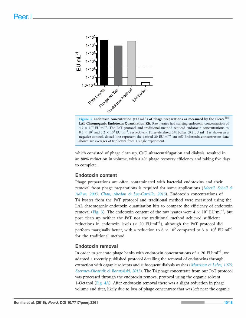
Endotoxin contentPhage preparations are often contaminated with bacterial endotoxins and their
removal from phage preparations is required for some applications (Merril, Scholl &
Adhya, 2003; Chan, Abedon & Loc-Carrillo, 2013). Endotoxin concentrations of
T4 lysates from the PoT protocol and traditional method were measured using the
LAL chromogenic endotoxin quantitation kits to compare the efficiency of endotoxin
removal (Fig. 3). The endotoxin content of the raw lysates were 4 � 104 EU·ml-1, but
post clean up neither the PoT nor the traditional method achieved sufficient
reductions in endotoxin levels (< 20 EU·ml-1), although the PoT protocol did
perform marginally better, with a reduction to 8 � 103 compared to 3 � 104 EU·ml-1
for the traditional method.
Endotoxin removalIn order to generate phage banks with endotoxin concentrations of < 20 EU·ml-1, we
adapted a recently published protocol detailing the removal of endotoxins through
extraction with organic solvents and subsequent dialysis washes (Morrison & Leive, 1975;
Szermer-Olearnik & Boraty�nski, 2015). The T4 phage concentrate from our PoT protocol
was processed through the endotoxin removal protocol using the organic solvent
1-Octanol (Fig. 4A). After endotoxin removal there was a slight reduction in phage
volume and titer, likely due to loss of phage concentrate that was left near the organic
Figure 3 Endotoxin concentration (EU·ml-1) of phage preparations as measured by the PierceTM
LAL Chromogenic Endotoxin Quantitation Kit. Raw lysates had starting endotoxin concentration of
4.7 � 104 EU·ml-1. The PoT protocol and traditional method reduced endotoxin concentrations to
8.3 � 103 and 3.2 � 104 EU·ml-1, respectively. Filter-sterilized SM buffer (0.2 EU·ml-1) is shown as a
negative control, dotted line represent the desired 20 EU·ml-1 cut off. Endotoxin concentration data
shown are averages of triplicates from a single experiment.
Bonilla et al. (2016), PeerJ, DOI 10.7717/peerj.2261 10/18

layer. Unfortunately, endotoxin quantification at this step was not possible due to residual
1-Octanol in the phage concentrate disabling LAL endotoxin test (Szermer-Olearnik &
Boraty�nski, 2015). In order to remove residual 1-Octanol, phage concentrates were split
evenly, processed through dialysis and speed vacuum (5 ml each) and endotoxin levels
quantified (Fig. 4B). Following dialysis washes, we were capable of producing a T4 phage
concentrate with 14 EU·ml-1, with a 30% increase in volume (likely due to water
movement to the higher ionic strength SM buffer), and a phage recovery efficiency of 60%
within four days. The major limitation with this method is the lengthy dialysis step
required to remove residual 1-Octanol solvents from the phage concentrate, taking
upwards of 48 h to complete. We tested a faster speed vacuum method to remove this
residual solvent, taking just 3 h to complete and producing a lysate with a slightly
Figure 4 PoT protocol processed for the removal of bacterial endotoxins by 1-Octanol and solvent
removal by dialysis and speed vacuum. (A) The PoT protocol processed for endotoxin removal by the
dialysis and speed vacuum methods generated a 7.2 mL (3.8 � 1010 PFU·ml-1) and 4.2 mL (4.7 � 1010
PFU·ml-1) phage banks, respectively. (B) Endotoxin concentrations of dialysis (14 EU·ml-1) and speed
vacuum (27 EU·ml-1) processed PoT phage banks. Quantification of 1-Octanol treated concentrate was
not possible due to residual solvent disabling the LAL quantification test, dotted line represent the
desired 20 EU·ml-1 cut off.
Figure 5 Stability of T4 phage processed through PoTunder high-use laboratory conditions. (A) The
mean titer of T4 phage stored at 4 �C in SM buffer, liquid nitrogen (LN2) in 5% (v/v) DMSO, LN2 in
50% (v/v) glycerol, -20 �C in 50% glycerol and -80 �C in 50% glycerol. Phage stocks were tittered
in duplicate under high-use conditions (full titers Fig. S4). (B) Negative stain transmission electron
micrograph of T4 phage processed through the PoT protocol (scale bar ¼ 100 nm).
Bonilla et al. (2016), PeerJ, DOI 10.7717/peerj.2261 11/18

increased endotoxin content of 27 EU·ml-1, with a 20% reduction in volume and a phage
recovery efficiency of 47%.
Phage banks and storagePhage banks were produced from purified concentrates to provide homogenous, high
titer, endotoxin reduced phage stock for repeat molecular testing and characterization.
Figure 6 Applicability of PoT for the generation of high-titer, homogenous, endotoxin reduced
phage banks from diverse phages. (A) T4�hoc phage that infects E. coli bacterial host propagated
by liquid lysate (2.2 � 1010 PFU·ml-1). (B) T3 phage that infects E. coli bacterial host propagated by
plate lysate (1.1 � 1010 PFU·ml-1). (C) T5 phage that infects E. coli bacterial host propagated by plate
lysate (2 � 109 PFU·ml-1). (D) Spp1 phage that infects B. subtilis Gram-positive bacterial host
propagated by plate lysate (1.5 � 109 PFU·ml-1). (E) Endotoxin concentrations of raw lysates,
PoT concentrate, dialysis and speed vacuum treated phage banks, dotted line represent the desired
20 EU·ml-1 cut off. Overall, a decrease in volume (ml) of the lysates was observed after each step of
the procedure.
Bonilla et al. (2016), PeerJ, DOI 10.7717/peerj.2261 12/18
As such it was important to ensure the stability of phage banks under repeated laboratory
use. We tested a range of conditions for the shot-term storage and repeated use of T4
phage banks, including 4 �C in SM buffer, liquid nitrogen in 50% v/v glycerol, liquid
nitrogen in 5% v/v DMSO, -20 �C in 50% v/v glycerol and -80 �C in 50% v/v glycerol
(Fig. 5A). Phage bank storage at 4 �C in SM buffer showed minimal reduction in titer for
short-term storage of high-use phage banks. Freezing of high-use phage banks is not
recommended, as the repeated freeze-thaw cycles from the frozen preparations likely
damaged phage, reducing titer. If long-term storage of phage banks is required, we
recommend storage in liquid nitrogen with 5% v/v DMSO with minimal freeze-thaws, as
this showed lowest loss of phage titer over a 3-month period. However, prior to use of
frozen phage banks, it is advised to wash phage lysate with SM buffer through an
ultrafiltration unit to remove any residual DMSO. Finally, T4 phage morphology and
structure following PoT protocol was determined by transmission electron microscopy
(TEM) (Fig. 5B). T4 phage showed tails that were not contracted and intact capsid
structures, indicating that PoT protocol did not negatively impact phage structure and
viability.
Applicability of Phage on Tap protocolAs the PoT protocol is based on an ultrafiltration methodology, which we believe is
broadly applicable to other tailed phages that can be isolate and cultured to high titer.
To test this, we processed four tailed phages, including T4�hoc–a Myoviridae infecting
E. coli; T3–a Podoviridae infecting E. coli; T5–a Siphoviridae infecting E. coli; and
Spp1–a Siphoviridae infecting the Gram-positive Bacillus subtilis, through the PoT
protocol (Fig. 6). Phages T3, T5 and Spp1 were propagated by plate lysates, while
T4�hoc phage was propagated by liquid lysate. All phages were processed through to a
concentrate with a > 50% phage recovery efficiency, followed by 1-Octanol endotoxin
removal and processing by dialysis and speed vacuum. All phages showed a large
reduction in endotoxin concentrations, but none were below the desired < 20 EU·ml-1
with the exception of the Spp1 phage, which infects the Gram-positive Bacillus subtilis
bacterial host. The Spp1 phage lysate produced an already low endotoxin lysate that we
further reduced to < 1 EU·ml-1. The dialysis method was more efficient than speed
vacuum at removing bacterial endotoxins, but took 48 h to complete. Comparatively,
the speed vacuum method took only 3 h to complete, producing a phage concentrate
with an average 2-fold higher endotoxin concentration than the dialysis method with a
comparable titer. Overall, all of the phages processed through the PoT protocol
generated high titer, homogenous phage banks with significantly reduced
endotoxin levels.
DISCUSSIONTraditional methods for the isolation and generation of phages lysates often involve
centrifugation, precipitation with PEG, ultracentrifugation in CsCl gradients, followed by
dialysis and storage. These procedures are lengthy and time consuming and generate
Bonilla et al. (2016), PeerJ, DOI 10.7717/peerj.2261 13/18

phage lysates with variable titer, endotoxin, and ionic concentration. Here we present a
fast and efficient method to produce homogeneous phage banks for laboratory testing
and molecular characterization. Our method focused on T4 phage, but is broadly
applicable to other phages that can be isolated in high titer (> 109 PFU·ml-1). Specifically,
the T4 phage was propagated with E. coli bacterial host in liquid lysate, purified by
centrifugation, 0.22 mm filtration and chloroform treatment, concentrated by
ultrafiltration centrifugation, and stored in large phage banks at 4 �C in buffer.
Unfortunately, neither the PoT protocol nor the traditional methods were effective at
significantly reducing bacterial endotoxins from phage preparations, and additional
purification steps were required.
Numerous methods and commercially available kits are available for the removal
of bacterial endotoxins (Merril et al., 1996; Boraty�nski et al., 2004; Merabishvili
et al., 2009; Oslizło et al., 2011; Branston, Wright & Keshavarz-Moore, 2015), yet
many of these are either highly specific, time consuming, laborious, or expensive.
Szermer-Olearnik & Boraty�nski (2015) recently proposed the use of an organic solvent
to successfully reduce bacterial endotoxin from phage lysates (< 20 EU·ml-1). The
method is cheap, broadly applicable, and capable of endotoxin removal regardless of
initial variations in titer and endotoxin levels, but does rely on multiple dialysis steps
that are time consuming (Morrison & Leive, 1975; Szermer-Olearnik & Boraty�nski,
2015). It may be possible to shorten the dialysis method through more frequent
changes in buffer solution, although this needs to be confirmed. Here we modify this
method by replacing the long dialysis steps with a speed vacuum step for the removal
of residual 1-octanol from the lysates. The speed vacuum modification was not as
efficient as the dialysis step for the removal of bacterial endotoxins, but was
significantly shorter to complete–taking just 3 h compared to the 48 h required for
dialysis. Using this modification we are able to produce homogenous, high-titer,
endotoxin-reduced phage banks within two days.
Phage are generally quite stable at high concentrations when stored at 4 �C, free ofbacterial debris, protected from light exposure, and in appropriately buffered solution
with a pH between 5–9 (Adams, 1959; Clark, 1962; Wommack et al., 1996; Jo�nczyk et al.,
2011). Although most tailed and filamentous phages can be easily stored under these
conditions for 5–10+ years, it is always best to determine the optimal storage conditions
for each phage of interest. Phage are generally sensitive to freezing, thawing, and
lyophilization and titer is known to vary with storage time (Clark, 1962; Clark & Klein,
1966; Clark & Geary, 1973; Jo�nczyk et al., 2011). After monitoring the degradation of
numerous phages for over 21 years, it was shown that phage storage at 4 �C and -80 �Cwas suboptimal compared to storage in liquid nitrogen (-196 �C) (Ackermann,
Tremblay & Moineau, 2004). However, due to their diversity and the difficulties associated
with accurate monitoring, the optimal conditions for long-term storage of phage
(> 10 years) remains uncertain. The purpose of PoT protocol was to generate
homogenous, high-titer phage banks for regular and consistent laboratory use. For this
purpose the storage of phage in SM buffer at 4 �C over three months showed no
Bonilla et al. (2016), PeerJ, DOI 10.7717/peerj.2261 14/18

noticeable depreciation in titer, which is consistent with previously described short-term
storage conditions.
The PoT protocol described here takes two days to produce high titer, homogenous,
endotoxin reduced phage banks for molecular characterizations. In comparison, the
traditional method can take upwards of five days to complete, is laborious, and generates a
lower titer phage lysate with high endotoxin levels. Traditional phage propagations suffer
from further variability in titer, salts, and bacterial contaminants between successive
lysates. Eliminating this variability through the use of PoT phage banks has been critical
for the accurate analysis and molecular investigations of phage within the context of
eukaryotic hosts (Barr et al., 2013; Barr et al., 2015). Phage banks can be easily stored at
4� C in SM buffer while maintaining their viability under continuous high-use. The PoT
protocol is efficient, can be easily completed in the laboratory, may be among the least
costly, and generates large homogenous phage banks for repeated use.
ACKNOWLEDGEMENTSWe thank the San Diego State University TEM Facility for the help with the TEM
analyses.
ADDITIONAL INFORMATION AND DECLARATIONS
FundingThis work was supported by the National Institutes of Health (Grant R01GM095384-01 to
F.R.), Grants NS047101 and R21AI094534 from the National Institute of General
Medical Sciences, and the Gordon and Betty Moore Foundation (Investigator Award
3781). J.J.B. received funding and support from San Diego State University. The funders
had no role in study design, data collection and analysis, decision to publish, or
preparation of the manuscript.
Grant DisclosuresThe following grant information was disclosed by the authors:
National Institutes of Health: R01GM095384-01 to F.R.
National Institute of General Medical Sciences: NS047101 and R21AI094534.
Gordon and Betty Moore Foundation: Investigator Award 3781.
Competing InterestsThe authors declare that they have no competing interests.
Author Contributions� Natasha Bonilla conceived and designed the experiments, performed the experiments,
analyzed the data, contributed reagents/materials/analysis tools, wrote the paper,
prepared figures and/or tables, reviewed drafts of the paper.
� Maria Isabel Rojas performed the experiments, reviewed drafts of the paper.
� Giuliano Netto Flores Cruz performed the experiments, reviewed drafts of the paper.
Bonilla et al. (2016), PeerJ, DOI 10.7717/peerj.2261 15/18

� Shr-Hau Hung contributed reagents/materials/analysis tools, reviewed drafts of
the paper.
� Forest Rohwer conceived and designed the experiments, contributed reagents/materials/
analysis tools, reviewed drafts of the paper.
� Jeremy J. Barr conceived and designed the experiments, performed the experiments,
analyzed the data, contributed reagents/materials/analysis tools, wrote the paper,
prepared figures and/or tables, reviewed drafts of the paper.
Data DepositionThe following information was supplied regarding data availability:
The raw data has been supplied as Supplemental Dataset Files.
Supplemental InformationSupplemental information for this article can be found online at http://dx.doi.org/
10.7717/peerj.2261#supplemental-information.
REFERENCESAbedon ST, Kuhl SJ, Blasdel BG, Kutter EM. 2011. Phage treatment of human infections.
Bacteriophage 1(2):66–85 DOI 10.4161/bact.1.2.15845.
Ackermann H-W, Tremblay D, Moineau S. 2004. Long-term bacteriophage preservation.
WFCC Newsletter 38:35–40.
Adams MH. 1959. Bacteriophages. New York: Interscience.
Alexander C, Rietschel ET. 2001. Invited review: bacterial lipopolysaccharides and innate
immunity. Journal of Endotoxin Research 7(3):167–202 DOI 10.1177/09680519010070030101.
Barr JJ, Auro R, Furlan M, Whiteson KL, Erb ML, Pogliano J, Stotland A, Wolkowicz R,
Cutting AS, Doran KS, Salamon P, Youle M, Rohwer F. 2013. Bacteriophage adhering to
mucus provide a non-host-derived immunity. Proceedings of the National Academy of Sciences of
the United States of America 110(26):10771–10776 DOI 10.1073/pnas.1305923110.
Barr JJ, Auro R, Sam-Soon N, Kassegne S, Peters G, Bonilla N, Hatay M, Mourtada S, Bailey B,
Youle M, Felts B, Baljon A, Nulton J, Salamon P, Rohwer F. 2015. Subdiffusive motion of
bacteriophage in mucosal surfaces increases the frequency of bacterial encounters. Proceedings
of the National Academy of Sciences of the United States of America 112(44):13675–13680
DOI 10.1073/pnas.1508355112.
Boraty�nski J, Syper D, Weber-Dabrowska B, qusiak-Szelachowska M, Pozniak G, Gorski A.
2004. Preparation of endotoxin-free bacteriophages. Cellular & Molecular Biology Letters
9(2):253–259.
Bourdin G, Schmitt B, Marvin Guy L, Germond J-E, Zuber S, Michot L, Reuteler G, Brussow H.
2014. Amplification and purification of T4-like Escherichia coli phages for phage therapy: from
laboratory to pilot scale. Applied and Environmental Microbiology 80(4):1469–1476
DOI 10.1128/AEM.03357-13.
Branston SD, Wright J, Keshavarz-Moore E. 2015. A non-chromatographic method for the
removal of endotoxins from bacteriophages. Biotechnology and Bioengineering 112(8):
1714–1719 DOI 10.1002/bit.25571.
Bruttin A, Brussow H. 2005. Human volunteers receiving Escherichia coli phage T4 orally: a safety
test of phage therapy. Antimicrobial Agents and Chemotherapy 49(7):2874–2878
DOI 10.1128/AAC.49.7.2874-2878.2005.
Bonilla et al. (2016), PeerJ, DOI 10.7717/peerj.2261 16/18
Carlson K. 2005. Appendix: working with bacteriophages: common techniques and
methodological approaches. In: Kutter E, Sulakvelidze A, eds. Bacteriophages: Biology and
Applications. Vol. 1. Boca Raton: CRC Press, 439–490.
Chan BK, Abedon ST, Loc-Carrillo C. 2013. Phage cocktails and the future of phage therapy.
Future Microbiology 8(6):769–783 DOI 10.2217/fmb.13.47.
Clark WA. 1962. Comparison of several methods for preserving bacteriophages. Applied
Environmental Microbiology 10(5):466–471.
Clark WA, Geary D. 1973. Preservation of bacteriophages by freezing and freeze-drying.
Cryobiology 10(5):351–360 DOI 10.1016/0011-2240(73)90057-6.
Clark WA, Klein A. 1966. The stability of bacteriophages in long term storage at liquid nitrogen
temperatures. Cryobiology 3(2):68–75 DOI 10.1016/S0011-2240(66)80002-0.
Erickson HP. 2009. Size and shape of protein molecules at the nanometer level determined by
sedimentation, gel filtration, and electron microscopy. Biological Procedures Online 11(1):32–51
DOI 10.1007/s12575-009-9008-x.
Gorbet MB, Sefton MV. 2005. Endotoxin: the uninvited guest. Biomaterials 26(34):6811–6817
DOI 10.1016/j.biomaterials.2005.04.063.
Jo�nczyk E, K1ak M, Miedzybrodzki R, Gorski A. 2011. The influence of external
factors on bacteriophages–review. Folia Microbiologica 56(3):191–200
DOI 10.1007/s12223-011-0039-8.
Magalhaes PO, Lopes AM, Mazzola PG, Rangel-Yagui C, Penna TCV, Pessoa A Jr. 2007.
Methods of endotoxin removal from biological preparations: a review. Journal of
Pharmacy & Pharaceutical Sciences 10(3):388–404.
Merabishvili M, Pirnay J-P, Verbeken G, Chanishvili N, Tediashvili M, Lashkhi N, Glonti T,
Krylov V, Mast J, Van Parys L, Lavigne R, Volckaert G, Mattheus W, Verween G, De Corte P,
Rose T, Jennes S, Zizi M, De Vos D, Vaneechoutte M. 2009. Quality-controlled small-scale
production of a well-defined bacteriophage cocktail for use in human clinical trials. PLoS ONE
4(3):e4944 DOI 10.1371/journal.pone.0004944.
Merril CR, Biswas B, Carlton R, Jensen NC, Creed GJ, Zullo S, Adhya S. 1996. Long-circulating
bacteriophage as antibacterial agents. Proceedings of the National Academy of Sciences of the
United States of America 93(8):3188–3192 DOI 10.1073/pnas.93.8.3188.
Merril CR, Scholl D, Adhya SL. 2003. The prospect for bacteriophage therapy in Western
medicine. Nature Reviews Drug Discovery 2(6):489–497 DOI 10.1038/nrd1111.
Morrison DC, Leive L. 1975. Fractions of lipopolysaccharide from Escherichia coli
O111:B4 prepared by two extraction procedures. Journal of Biological Chemistry
250:2911–2919.
Morrison DC, Ulevitch RJ. 1978. The effects of bacterial endotoxins on host mediation
systems. A review. American Journal of Pathology 93(2):526–618.
Osliz1o A, Miernikiewicz P, Piotrowicz A, Owczarek B, Kopciuch A, Figura G, Dabrowska K.
2011. Purification of phage display-modified bacteriophage T4 by affinity chromatography.
BMC Biotechnology 11(1):59 DOI 10.1186/1472-6750-11-59.
Pabst MJ, Pabst KM, Handsman DB, Beranova-Giorgianni S, Giorgianni F. 2008. Proteome of
monocyte priming by lipopolysaccharide, including changes in interleukin-1beta and leukocyte
elastase inhibitor. Proteome Science 6(1):13 DOI 10.1186/1477-5956-6-13.
Raetz CRH, Reynolds CM, Trent MS, Bishop RE. 2007. Lipid a modification systems in
gram-negative bacteria. Annual Review of Biochemistry 76(1):295–329
DOI 10.1146/annurev.biochem.76.010307.145803.
Bonilla et al. (2016), PeerJ, DOI 10.7717/peerj.2261 17/18
Rietschel E, Kirikae T, Schade F, Mamat U, Schmidt G, Loppnow H, Ulmer A, Zahringer U,
Seydel U, Di Padova F. 1994. Bacterial endotoxin: molecular relationships of structure to
activity and function. FASEB Journal 8(2):217–225.
Seeley ND, Primrose SB. 1982. A review: the isolation of bacteriophages from the environment.
Journal of Applied Bacteriology 53(1):1–17 DOI 10.1111/j.1365-2672.1982.tb04729.x.
Suttle CA, Chan AM, Cottrell MT. 1991. Use of ultrafiltration to isolate viruses from seawater
which are pathogens of marine Phytoplankton. Applied Environmental Microbiology
57(3):721–726.
Szermer-Olearnik B, Boraty�nski J. 2015. Removal of endotoxins from bacteriophage
preparations by extraction with organic solvents. PLoS ONE 10(3):e122672
DOI 10.1371/journal.pone.0122672.
Wommack KE, Hill RT, Muller TA, Colwell RR. 1996. Effects of sunlight on bacteriophage
viability and structure. Applied and Environmental Microbiology 62(4):1336–1341.
Yamamoto KR, Alberts BM, Benzinger R, Lawhorne L, Treiber G. 1970. Rapid bacteriophage
sedimentation in the presence of polyethylene glycol and its application to large-scale virus
purification. Virology 40(3):734–744 DOI 10.1016/0042-6822(70)90218-7.
Bonilla et al. (2016), PeerJ, DOI 10.7717/peerj.2261 18/18