pharmacol rev 55:509–550, 2003 printed in u.s.a...
TRANSCRIPT

Molecular Mechanisms and Therapeutical Implicationsof Intramembrane Receptor/Receptor Interactions
among Heptahelical Receptors with Examples from theStriatopallidal GABA Neurons
LUIGI F. AGNATI,1 SERGI FERRE, CARME LLUIS, RAFAEL FRANCO, AND KJELL FUXE
University of Modena, Modena, Italy; National Institute on Drug Abuse, National Institutes of Health, Department of Health and HumanServices, Baltimore, Maryland; University of Barcelona, Barcelona, Spain; Department of Neuroscience, Karolinska Institutet,
Stockholm, Sweden
Abstract . . . . . . . . . . . . . . . . . . . . . . . . . . . . . . . . . . . . . . . . . . . . . . . . . . . . . . . . . . . . . . . . . . . . . . . . . . . . . . . 510I. Experimental evidence on protein/protein interactions involving G protein-coupled receptor in the
central nervous system . . . . . . . . . . . . . . . . . . . . . . . . . . . . . . . . . . . . . . . . . . . . . . . . . . . . . . . . . . . . . . . . . . 510A. Early indications for intramembrane receptor/receptor interactions involving G protein-coupled
receptors . . . . . . . . . . . . . . . . . . . . . . . . . . . . . . . . . . . . . . . . . . . . . . . . . . . . . . . . . . . . . . . . . . . . . . . . . . . 510B. G protein-coupled receptors homo- and heteromerization . . . . . . . . . . . . . . . . . . . . . . . . . . . . . . . . 512
1. Homomerization of G protein-coupled receptors . . . . . . . . . . . . . . . . . . . . . . . . . . . . . . . . . . . . . . 5122. Heteromeric complexes involving G protein-coupled receptors . . . . . . . . . . . . . . . . . . . . . . . . . 514
a. The GABAB receptor heterodimer . . . . . . . . . . . . . . . . . . . . . . . . . . . . . . . . . . . . . . . . . . . . . . . 514b. Heteromerization of d and k opioid receptors and of m and d opioid receptors. . . . . . . . . 515c. The serotonin 5-HT1D/5-HT1B receptor heteromer . . . . . . . . . . . . . . . . . . . . . . . . . . . . . . . . . 515d. The dopamine D2/D3 receptor heteromer . . . . . . . . . . . . . . . . . . . . . . . . . . . . . . . . . . . . . . . . . 516e. The somatostatin SSTR5/SSTR1 receptor heteromer . . . . . . . . . . . . . . . . . . . . . . . . . . . . . . . 516f. The somatostatin SSTR5 and dopamine D2 heteromeric receptor complex. . . . . . . . . . . . 516g. The adenosine A1 and dopamine D1 heteromeric receptor complex . . . . . . . . . . . . . . . . . . 517h. The metabotropic glutamate mGluR1a and adenosine A1 heteromeric receptor complex . . 518i. The purinergic P2Y1 and adenosine A1 heteromeric receptor complex . . . . . . . . . . . . . . . . 519j. The adenosine A2A and dopamine D2 heteromeric receptor complex . . . . . . . . . . . . . . . . . 519
k. The metabotropic glutamate mGlu5 and adenosine A2A heteromeric receptorcomplex . . . . . . . . . . . . . . . . . . . . . . . . . . . . . . . . . . . . . . . . . . . . . . . . . . . . . . . . . . . . . . . . . . . . . . . 520
l. The bradykinin B2 and angiotensin AT1 heteromeric receptor complex . . . . . . . . . . . . . . 521m. Other heteromeric G protein-coupled receptor complexes. . . . . . . . . . . . . . . . . . . . . . . . . . . 522
C. Direct protein/protein interactions between G protein-coupled receptors and multisubunitligand-gated ion channels . . . . . . . . . . . . . . . . . . . . . . . . . . . . . . . . . . . . . . . . . . . . . . . . . . . . . . . . . . . . 5221. The GABAA and dopamine D5 heteromeric receptor complex . . . . . . . . . . . . . . . . . . . . . . . . . . 522
D. Oligomeric complexes containing G protein-coupled receptors and receptor tyrosine kinases. 524E. Oligomeric complexes containing G protein-coupled receptors and receptor activity modifying
proteins . . . . . . . . . . . . . . . . . . . . . . . . . . . . . . . . . . . . . . . . . . . . . . . . . . . . . . . . . . . . . . . . . . . . . . . . . . . . 5241. Receptor activity modifying transmembrane proteins . . . . . . . . . . . . . . . . . . . . . . . . . . . . . . . . . 524
a. The calcitonin receptor family/RAMP1–3 heteromeric complexes. . . . . . . . . . . . . . . . . . . . 524b. The dopamine D1 receptor/calcyon heteromeric complex. . . . . . . . . . . . . . . . . . . . . . . . . . . . 525
2. Receptor activity modifying cytosolic proteins. . . . . . . . . . . . . . . . . . . . . . . . . . . . . . . . . . . . . . . . 526a. The adenosine A1 receptor and adenosine deaminase heteromeric complex. . . . . . . . . . . 526b. The adenosine A1 receptor and hsc73 heteromeric complex . . . . . . . . . . . . . . . . . . . . . . . . . 526
1Dedicated to Prof. G. L. Gessa.Address correspondence to: Kjell Fuxe, Department of Neuroscience, Karolinska Institutet, 171 77 Stockholm, Sweden. E-mail:
[email protected], publication date, and citation information can be found at http://pharmrev.aspetjournals.org.DOI: 10.1124/pr.55.3.2.
0031-6997/03/5503-509–550$7.00PHARMACOLOGICAL REVIEWS Vol. 55, No. 3U.S. Government work not protected by U.S. copyright 30303/1082283Pharmacol Rev 55:509–550, 2003 Printed in U.S.A
509
Copyright 2003 by the American Society for Pharmacology and Experimental Therapeutics.
Pharmrev Fast Forward. Published on July 17, 2003 as DOI:10.1124/pr.55.3.2
II. On the functional implications of receptor/receptor interactions . . . . . . . . . . . . . . . . . . . . . . . . . . . . . 527A. The context of the present discussion . . . . . . . . . . . . . . . . . . . . . . . . . . . . . . . . . . . . . . . . . . . . . . . . . . 527B. Structural basis of receptor function. . . . . . . . . . . . . . . . . . . . . . . . . . . . . . . . . . . . . . . . . . . . . . . . . . . 527
1. Conformational diversity . . . . . . . . . . . . . . . . . . . . . . . . . . . . . . . . . . . . . . . . . . . . . . . . . . . . . . . . . . 5272. Oligomeric diversity: the receptor mosaic. . . . . . . . . . . . . . . . . . . . . . . . . . . . . . . . . . . . . . . . . . . . 5293. The role of the receptor mosaic in learning and memory . . . . . . . . . . . . . . . . . . . . . . . . . . . . . . 530
C. Communication processes in the cell. . . . . . . . . . . . . . . . . . . . . . . . . . . . . . . . . . . . . . . . . . . . . . . . . . . 530D. Receptor/receptor interactions in the striatopallidal GABA neurons: implications for Parkinson’s
disease, schizophrenia, and drug addiction . . . . . . . . . . . . . . . . . . . . . . . . . . . . . . . . . . . . . . . . . . . . . 5331. Localization of adenosine A2A, dopamine D2 and glutamate metabotropic mGluR5 receptors
in the GABA striatopallidal neuron. . . . . . . . . . . . . . . . . . . . . . . . . . . . . . . . . . . . . . . . . . . . . . . . . 5332. Interactions between adenosine A2A, dopamine D2 and glutamate metabotropic mGluR5
receptors in the GABA striatopallidal neuron: biochemical-cellular level. . . . . . . . . . . . . . . . 5353. Interactions between adenosine A2A, dopamine D2 and glutamate metabotropic mGluR5
receptors in the GABA striatopallidal neuron: physiological-behavioral level . . . . . . . . . . . . 5384. Interactions between adenosine, dopamine, and glutamate metabotropic receptors in the
GABAergic striatoentopeduncular and striatonigral neurons . . . . . . . . . . . . . . . . . . . . . . . . . . 541III. Implications of receptor/receptor interactions for drug development . . . . . . . . . . . . . . . . . . . . . . . . . . 541
A. The ground for novel therapeutical interventions . . . . . . . . . . . . . . . . . . . . . . . . . . . . . . . . . . . . . . . 541B. Theoretical strategies to target receptor complexes. . . . . . . . . . . . . . . . . . . . . . . . . . . . . . . . . . . . . . 541C. Possible targets for drugs acting on heteromeric receptor complexes . . . . . . . . . . . . . . . . . . . . . . 542Acknowledgments. . . . . . . . . . . . . . . . . . . . . . . . . . . . . . . . . . . . . . . . . . . . . . . . . . . . . . . . . . . . . . . . . . . . . . . 543References . . . . . . . . . . . . . . . . . . . . . . . . . . . . . . . . . . . . . . . . . . . . . . . . . . . . . . . . . . . . . . . . . . . . . . . . . . . . . 543
Abstract——The molecular basis for the known in-tramembrane receptor/receptor interactions among Gprotein-coupled receptors was postulated to be het-eromerization based on receptor subtype-specific in-teractions between different types of receptor ho-momers. The discovery of GABAB heterodimersstarted this field rapidly followed by the discovery ofheteromerization among isoreceptors of several G pro-tein-coupled receptors such as d/k opioid receptors.Heteromerization was also discovered among distincttypes of G protein-coupled receptors with the initialdemonstration of somatostatin SSTR5/dopamine D2and adenosine A1/dopamine D1 heteromeric receptorcomplexes. The functional meaning of these hetero-meric complexes is to achieve direct or indirect (viaadapter proteins) intramembrane receptor/receptorinteractions in the complex. G protein-coupled recep-tors also form heteromeric complexes involving direct
interactions with ion channel receptors, the best ex-ample being the GABAA/dopamine D5 receptor hetero-merization, as well as with receptor tyrosine kinasesand with receptor activity modulating proteins. As anexample, adenosine, dopamine, and glutamatemetabotropic receptor/receptor interactions in thestriatopallidal GABA neurons are discussed as well astheir relevance for Parkinson’s disease, schizophre-nia, and drug dependence. The heterodimer is onlyone type of heteromeric complex, and the evidence isequally compatible with the existence of higher orderheteromeric complexes, where also adapter proteinssuch as homer proteins and scaffolding proteins canexist. These complexes may assist in the process oflinking G protein-coupled receptors and ion channelreceptors together in a receptor mosaic that may havespecial integrative value and may constitute the mo-lecular basis for some forms of learning and memory.
I. Experimental Evidence on Protein/ProteinInteractions Involving G Protein-CoupledReceptors in the Central Nervous System
A. Early Indications for IntramembraneReceptor/Receptor Interactions Involving G Protein-Coupled Receptors
Emerging evidence shows that G protein-coupled re-ceptors (GPCR2) can form homo- and heteromers (Bou-
2Abbreviations: GPCR, G protein-coupled receptor; ADA, adeno-sine deaminase; ADM, adrenomedullin; AT, angiotensin; BRET, bi-oluminescence resonance energy transfer; CaMK, Ca21/calmodulin-dependent protein kinases; CCK, cholecystokinin; CGRP, calcitonin
gene-related peptide; CHO, Chinese hamster ovary; CRE, cAMPresponse element; CREB, cAMP response element-binding protein;CRL, calcitonin receptor-like; DARPP-32, dopamine and cyclic aden-osine 39,59-monophosphate-regulated phosphoprotein, 32 kDa; EGF,epidermal growth factor; FRET, fluorescence resonance energytransfer; Gal, galanin; GRK, GPCR kinase; GST, glutathione S-transferase; GTPgS, guanosine 59-O-(3-thio)triphosphate; HEK, hu-man embryonic kidney; 5-HT, 5-hydroxytryptamine (serotonin); IP3,inositol 1,4,5-trisphosphate; MAPK, mitogen-activated protein ki-nase; MEK, MAPK/extracellular-regulated kinase; MPTP, 1-methyl-4-phenyl-1,2,3,6-tetrahydropyridine; NK, neurokinin; NMDA, N-methyl-D-aspartate; NPY, neuropeptide Y; NT, neurotensin; PAGE,polyacrylamide gel electrophoresis; PAR, protease-activated recep-tor; PCP, phencyclidine; PLC, phospholipase C; PKA, protein kinaseA; PKC, protein kinase C; PP-1, protein phosphatase-1; PSD,
510 AGNATI ET AL.

vier, 2001; Marshall, 2001). It all began in 1979–1980 insearch of an explanation of where all the recently dis-covered neuropeptides in the brain could integrate theirmessages with those of classical transmitters such asthe monoamines. Luigi F. Agnati and Kjell Fuxe postu-lated that an intramembrane interaction between neu-ropeptide and monoamine receptors could be involved.The first observations were published in 1980 (Agnati etal., 1980), showing that substance P could modulate thehigh-affinity serotonin binding sites in spinal cord mem-brane preparations using biochemical binding tech-niques (also, see Agnati et al., 1983b). The same year, aninteresting paper was published by Maggi et al. (1980)showing that the b adrenergic receptor agonist isopro-terenol could increase a2 adrenergic receptor binding incortical slices, supporting the concept of intramembranereceptor/receptor interactions of GPCR, in this caseamong isoreceptors. Subsequently, in 1981 the existenceof cholecystokinin (CCK) receptor/dopamine D2 receptorinteractions using biochemical binding techniques wasindicated since CCK-8 could modulate the dopamine D2receptor antagonist and agonist binding sites in striatalmembrane preparations (Fuxe et al., 1981, 1983b; Ag-nati et al., 1983a,b, 1985). Further evidence for receptor/receptor interactions came in 1982 from Lundberg, Bar-tfai, and colleagues (Lundberg et al., 1982) and fromZarbin and colleagues (Zarbin et al., 1982). Using thesame type of approach, a large number of papers werepublished in 1983 that suggested the existence of in-tramembrane receptor/receptor interactions betweendifferent GPCR (Fuxe et al., 1983b; Agnati et al., 1984;Fuxe and Agnati, 1985). Those included neurotensin(NT receptor)/D2 (Agnati et al., 1983c; Nemeroff, 1986;Von Euler and Fuxe, 1987; Von Euler, 1991), CCKB/serotonin 5-HT2 (Agnati et al., 1983a, 1985), vasoactiveintestinal peptide (VIP) receptor/serotonin 5-HT1 (Ros-tene et al., 1983a,b), neuropeptide Y (NPY) receptor/a2adrenergic (Agnati et al., 1983d) and neurokinin NK1/5-HT1 receptor/receptor interactions (Agnati et al., 1983e).Subsequently in the 1980s, indications for glutamatereceptor/D2 receptor interactions (Fuxe et al., 1984)were obtained in striatal membrane preparations afterearlier observations had indicated the existence of inter-actions at the membrane level among glutamate recep-tor subtypes (Fuxe et al., 1983c). This early research ledto the following postulation in the opening address ofFuxe and Agnati at the International Wenner-GrenSymposium on receptor/receptor interactions in 1986(Fuxe and Agnati, 1987) “. . . we will find out that somesophisticated elaborations are performed at the mem-brane level, via interactions within and among differentclasses of macromolecules (such as receptors, ion pumps,ion channels)” (also, see Agnati et al., 1988). In 1988,
evidence for galanin (Gal) receptor/serotonin 5-HT1A re-ceptor interactions in limbic cortical membranes (Fuxeet al., 1988a), as well as for angiotensin II receptor(AT1)/a2 adrenergic receptor interactions (Fuxe et al.,1988b) in the medulla oblongata membrane prepara-tions were obtained. In the early 1990s, adenosine A2Areceptor/D2 receptor interactions (Ferre et al., 1991d,1993; Ferre and Fuxe, 1992) were demonstrated in stri-atal membrane preparations.
Thus, not only neuropeptide and monoamine recep-tors were involved in intramembrane receptor/receptorinteractions but also certain types of glutamate andadenosine receptors (Agnati et al., 1986, 1990, 1993;Harfstrand et al., 1988; Tanganelli et al., 1989, 1990,1993; Von Euler et al., 1989; Fuxe et al., 1990a–c, 1991,1992a–c; Ferre et al., 1992, 1993b; Fior et al., 1993;Yang et al., 1994b). These results were all obtained atthe recognition site of the receptors, using saturationand competition binding experiments. The modulationof binding could be shown as changes in KD and Bmaxvalues (saturation analysis) and as KL, KH, and RHvalues (competition analysis) allowing a determinationof modulation of the high- versus the low-affinity statesof the receptor. An indication of an effect on the Gprotein-coupling and thus on the efficacy of the modu-lated receptor could be obtained by studying how, e.g.,the modulator could control the GTP-induced disappear-ance of the high-affinity state of the receptor (reductionof the RH values). This would imply a G protein activa-tion with formation of Ga-GTP and bg dimers associatedwith a cross-regulation of the GPCR with a disappear-ance of the high-affinity state of the receptor.
In this period, the above work was extended to showmultiple receptor/receptor interactions. Thus, evidencewas obtained for a dopamine D1 receptor involvement inthe CCKB receptor/D2 receptor interaction. Coactivationof D1 and D2 receptors led to an enhancement of theaffinity of D2 receptor agonist sites by CCK-8, instead ofa reduced affinity of D2 receptor agonist sites observedwithout D1 receptor stimulation (Li et al., 1994a). Theseresults are in line with the findings of Seeman et al.(1989) suggesting reciprocal interactions between D1and D2 receptors in striatal homogenates. Thus, theremay exist striatal nerve cell populations where in-tramembrane multiple CCKB receptor/D1 receptor/D2receptor interactions can take place (Agnati et al., 1982).
In the early 1990s, evidence was also obtained thatstriatal NT receptors involved in the G protein-indepen-dent antagonistic regulation of striatal D2 receptors(Von Euler et al., 1991) may represent a novel type of ahigh-affinity NT receptor. This was suggested in view ofthe rank order of potency found among COOH-terminalNT fragments, neuromedin N, and NT in this responseversus the rank order of potency found at the clonedhigh-affinity NT receptors (NT1 receptors) (Li et al.,1993a,b). These effects were stronger in striatal sections(Li et al., 1994b), and recently, the NT-induced reduc-
postsynaptic density protein; RAMP, receptor activity-modifyingprotein; RTK, receptor tyrosine kinase; SST, somatostatin; TCF/Elk-1, ternary complex factor/Elk-1; TM, transmembrane; trunc,truncated; VT, volume transmission; WT, wiring transmission.
RECEPTOR/RECEPTOR INTERACTIONS VIA HETEROMERIZATION 511

tion of D2 receptor affinity in striatal sections has beenfound to be blocked by a NT1-like antagonist (Diaz-Cabiale et al., 2002a).
In 1993 (Zoli et al., 1993), the hypothesis was intro-duced that the molecular mechanism for these largenumbers of intramembrane receptor/receptor interac-tions among GPCR could be the formation of hetero-meric complexes, the simplest being a heterodimer. Thisconcept was based on the indication at the time thatdimerization upon agonist activation may be a generalphenomenon essential for receptor activation (Hollen-berg, 1991), the best example being the dimerization oftyrosine kinase receptors (Schlessinger 1988, 2000; Hell-din 1995). Thus, it was assumed that GPCR exist mainlyas homodimers that interact with other types of ho-modimers to form heterodimers. The relative propor-tions of homo- and heterodimers would be determined bythe concentrations of the two transmitters, the densityof the two receptors and their distribution patterns, andthe unique features of each receptor/receptor interaction(Zoli et al., 1993). In fact, early evidence obtained onpurified GPCR by, e.g., Venter and Fraser (1983) andConn et al. (1982) indicated that the functional GPCRwere in a dimeric state. The same year that our reviewarticle appeared, the first evidence was published thatGPCR can exist as dimers (Ng et al., 1993). Thus, the5-HT1B receptor in Sf9 cells was in the immunoblotanalysis shown to exist as dimers and monomers. Fi-nally, it should be mentioned that in 1982 we had intro-duced the receptor mosaic hypothesis of learning andmemory based on the formation of membrane receptorclusters and thus of high order oligomeric receptor com-plexes (Agnati et al., 2002). It was postulated (Agnati etal., 1982; Zoli et al., 1993) that the formation and/orstabilization of the heteromeric complexes of GPCRcould be enhanced by associated (adapter) proteins es-pecially in the synaptic membranes. It must be notedthat most of the GPCR are located in extrasynapticmembranes and therefore the potential heteromericcomplexes discussed above may also be reached by vol-ume transmission (VT) signals (Agnati and Fuxe, 2000).
Recent experimental data exist confirming this hy-pothesis, and they are described in this review (see Ta-ble 1). Despite all this novel experimental evidence,there are many questions regarding the molecular mech-anism of receptor heteromerization and among them themapping of the residues involved in the interaction inthe case of direct interactions and the identification ofscaffolding and adapter proteins in the case of indirectinteractions. It should be noted that both types of inter-action, direct and indirect, are likely to occur.
B. G Protein-Coupled Receptors Homo- andHeteromerization
1. Homomerization of G Protein-Coupled Recep-tors. A number of new approaches made it possible toobtain convincing evidence for the existence of ho-
momers of many types of GPCR. Those include comple-mentary chimeras, coimmunoprecipitation with differ-entially epitope-tagged receptors, the use of sodiumdodecyl sulfate-polyacrylamide gel electrophoresis(SDS-PAGE), often in combination with covalent cross-linking, and finally biophysical methods, namely biolu-minescence resonance energy transfer (BRET) and flu-orescence resonance energy transfer (FRET).
In the mid 1980s, evidence for dimerization was ob-tained with experiments with photo-affinity labeling,radiation inactivation, cross-linking, and hydrodynamicanalysis (Fraser and Venter, 1982; Avissar et al., 1983;Herberg et al., 1984; Peterson et al., 1986). However, itwas the work of Maggio, Wess, and colleagues (Maggioet al., 1993) that offered strong indications for the exis-tence of dimers, with the transcomplementation resultsobtained using cholinergic M3 muscarinic receptor/a2adrenergic receptor chimeras. In fact, when a chimeraconsisting of 5TM regions of one receptor and two of theother was expressed, there was a lack of ligand bindingand of function that was recovered after coexpression ofthe two types of chimeras. This finding had a majorimpact, and the interpretation that the functionaltranscomplementation was caused by intermolecular in-teractions leading to the formation of a dimeric complexwas early on accepted (Monnot et al., 1996). The earlywork in the mid-1970s demonstrating negative cooper-ativity in b-adrenergic receptors, opening up the possi-bility of dimer formation should also be mentioned (Lim-bird et al., 1975; Limbird and Lefkowitz, 1976). Morerecently, structural implications for V2 vasopressin re-ceptor oligomerization have been given by Schultz et al.(2000) from functional reconstitution studies.
The SDS-PAGE approach provided one of the firstdemonstrations of GPCR dimers, namely of 5-HT1B, D1,and D2 receptor homomers (Ng et al., 1993, 1994a,b,1996; George et al., 1998; Zawarynski et al., 1998).These observations are based on the fact that severalhomomers are resistant to the denaturation propertiesof SDS. Thus, it was possible early on for George,O’Dowd, and colleagues to observe upon SDS-PAGE thatthe 5-HT1B, D1, and D2 receptors expressed in cell linesformed molecular species not only corresponding tomonomers but also to dimers (Ng et al., 1993, 1994a,b;Lee et al., 2000). These dimeric and even higher orderoligomeric complexes were not caused by glycosylation ofmonomers nor to the presence of G proteins (Lee et al.,2000). Treatment with covalent cross-linkers before sol-ubilization increased the proportion of dimeric com-plexes and facilitated the demonstration of dimers inimmunoblots (Hebert et al., 1996; Romano et al., 1996).It should be considered that monomers at least in somecases could represent the disruption of dimers or higheroligomeric complexes (Lee et al., 2000). Also by using theSDS-PAGE strategy, the existence of D2 and A1 recep-tors homomers was demonstrated, for the first time, inbrain tissue, showing their existence in situ and not only
512 AGNATI ET AL.

in cell lines where artificially high levels of receptors areexpressed (Ciruela et al., 1995; Ng et al., 1996).
The coimmunoprecipitation approach was first de-scribed in the article by Bouvier and colleagues in 1996(Hebert et al., 1996) on b2 adrenergic receptors withcoexpression of differentially tagged b2 adrenergic re-ceptors. This coimmunoprecipitation was taken as evi-dence for the existence of a b2 adrenergic receptor dimer.Peptides corresponding to TM6 were found to disruptthe dimerization as well as receptor activation indicat-ing a participation of the hydrophobic forces in the TM6region in the dimerization interface (Hebert et al., 1996;Ng et al., 1996). The focus on the TM6 came from thework of Engelman et al. (Lemmon et al., 1992; Lemmonand Engelman, 1994), showing that dimerization isdriven by specific interactions among TM a-helices. Anumber of GPCR homomers have been demonstrated
with this approach, e.g., the metabotropic glutamatereceptor (mGlu5) homomer, where the disulfide bridgesbetween the large extracellular NH2-terminal domainsplay a role in the formation of the homomer (Romano etal., 1996; Bouvier, 2001).
The techniques of BRET and FRET imply as closedistances as 5 to 10 nM between donor and acceptor forenergy transfer. In FRET both the donor and acceptorare fluorescent molecules, whereas for BRET the donoris bioluminescent and the acceptors fluorescent. Thesetechniques have been very valuable in detecting dimersin living cells without the risk of solubilization artifacts(for details on methodology, see Bouvier, 2001). In 2000,the BRET approach could for the first time demonstratehomomerization of b2 adrenergic receptors in living cells(Angers et al., 2000) independently, and at the sametime, the FRET procedure revealed the homomerization
TABLE 1Intramembrane receptor-receptor interactions via heteromerization
Type of Receptors Involved Possible Mechanism forInteraction
Changes at RecognitionLevel
Changes at SignalingLevel
Changes in ReceptorTrafficking
Possible TherapeuticImplications
GABABR1/GABABR2 Coiled-coil interaction atCOOH terminus(heterodimers arepreferred)
Increased potency ofagonists (R1, R2)
Essential forsignaling (R1, R2)
Essential for cellsurface expression(R1)
Anticonvulsive therapy
d opioid/k opioid Direct interaction(heterodimers arepreferred)
High affinity forunselective ligands
Synergisticactivation ofMAPK
Reduced d Rinternalization
Pain relief, drugdependence
d opioid/m opioid Direct interaction(heterodimers arepreferred)
Increased affinity forcertain enkpeptides
Altered selection ofG protein (e.g.,Gz)
Unknown Pain relief, drugdependence
5-HT1B/5-HT1D Direct interaction(heterodimers arepreferred)
Unknown Unknown Unknown Antidepressanttreatment
D2/D3 Direct interaction No clear-cut changes Increased couplingof D3 to AC
Unknown Antipsychotic treatment
SSTR5/SSTR1 Direct interaction Up-regulation ofSSTR1 agonistbinding
Sensitization ofSSTR1 afterSSTR5 activation
Unknown Unknown
SSTR5/D2 Agonist-dependentdirect interaction
Positive reciprocalaffinity regulation
Enhancement ofsignaling
Unknown Antiparkinsoniantreatment
A1/D1 Agonist regulation ofheteromerization:ADA dependency
Disappearance ofD1-R high affinitystate
Desensitization ofD1-R after A1/D1agonist treatment
A1-inducedcoaggregation ofA1-R/D1-R
Antiparkinsoniantreatment drugdependence
mGLU-R1a/A1 Involvement of mGLU-R1a COOH terminus:Homer dependency?
Unknown Reciprocalenhancement ofCa21 signaling
Unknown Neuroprotection
P2Y1/A1 Direct interaction Novel A1-R bindingpocket?
Altered selection ofG protein
Unknown Neuroprotection
A2A/D2 Possible involvement ofD2 5,6 TM domainsand intracellular loop3
Reduced D2-Raffinity, especiallyhigh-affinity state
Reduced D2-R Ca21
and cAMPsignaling
Coaggregation,cointernalization,codesensitization
Antiparkinsonian,antipsychotic,antidyskinetictreatment
A2A/mGLU-R5 Homer dependency? Unknown Synergism for c-Fosexpression
Unknown Antiparkinsonian,antipsychotic,antidyskinetic drugdependence treatment
B2/AT1 Unknown (no adapterproteins)
Complex regulationof AT1-R affinity
Increase in AT1-Rcoupling to Gprotein
Altered receptortrafficking
Antihypertensivetreatment
D5/GABAA COOH-terminal g2intracellular loop 2,(agonist coactivation-dependent)
No changes in D5-Rbinding
Reduction ofGABAA-dependentcurrents and ofD5-R signaling
Agonist inducedcotrafficking
Antipsychotic treatment
b2/EGF-R Multiprotein complex(involvement of b-arrestin) agonistdependency
Unknown Transactivation ofEGF-R
Favoring of clathrin-mediatedendocytosis
Neuroprotection,neuroplasticity
RECEPTOR/RECEPTOR INTERACTIONS VIA HETEROMERIZATION 513

of the yeast a mating factor in living cells (Overton andBlumer, 2000). In 2001, using the FRET and BRETtechnologies, constitutive homo-oligomerization could bedemonstrated for d opioid receptors in intact cells(McVey et al., 2001). The same was also found to be truefor the thyrotropin-releasing hormone (TRH) receptorsusing the BRET technique (Kroeger et al., 2001). WithFRET technique, the somatostatin receptor subtypeshave been shown to assemble, e.g., as homomers (Roch-eville et al., 2000b) using differential epitope taggingand fluorescently labeled antibodies against theepitopes.
Together with results from coimmunoprecipitation ex-periments these results indicate that several, if notmany, GPCR undergo constitutive homomerization, i.e.,the basal state of the GPCR may be the dimer. It re-mains to be shown if the existence of constitutive dimerscan help explain the constitutive, agonist independent,activity of several GPCR. We have, e.g., recently ob-served that D2 receptor antagonists do not affect or evenreduce the D2 receptor clustering (increased by D2 re-ceptor agonists) in the basal state of CHO cell linesexpressing human D2L receptors on their surface. Suchan effect of the D2 receptor clustering by a D2 receptorantagonist in the absence of a dopamine receptor agonistcould at least in part explain an inverse D2 agonistactivity of the D2 antagonist (L. F. Agnati, S. Ferre, R.Franco, and K. Fuxe, unpublished data). Therefore, theaction of the agonist at GPCR may sometimes be toproduce a conformational change in the basal homomerleading to the development of the active state. Again itshould be emphasized that the basal state instead some-times may be represented by a monomeric or higheroligomeric form of the GPCR.
2. Heteromeric Complexes Involving G Protein-Cou-pled Receptors. The existence of heteromers of GPCRwas postulated in the 1993 Molecular Neurobiology re-view article (Zoli et al., 1993) to give a molecular basisfor the large amount of evidence we had obtained on theexistence of receptor/receptor interactions among GPCR(see above). It was therefore inspiring when the firstevidence of the existence of a GPCR heteromer, namelythe GABAB heteromer, came in 1998/99 (Jones et al.,1998; Kaupman et al., 1998; White et al., 1998; Kuner etal., 1999; Marshall et al., 1999; Ng et al., 1999). The fieldof heteromerization has been excellently summarized byMarshall (2001) and by Bouvier (2001). Here some keyexamples will be discussed together with the evidencesuggesting that the heterodimerizaton and hetero-oli-gomerization may be the main molecular basis for thepreviously observed receptor/receptor interactionsamong GPCR (Fuxe and Agnati, 1985, 1987; Agnati etal., 1993; Zoli et al., 1993; Fuxe et al., 1995, 1996, 1998;Ferre et al., 1997). The functional relevance, the patho-logical implications and the relevance for new drug de-velopment (see Agnati et al., 1986, 1990; Fuxe et al.,
1989, 1998) of certain selected receptor heteromers arealso discussed.
One question that arises out of the recent reports ishow to understand the architecture of cross-talk amongheteromeric complexes. It is interesting to ask, for in-stance, how the strongly antagonistic A2A/D2 intramem-brane receptor/receptor interaction through its hetero-meric complex (Hillion et al., 2002) becomes integratedwith the strongly facilitatory SSTR5/D2 intramembranereceptor/receptor interaction through its heteromericcomplex (Rocheville et al., 2000a). Do these heteromericcomplexes directly interact? Are they part of the samemolecular circuits of the same striatal nerve cell or arethey independently located in different membrane do-mains of the same cell or even in different striatal nervecell populations? These will be important studies to per-form in order to understand the integration of transmit-ter signals in the striatum and in general in the brain.How do the various heteromeric complexes of GPCRinteract at membrane level and downstream at intracy-toplasmatic level? How are these two levels of interac-tions integrated? These are fundamental questions to beanswered and a possible heuristic frame to tackle thesequestions is discussed below (Section II.C.).
a. The GABAB Receptor Heterodimer. The first evi-dence that two subtypes of GABAB receptors, GABABR1and GABABR2, undergo heterodimerization and thatthis process is essential for the cell surface expression ofthe functional receptor was given in three studies pub-lished simultaneously in December 1998 (Kaupmann etal., 1998; Jones et al., 1998; White et al., 1998). This wasalso demonstrated by Kuner et al. and Ng et al. inJanuary 1999 (see also Gordon et al., 1999; Mitrovic etal., 2000). The physiological relevance of these findingsis supported by the demonstration of coimmunoprecipi-tation in cerebral cortex membranes of GABABR1 (a orb) with GABABR2 proteins, of their colocalization indendritic spines (Kaupmann et al., 1998) and of substan-tial degree of coexpression of GABABR1 and GABABR2mRNA levels in many nerve cell populations (Kuner etal., 1999). Changes also occurred at the recognition sitelevel as a result of the heterodimerization, since thepotency of agonists and partial agonists became in-creased (Kaupmann et al., 1988).
The most dramatic change is, however, that theformed GABAB receptor heteromer, unlike its mono-meric components, can become functional and couple tothe G protein leading to regulation of the inwardly rec-tifying K1 channels, the Ca21 channels, and adenylylcyclase (Alger and Nicoll, 1979; Bettler et al., 1998).Thus, it seems as if the predominant native GABAB
receptor is the GABAB receptor heteromer. Evidence hasbeen presented that the COOH-terminal domain is in-volved in the formation of this heterodimer by a coiled-coil interaction (Kammerer et al., 1999; Kuner et al.,1999).
514 AGNATI ET AL.
In 2000, the important observation was made that thecoiled-coil interaction at the COOH-terminal domainblocks a retention motif for the endoplasmic reticulum ofthe GABABR1 receptor (Margeta-Mitrovic et al., 2000).The masking of this motif allows the heterodimers totravel to the cell surface. However, even if in the mutantGABABR1 the retention motif had been removed andthis mutant receptor could be expressed on the cell sur-face, it still remained functionally inactive underlining aprobable crucial role of GABAB receptor heterodimeriza-tion in signaling.
These important findings give a clear example of thefunctional relevance of intramembrane receptor/recep-tor interactions through heteromerization, namely inreceptor trafficking, including receptor maturation andreceptor cell surface expression, and in receptor signal-ing, i.e., in G protein coupling and in increased bindingpotency of agonists and partial agonists, in line withprevious work on receptor/receptor interactions (seeFuxe and Agnati, 1985, 1987; Zoli et al., 1993).
It is well known that GABAB receptors play a distinctrole in modulating the neuronal networks, and agonistdrugs acting on these receptors appear to have inter aliaanticonvulsive and anxiolytic properties. It is thereforeof substantial interest that the anticonvulsive compoundgabapentin is a selective agonist at the GABABR1a/GABABR2 heteromer (Ng et al., 2001). This is an exam-ple of how the molecular composition of the heteromerdetermines its pharmacological profile and gives rise toa novel GABAB receptor agonist selective for a certaintype of GABAB receptor heteromer dependent on thesplice variant involved. In this study, these results werecorrelated with a selective ability to increase postsynap-tic GABAB receptor signaling (opening of inwardly rec-tifying K1 channels) without altering GABA transmis-sion at the presynaptic level (Ng et al., 2001). This noveltype of pharmacological selectivity based on unique het-eromers may therefore have considerable potential fordrug development. It serves to show the pharmacologi-cal relevance of intramembrane receptor/receptor inter-actions that may give rise to novel receptor subtypeswith a unique pharmacology based on the composition ofthe heteromer formed changing the biochemical charac-teristics of the binding pocket of the receptor.
b. Heteromerization of d and k Opioid Receptors and ofm and d Opioid Receptors. After the discovery of theGABAB receptor heterodimers the discovery of the d/kopioid receptor dimer came next (Jordan and Devi 1999;Jordan et al., 2000) followed by another interesting pa-per on m/d opioid receptor heteromerization by George etal., (2000). In this case, however, the two receptors of theheterodimer were functional on their own and couldreach the cell surface without heteromerization withanother opioid receptor subtype.
These discoveries were in a way expected, since earlywork had given indications for the existence of a m/dopioid receptor complex (Rothmann et al., 1988; Schof-
felmeer et al., 1990). Furthermore, the opiate receptorfield had for some time discussed the possibility thatheterodimerization among the cloned m, d, and k opioidreceptors could explain the existence of more than threeopioid receptor subtypes as characterized pharmacolog-ically (Kieffer, 1999). The d/k opioid heteromer (Jordanand Devi, 1999; Jordan et al., 2000) was shown to havea unique pharmacology with high affinity for ratherunselective ligands but very little affinity for d- andk-selective compounds. Nevertheless, the selective d andk agonists, when given at the same time, bound syner-gistically to the heterodimers associated with a syner-gistic activation of the mitogen-activated protein kinase(MAPK). Thus, a novel subtype of opioid receptor bind-ing pocket may have appeared through this receptor/receptor interaction via d/k opioid receptor heterodimer-ization. Also, the d and k heteromer had consequencesfor the agonist-induced internalization of the d opioidreceptor, which became reduced. Thus, another func-tional role of this intramembrane receptor/receptor in-teraction through heteromerization may be the controlof receptor internalization.
The m/d opioid receptor heteromer, demonstrated byGeorge et al. (2000), also showed changes in the phar-macological properties at the recognition site with re-duced affinity for selective agonists and increased affin-ity for certain enkephalin peptides. Can in fact the m/dheteromer be the target of distinct enkephalin peptides?Of special interest was the demonstration that the m/dheterodimer, unlike the m and d receptors when ex-pressed alone, could become coupled to G proteins resis-tent to pertussis toxin, like GZ. Thus, the G proteincoupling has become markedly altered in the heteromer.In fact, the major function of this intramembrane recep-tor/receptor interaction based on m/d heteromerizationmay be a change in the selection of G protein couplinginvolving a conformational change in the G protein in-terface of the m/d receptor heteromer. The other func-tional change is altered binding properties of the recog-nition site seen as a novel pattern of ligand bindingbased on affinity changes; a novel binding pocket seemsto have appeared (see Levac et al., 2002).
c. The Serotonin 5-HT1D/5-HT1B Receptor Heteromer.This is an interesting demonstration of how two receptorsubtypes of the type A receptor family (rhodopsin-likeGPCR) when coexpressed preferentially form hetero-mers (Xie et al., 1999) without homomers. In contrast,when the two receptor subtypes were expressed alonehomomers were formed. It is of substantial interest thatthe two receptor subtypes when coexpressed prefer theheteromer, since it indicates that at least in some casesthere is a markedly displaced equilibrium between ho-momers and heteromers, since the heteromer is soclearly preferred. In this example the formation of theheteromer was not associated with a change in the bind-ing pocket, and the functional relevance of this receptor/receptor interaction still remains to be determined.
RECEPTOR/RECEPTOR INTERACTIONS VIA HETEROMERIZATION 515

Based on their expression patterns in the brain it seemsas if they form different types of complexes in the brain,namely 5-HT1B receptor and 5-HT1D receptor homomersand 5-HT1B/5-HT1D receptor heteromers.
d. The Dopamine D2/D3 Receptor Heteromer. The D2and D3 receptors are known to exist as monomers andhomomers (Nimchinsky et al., 1997; see Lee et al., 2000).In 2001, the evidence also came that D2 and D3 receptorscan form heteromers with unique functional properties(Scarselli et al., 2001). Coimmunoprecipitation experi-ments using differentially tagged D2 and D3 receptorsshowed that D2 and D3 receptors in HEK-293 cells canform a heteromeric complex. Furthermore, they wereable to demonstrate that heterologous cotransfected do-pamine receptor fragments [D2 trunc (TM1–5)/D3 tail(TM6–7); D3 trunc (TM1–5)/D2 tail (TM6–7)] could formfunctional dopamine receptors that bound dopamineagonists and antagonists with a different pharmacolog-ical profile compared with native D2 and D3 receptors,with the highest affinity of all being found with the D3trunc/D2 tail fragment combination. Thus, split D2/D3heteromers may be formed through the domain-swap-ping mechanism as proposed by Gouldson, Reynolds,and colleagues (Gouldson et al., 1998, 2000) based interalia on demonstrations of functional complementationbetween chimeras of a2 adrenergic and M3 muscarinicreceptors (Maggio et al., 1993).
In agreement with the formation of D2/D3 heteromersin cells, these types of D2 and D3 receptor fragments,when coexpressed with native D2 and D3 receptors, re-duced the expression of native dopamine receptors indi-cating fragment/native receptor complex formation.However, not only are D2 trunc/D3 tail and D3 trunc/D2tail receptors able to bind ligands but they can alsocouple in an inhibitory way to adenylyl cyclase and tothe same extent as the native D2 receptor. It is also ofsubstantial interest that the D3 receptor under condi-tions in which it cannot inhibit adenylate cyclase VI(Robinsson and Caron, 1997) can develop such a cou-pling by cotransfection with D2 receptors. It is thereforepossible that in the D2/D3 heteromeric receptor complexformed, the D2 receptor can make possible the G proteincoupling of the D3 receptor to adenylyl cyclase VI. Alter-natively, the D3 receptor binding pocket upon activationby D3 agonists can, through conformational changes,transfer the D2 pocket of the heteromer into an activatedstate, leading to Gi activation and adenylyl cyclase VIinhibition. Finally, in the case of adenylyl cyclase Vactivity, the coexpression of D2 and D3 receptors evenresulted in an increased potency of the D3 receptor ag-onist to inhibit this adenylyl cyclase compared withwild-type D2 receptors when activated by D2 agonists.One function of the D2/D3 heteromeric receptor complexmay therefore be to allow a stronger inhibitory couplingof the D3 receptors to adenylyl cyclase.
Colocalization of D2 and D3 receptors has been dem-onstrated in nerve cells of the basal ganglia (Le Moine
and Bloch, 1996; Gurevich and Joyce, 1999) showingthat there is the potential to form functional D2/D3 het-eromers also in vivo.
e. The Somatostatin SSTR5/SSTR1 Receptor Hetero-mer. The study by Rocheville et al. (2000b) gives a fineillustration of intramembrane receptor/receptor interac-tions and their functional relevance and of the relation-ship of monomers, homomers, and heteromers amongfive somatostatin receptor subtypes. Using FRET anal-ysis, the human somatostatin receptor subtype SSTR5was shown to exist as a monomer in the basal state,which upon agonist activation was converted into a ho-momer. The data suggested that the agonist-induceddimerization of SSTR5 receptors was essential for sig-naling. Agonist-induced heteromerization of SSTR5 andSSTR1 receptors could also be demonstrated, which ap-peared to be subtype specific.
The suggestion was made that the reported high levelof basal homomer expression of GPCR could be due toreceptor overexpression. The intramembrane SSTR5and SSTR1 receptor/receptor interactions through het-eromerization was shown to have important functionalconsequences for the participating receptors. Besidesthe changes in agonist affinity that usually develop uponchanges in the oligomeric state, marked alterations inagonist-dependent internalization and an up-regulationof SSTR1 receptors occurred through formation of a het-eromer with SSTR5 receptors. Thus, the SSTR1 receptoronly underwent agonist-induced internalization as aheterodimer with the SSTR5 receptor. Furthermore, theheteromerization allowed a somatostatin receptor ago-nist (not binding to SSTR1) to induce up-regulation ofagonist binding at the SSTR1 receptor. SSTR5 receptorsignaling via its G protein is probably not involved inthis response of the heteromer, since the COOH-termi-nal tail of the SSTR5 receptor had been deleted abolish-ing adenylyl cyclase regulation. These results are ofsubstantial interest since in this way the desensitizationof activated somatostatin receptor subtypes can be com-pensated for by an up-regulation of the nonactivatedsomatostatin receptor subtypes, such as SSTR1, madepossible through the heteromerization. Thus, anotherfunctional meaning of intramembrane receptor/receptorinteractions via heteromerization may be the sensitiza-tion of one isoreceptor as the other isoreceptor of theheteromer undergoes desensitization.
f. The Somatostatin SSTR5 and Dopamine D2 Hetero-meric Receptor Complex. The discovery of this in-tramembrane receptor/receptor interaction through het-ero-oligomerization gave a novel way to understand thewell known somatostatin/dopamine interactions in thebrain involved, e.g., in the control of motor activity(Cohn and Cohn, 1975; Havlicek et al., 1976; Kastin etal., 1978; Chneiweiss et al., 1985; Glowinski and Pre-mont, 1985; Martin-Iverson et al., 1986; Leblanc et al.,1988; Izquierdo-Claros et al., 1997; Rodriguez-Sanchezet al., 1997). In this case the hetero-oligomerization in-
516 AGNATI ET AL.
volved distinct GPCR and not isoreceptors having thesame or similar endogenous ligands. It was by means ofphotobleaching FRET microscopy that the directSSTR5/D2 receptor/receptor interaction could be deter-mined, and oligomerization was hardly observed in thebasal state but only after treatment with either agonist.Simultaneous treatment with the two types of agoniststogether had no further action (Rocheville et al., 2000a).It is known that D2 receptor homomers exist in the basalstate (Lee et al., 2000), and somatostatin homomers areinduced by somatostatin receptor agonists (see above). Itremains to be determined whether heterodimers areformed or larger oligomeric complexes in the case of theSSTR5/D2 heteromerization.
The functional meaning of this direct intramembranereceptor/receptor interaction appeared to be severalfold(Rocheville et al., 2000a). First, the binding pocket ofSSTR5 receptor was markedly altered, since a 30-foldincrease in affinity was found upon D2 receptor agonistactivation, whereas D2 receptor antagonists reduced theaffinity of SSTR5 receptors for the somatostatin agonistSST-14. Thus, different conformational states (agonist-antagonist states) of the D2 receptor have a substantialmodulatory action on the binding pocket of the SSTR5.The interaction at the recognition site level was alsoreciprocal since the somatostatin agonist enhanced theaffinity of the D2 receptor for antagonists. Second, the Gprotein coupling of the SSTR5 receptor was enhanced bythe D2 receptor activation, since the reduction of SSTR5receptor agonist binding by GTPgS was enhanced by theD2 agonist. Furthermore, the inhibitory responses oncAMP accumulation were significantly enhanced by si-multaneous agonist treatments, emphasizing the en-hancement of the functional activity through the hetero-oligomer formed and the associated conformationalchanges induced by agonists in this complex. In fact,these functional changes in the intramembrane recep-tor/receptor interaction induced by SSTR5 and D2 recep-tor agonists may explain the increased somatostatin-and D2 receptor-mediated neurotransmission found invivo after somatostatin or dopamine agonist treatments.
In this study, Rocheville et al. (2000a) also used themutant D 318-SSTR5 receptor, with a COOH-terminaltail deletion. This mutant SSTR5 receptor had been pre-viously shown to bind somatostatin agonists with un-changed affinity, but different than the wild-type SSTR5receptor, it is not able to produce inhibition of forskolin-induced cAMP accumulation (Hukovic et al., 1998). Theinteresting finding was that cotransfection with D2 re-ceptors and mutant D 318-SSTR5 receptors could restorethe somatostatin agonist signaling to the adenylate cy-clase provided the D2 recognition site was not blocked bya D2 antagonist. These observations can be explained bythe formation of a hetero-oligomer in the CHO-k cellsused, in which the SSTR5 binding pocket when activatedby agonists can signal via a conformational change inthe dopamine D2 binding pocket. This would lead to a
coupling of the D2 recognition site to the Gi proteinfollowed by its activation and subsequent inhibition ofadenylate cyclase. This conformational change cannotoccur when the D2 binding pocket is in an antagonisticbinding state. It indicates in fact that the SSTR5 recep-tor can signal via a conformational change in the D2receptor similar to that produced by the D2 agonist. Inother words, a cross-activation of the D2 receptor canoccur in the absence of dopamine by a direct receptor/receptor interaction in the receptor interfaces of thehetero-oligomer. Thus, the activated SST receptor can-not only modulate the activated D2 receptor/Gi proteincoupling to adenylate cyclase but also produce a consti-tutive activity of the D2 receptor when it is not lockedinto an antagonistic state. From another perspective, itrepresents an example of how a mutant GPCR can res-cue its signaling by activating another receptor coupledto the same type of G protein.
g. The Adenosine A1 and Dopamine D1 HeteromericReceptor Complex. The article on A1/D1 heteromerscame out a couple of months after the appearance of theSST5/D2 receptor oligomer article and gives another ex-ample of heteromerization between distinct GPCR(Gines et al., 2000). A number of morphological andneurochemical observations indicate that adenosine A1and dopamine D1 receptor/receptor interactions exist inthe basal ganglia (Ferre et al., 1994b, 1996a,b, 1997;Fuxe et al., 1998, 2002; Franco et al., 2001) and colocal-ization of A1 and D1 receptors exists in primary corticalcultures (Gines et al., 2000). The article by Gines et al.(2000) gives the first evidence that this receptor/receptorinteraction can involve A1/D1 heteromeric receptor com-plexes since such complexes could be demonstrated incotransfected A1/D1 fibroblast Ltk2 cells by means ofcoimmunoprecipitation. Thus, the previously found A1receptor-induced uncoupling of the D1 receptor, demon-strated as the A1 receptor-induced disappearance of thehigh-affinity D1 receptor agonist binding sites in mem-brane preparations (Ferre et al., 1994b, 1998; Fuxe etal., 1998), could be the result of a physical interaction ofthe A1 receptor with the D1 receptor in this heteromericcomplex, leading to an uncoupling of the D1 receptor toits Gs-like protein in this functionally interacting het-eromeric complex. The coimmunoprecipitation analysisdemonstrates its existence already in the basal state andthe specificity by the failure to show A1/ D2 receptorheteromerization in A1/ D2 receptor cotransfected fibro-blast cells.
However, A1/D1 receptor heteromerization in the co-transfected fibroblast cells was strongly reduced by theD1 receptor agonist treatment, showing an agonist de-pendence, and simultaneous D1 and A1 receptor agonisttreatment blocked this disruption of the heteromericcomplex. Thus, like the Rocheville et al. (2000a) study,this study shows how agonists alone or simultaneoustreatment lead to conformational changes in their re-spective binding pockets that are transmitted to the
RECEPTOR/RECEPTOR INTERACTIONS VIA HETEROMERIZATION 517

heteromeric interface and results in strengthening ordisruption of the complex. In this case, the physicalinteraction is maintained when the A1 and D1 receptorbinding pockets are simultaneously activated by ago-nists allowing the antagonistic intramembrane receptor/receptor interaction to take place, namely the G proteinuncoupling with the disappearance of the high-affinitystate of the D1 receptor for agonists. One functionalmeaning of this intramembrane receptor/receptor inter-action is therefore uncoupling of the D1 receptor from Gsprotein. This is in sharp contrast to the enhanced func-tional activity of the SSTR5/D2 receptor oligomers, espe-cially after combined agonist treatment (Rocheville etal., 2000a).
The A1/D1 heteromeric receptor complex may there-fore give the molecular basis for the well documentedantagonistic A1/D1 receptor/receptor interactions foundin the neuronal networks of the brain (Ferre et al., 1997;Fuxe et al., 1998, 2002; Franco et al., 2000, 2001). TheA1/D1 receptor heteromerization also appears to have animpact on receptor trafficking (Gines et al., 2000). Thus,an A1 receptor agonist, after 3 h of exposure, produced acoaggregation of A1 and D1 receptors. On the other hand,a D1 receptor agonist after 3 h of exposure only producedan aggregation of D1 receptor immunoreactivity with alack of coaggregation in agreement with the ability ofthe D1 receptor agonist to disrupt A1/D1 receptor hetero-merization (see above). The D1 receptor signaling re-mained unaffected by the formation of D1 receptor orA1/D1 receptor clusters, as seen in terms of an un-changed D1 receptor-stimulated cAMP accumulationand thus with no signs of D1 receptor desensitization. Incontrast, combined A1 and D1 receptor agonist treat-ments under the same conditions did not result in theformation of A1/D1 receptor clusters, but the diffuseA1/D1 receptor colocalization was maintained. Further-more, now signs of D1 receptor desensitization developedas seen from reductions in D1 receptor-induced increasesof cAMP levels. Thus, essential features of D1 receptordesensitization may be a maintained heteromerizationwith no A1/D1 receptor coaggregates formed after pro-longed combined exposure to A1 and D1 receptor ago-nists with no indications of receptor internalization. Itseems possible that the D1 receptor desensitization maybe mainly caused by a prolonged allosteric change in theD1 receptor brought about by the A1/D1 receptor/recep-tor interaction within the heteromeric complex, whichcould be related to subsequent phosphorylation changesand/or association with b-arrestin-like molecules(Lefkowitz, 2000; McDonald and Lefkowitz, 2001), lead-ing overall to a reduced D1 receptor/Gs coupling. Thus, itmay be suggested that the intramembrane A1/D1 recep-tor/receptor interaction in this heteromeric complex isrelevant not only for acute antagonism of D1 receptorsignaling but also for a persistent long-term antagonismof D1 signaling to the Gs protein. The details of thecomposition and stoichiometry of the A1/D1 heteromeric
receptor complex is unknown, and A1 and D1 receptorsare known to exist as monomers and homomers (Ciruelaet al., 1995; Franco et al., 2000; Lee et al., 2000). It isunknown if heteromers are preferred when A1 and D1receptors are coexpressed in the same cells.
h. The Metabotropic Glutamate mGluR1a and Adeno-sine A1 Heteromeric Receptor Complex. There is evi-dence that the group I metabotropic glutamate receptormGluR1a and adenosine A1 receptors colocalize in cer-tain types of cerebellar neurons and that functional in-teractions occur between adenosine and glutamate re-ceptors in the brain (Ferre et al., 1999a; Ciruela et al.,2001a,b). Coimmunoprecipitation experiments on solu-ble extracts from the rat cerebellum synaptosomes haveshown that mGluR1a receptors can coimmunoprecipitatewith anti-A1 receptor antibodies. Thus, mGluR1a and A1receptors may exist as heteromers in certain cell popu-lations of the cerebellum (Ciruela et al., 2001a). Subse-quent coimmunoprecipitation studies on transiently co-transfected HEK-293 cells showed that mGluR1a/A1heteromeric receptor complexes exist also in these cells.The receptor subtype specificity was shown by the fail-ure of the COOH-terminal splice variant, the mGluR1b
to immunoprecipitate with the A1 receptor, indicatingthe involvement of the COOH-terminal tail in the for-mation of this heteromeric complex. It is unknown howthis heteromeric complex relates to mGluR1 receptordimer, where the interface modulates the glutamatebinding site of the extracellular region of the receptor(Kunishima et al., 2000).
The functional role of this heteromerization was espe-cially studied in the HEK-293 cells. After cotransfectionof the mGluR1a and A1 receptors, it was found thatquisqualic acid substantially enhanced the increase inCa21 signaling produced by A1 receptor activation, andthe same was true when A1 receptor modulation ofmGluR1a receptor function was studied. Thus, it seemsas if heteromerization led to the development of syner-gistic responses in Ca21 signaling upon simultaneousactivation of the receptors within the mGluR1a/A1 recep-tor heteromer. Also in primary cortical cultures, where ahigh colocalization was observed at dendritic locations, asynergistic interaction was found in terms of a reductionof NMDA-induced neurotoxicity (Ciruela et al., 2001a,b).A reduced hypoxic neuroprotection has been observed inmice lacking A1 receptor (Johansson et al., 2001). Itseems possible that the mGluR1a/A1 receptor heteromer-ization can take place indirectly, since the COOH termi-nus interacts with specific targeting proteins. Thus, theprotein Homer-1a with an enabled VASP homology1-like domain binds to the COOH-terminal of mGluR1a
receptor. Homer-1c can link together proteins with aproline-rich motif (PPSPF), since it binds to this motif(Tu et al., 1999). It is of substantial interest that this ora similar motif is found in both the COOH-terminal partof mGluR1a and in the COOH-terminal part of the A1receptor. Thus, the Homer can be an important part of
518 AGNATI ET AL.

this heteromeric complex as an adapter protein (Xiao etal., 2000; Ciruela et al., 2000). It should also be noticedthat the Homer can also link mGluR1a to Shank, since italso contains a similar motif (PPEEF). Shank is a scaf-folding multimeric postsynaptic protein that may bringthe mGluR1a to the appropriate location on the cellsurface and is part of the NMDA receptor-associatedPSD-95 complex (Naisbitt et al., 1999; Tu et al., 1999).
i. The Purinergic P2Y1 and Adenosine A1 HeteromericReceptor Complex. In 2001, the fascinating finding wasmade in coimmunoprecipitation experiments on cotrans-fected HEK-293 T cells that Gi/o-coupled P1 purinoceptoradenosine A1 receptors can form a heteromeric complexwith Gq-coupled P2 purinoceptor ATP P2Y1 receptors(Yoshioka et al., 2001), showing less than a 5% homologywith each other (Fredholm et al., 1994). The COOH-terminal part of A1 receptors was shown not to be in-volved in this type of heteromerization. In agreementimmunofluorescence studies with confocal imagingshowed a marked colocalization of the A1 and P2Y1 re-ceptors. Furthermore, A1 receptors coimmunoprecipi-tated with P2Y1 receptors indicating that heteromeriza-tion between P1 and P2 receptor subtypes could be arather widespread mechanism for the immediate cross-talk between, e.g., inhibitory A1 receptors and excitatoryP2Y1 receptors.
A marked change in the signaling was found in theheteromeric complex. Thus, it became possible for theP2Y1 receptor agonist ADPbS to induce signaling via theGi/o protein coupled to the A1 receptor, an action blockedby pertussis toxin and the A1 receptor antagonist but notby the P2Y1 receptor antagonist.
These interesting results seemed to be explained bythe development of an ability of the ADPbS to reduce3H-labeled (2)-N6-phenylisopropyladenosine binding ofthe heteromeric complex in the high-affinity range. Itwas therefore suggested that the A1 receptor ligandpocket of the heteromer had markedly changed so as tobind the P2Y1R agonist associated with activation of theGi/o protein. Thus, it was correctly suggested that theheteromeric association produces a P2Y1-like A1 recep-tor. However, it is also possible to propose another mo-lecular mechanism. Thus, there could exist an A1/P2Y1receptor/receptor interaction at the recognition site levelthat changes the pharmacology of both the A1 and P2Y1receptor binding pockets. The P2Y1 agonist-induced con-formational change in the P2Y1 receptor binding pocketnow not only leads to an activation of Gq proteins butalso to a change in the conformation of the A1 receptor,converting it into an agonist state capable of turning onthe Gi/o protein. Such a conformational change may nolonger occur when the A1 receptor binding pocket isoccupied by the A1 receptor antagonist locking it into anantagonist state. The P2Y1 receptor antagonist may notblock the action of the P2Y1 receptor agonist since itseems possible that in the heteromeric complex the an-tagonist used may not have sufficient affinity for the
P2Y1 receptor binding pocket. The possible existence ofthe P2Y1/A1 heteromeric receptor complex in the braincould explain the demonstration of theophylline-sensi-tive P2Y receptors (Mendoza-Fernandez et al., 2000).This P2Y1/A1 heteromeric receptor complex is of substan-tial interest, since it allows the excitatory ATP receptorP2Y1 upon activation to immediately activate in parallelthe inhibitory A1R mechanism. In this way, the excita-tion and increased energy expenditure brought about bythe ATP P2Y1 receptor activation begins to be counter-acted even at a moment when the extracellular ATP hasnot been broken down to adenosine, the major ligand forthe A1 receptor (Fredholm, 1995a,b; Ferre and Fuxe,2000; Fredholm et al., 2001).
j. The Adenosine A2A and Dopamine D2 HeteromericReceptor Complex. In 1991, the antagonistic A2A/D2receptor/receptor interaction was demonstrated in stri-atal membrane preparations with A2A receptors reduc-ing the affinity of D2 receptors, especially in the high-affinity state, for agonists (Ferre et al., 1991d). Thisoffered a novel mechanism for the reported antagonisticadenosine/dopamine interactions found in the brain(Ferre, 1992, 1997; Fuxe et al., 1993, 1998; Lepiku et al.,1997). The molecular mechanism was proposed to be oneof heteromerization of A2A/D2 receptors (Zoli et al.,1993). The same antagonistic intramembrane modula-tion of D2 receptor recognition mechanisms by A2A re-ceptor activation was observed in different cell linesstably cotransfected with different species and isoformsof A2A and D2 receptors. These were a native A2A recep-tor/human D2L receptor neuroblastoma cell line (Salimet al., 2000), a dog A2A receptor/human D2L receptorLtk2 fibroblast cell line (Snaprud et al., 1994; Yang etal., 1995; Dasgupta et al., 1996a), and a human A2Areceptor/rat D2S receptor CHO cell line (Kull et al.,1999). This indicated that the same type of intramem-brane A2A/D2 receptor/receptor interaction occurs in allcell types and that both D2L and D2S receptors couldundergo the same modulation by A2A receptor activa-tion, at least at the recognition site level. The specificityis demonstrated by the failure of A1 receptor agonists toalter the affinity of the D2 receptors (Ferre et al., 1991d).Hillion et al. (2002) have recently reported, based oncoimmunoprecipitation experiments, that heteromeriza-tion of human A2A and human D2L receptors exists inthe basal state in neuroblastoma SH-SY5Y cells stablytransfected with D2L receptors and containing nativeA2A receptors and in fibroblast Ltk2 cells stably trans-fected with human D2L receptors and transiently trans-fected with tagged dog A2A receptors. There also exists ahigh degree of colocalization of D2 and A2A receptors inthese cotransfected cells and in primary cultures of ratstriatal neurons. The existence of monomers and ho-momers versus the heteromeric complexes in these co-transfected cells remains to be determined as well as theexistence of the simplest heteromeric complex, theA2A/D2 heterodimer. Again, it should be emphasized
RECEPTOR/RECEPTOR INTERACTIONS VIA HETEROMERIZATION 519

that this heteromeric complex exists in the absence ofexogenous agonists and the specificity of the A2A/D2receptor heteromerization is shown by the absence ofA2A/D1 receptor coimmunoprecipitation in cells express-ing D1 receptors and tagged A2A receptors. One func-tional meaning of this intramembrane receptor/receptorinteraction through heteromerization is then to reducethe affinity of the high-affinity agonist state of D2 recep-tors. Another meaning is to counteract D2 receptor Gprotein coupling, since the A2A agonist counteracts theGTP analog-induced disappearance of D2 receptors inthe high-affinity state (RH) through a site of actionindependent of the GTP binding site (Ferre et al.,1993b). Thus, the essence of this A2A/D2 receptor hetero-merization may be to convert the D2 receptor into a stateof strongly reduced functional activity. In line with thisview A2A receptor activation counteracts D2 receptor-induced intracellular Ca21 responses (Salim et al., 2000)and D2 receptor-mediated inhibition of cAMP formation(Kull et al., 1999; Hillion et al., 2002). Based on studiesof D1/D2 receptor chimeras (Kozell et al., 1994; Kozelland Neve, 1997; Torvinen et al., 2001) where the 5th and6th TM domain plus the IC loop 3 of the D2 receptor hasbeen replaced by the corresponding domain of the D1receptor, it is likely that these D2 receptor domains arepart of the A2A/D2 interface, since the affinity of thisD1/D2 receptor chimera for dopamine can no longer bemodulated by the A2A receptor agonist activation (Tor-vinen et al., 2001). So far it has not been possible to showa reciprocal A2A/D2 receptor affinity regulation by whichthe D2 receptor upon activation controls the agonistaffinity of the A2A receptor.
The A2A/D2 receptor intramembrane receptor/receptorinteraction through heteromerization also has an impacton receptor trafficking (Hillion et al., 2002). Thus, coag-gregation of D2 and A2A receptors in the cell membraneof neuroblastoma cells could be demonstrated after A2Aor D2 receptor agonist treatment for 3 h by means ofimmunocytochemistry in combination with confocal im-age analysis of nonpermeabilized cells. The D2 receptoragonist-induced aggregation of A2A receptors was absentin parental neuroblastoma cells, with a very reducedexpression of D2 receptors. The increased developmentof the A2A/D2 receptor coaggregates on the cell mem-brane after prolonged A2A or D2 agonist treatment wasassociated with a failure of the A2A receptor agonist toincrease cAMP levels. Thus, the A2A/D2 receptor coag-gregates that developed were associated with the ap-pearance of both homologous and D2 receptor-mediatedheterologous desensitization of A2A receptors.
In contrast, the D2 receptor did not desensitize underthese conditions in terms of inhibition of forskolin-in-duced cAMP accumulation, possibly related to the sub-stantially higher density of D2 receptors, several ofwhich could represent spare receptors. A high degree ofcolocalization of A2A and D2 receptors was also found incultured striatal neurons and also here the A2A agonist
or the D2 agonist after a prolonged exposure could in-duce coaggregates of A2A/D2 receptors.
Evidence for coaggregation followed by cointernaliza-tion of A2A/D2 receptors was observed after prolongedcotreatment of the neuroblastoma cells with A2A and D2receptor agonists. Thus, under these conditions an in-crease in the uneven distribution of the A2A/D2 receptorimmunoreactivity on the membrane was found associ-ated with a marked reduction of the intensity of theimmunoreactivity over the A2A/D2 receptor coaggre-gates. The cointernalization of A2A/D2 receptors couldalso be directly demonstrated by incubating fluorescent-labeled D2 and A2A receptor antibodies together withA2A and D2 receptor agonists at 4°C for 2 h followed byincubation for 3 h at 37°C, allowing the labeled A2A/D2receptors to internalize under the influence of the twoagonists. Such a synergism with regard to coaggregationand cointernalization of A2A/D2 receptors could not bedemonstrated in primary striatal neurons.
It is of substantial interest that in the cAMP accumu-lation experiments on the neuroblastoma cells, com-bined agonist treatment was associated with the devel-opment of a D2 receptor desensitization as seen from thereduced inhibition by D2 receptor activation of the fors-kolin-induced cAMP accumulation (Hillion et al., 2002).
Thus, the A2A/D2 receptor heteromerization appearsto be involved in the coaggregation, cointernalization,and codesensitization of the A2A and D2 receptors (Hil-lion et al., 2002). Finally, this intramembrane A2A/D2receptor/receptor interaction through heteromerizationmay help understand the cross-tolerance and cross-sen-sitization found in vivo between dopamine agonists anddrugs acting at A2A receptors (Garrett and Holtzman,1994; Fenu et al., 2000) and also the reduced antipar-kinsonian activity and the dyskinesias found afterchronic intermittent L-DOPA treatment (Zeng et al.,2000).
k. The Metabotropic Glutamate mGlu5 and AdenosineA2A Heteromeric Receptor Complex. In 2002, Ferre etal. were able to demonstrate in cotransfected HEK-293cells a substantial overlap in the distribution of differ-entially tagged A2A and the group I metabotropic gluta-mate receptor mGluR5 receptors. Furthermore, in thesetransiently cotransfected cells (cDNAs for Flag A2A re-ceptor and hemagglutinin-mGluR5 receptor) coimmuno-precipitation experiments showed that the mGluR5 andA2A receptors formed heteromeric complexes that ap-peared to be selective since such complexes were notformed between mGluR5 and mGluR1b. Importantly,A2A/mGluR5 heteromeric complexes were also demon-strated in rat striatal membrane preparations with co-immunoprecipitation experiments (Ferre et al., 2002).
These findings are of special interest, since in thestriatum the A2A and mGluR5 receptors seem to have asimilar distribution pattern in the striatopallidal GABAneurons with a perisynaptic localization to asymmetricpostsynaptic, putative glutamatergic synapses (see Sec-
520 AGNATI ET AL.

tion II.D.). Furthermore, in behavioral studies A2A andmGluR5 receptor agonists synergize in counteracting D2receptor-induced turning behavior at supersensitive do-pamine receptors (Popoli et al., 2001) and, in biochemi-cal studies, both A2A and mGluR5 receptor agonists re-duced the affinity of D2 receptor high-affinity agonistbinding sites (Ferre et al., 1999a; Rimondini et al., 1999;Popoli et al., 2001). At the moment, it is unknownwhether the A2A receptor and the mGluR5 are linkedtogether via direct heteromerization or whether, e.g., thecytosolic Homer proteins are involved that can bind tothe COOH-terminal part of mGluR5 and produce theirclustering. The Shank proteins having a scaffolding rolewith multiple protein/protein interaction motives suchas proline-rich regions and PDZ domains could also beinvolved (Milligan and White, 2001), especially sincethey participate in linking together the mGluR5 with theNMDA receptors (Sheng and Kim, 2000).
The A2A/mGluR5 heteromeric receptor complex in thecotransfected HEK-293 cells failed to show synergism inCa21 mobilization and cAMP accumulation. Neverthe-less, a substantial synergism was found after coagonisttreatments in terms of MAPK [extracellular signal-reg-ulated kinase 1/2 (ERK 1/2)] and c-fos expression in thecotransfected HEK-293 cells (Ferre et al., 2002). It ispresently unknown how signals from the heteromer canbring about this strong synergistic functional interac-tion that was also observed in the striatum in vivo aftercombined A2A and mGluR5 agonist treatments (see Sec-tion II.D.). Thus, mGluR5 and A2A receptor may mediateglutamate adenosine synergism in case of c-fos expres-sion in the striatum involving the A2A/mGluR5 hetero-meric receptor complex. There is a distinct possibilitythat the combined activation of the two receptors of theA2A/mGluR5 heteromeric complex may lead to reduceddesensitization of the mGluR5 by allowing an increaseddephosphorylation to develop thanks to increased acti-vation and/or availability of protein phosphatase 2B atthe mGluR5 (Cho and Bashir, 2002; Dale et al., 2002).
It seems likely that the demonstrated synergism inrat striatal expression of c-fos has important functionalconsequences, since it was matched by a synergism ofthe mGluR5 receptor agonist CHPG and of the A2A re-ceptor agonist CGS 21680 to counteract phencyclidine-induced motor activity in rats, which is a behavioralresponse known to be highly dependent on D2 receptorfunction. It seems possible that the combined activationof the A2A and mGluR5 receptors in the striatum maycounteract the well known strong tonic D2 receptor-mediated inhibition of adenylyl cyclase and expressionof immediate-early genes in the striatopallidal GABAer-gic neurons (see Section II.D.). Since immediate-earlygenes are involved in the connection between short- tolong-term adaptive neuronal responses, the A2A/mGluR5heteromeric receptor complex may have a role in striatalplasticity inter alia long-term depression and potentia-tion as well as in the sensitization to psychostimulants
linked to dopamine-independent c-fos expression (seeSection II.D.). Finally, chronic but not acute treatmentwith a mGluR5 antagonist can reverse a kinetic deficit ina 6-OH-dopamine model of Parkinson’s disease (Breysseet al., 2002). This may be related to an altered trafficingof the A2A/mGluR5 heteromer, leading to its internaliza-tion and/or redistribution allowing a dominance of D2signaling.
l. The Bradykinin B2 and Angiotensin AT1 Hetero-meric Receptor Complex. The first indications of thepossible existence of a bradykinin/angiotensin II recep-tor/receptor interaction was obtained by quantitativereceptor autoradiography in the nucleus tractus soli-tarius of the rat brain, a central cardiovascular center(Fior et al., 1993). The findings suggested that in thenucleus tractus solitarius bradykinin B2 receptors wereinvolved in modulating in a differential way the affinityof the high and low affinity binding sites of the angio-tensin II (AT1) receptors without effects on the Bmaxvalues of the AT1 agonist binding sites. Thus, the affin-ity of the high-affinity agonist state of the AT1 receptorswas reduced by bradykinin while bradykinin increasedthe affinity of the low affinity agonist state using agonistand antagonist radioligands for the AT1 receptor. It wassuggested that this receptor/receptor interaction cancontribute to the central vasopressor activity of brady-kinin by reducing and increasing AT1-mediated trans-mission at high and low affinity agonist states, consid-ered to be involved in vasodepressor and vasopressoractivity, respectively (Fior et al., 1993). However, an-other interpretation of the results from the competitionexperiments with an iodinated AT1 receptor antagonistversus angiotensin II (revealing mainly the low affinityagonist component) is that bradykinin reduces the affin-ity of the AT1 receptor antagonist binding sites, allowingan improved competition by angiotensin II seen as areduction in the IC50 values. Overall it may be consid-ered that the antagonist state of the AT1 receptor can bedifferentially regulated by B2 receptor activation versusthe agonist state. The modulation of the AT1 receptorantagonist binding sites by bradykinin, however, stillremains to be determined.
Recently the discovery was made that angiotensinAT1 and bradykinin B2 receptors form heteromers insmooth muscle cells and HEK-293 cells, coexpressingAT1 and B2 receptors (AbdAlla et al., 2000) indicatingthat this may be the molecular basis for the intramem-brane receptor/receptor interactions previously observedbetween these two receptors.
Immuno-affinity chromatography was performed onproteins from smooth muscle cells and AT1 receptordimers were coenriched with the anti-B2 receptor anti-bodies. Since bradykinin and angiotensin II had beencross-linked to the B2 and AT1 receptor antibodies beforeimmuno-affinity chromatography, the results suggestedthat high-affinity AT1 and B2 receptors form hetero-meric complexes on smooth muscle cells. The HEK-293
RECEPTOR/RECEPTOR INTERACTIONS VIA HETEROMERIZATION 521

cells, when expressing only one of the two receptorsshowed only a monomeric form, but when coexpressingthe AT1 and B2 receptors a heteromer was demon-strated, consisting of the AT1 and B2 receptors. Thestable AT1/B2 receptor heteromer could be demonstratedby SDS-PAGE (nonreducing conditions) and was notdependent on agonists but on the density of the tworeceptors. Thus, it seems likely that intramembranereceptor/receptor interactions reported earlier (Fior etal., 1993) reflect agonist-induced conformationalchanges in the binding pockets of preformed heteromersleading to alterations in ligand affinity of the otherbinding pocket. The most impressive finding in the ar-ticle from AbdAlla et al. (2001) was the increase in theAT1 receptor/G protein coupling in the AT1/B2 receptorheteromer. This was seen, e.g., by the increased degreeof AT1-stimulated redistribution of Ga protein into thecytosol, by the marked increase of angiotensin II-stimu-lated GTPgS binding and the substantial increases ininositol phosphates. An elegant analysis with B2 recep-tor mutants demonstrated that the AT1 signal increasein the heteromer was dependent on the G protein inter-face of the B2 receptors but not on the binding of brady-kinin to the B2 receptors. The heteromer was, however,formed independently of interference with G proteincoupling and with bradykinin binding. Thus, an impor-tant functional meaning of the heteromer in this case isthe enhancement of AT1 receptor/G protein coupling andthus of AT1 receptor signaling. It has also been shownthat the increased presence of the AT1/B2 receptor het-eromer may contribute to the development of angioten-sin II hypersensitivity in preeclampsia (AbdAlla et al.,2001).
Still another functional meaning may be a change inreceptor trafficking, since the AT1/B2 receptor hetero-mer becomes internalized through a dynamin-depen-dent pathway in contrast to the case when they areexpressed alone (dynamin- and clathrin-independentpathway).
m. Other Heteromeric G Protein-Coupled ReceptorComplexes. It has been reported that also b2 adrener-gic receptors and d opioid or k opioid receptors can un-dergo heteromerization using coimmunoprecipitationtechnology (Jordan et al., 2001). Furthermore, protease-activated receptors PAR3 and PAR4 can also form het-eromers, where PAR3 is a cofactor for PAR4 activation bythrombin in platelets (Nakanishi-Matsui et al., 2000).
A large number of functional receptor/receptor inter-actions exists among GPCR in the brain for which evi-dence of heteromerization is still lacking. This is becausethere has been no time so far to perform such studies butmany are on the way. Based on the evidence for in-tramembrane receptor/receptor interactions at the rec-ognition level, the following GPCR heteromerizationsare postulated: NK1/5-HT1, NT receptor/D2, CCKB/D2,A2A/D3, Gal receptor/5-HT1A, Gal receptor/a2, NPY re-ceptor/a2, AT1/a2, NPY receptor/AT1, B2/a2, and oxyto-
cin receptor/a2 receptor/receptor heteromerizations (seeprevious citations in the Introduction, and Harfstrand etal., 1988; Aguirre et al., 1991; Hedlund et al., 1991, 1994;Fior et al., 1994; Yang et al., 1994a, 1996; Fior and Fuxe,1995; Li et al., 1995a,b; Dasgupta et al., 1996b; Hedlundand Fuxe, 1996; Ferraro et al., 1997; Diaz-Cabiale et al.,2000a–e, 2001; Tanganelli et al., 2001). Finally, musca-rinic acetylcholine receptor heterodimerization may alsoexist (Chiacchio et al., 2000).
C. Direct Protein/Protein Interactions between GProtein-Coupled Receptors and Multisubunit Ligand-Gated Ion Channels
1. The GABAA and Dopamine D5 Heteromeric ReceptorComplex. The direct protein/protein interaction be-tween GABAA and dopamine D5 receptors was reportedin an impressive paper by Liu et al. (2000). A colocaliza-tion of GABAA and D5 receptors was demonstrated incultured hippocampal neurons by means of immunoflu-orescence studies in combination with confocal laser mi-croscopy. This was in line with previous work indicatingthat D5 receptors in hippocampal neurons exist on den-dritic shafts and in the axon hillock, regions rich inGABA synapses (Bergson et al., 1995; Nusser et al.,1995).
In Western blots, hippocampal GABAA receptorscould be demonstrated after affinity precipitation withD5 but not D1 receptor COOH-terminal GST fusion pro-teins. Furthermore, GST fusion proteins with theGABAA receptor g2 (short) second intracellular loop pre-cipitated the hippocampal D5 but not D1 receptors.These results indicated a physical interaction betweenthe COOH-terminal part of the D5 receptor and theGABAA receptor g2 subunit, more precisely the secondintracellular loop. GABAA/D5 receptor heteromerizationwas further demonstrated in coimmunoprecipitation ex-periments. In blot overlay assays, it was shown that theSDS-PAGE separated GST fusion protein of the g2 sub-unit but no other subunit could directly bind the in vitrotranslated [35S]methionine-labeled D5 COOH-terminalpeptide. Likewise, the [35S]methionine-labeled secondintracellular loop of the g2 subunit could directly bind tothe GST-D5 receptor-COOH-terminal fusion protein. Itshould also be considered that the GST fusion protein ofthe second intracellular loop of the b2 subunit could notbind to the D5 receptor COOH-terminal part in the blotoverlay assay in spite of the fact that this part canimmunoprecipitate D5 receptors. This interaction maytherefore be indirect via associated proteins or involveother parts than the COOH terminus of the D5 receptor.However, this part of the GABAA receptor may also havea role in the formation of the GABAA/D5 heteromericreceptor complex. It is of interest that there exists aGABAA receptor-associated protein that can interferewith the ability of the D5 receptor COOH-terminal tail tointeract with the g2 intracellular loop (Wang, 2002).
522 AGNATI ET AL.

Studies in HEK-293 cells demonstrated that agonistcoactivation of D5 and GABAA receptors (transient coex-pression of GABAa1, b2, and g2 receptor subunit) wasnecessary for the coimmunoprecipitation to take place.Thus, agonist-induced changes in the second intracellu-lar loop of the g subunit and in the COOH-terminal partof D5 receptor are essential for the formation of thisheteromeric complex. One functional meaning of thisheteromerization appears to be to allow mutually inhib-itory cross-talk to take place. Thus, in cotransfectedHEK-293 cells the D5 but not D1 receptor activationreduced by 30% the GABAA currents by decreasing theslope of the current-voltage curve. These changes werebrought about by a cAMP-independent mechanism andwere by means of D1/D5 receptor chimeras shown to beentirely dependent on the D5 receptor COOH-termi-nal/g2 interaction in the heteromeric complex formed.Thus, these results indicate that D5 can reduce the syn-aptic strength over GABAA receptors via this complex.In agreement with this view, electrophysiological stud-ies in hippocampal slices showed that a D1/D5 agonistcould reduce the amplitude of the GABAA receptor-me-diated miniature inhibitory postsynaptic currents inde-pendent of protein kinase C (PKC) and PKA. The injec-tion of a GST-encoded D5 COOH-terminal peptide intothe recorded neuron prevented this action. This hetero-meric complex may therefore be of relevance in vivo,where it may control the synaptic strength of theGABAA receptor.
Signaling over the D5 receptor was in turn modulatedby the GABAA receptor in an inhibitory way. Thus, incells coexpressing the two receptors the GABAA receptorreduced the maximal dopamine activation of the adenyl-ate cyclase by 45%, which was selective for the D5 versusthe D1 receptor. This action did not involve changes inthe D5 recognition site as shown by absence of changesin dopamine receptor agonist and antagonist affinity norwere any changes in Bmax values of D5 antagonist bind-ing sites observed. Instead, it seems as if the GABAAactivation of this heteromeric complex reduces the D5receptor/Gs protein coupling through the g2 intracellularloop 2/D5 receptor COOH-terminal physical interaction.Thus, expression of minigenes encoding the g2 intracel-lular loop 2 sequences or the D5 receptor COOH-termi-nal sequences blocked the GABAA modulation of D5-induced cAMP accumulation. The use of D1/D5 chimerasgave further evidence for the crucial involvement of theagonist-induced D5 receptor COOH-terminal/g2 complexin the regulation of the G protein coupling of the D5receptor.
Another functional meaning of this dynamic hetero-meric complex may be a role in receptor trafficking(Wang, 2002). Thus, there exist indications that agonistactivation of either the GABAA or the D5 receptor makespossible endocytosis of both receptors and thus cotraf-ficking (Wang, 2002). It should also be considered thatthe g2 subunit may be essential for the clustering of the
postsynaptic GABAA receptors. A potential role of thisGABAA receptor/D5 receptor complex in the pathophys-iology of schizophrenia was also postulated in view of thefact that alterations in D1/D5 and GABAA g2 containingreceptors and their functions may exist in the schizo-phrenic brain (Goldman-Rakic and Selemon, 1997;Okubo et al., 1997; Huntsman et al., 1998; Keverne,1999).
Very recently Liu and collaborators (personal commu-nication) have obtained evidence that D1 receptors candirectly interact with NMDA receptors via protein/pro-tein interaction. Thus, NMDA/D1 heteromerization mayhave a role in the regulation of glutamate transmission.The demonstration of the GABAA/D5 and possiblyNMDA/D1 receptor complexes are very exciting, sincethey open up a new way to understand how volumetransmission (VT) and wiring transmission (WT) signalscan become integrated (Agnati et al., 1987; Zoli et al.,1993; Agnati and Fuxe, 2000). Thus, the GABAA andNMDA receptors are classical fast synaptic receptorsoperating via regulation of their ion channels, whereasthe GPCR are slow and mainly located extrasynapticallyand reached by VT signals in the extracellular space.Heteromerization of ion channel receptors and GPCRoffers a new mechanism for the integration of WT andVT and how to control synaptic strength of crucial im-portance for learning and memory (Abel and Kandel,1998; Agnati et al., 2002a,b). It may be mentioned thatseveral years ago we obtained indications that GABAA
receptor agonists could modulate the binding character-istics of D2-like receptors in striatal membrane prepa-rations (Perez de la Mora et al., 1997). Thus, the GABAA
agonist muscimol reduced the affinity of the high-affin-ity D2 receptor agonist sites as shown in competitionexperiments with [3H]raclopride versus dopamine.Thus, GABAA/D2 heteromeric receptor complexes mayalso exist, since such interactions at the recognition levelhave been regarded (Zoli et al., 1993) as biochemicalindicators of the existence of a heteromeric complex, inthis case between GABAA and D2 receptors. It is also ofinterest that early on we could report the ability ofagonist-activated a2 adrenergic and D1 receptors to sub-stantially modulate [3H]nicotine binding in membranepreparations from the tel-diencephalic regions (Fuxe etal., 1988c, 1989). In view of the above, it seems relevantto test the existence also of heteromeric complexes be-tween nicotinic and a2 adrenergic receptors and betweennicotinic and D1 receptors involved in the control ofallosteric mechanisms at nicotinic acetylcholine recep-tors (Changeux and Edelstein, 2001). It is also of sub-stantial interest that cross-inhibition between certaintransmitter-gated cation channels (ATP-gated P2X2 anda3b4 nicotinic channels) has been shown to exist upontheir coactivation (Khakh et al., 2000) probably reflect-ing heteromerization between these two ion channel re-ceptors when coactivated.
RECEPTOR/RECEPTOR INTERACTIONS VIA HETEROMERIZATION 523
D. Oligomeric Complexes Containing G Protein-Coupled Receptors and Receptor Tyrosine Kinases
Recently, it has been reported that the epidermal growthfactor (EGF) receptors can become associated with growthhormone (GH) receptors and with b2 adrenergic receptorsupon their stimulation by GH and b2 receptor agonists,respectively (Yamauchi et al., 1997; Maudsley et al., 2000).Evidence was provided that their physical association re-sulted in a transactivation of the EGF receptor. As anexample, we will focus on how the b2 adrenergic receptorcan produce EGF receptor dimerization, tyrosine auto-phosphorylation, and EGF receptor internalization (Maud-sley et al., 2000) leading to MAPK activation. A prerequi-site for such a RTK transactivation appears to be the b2agonist-induced formation of a multiprotein complex con-taining not only the b2 adrenergic receptor and the EGFreceptor but also b-arrestin and c-Src, a nonreceptor ty-rosine kinase. The coimmunoprecipitation experimentsdemonstrated the b2 adrenergic/EGF heteromeric receptorcomplex that rapidly formed with a peak within minutesafter b2 agonist treatment correlated with the b2 agonist-induced MAPK activation. This complex could be detectedalso under basal conditions probably related to a constitu-tive activity of the b adrenergic receptor, since this basal b2adrenergic/EGF receptor complex was markedly reducedby a b2 adrenergic receptor inverse agonist. It was shownthat Src kinase inhibitors blocked the formation of the b2adrenergic/EGF receptor complex and MAPK activation,indicating a critical role of Src catalytic activity. Further-more, b-arrestin recruits c-Src protein to the b2 adrenergicreceptor (Luttrell et al., 1999) after b-arrestin has becomelinked to the b2 adrenergic receptor through its agonist-induced phosphorylation via G protein receptor kinase. Itwas noticed that the transactivation of the EGFR by b2adrenergic receptor was blocked by inhibition of clathrin-mediated endocytosis. It may therefore be that the clath-rin-coated pits can represent microdomains for optimizingthe signaling among the assembled proteins, leading to theRAS-dependent activation of Raf after the transactivationof the EGF receptor has occurred. As pointed out by Maud-sley et al. (2000), these results open up an important mech-anism for how GPCRs and also cytokine receptors (Phon-phok and Rosenthal, 1991; Quijano et al., 1998) maycontrol trophic signaling, namely through agonist-inducedheteromeric complexes with EGF receptors leading to reg-ulation of its transactivation followed by MAPK activation.These studies are excellent examples of how multiproteincomplexes form a crucial role also in trophic signaling.
E. Oligomeric Complexes Containing G Protein-CoupledReceptors and Receptor Activity-Modifying Proteins
1. Receptor Activity-Modifying TransmembraneProteins.
a. The Calcitonin Receptor Family/RAMP1–3 Hetero-meric Complexes. The calcitonin family peptides suchas calcitonin, calcitonin gene-related peptides (CGRP),
adrenomedullin, and amylin act via GPCR. Among oth-ers, the calcitonin-receptor-like (CRL) receptor wascloned but could not bind CGRP peptides. An attemptwas therefore made to clone the gene for the CGRPreceptor by expression cloning (McLatchie et al., 1998).In this study, a single cDNA was finally shown to mark-edly enhance the responses at an endogenous CGRPreceptor in Xenopus oocytes. The cDNA was shown toencode a protein with a single TM domain and an extra-cellular NH2 terminus (RAMP1). Expression of this pro-tein selectively enhanced CGRP-induced actions in theoocytes. This was the way the RAMP1 was discovered(McLatchie et al., 1998).
Subsequent experiments on cell lines (HEK-293 Tcells) demonstrated that coexpression of RAMP1 wasnecessary for CRL receptor ligand binding (increases inbinding of 125I-CGRP1) and function in terms of cAMPaccumulation. The question was how RAMP1 broughtabout the development of a functional CGRP receptorbased on coexpression of CRL receptor and RAMP1. Bymeans of epitope-tagged CRL receptor and RAMP1 incombination with fluorescence-activated cell sorting, itwas shown that their coexpression made possible the cellsurface expression of both receptors. It was of substan-tial interest that SDS-PAGE could show cross-linking of125I-CGRP1 to two proteins (66,000 and 17,000 bands)from the surface of cotransfected HEK-293 T cells. Thetwo bands seemed to correspond to the native CGRPreceptor and RAMP1, respectively, which may form aneasily disrupted heteromeric complex according to thesefindings. Further experiments demonstrated that thecoexpression of epitope-tagged RAMP1 and CRL recep-tor led to the disappearance of a 58-kDa band found withthe expression of CRL receptor alone with the appear-ance of a diffuse 66-kDa band correlating in size with theband cross-linked to 125I-CGRP1. This additional in-crease in size by 8 kDa could not be due to an associationwith the epitope-tagged RAMP1 having a size of Mr 14kDa. Instead, experiments with endoglycosidases F andH indicated that it was related to a terminal glycosyla-tion of CRL receptor, not found when it was expressedalone and subject only to core glycosylation. The termi-nal glycosylation of CRL receptor found in the presenceof RAMP1 indicates that CRL receptor now can be ex-pressed on the cell surface as a mature glycoprotein,capable of being a CGRP receptor. Nevertheless, itseems likely that also the RAMP1 coexpressed with theterminally glycosylated CRL receptor on the cell surfaceand capable of becoming cross-linked with 125I-CGRP1can contribute to regulation of CRL receptor ligand se-lectivity and function by physical and/or indirect inter-actions. It has also recently been observed that multipleamylin receptors can be formed by RAMP interactionswith the calcitonin receptor gene product (Christopouloset al., 1999).
Two other RAMP proteins called RAMP2 and RAMP3were also discovered (McLatchie et al., 1998), and it was
524 AGNATI ET AL.

found that RAMP2 and CRL receptor could generate areceptor for adrenomedullin (ADM). It was found thatRAMP2 allowed the transport to and expression of acore-glycosylated CRL receptor (the 58-kDa protein) onthe cell surface. This was shown not to be a regularCGRP receptor. It was elegantly demonstrated that oo-cytes expressing RAMP2 and CRL receptors substan-tially respond to low concentrations of ADM fragmentsbut only weakly to CGRP. In HEK-293 T cells coexpress-ing these proteins, specific ADM binding and ADM-me-diated increases of cAMP accumulation could be demon-strated. Thus, RAMP2-transported CRL receptorbecomes ADM receptors on the cell surface. Based onthis work it seems as if the functional meaning of thevarious RAMPs is severalfold, namely to transport theCRL receptor to the cell surface, to differentially glyco-sylate the CRL receptor and to interact differentiallywith the CRL receptor on the cell surface, which mayinvolve heteromeric complexes. All these processes maylead to the development of receptor diversity with mark-edly different ligand specificities, and CGRP, ADM, andamylin receptor subtypes (see Chen et al., 1997; Perry etal., 1997) can be formed based on the CRL receptor andcalcitonin receptor interactions with different types ofRAMPs. Thus, CRL receptor and possibly other GPCRcan markedly alter its binding pocket by interactionswith single TM domain proteins. The role of core andterminal glycosylation versus receptor/RAMP proteininteractions resulting in putative heteromeric com-plexes remain to be determined. When reading aboutRAMPs one becomes aware of the possibility that manyreceptors may not function in their own right but mainlyas partners in heteromeric complexes and in associationwith other membrane-associated proteins.
b. The Dopamine D1 Receptor/Calcyon HeteromericComplex. In an attempt to understand how D1 recep-tors can couple to multiple G proteins, the groupsaround Goldman-Rakic and Bergson began searching forD1 receptor-interacting proteins with a yeast two-hybridscreen, using the COOH-terminal part of the human D1
receptor as bait (Lezcano et al., 2000). They found a24-kDa single transmembrane protein, named calcyon,that could interact with the D1 receptor and produceenhancement of D1 receptor-induced Ca21 signaling.The calcyon may therefore be regarded as a RAMPwhere the interaction is focused on the G protein cou-pling and not on the binding pocket selectivity as de-scribed above for RAMP1–2. The calcyon appears tohave an NH2 terminus located extracellularly and aCOOH terminus intracellularly located domain like theRAMP1–3 and also contains N-linked oligosaccharides.Immunocytochemistry demonstrated that the D1 recep-tor and calcyon colocated in the same population of py-ramidal cells of the cerebral cortex and in a subpopula-tion of D1 receptor-containing striatal neurons. It is ofinterest that both calcyon and D1 receptors were located
perisynaptically in dendritic spines at a postsynapticlocation.
Coimmunoprecipitation experiments indicated thatD1 receptor and calcyon formed a heteromeric complexin HEK-293 cells. Furthermore, using a GST fusion pro-tein with the D1 receptor bait sequence (GSTD1) and abacterial fusion protein with the COOH-terminal part ofcalcyon (S-calcyon), it could be shown that GSTD1 be-came bound to S-calcyon. Thus, a direct COOH-terminalinteraction may be involved in the formation of thiscalcyon/ D1 receptor heteromeric complex, where the D1receptor COOH-terminal sequences 421–435 appearcrucial since this peptide prevented the binding.
The formation of the calcyon/D1 receptor heteromericcomplex resulted in a marked change in D1 receptorsignaling in HEK-293 cells. After priming by activationof ATP P2Y receptors but not otherwise, the D1 receptoragonist SKF 81297 produced a rapid increase in Ca21
signaling dependent on release from intracellular Ca21
stores provided that transfection with calcyon had beenperformed. This Ca21 response was similar in size tothat produced by the P2Y receptor linked to Gq/11.
These results can be explained by assuming that theP2Y activation can increase the coupling of the D1 recep-tor to the Gq/11 protein, an increase that may be furtherstrengthened by the formation of the D1 receptor/calcyoncomplex. In contrast, the D1 receptor signaling over theGs to adenylyl cyclase leading to increases of cAMPaccumulation was unaltered by the formation of thiscomplex. Thus, the dual signaling of the D1 receptorwith involvement also of Ca21 signaling via Gq/11 be-comes more pronounced by this heteromeric complexwith calcyon.
Formation of this complex appeared to be inhibited byexpression of D1
421–435 in the cells, which reduced theD1 receptor-stimulated Ca21 responses probably by com-peting with the D1 receptor for calcyon. The heteromericcomplex formation is increased by the D1 receptor acti-vation and reduced by a PKC inhibitor. In fact, theCOOH-terminal part of the calcyon can become phos-phorylated by PKC and may bind phosphoinositol-4,5-biphosphate. These observations give an increased un-derstanding to the importance of the direct interactionbetween the COOH-terminal parts of D1 receptor andcalcyon for the increased Gq/11 protein coupling of theD1 receptor with activation of phospholipase C (PLC)and formation of inositol 1,4,5-trisphosphate (IP3) (seeSection II.D. for details of a Gq-mediated signaling).
It is of substantial interest that stimulation of endog-enous muscarinic Gq/11-coupled M1 receptors, like ATPP2Y activation, prior to D1 receptor activation also madepossible a strong D1 receptor-induced increase in Ca21
signaling by the heteromeric D1 receptor/calcyon com-plex. It seems possible that the mechanism underlyingthe primary actions of ATP and M1 receptor activationcan be severalfold with the end result being an increasedGq/11 coupling of the D1 receptor. Thus, the indication of
RECEPTOR/RECEPTOR INTERACTIONS VIA HETEROMERIZATION 525

multiple receptor interactions with formation of highorder heteromeric complexes containing ATP and D1receptors and calcyon or M1 and D1 receptors and cal-cyon may be considered leading to the increase of D1receptor coupling to the Gq/11 protein. The activation ofintracellular phosphorylation cascades involving, e.g.,PKC can also be involved with phosphorylation of theCOOH-terminal calcyon, especially since the primingaction have so far only been observed with GPCR cou-pled to Gq/11.
The pioneering work of Goldman-Rakic has demon-strated the prominent role of the D1 receptor in thecerebral cortex in the modulation of glutamate receptorsignaling during working memory operation (Williamsand Goldman-Rakic, 1995). This work will thereforehave relevance for understanding cortical plasticity. It isalso inspiring that M1 receptors, like D1 receptors, arelocated on dendritic spines of pyramidal nerve cells (Mr-zljak et al., 1993). Thus, M1/D1 receptor/receptor inter-action via possible heteromeric complexes may thereforeexist in dendritic spines and have a functional relevance(Wang and McGinty, 1997).
2. Receptor Activity-Modifying Cytosolic Proteins.a. The Adenosine A1 Receptor and Adenosine Deami-
nase Heteromeric Complex. Franco and collaborators(Franco et al., 1997, 1999, 2000) have obtained evidencethat adenosine deaminase (ADA) can form a heteromericcomplex with adenosine A1 receptors. ADA is an enzymecapable of converting adenosine in inosine. ADA is alsoa multifunctional protein appearing on the cell surfaceanchored to different proteins (Lluis et al., 1998; Mira-bet et al., 1999, Herrera et al., 2001). It can therefore actenzymatically but also extraenzymatically (Franco etal., 1997, 1998) as in the case of the formation of ADA/A1receptor complexes. Formation of ADA/A1 receptor com-plexes was demonstrated in experiments involving con-focal laser microscopy, coimmunoprecipitation, and af-finity chromatography. Thus, ADA and A1 receptorscoimmunoprecipitated and A1 receptors were retained ina matrix of ADA-Sepharose. Finally A1 receptors colo-calized with ADA on cell membranes, including cell sur-face cortical neurons (primary culture; Ruiz et al., 2000).The binding of ADA to the A1 receptors appears to beessential for the high-affinity agonist binding of A1 re-ceptors, giving a functional role of this physical interac-tion in A1 receptor-mediated transmission (Ciruela etal., 1996; Saura et al., 1996, 1998). Thus, ADA has a rolenot only as a degradative ectoenzyme but also as an A1receptor activity-modulating protein. It therefore be-came of interest to study a possible role of ADA in theA1/D1 heteromeric receptor complex. In a recent study(Torvinen et al., 2002), using the same A1/D1 receptor-cotransfected fibroblast cell line as described above, ev-idence was obtained that in nonpermeabilized A1/D1receptor-cotransfected cells, but not in cells only trans-fected with D1 receptor, ADA exists on the plasma mem-brane. These results indicated that ADA can be targeted
to the membrane by A1 receptors but not by D1 recep-tors. Futhermore, A1 receptor agonist (R-PIA, 100 mM,3 h) preincubation resulted in coaggregations of both A1receptors and ADA and D1 receptors and ADA in theA1/D1 receptor-cotransfected fibroblast cells. These re-sults suggested that after A1 receptor agonist treatmentwith maintained A1/D1 heteromerization coaggregatesare formed that contain high-order molecular structures(Torvinen et al., 2002) involving ADA/A1 receptor/D1receptor heteromeric complexes and other interactingproteins that have a special functional role, especially inreceptor trafficking. However, ADA does not seem to belinked directly to D1 receptors. In line with this view,ADA/D1 receptor aggregates are no longer present afterD1 receptor agonist pretreatment (SKF 38593, 10 mM,3 h), disrupting the A1/D1 receptor heteromerizationleading to aggregated D1 receptor alone, while ADA/A1receptor immunoreactivity remain diffusely colocalized(Torvinen et al., 2002). The impact of the ADA/A1 recep-tor complex on D1 receptor signaling was demonstratedby the blockade of the A1 agonist-induced uncoupling ofthe D1 receptor by the irreversible blockade of ADAfunction using deoxycoformycin. This counteraction wasunrelated to the rise of endogenous adenosine levels(Torvinen et al., 2002). Thus, ADA is part of the A1/D1heteromeric receptor complex but directly linked only tothe A1 receptor, where it makes possible the high-affin-ity state of the A1 receptor for agonists, allowing it tomodulate the operation of the D1 receptor.
b. The Adenosine A1 Receptor and hsc73 HeteromericComplex. Sarrio et al. (2000) demonstrated that aden-osine A1 receptors interact with a protein of the family ofheat shock proteins. By affinity chromatography theheat shock cognate protein hsc73 was identified as acytosolic component able to interact with the third in-tracellular loop of the receptor. As demonstrated by sur-face plasmon resonance, purified A1 receptors interactspecifically with hsc73 with a dissociation constant inthe nanomolar range (0.5 6 0.1 nM). The hsc73/A1 re-ceptor interaction leads to a marked reduction in theaffinity of A1 receptor agonist ligands, a reduction of A1receptor antagonist binding, and prevents activation ofG proteins, as deduced from [35S]GTPgS binding assays.Interestingly, this effect on A1 receptor agonist bindingwas stronger than that exerted by guanine nucleotideanalogs, which uncouple receptors from G proteins, andwas completely prevented by ADA, which interacts withthe extracellular domains of A1 receptors (see above). Asassessed by immunoprecipitation, a high percentage ofA1 receptors in cell lysates are coupled to hsc73.
Members of the hsp70 family interact with a numberof cellular proteins. Due to the molecular chaperonefunction of hsp70 proteins, they appear capable of rec-ognizing “non-native” forms of proteins. This is not thecase for A1 receptors for some reasons. One is due to thefact that solubilized A1 receptors are very sensitive tothe composition of the medium, which affects ligand
526 AGNATI ET AL.

binding to the soluble molecule. Thus, a precise combi-nation of detergent and salts is required to achieve ahigh recovery of binding sites in solubilized preparationsof A1 receptors. The strong effect of nanomolar concen-trations of hsc73 upon ligand binding to purified solubleA1 receptors is evidence for a specific interaction be-tween hsc73 and functional A1 receptors. The specificityof the interaction has also been demonstrated in pri-mary cultures of neurons where other GPCR (A2A recep-tors, A2B receptors, or metabotropic glutamate mGluR4receptors) do not colocalize with hsc73.
On the other hand, colocalization between A1 recep-tors and hsc73 is not restricted to a specific zone of thecell, even in cells naturally expressing the proteins, andthis is evidence that the interaction occurs with com-pletely folded functional receptors. Apart from the reg-ulatory role of the interaction in ligand binding, thereare data supporting the idea that hsc73 is relevant forthe trafficking of the A1 receptors. As a matter of fact,colocalization between hsc73 and A1 receptors was de-tected in specific regions of rat cerebellum and in thenerve cell bodies of cortical neurons but not in dendritesor synapses. Moreover, it seems that agonist-inducedreceptor internalization leads to the endocytosis of A1receptors by two qualitatively different vesicle types,one in which A1 receptors and hsc73 colocalize and an-other in which hsc73 is absent. These results open theinteresting possiblity that the signaling and traffickingof GPCR may be regulated by heat shock proteins.
The novel findings presented by Sarrio et al. (2000)suggest a specific role for hsp70 proteins in regulating theactivation and operation of A1 receptors. Although a rele-vant role for chaperones in signaling by steroid hormonereceptors has already been demonstrated (Bohen et al.,1995; Caplan et al., 1995), our results are the first evidencesuggesting a control by hsc73 of signaling via plasma mem-brane GPCR. It should also be noted that this member ofthe GPCR can be regulated differently by a protein inter-acting with extracellular domains of the receptor (ADA,see above) and by a cytosolic protein interacting with thethird intracellular loop of the receptor (hsc73).
II. On the Functional Implications of Receptor/Receptor Interactions
A. The Context of the Present Discussion
The existence of homodimers, heterodimers, homo-oligomers, and hetero-oligomers of GPCR provides thestructural framework to explain the function of GPCR ina variety of systems. From a biochemical point of view,the formation of GPCR homo- and heteromers explainssome of the data on ligand binding and of cross-talk thathave been reported for many years in the literature andthat were interpreted in an erroneous or incompleteway. The knowledge of the molecular mechanisms un-derlying receptor function introduces, however, com-plexities derived from the fact that different conforma-
tions of a single receptor may arise and that receptormolecules with different conformation and from differ-ent receptors can interact to give rise to multiple oligo-meric structures in specific membrane microdomains.The role of structural diversity in GPCR function will bethe topic covered first in this section (Gutkind et al.,1998; Bockaert and Pin, 1999; Gether, 2000; Heuss andGerber, 2000; Lefkowitz, 2000).
On the other hand, it is important to give an exampleof how heteromerization of the receptors can improveour understanding of how signals are integrated in agiven system. Since substantial indirect evidence forreceptor/receptor interactions has been provided, study-ing receptors for neurotransmitters and recent evidencefor heteromeric receptor/receptor interactions has beengiven in the central nervous system, a complete andcomprehensive account of functional implications of cer-tain receptor/receptor interactions occurring in the basalganglia is provided. Thus, the interactions betweenadenosine, dopamine, and glutamate metabotropic re-ceptors in the GABAergic striatopallidal neurons will becovered in full. It should be noted that the heteromer-ization concept gives new therapeutic directions fortreatment of diseases involving inter alia this brain re-gion, such as Parkinson’s disease, schizophrenia, anddrug addiction.
B. Structural Basis of Receptor Function
1. Conformational Diversity. It seems likely that thereceptor can assume not only two conformations relatedto two functional states: active (R*) versus inactive (R)receptor, but rather several slightly different conforma-tions. In fact, proteins can assume a large number ofslightly different structures each of which with poten-tially different biochemical characteristics.
The pharmacology of GPCR has led to the frequentfinding of negative cooperativity in agonist binding. Infact, the work of Limbird et al. (1975) and Limbird andLefkowitz (1976) gave clear evidence for negative coop-erativity for binding of adrenergic agonists to b adren-ergic receptors, which was further demonstrated for avariety of GPCR. In the case of catalytic proteins anegative cooperativity could be explained only in multi-meric enzymes and assuming that there was intercom-munication between the enzyme subunits. In a scenariowhere GPCRs were assumed to be present as monomersin the membranes, negative cooperativity was difficultto explain. One possible explanation for this apparentnegative cooperativity was the assumption that a givenreceptor can exist in two conformational states withdifferent affinity for the agonist. It is frequently as-sumed that the high-affinity form is due to the confor-mation of the receptor/G protein complex whereas thelow affinity form is due to the conformation of the recep-tor uncoupled from the G protein. These coupled anduncoupled receptor states would correspond to the func-tionally active (R*) and inactive forms (R). The view that
RECEPTOR/RECEPTOR INTERACTIONS VIA HETEROMERIZATION 527

some of the GPCRs displaying negative cooperativityexisted in membranes as two totally independent con-formations had to change by the finding that the affinityconstant deduced from equilibrium binding or kineticbinding experiments were different (Casado et al., 1991).This should not be the case for two independent affinitystates.
The existence of receptor dimers would lead to anexplanation of the kinetic data on agonist binding as-suming that there is negative cooperativity among thetwo interacting receptor monomers. The truth may benot as simple as that. The occurrence of clusteringclearly suggests that G protein-coupled receptors formhigh order molecular structures, in which multimers ofthe receptors and probably other interacting proteinsform functional complexes (Fuxe and Agnati, 1987).Therefore, negative cooperativity has to be explainedtaking into account the variety of conformations inwhich a given GPCR can exist in the cell membrane.There is a model for GPCR operation that takes intoaccount that different GPCR molecules can display dif-ferent conformations at a given time and that agonistbinding can change this conformational pattern. Thecluster-arranged cooperative model, which accounts forthe kinetics of ligand binding to adenosine A1 receptors(Franco et al., 1996), shows that high- and low-affinitysites are a consequence of the negative cooperativity ofagonist binding and may not be related to the content offree and G protein-coupled species. Conceptually thismodel takes advantage of the fact that GPCRs are notisolated proteins but linked with other components ofthe membrane. This intramembrane intercommunica-tion is the basis of the observed negative cooperativityand can even participate in the multiple processes in-volved in ligand-induced desensitization. Assuming thatthe GPCR communicate with each other in the mem-brane, the idea of the model is that agonist bindingdecreases the affinity of receptors that are not yet inter-acting with the ligand. The molecular mechanisms ofthis effect are multiple and probably vary from GPCR toGPCR. The validity of the model was proven with A1adenosine receptors, which cluster after agonist binding(Franco et al., 1996). For this receptor, it is likely thatclustering is one of the factors affecting the conformationof the receptor in such a way that it decreases theaffinity for the agonist. This is also the reason that themodel was named as cluster-arranged cooperativemodel. The model considers that the cluster may beformed by molecules other than the A1 receptor itself,and therefore, it is open to heteromeric and multimericinteraction involving two or more proteins and evenmembrane lipids (Franco et al., 1996). This broad con-formational spectrum that is possible for GPCR confersfunctional plasticity of GPCR since the conformationalpattern and thus the energy landscape in response to avariety of effectors, chemical and also physical stimuliwill indeed vary. These physicochemical-induced influ-
ences on the GPCR energy landscape is homologous towhat happens in computational processes (Frauenfelderet al., 1991). Therefore, GPCRs are capable of detectingthe specific features of the cellular microenvironmentsin which they are embedded to assume a certain prefer-ential conformation, which in turn affects function (Fig.1). Thus, the following can be proposed (Agnati et al.,2002a,b):
• GCPR conformation at a given circumstance is thetransient result of the chemical-physical factorsacting upon the receptor and coming simulta-neously from the extracellular space (Ciruela et al.,1996; Saura et al., 1996, 1998), the membrane(Casado et al., 1990, 1991, Gines et al., 2001), andthe intracellular space (Sarrio et al., 2000).
• Ligand induces conformational changes and confor-mational changes in the receptor would affect li-gand binding and, therefore, signaling (Franco etal., 1996).
• The chemical-physical inputs can lead to a confor-mation that weakens the intramolecular stabilizinginteractions favoring constitutive ligand-indepen-dent activation of the receptor [see the so-called“protonation hypothesis” for GPCR conformation(Gouldson et al., 2000)].
• The GPCR conformation may affect signaling; thatis, one GPCR conformation may favor the activationby the receptor of certain particular decoding path-ways (Kenakin, 1995, 1997). It should be noted thatsignaling is also dependent on the G protein inter-acting with the GPCR, but again, it is reasonable to
FIG. 1. Schematic representation of the GPCR as a sensor of theextracellular, intramembrane, and intracellular microenvironments.Thus, at least three aspects should be considered: first, the steric confor-mation of a protein is deeply affected by the microenvironment in whichit is embedded, and the GPCR spans three different microenvironments(extracellular, intramembrane, and intracellular microenvironments);second, the GPCR has two long tails (NH2 terminus, COOH terminus)that can very easily change shape as a result, e.g., of the ionic force oftheir respective microenvironment; third, changes in GPCR conformationcan deeply affect both its ligand binding characteristics and its binding tothe G proteins. NT1, neurotransmitter 1; NT2, neurotransmitter 2; R1,receptor 1; R2, receptor 2.
528 AGNATI ET AL.

assume that a given G protein interacts preferen-tially with a given conformation of the receptor(Cordeaux et al., 2000).
• The GPCR conformation may affect receptor turn-over, and thus signaling and receptor trafficking(Gines et al., 2000; Hillion et al., 2002).
• As a result of the above, the conformational patternand thus the energy landscape is the key for theoutput in terms of function.
2. Oligomeric Diversity: The Receptor Mosaic Afterthe reports indicating the existence of heterodimers andheteromers of GPCR, the present data on receptor/re-ceptor interactions cannot distinguish between domain-swapped dimers/heteromers and contact dimers/hetero-mers (Gouldson et al., 2000; Schulz et al., 2000). It is ouropinion that very likely both types of interactions (do-main-swapping and domain contact) can occur allowingthe formation of high order hetero-oligomers. The prev-alence of one of the two ways may depend on the receptortype and on the chemical-physical environments inwhich the interacting receptors are embedded that affectGPCR conformations (see above).
Recent papers have carefully discussed the mechanisticaspects of receptor/receptor interactions (Gouldson et al.,2000). Based on this work, it is possible to distinguish twopseudoindependent units in the GPCR; thus the NH2-ter-minal and helices 1 through 5 constitute the A-GPCRdomain, whereas helices 6 and 7 through to the COOHterminus constitute the B-GPCR domain. The two A and Bdomains are connected by a hinge loop (ICL3) that is,frequently, the longest loop in GPCR and therefore verywell suited to allow reciprocal movements of the two A andB-GPCR domains. It has been suggested that helices 5 and6 form the dimerization interface and the 5 and 6 domain-swapped dimer may be the active (R*) form of the receptorthat interacts with the G protein. Examination of variousstructures of adrenergic receptors by means of moleculardynamics suggests that the role of the agonist may be thatof stabilizing the 5 and 6 dimer through conformationalchanges in helices 5 and 6. One problem is the dilemmawhether agonist promotes dimerization or dimers are al-ready preformed. It has been suggested that dimers arepreformed and merely rearranged in the presence of theagonist (Gouldson et al., 2000). Although this can happenfor some receptors (Gines et al., 2000; Hillion et al., 2002),current evidence indicates that agonists affect markedlythe oligomerization state of some GPCR (see Table 1 andFranco et al., 1996). It is important to underline that func-tional sites have been identified on the external face ofhelix 2, which could be involved in the formation of het-erodimers and thus in the formation of high order hetero-oligomers as well as in heterodimerization processes withother proteins.
This view is in agreement with the receptor mosaichypothesis suggested in 1982 (Agnati et al., 1982, 1990,2002, 2003; Fuxe et al., 1983a; Zoli et al., 1996), with the
cluster arranged cooperative model and with the factthat quite often agonists lead to clustering of GPCR cellsurface receptors (Franco et al., 1996). Taking into ac-count that oligomeric complexes are likely composed ofordered conglomerates of a number of different mem-brane, extracellular and intracellular proteins, suchclusters were viewed as computational units having animportant role in the information handling by the celland were defined by Agnati et al. (1982) as a “receptormosaic” or, more generally, a “protein mosaic” that maycontain also other plasma membrane-associated pro-teins (Fuxe and Agnati, 1985; Agnati et al., 2002, 2003).
It is reasonable to think that the composition, geom-etry, and characteristics of this protein mosaic will de-pend on the above-mentioned physical-chemical factors,and in turn, the characteristics of the mosaic will targetspecific patterns of signaling pathways. Therefore, acti-vation of one receptor would lead to different cell re-sponses depending upon the nature of the mosaic wherethe receptor is located. It is evident that different cells,or the same cell in different conditions (activation, mi-tosis, etc.) could lead to different signaling scenarios fora given receptor since the composition and function ofthe mosaic will depend on the pattern of trafficking andenergy landscape (conformational state) of the proteinsforming the mosaic (Agnati et al., 2003).
Some basic features of a receptor mosaic with func-tional consequences are described as follows (Agnati etal., 2002):
• A receptor cluster works as a receptor mosaic ifand only if the cluster of proteins (heteromericreceptor complex involving GPCR and/or otherproteins as ion channel receptors, tyrosine kinasereceptors or receptor activity-modulating pro-teins) is such that activation of a receptor modu-lates the biochemical/functional features of atleast another receptor of the cluster, i.e., if andonly if receptor/receptor interactions are in oper-ation in the cluster of molecules, where adapterprotein may play an important role in stabilizingthe receptor mosaics.
• The fluctuation of each receptor (of the receptormosaic) among its possible conformational states isconditioned by the conformations of the other recep-tors in the receptor mosaic (receptor/receptor inter-actions). Hence, each receptor will respond to itsligand in a way that depends on its conformationand thus on its interactions with the other receptorsof the macromolecular complex.
• The receptor mosaic is connected with effector pro-teins (e.g., G proteins, ion channels) and works asan integrative unit.
It should be noted that proteins can work as modulescapable of building up supra molecular complexes (Paw-son, 1995; Pawson and Scott, 1997; Pawson et al., 2001).
RECEPTOR/RECEPTOR INTERACTIONS VIA HETEROMERIZATION 529
This can be used for a variety of molecular events, forinstance, to “hide” functional groups of a protein or tohave emergent complex functions performed by proteinaggregates. On the other hand, protein/protein associa-tion is, inter alia, strongly regulated by protein phos-phorylation (Huganir and Greengard, 1987; Greengardet al., 1998, 1999; Hunter, 2000). Examples of protein/protein interactions are the receptor/receptor interac-tions, the assembly of abg subunits of the G proteins,and those interactions occurring along the MAP kinasepathways (Impey et al., 1999). Some of the possibleinteracting proteins can work indirectly as “scaffolds”(Pawson, 1995; Luttrell et al., 1999, 2001; Kohout et al.,2001; Miller and Lefkowitz, 2001). These proteins do nothave the capability to transfer or elaborate information,but provide a scaffold along which other proteins trans-fer and/or elaborate information in an ordered and effi-cient fashion. For example, they provide a scaffold alongwhich a series of enzymes process their substrates in awell defined sequence and with an efficiency, specificity,and rate that is clearly not possible in a freely diffusingsystem. These scaffolding proteins have indeed a rele-vant role that must be taken into account to understandhow recognition occurs and signaling is transmitted.They may even be involved in processes such as thoserelated to memory in the central nervous system.
Aspects on receptor activation should be revised in thelight of the receptor plasticity and especially in light ofreceptor/receptor interactions. The different models ofreceptor activation that have been proposed (Kenakin,1995, 1997) such as the two-state model versus the mul-tistate model and the conformation selection versus con-formation induction of the receptor binding site are stilluseful. The cluster arranged cooperative model (seeabove) has given a new perspective on the ligand bindingand activation of GPCR (Franco et al., 1996). However,on the basis of the recent evidence on GPCR plasticityand on the suspected complexity of receptor mosaics, anew more global model should be devised. This modelshould take into account that the receptor recognizesand decodes the neurotransmitters or other physical-chemical signals according to the inputs that it receivesfrom the other proteins of the membrane (in particularthe other receptors of the cluster) and from the extracel-lular and intracellular signals impinging on it. On theother hand, the receptor mosaic concept can help inunderstanding better at a molecular level how complexintegrative tasks performed by neurons (such as mem-ory, see below) can take advantage of molecular circuitslocated at the plasma membrane level.
3. The Role of the Receptor Mosaic in Learning andMemory. Grant and Husi (2001) have elegantly de-scribed the synaptic multiprotein complexes associatedwith the NMDA receptors and the PSD-95 (postsynapticdensity protein 95) and how they can process informa-tion and encode memories (Migaud et al., 1998). In re-cent articles (Agnati et al., 2002a,b) building on earlier
articles (Agnati et al., 1982; Fuxe et al., 1983a), we havepostulated the existence of three-dimensional molecularcircuits built up of intramembrane receptor mosaics, ionchannels, and G proteins linked to cytosolic protein net-works of protein kinases, protein phosphatases, scaffold,and anchoring proteins (Weng et al., 1999). These three-dimensional molecular circuits can store information innerve cells, where engram formation may depend on theresetting of receptor mosaics (higher order hetero-oligo-meric complexes). Transient stabilization of the receptormosaic is postulated to result in short-term memoryassociated with an appropriate change of synapticweight. Long-term memory, i.e., engram consolidation,may according to our hypothesis be caused by the abilityof the molecular circuits involved to form intracellularsignals translocating to the nucleus and activating alarge number of transcriptional factors leading also toinduction of various immediate early genes. In this wayinter alia a number of postulated unique adapter pro-teins can be formed that will stabilize the receptor mo-saic and lead to the consolidation of the receptor mosaicso that it becomes a long-lived heteromeric receptor com-plex (Agnati et al., 2002a,b). It will then, when againactivated, induce ion channel activity and protein kinaseand phosphatase cascades in its molecular circuit thatwill develop the synaptic weight at the time of the per-ception of the event to be remembered, and the retrievalof the engram can take place, since the appropriatepattern of firing rate in the nerve cell can occur. It mustbe underlined that this theory on the molecular basis oflearning and memory is in agreement with the Hebbianrule stating that learning is associated with simulta-neous changes in pre- and postsynaptic activity. Accord-ing to our theory, the change of presynaptic activity willcause the specific change of postsynaptic activity byreorganizing the receptor mosaics of the postsynapticmembrane, leading to novel or altered molecular circuitsthat upon integration with other available three-dimen-sional molecular circuits in the nerve cell will produce achange of the firing rate pattern that is linked to thechange in presynaptic activity.
The development of long-lived heteromeric receptorcomplexes of high order may, in the nerve cell mem-brane, therefore be the molecular basis for learning,memory, and retrieval (Hobb, 1949). Here the importantwork on cooperativity in biological membranes and ofclustered receptors must also be mentioned (Changeuxet al., 1967; Duke and Bray, 1999) and is in line with thishypothesis.
C. Communication Processes in the Cell
The concept to be introduced here is derived in partfrom studies on the communication processes at thelevel of neural networks (Agnati and Fuxe, 2000). Thus,the communication within a compartment and betweencompartments in the cell may occur via WT as well asvia VT. In the first instance, there is the channeling of
530 AGNATI ET AL.

the information along some sort of physical delimitedpathway (the information channel), e.g., a set of proteinsinterconnected or working along scaffold proteins (Wenget al., 1999). In the second instance, there is the diffu-sion of signals in the cytoplasm (possibly along prefer-ential diffusion pathways) to reach the proper target,where the signal is recognized (bound) by the targetmolecule, and the message is decoded (Fig. 2). AlthoughVT is very likely important both for communication be-tween different compartments and within compart-ments, WT is mainly used for communication inside acompartment. It should also be noticed that to get WT,besides molecular scaffolds, the cell can use other spe-cific nonleaking routes such as the directional transportof signaling proteins via filament-bound motor proteinsor via the vesicular transport.
The modular nature of the signaling pathways andthe process of protein/protein interactions (Edwards and
Scott 2000; Fraser et al., 2000, Grant and Blackstock,2001; Marinissen and Gurkind, 2001; Pawson et al.,2001) lead to “wired molecular circuits” that can betraced from the plasma membrane compartment,through the cytoplasm, toward the nuclear compart-ment. Receptor mosaics represent not simple inputs tothese “vertical molecular circuits” but rather “horizontalmolecular circuits” capable of integrating different sig-nals already at the plasma membrane level (Fig. 3). Infact, due to the spatial/temporal contiguity of the trans-ductional processes not a single (elementary) responsebut a complex activation of several intracellular signal-ing pathways occurs. These pathways cross-talk, in acontrolled fashion, at several levels. This leads to bio-chemical/ionic responses that are the result of integra-tions at several crucial levels (“nodal points” of the sig-naling pathways) and thus to a “syndromic response” bythe cell (Figs. 2 and 4). It should be noticed that, accord-ing to this view, the receptor mosaic has to be consideredas a nodal point of signaling pathways at the membranelevel.
As pointed out above, intracellular signaling path-ways can use both WT and VT. In addition, cross-talksamong signaling pathways are made possible by bothWT and VT. In fact, there are nodal points where VTmessages can enter a “wired signaling pathway” as wellas there are nodal points where several wired signalingpathways converge. Via this cross-talk among intracel-
FIG. 2. Two modes for the communication process inside of a cell areillustrated, the WT and the VT. The WT is the transmission of signalsalong a nonleaking channel (e.g., assembled protein modules that work asa “wire”), whereas VT is the transmission of signals along leaking chan-nels (i.e., free diffusion of signals in the cytoplasm medium). Sites (nodalpoints) where integration of WT and VT signals can take place areindicated. Thus, it may be underlined that the pattern according to whichsignaling pathways converge toward nodal points should be identifiedsince each of these potentially different arrangements represents animportant feature of the handling of information by the cell (see also Fig.4). How a “syndromic response” is generated is also shown.
FIG. 3. Signaling pathways inside the cell and how a preferentialhorizontal (i.e., in the plane of the membrane) versus a vertical (i.e.,through the cytoplasm toward the nuclear compartment) elaboration canbe distinguished. For further details, see text.
RECEPTOR/RECEPTOR INTERACTIONS VIA HETEROMERIZATION 531

lular signaling pathways, integrative units can beformed that very easily can be assembled and disassem-bled, according to the needs of the cell.
Thus, both WT and VT are used to create intracellularmolecular circuits. For example, the MAPK cascade ismainly WT. However, the terminal MAPK, once acti-vated, migrates (VT) into the nucleus where it phosphor-ylates and in this way activates transcription factors.
There are some potential arrangements of high func-tional significance to be considered. In fact, it can behypothesized that the following instances are all theo-retically possible:
• The WT signaling pathways are completely pre-cabled and they need only to be activated at somenodal point (e.g., at the plasma membrane receptorsite) to transmit (and/or elaborate) information.
• The WT signaling pathways are partially precabledand assembled when needed.
• The WT signaling pathways are to be entirely ca-bled.
The same logical scheme may hold for receptor mosa-ics. In fact, as discussed above, the following can besurmised.
• The receptor mosaic is already present at theplasma membrane level.
• The receptor mosaic is assembled from homo-het-erodimers or simple oligomers, when needed.
These concepts may allow tackling, in a new way,some problems concerning the signaling processeswithin a cell and the generation of the cell response. Infact, by applying the concept of WT and VT and takinginto proper consideration the hypothesis on the informa-tional role of the receptor mosaic, it is possible to explainhow the signaling pathways can operate with a limitednumber of components as follows:
• with selectivity of the signaling pathways, notwith-standing the fact that common signals and commonprotein modules are used in the various signalingpathways; and
• with a complexity of the biological response in spiteof the activation of a single signaling pathway. Infact, as pointed out for neural networks, it is ouropinion that it would be better to speak of “syn-dromic responses” rather than of a single response(elementary responses).
• In this way we can also understand the extraordi-nary capability of the cell to adapt to the highlyvariable challenging conditions in which the cellmust live and operate in a finalistic fashion.
As shown in Fig. 1, the adaptation of the cell to chal-lenges starts at the level of the membrane where thereceptors can modify their conformation in response tothe chemical-physical environments with which they arein contact. The binding of the neurotransmitter to itsreceptor (or of the neurotransmitters to the varioustypes of receptors) will trigger the activation or modula-tion not only along one single signaling pathway butalong several pathways. In this way the intracellularmachinery is tuned in a form that can be described notwith a “branch” (i.e., a single signaling pathway) butwith a “tree” (see Fig. 2) where some main branches canbe recognized but where the entire tree is important forthe biological response of the cell (syndromic response).Selectivity is obtained since WT allows the direct and“private” link to the input site (e.g., the receptor) withthe target (e.g., an enzyme).
It should be noted that in a nodal point (Fig. 4), it ispossible to have summations as well subtractions of WTand VT signals. Thus, the output of the nodal point is theresult of such an integration. Furthermore, it should benoted that the integration in the nodal point follows atemporal code that depends on the timing of the sum-mations/subtractions among the different signals, sincefor each of these signals the progression toward thenodal point is different depending on the length andspeed of its transmission along the respective signalingpathway.
FIG. 4. The scheme shows the convergence of several signaling path-ways (SPi; i 5 5) to a nodal point where the integration process of thevarious messages occurs. The output of the nodal point is a function of theintegration process of the messages (f 5 SPi). The figure shows also thepotential importance of the temporal coding in the integration process bythe different delays (and hence for the output) in the arrival of the varioussignals to the nodal point (see also Fig. 2).
532 AGNATI ET AL.

A peculiar aspect of this process of integration at thenodal points is its dependence on the assembly/disas-sembly of the single modules (proteins) of the wire thatforms the WT signaling pathway in question or on thecharacteristic of the diffusion of signals in the case of VTsignaling pathways. In the case of WT, sometimes piecesof the wire can continue to operate: see, e.g., Raf-MEK-MAPK that goes on to operate also after Raf dissociatesfrom Ras-GDP (Egan and Weinberg, 1993).
This possibility of assembling prewired segments ofthe various signaling pathways could, at least in part,explain the extraordinary capability of the cell topromptly adapt its responses to highly variable chal-lenging conditions.
D. Receptor/Receptor Interactions in the StriatopallidalGABA Neurons: Implications for Parkinson’s Disease,Schizophrenia, and Drug Addiction
1. Localization of Adenosine A2A, Dopamine D2, andGlutamate Metabotropic mGluR5 Receptors in the GABAStriatopallidal Neuron. The striatum is the main in-put structure of the basal ganglia and a key componentof the motor and limbic systems. On the basis of itsafferent and efferent connections the striatum is cur-rently subdivided into two parts, the dorsal and theventral striatum. Accordingly, the dorsal striatum ismainly represented by the dorsolateral part of the nu-cleus caudate putamen and the ventral striatum is madeof the ventromedial caudate putamen, the nucleus ac-cumbens (with its two compartments, shell and core)and the olfactory tubercle (Heimer et al., 1995; Gerfenand Wilson, 1996). The basic intrastriatal circuitry isquite simple, only one type of neuron, the projectingGABAergic neuron, constitutes more that 90% of thestriatal neuronal population. The GABAergic efferentneurons mainly convey information carried by dopami-nergic mesencephalic cells, which are located in the sub-stantia nigra and ventral tegmental area, and by gluta-matergic cells located in cortical and limbic areas, likethe amygdala and hippocampus (Heimer et al., 1995;Gerfen and Wilson, 1996) (Fig. 5).
There are two subtypes of striatal GABAergic efferentneurons that give rise to the two dorsal striatal efferentsystems, which connect the dorsal striatum with theoutput structures of the basal ganglia, the substantianigra pars reticulata, and the entopeduncular nucleus(Alexander and Crutcher, 1990; Heimer et al., 1995;Gerfen and Wilson, 1996). These are called direct andindirect pathways. The direct pathway is made of stria-tonigral and striatoentopeduncular neurons. The indi-rect pathway consists of the striatopallidal GABAergicneurons, pallido-subthalamic GABAergic neurons, andglutamatergic neurons, which connect the subthalamicnucleus with the output structures. Pallidal GABAergicneurons also project directly to the output structureswithout using the subthalamic nucleus relay (Fig. 5).The striatopallidal GABAergic neurons contain the pep-
tide enkephalin and dopamine receptors predominantlyof the D2 subtype. On the other hand, the striatonigraland striatoentopeduncular GABAergic neurons containthe peptides dynorphin and substance P and dopaminereceptors predominantly of the D1 subtype. Stimulationof the direct pathway results in motor activation andstimulation of the indirect pathway produces motor in-hibition. Dopamine, or dopamine agonists, will inducemotor activation by activating the direct pathway (act-ing on stimulatory D1 receptors) and by depressing theindirect pathway (acting on inhibitory D2 receptors) (Al-exander and Crutcher, 1990; Gerfen and Wilson, 1996).In Parkinson’s disease a preferential degeneration of thenigrostriatal dopaminergic system produces striatal do-pamine depletion with the consequent impairment of thefunctioning of these circuits, which is associated with
FIG. 5. Scheme of the basal ganglia circuitry involving the caudate-putamen as the striatal component and showing the two striatal efferentpathways (see text). DA, dopaminergic neuron; GABA, GABAergic neu-ron; GLU, glutamatergic neuron; EPN, entopeduncular nucleus (internalsegment of the globus pallidus in primates); GP, globus pallidus (externalsegment of the globus pallidus in primates); SNc, substantia nigra parscompacta; SNr, substantia nigra pars reticulata; STN, subthalamic nu-cleus; THAL, thalamus.
RECEPTOR/RECEPTOR INTERACTIONS VIA HETEROMERIZATION 533
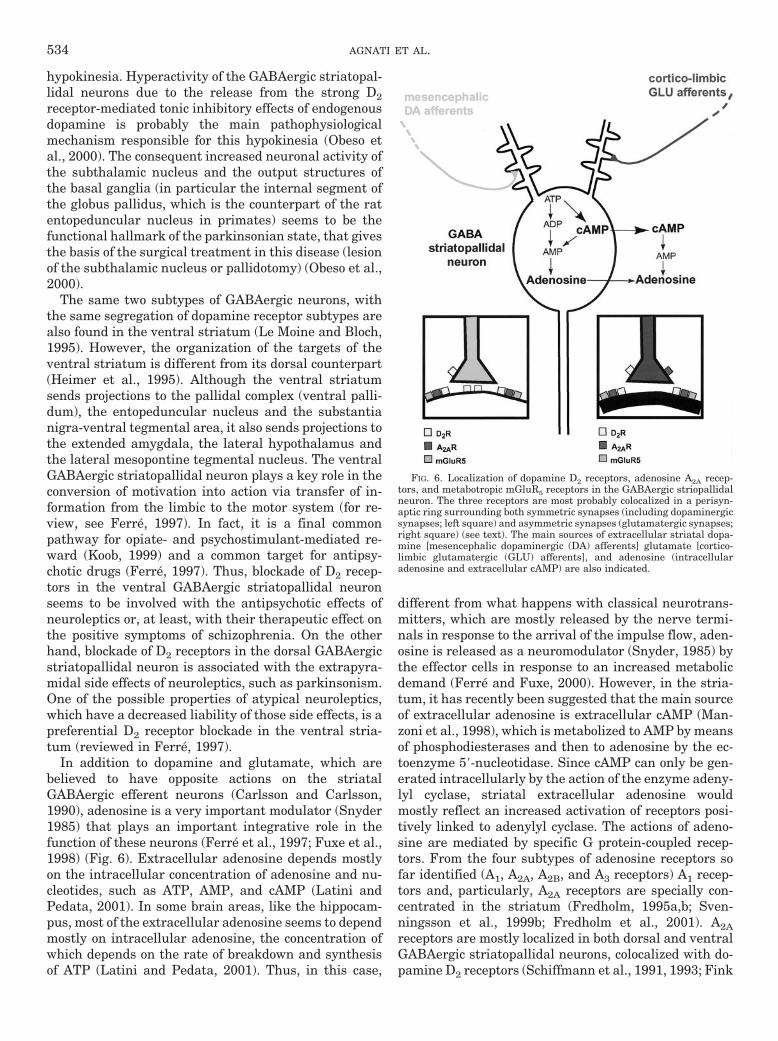
hypokinesia. Hyperactivity of the GABAergic striatopal-lidal neurons due to the release from the strong D2receptor-mediated tonic inhibitory effects of endogenousdopamine is probably the main pathophysiologicalmechanism responsible for this hypokinesia (Obeso etal., 2000). The consequent increased neuronal activity ofthe subthalamic nucleus and the output structures ofthe basal ganglia (in particular the internal segment ofthe globus pallidus, which is the counterpart of the ratentopeduncular nucleus in primates) seems to be thefunctional hallmark of the parkinsonian state, that givesthe basis of the surgical treatment in this disease (lesionof the subthalamic nucleus or pallidotomy) (Obeso et al.,2000).
The same two subtypes of GABAergic neurons, withthe same segregation of dopamine receptor subtypes arealso found in the ventral striatum (Le Moine and Bloch,1995). However, the organization of the targets of theventral striatum is different from its dorsal counterpart(Heimer et al., 1995). Although the ventral striatumsends projections to the pallidal complex (ventral palli-dum), the entopeduncular nucleus and the substantianigra-ventral tegmental area, it also sends projections tothe extended amygdala, the lateral hypothalamus andthe lateral mesopontine tegmental nucleus. The ventralGABAergic striatopallidal neuron plays a key role in theconversion of motivation into action via transfer of in-formation from the limbic to the motor system (for re-view, see Ferre, 1997). In fact, it is a final commonpathway for opiate- and psychostimulant-mediated re-ward (Koob, 1999) and a common target for antipsy-chotic drugs (Ferre, 1997). Thus, blockade of D2 recep-tors in the ventral GABAergic striatopallidal neuronseems to be involved with the antipsychotic effects ofneuroleptics or, at least, with their therapeutic effect onthe positive symptoms of schizophrenia. On the otherhand, blockade of D2 receptors in the dorsal GABAergicstriatopallidal neuron is associated with the extrapyra-midal side effects of neuroleptics, such as parkinsonism.One of the possible properties of atypical neuroleptics,which have a decreased liability of those side effects, is apreferential D2 receptor blockade in the ventral stria-tum (reviewed in Ferre, 1997).
In addition to dopamine and glutamate, which arebelieved to have opposite actions on the striatalGABAergic efferent neurons (Carlsson and Carlsson,1990), adenosine is a very important modulator (Snyder1985) that plays an important integrative role in thefunction of these neurons (Ferre et al., 1997; Fuxe et al.,1998) (Fig. 6). Extracellular adenosine depends mostlyon the intracellular concentration of adenosine and nu-cleotides, such as ATP, AMP, and cAMP (Latini andPedata, 2001). In some brain areas, like the hippocam-pus, most of the extracellular adenosine seems to dependmostly on intracellular adenosine, the concentration ofwhich depends on the rate of breakdown and synthesisof ATP (Latini and Pedata, 2001). Thus, in this case,
different from what happens with classical neurotrans-mitters, which are mostly released by the nerve termi-nals in response to the arrival of the impulse flow, aden-osine is released as a neuromodulator (Snyder, 1985) bythe effector cells in response to an increased metabolicdemand (Ferre and Fuxe, 2000). However, in the stria-tum, it has recently been suggested that the main sourceof extracellular adenosine is extracellular cAMP (Man-zoni et al., 1998), which is metabolized to AMP by meansof phosphodiesterases and then to adenosine by the ec-toenzyme 59-nucleotidase. Since cAMP can only be gen-erated intracellularly by the action of the enzyme adeny-lyl cyclase, striatal extracellular adenosine wouldmostly reflect an increased activation of receptors posi-tively linked to adenylyl cyclase. The actions of adeno-sine are mediated by specific G protein-coupled recep-tors. From the four subtypes of adenosine receptors sofar identified (A1, A2A, A2B, and A3 receptors) A1 recep-tors and, particularly, A2A receptors are specially con-centrated in the striatum (Fredholm, 1995a,b; Sven-ningsson et al., 1999b; Fredholm et al., 2001). A2Areceptors are mostly localized in both dorsal and ventralGABAergic striatopallidal neurons, colocalized with do-pamine D2 receptors (Schiffmann et al., 1991, 1993; Fink
FIG. 6. Localization of dopamine D2 receptors, adenosine A2A recep-tors, and metabotropic mGluR5 receptors in the GABAergic striopallidalneuron. The three receptors are most probably colocalized in a perisyn-aptic ring surrounding both symmetric synapses (including dopaminergicsynapses; left square) and asymmetric synapses (glutamatergic synapses;right square) (see text). The main sources of extracellular striatal dopa-mine [mesencephalic dopaminergic (DA) afferents] glutamate [cortico-limbic glutamatergic (GLU) afferents], and adenosine (intracellularadenosine and extracellular cAMP) are also indicated.
534 AGNATI ET AL.

et al., 1992; Svenningsson et al., 1997, 1999b). On theother hand, adenosine A1 receptors are localized in allthe striatal neuronal elements and dopamine D1 recep-tors in the GABAergic striatonigral-striatoentopeduncu-lar neurons are colocalized with adenosine receptors ofthe A1 subtype (Ferre et al., 1996).
An important amount of evidence exists for the exis-tence of specific antagonistic A2A/D2 receptor interac-tions that modulate the function of GABAergic striato-pallidal neurons (Ferre et al., 1993a, 1997; Fuxe et al.,1998). Furthermore, specific antagonistic A1/D1 receptorinteractions modulate the function of GABAergic stria-tonigral-striatoentopeduncular neurons (Ferre et al.,1997; Fuxe et al., 1998). It has been suggested that thesereceptor/receptor interactions are involved in the motoreffects of adenosine agonists and antagonists (like thenonselective adenosine antagonist caffeine) and thatthey can provide a new therapeutic approach for thetreatment of basal ganglia disorders and schizophrenia(Ferre et al., 1992, 1994a,b, 1997, 2001; Ferre, 1997;Lillrank et al., 1999; Fuxe et al., 2001). Here we willreview the more relevant findings on the A2A/D2 recep-tor interactions as well as the recent data on the multi-ple interactions between these receptors and the group Imetabotropic glutamate receptor mGluR5.
Glutamate acts on both ionotropic and metabotropic Gprotein-coupled receptors. Molecular and pharmacologi-cal characterization studies have currently divided themetabotropic glutamate receptor family into threegroups (I to III) (Pin and Duvoisin, 1995; Bockhaert andPin, 1999; Hermans and Challiss, 2001). Group I in-cludes mGluR1 and mGluR5 receptors, the latter ofwhich is particularly expressed in the striatum, espe-cially in the striatal GABAergic efferent neurons (Shige-moto et al., 1993; Testa et al., 1995; Tallaksen-Green etal., 1998). The ultrastructural analysis of the localiza-tion of mGluR5 receptors in the striatopallidal complexin primates demonstrated that mGluR5 receptor immu-noreactivity is commonly found perisynaptically toasymmetric (glutamatergic) postsynaptic synapses (Fig.6) but also at symmetric synapses formed by tyrosinehydroxylase-immunoreactive (dopaminergic) terminals(Smith et al., 2000). The recent ultrastructural analysisof the localization of A2A receptors in the rat striatumshowed a very similar localization to that described formGluR5 receptors in primates. Thus, A2A receptors aremostly localized postsynaptically in the dendrites anddendritic spines of the striatal GABAergic neurons. Im-portantly, A2A receptor-immunoreactivity was observedprimarily at asymmetric synapses (Rosin et al., 1998;Hettinger et al., 2001). Although less frequently A2Areceptor immunoreactivity was also found presynapti-cally at asymmetric synapses and postsynaptically atsymmetric synapses (Hettinger et al., 2001). Finally, inthe rat striatum D2 receptor immunoreactivity has beenfound both presynaptically (in both dopaminergic andglutamatergic terminals) and postsynaptically. At the
postsynaptic level, D2 receptor immunoreactivity hasnot only been observed at symmetric synapses but alsoperisynaptically at asymmetric synapses (Yung et al.,1995; Delle Donne et al., 1997). In summary, these mor-phological findings strongly suggest that the three re-ceptors, A2A, mGluR5, and D2, are colocalized in thestriatopallidal GABAergic efferent neuron, postsynapti-cally and perisynaptically at dopaminergic and glutama-tergic synapses (Fig. 6). Dopamine and glutamate maybe able to reach this localization, not only by spilloverfrom the respective nerve terminals, but also by longerdistance VT. Finally, the colocalization gives a morpho-logical frame for the existence of multiple A2A/D2/mGluR5 receptor interactions.
2. Interactions between Adenosine A2A, Dopamine D2,and Glutamate Metabotropic mGluR5 Receptors in theGABA Striatopallidal Neuron: Biochemical-CellularLevel. As previously discussed, evidence for the exis-tence of physical interactions between A2A and D2 recep-tors and between A2A and mGluR5 receptors has beenobtained with coimmunprecipitation experiments in celllines that express, constitutively or after stable or tran-sient cotransfection, the corresponding receptors (Ferreet al., 2002; Hillion et al., 2002). A2A/mGluR5 hetero-meric complexes have also been demonstrated in ratstriatal membrane preparations with coimmunoprecipi-tation experiments (Ferre et al., 2002). Different behav-ioral and biochemical models have demonstrated theexistence of functionally significant antagonistic A2A/D2receptor and mGluR5/D2 receptor interactions and syn-ergistic A2A/mGluR5 receptor interactions. At the bio-chemical level, by using membrane preparations of ratstriatum or of cell lines expressing the correspondingreceptors and by using receptor autoradiography in ratand human striatal sections, it has been shown thatstimulation of A2A receptors decreases the affinity of D2receptors for dopamine or dopamine agonists. These re-sults were shown, e.g., as an increase in KH or IC50 incompetition experiments of dopamine versus a radioac-tively labeled D2 receptor antagonist or as an increase inKD in saturation experiments with tritiated dopamine orquinpirole (Ferre et al., 1991d, 1994a,b, 1999a; Das-gupta et al., 1996a; Dixon et al., 1997; Lepiku et al.,1997; Kull et al., 1999; Franco et al., 2000; Salim et al.,2000; Dıaz-Cabiale et al., 2001). In receptor autoradiog-raphy experiments, the A2A receptor-mediated modula-tion of D2 receptor binding properties was found to bestronger in the ventral compared with the dorsal stria-tum (Ferre et al., 1994b; Dıaz-Cabiale et al., 2001). Do-pamine receptors of the D3 subtype are structurally andpharmacologically very similar to D2R and they are spe-cially concentrated in the ventral striatum (Missale etal., 1998). Therefore, it was suggested that the strongerA2A/D2 receptor interaction in the ventral striatummight be related to the action of A2A receptor agonists onD3 receptors, in addition to D2 receptors, as suggested bythe demonstration of the A2A receptor agonist-mediated
RECEPTOR/RECEPTOR INTERACTIONS VIA HETEROMERIZATION 535

modulation of D3 receptor agonist binding in vivo (Hille-fors et al., 1999). Also, in membrane preparations fromrat striatum, nonselective stimulation of group I gluta-mate metabotropic receptors or selective stimulation ofmGluR5 receptors decreased the affinity of D2 receptorsfor dopamine (Ferre et al., 1999a; Rimondini et al., 1999,Popoli et al., 2001). Finally, costimulation of A2A recep-tors and group I glutamate metabotropic receptors ormGluR5 receptors exerted a synergistic effect on themodulation of the binding characteristics of D2 receptors(Ferre et al., 1999a; Rimondini et al., 1999; Popoli et al.,2001).
In addition to the intramembrane receptor/receptorinteractions, the existence of A2A/D2 and A2A/mGluR5heteromeric receptor complexes provides the possibilityfor close cross-talk between A2A, D2, and mGluR5 recep-tors. This would synchronize the activation of these re-ceptors to interact at the second-messenger level andbeyond. The major signal transduction pathway used byA2A receptors depends on the activation of adenylyl cy-clase, by means of Gs/Golf coupling (Kull et al., 1999,2000). A2A receptor-mediated adenylyl cyclase activa-tion generates cAMP, which activates a cAMP-depen-dent protein kinase (PKA), which in turn regulates thestate of phosphorylation of various substrate proteins(Fig. 7). One of those proteins, DARPP-32 (dopamineand cyclic adenosine 39,59-monophosphate-regulatedphophoprotein, 32 kDa) is expressed in very high con-centration in the GABAergic efferent neurons (Green-gard et al., 1998, 1999). PKA-induced phosphorylationat a single site (Thr-34 of the rat sequence) convertsDARPP-32 into a potent and selective inhibitor of pro-tein phosphatase-1 (PP-1) (Greengard et al., 1999). Fur-thermore, DARPP-32 is also phosphorylated to a highstoichiometry under basal conditions at another site,Thr-75, which converts it into an inhibitor of PKA (Nishiet al., 2000). PKA stimulates protein phosphatase-2A,which is the predominant phosphatase responsible fordephosphorylation of phospho-Thr-75-DARPP-32 (Nishiet al., 2000). It has been demonstrated that the removalof the inhibitory constraint on PKA by dephosphoryla-tion of phospho-Thr-75-DARPP-32 provides a mecha-nism of amplification of the PKA signal transductionpathway (Nishi et al., 2000). In striatal slices, Fisoneand coworkers have demonstrated that stimulation ofA2A receptors produces phosphorylation of DARPP-32 atThr-34 and dephosphorylation of DARPP-32 at Thr-75(Svenningsson et al., 1998, 2000; Lindskog et al., 1999,2002). Another protein phosophorylated by PKA is theconstitutive transcription factor CREB (cAMP responseelement-binding protein). Induction of cAMP liberatesthe catalytic subunits of PKA, which diffuse into thenucleus and induce cellular gene expression by phos-phorylating CREB at a serine residue (Ser-133) (Mayrand Montminy, 2001). CREB activity declines after acouple of hours of continuous stimulation, due to dephos-phorylation at Ser-133 by PP-1 (Mayr and Montminy,
2001). Thus, through PKA activation A2AR stimulationin the GABAergic striatopallidal neurons can potentiallyproduce a sustained increase in the transcription ofsome CREB-modulated genes by a mechanism involvingincreased CREB phosphorylation and decreased CREBdephosphorylation (through phospho-Thr-34-DARPP-32-mediated inhibition of PP-1 activity) (Fig. 7). Theimmediate-early gene c-fos and the preproenkephalinand neurotensin genes are very well studied targetgenes, whose promoters contain consensus sites forCREB-P binding (Borsook and Hyman, 1995; Evers etal., 1995; Hughes and Dragunow, 1995; Le et al., 1997;Herdegen and Leah, 1998). Recent studies have shown
FIG. 7. Possible mechanisms responsible for the multiple biochemicalinteractions between dopamine D2 receptors, adenosine A2A receptors,and metabotropic mGluR5 receptors. Antagonistic A2A receptor/D2 recep-tor and mGluR5/D2 receptor interactions have been demonstrated at theintramembrane level and an antagonistic D2/A2A receptor interaction hasbeen found at the adenylyl cyclase level. MAPKs seem to be the maincoincidental signal involved in the synergistic A2A/mGluR5 interactiondemonstrated at the level of c-fos expression. Furthermore, other levels ofinteraction upstream to MAPK could be the nonreceptor tyrosine kinaseSrc or other enzymes recently demonstrated to be activated by G protein-coupled receptors through G protein-independent signaling. This A2A/mGluR5 interaction allows the counteraction of the strong tonic D2 re-ceptor-mediated inhibition of A2A receptor signaling. Broken arrows:antagonistic interactions or inhibitory effects (see text). A cyclase, adeny-lyl cyclase; DARPP-32 (dopamine and cAMP-regulated phosphoprotein,32 kDa); DARPP-32-P(Thr-34) and DARPP-32-P(Thr-75): DARPP-32-phosphorylated at threonine residues 34 and 75, respectively.
536 AGNATI ET AL.

that A2A receptor stimulation can, under certain condi-tions, increase the expression of c-fos (and other imme-diate-early genes), preproenkephalin, and neurotensingenes (Fig. 7) (see below).
One of the main fully documented signaling effects ofD2 receptors is adenylyl cyclase inhibition by means ofits coupling to Gi/o proteins (for review, see Missale etal., 1998). Also dependent on Gi/o coupling, but indepen-dent of cAMP, D2 receptors have been shown to modu-late the activity of K1 channels. In many preparations,including acutely dissociated striatal neurons (Freed-man and Weight, 1988), D2 receptor stimulation hasbeen shown to increase outward K1 currents, leading tocell hyperpolarization (Missale et al., 1998). Further-more, other less well characterized cAMP-independentsignaling pathways used by D2 receptors, such as acti-vation of PLC, inhibition of inward Ca21 currents, ara-chidonic acid release, inhibition of Na1-K1-ATPase andMAPK activation, have been reported (Missale et al.,1998; Yan et al., 1999). A strong antagonistic interactionbetween A2A receptors and D2 receptors at the adenylylcyclase level has been demonstrated in different celllines (Kull et al., 1999; Hillion et al., 2002) (Fig. 7). InCHO cells stably cotransfected with A2A and D2 receptorcDNAs, stimulation of A2A receptors produced a strongstimulation of cAMP, CREB phosphorylation and in-creased c-fos expression (Kull et al., 1999). A selectiveD2R agonist dose dependently counteracted these effectsand a complete blockade was attained at low concentra-tions of the D2R agonist (Kull et al., 1999). Similarly, instriatal slices a D2 receptor agonist completely counter-acted Thr-34 phosphorylation of DARPP-32 by A2A re-ceptor stimulation (Lindskog et al., 1999).
In the striatum, D2 receptors are tonically stimulatedby basal endogenous levels of dopamine (see above).Many experimental results indicate that this tonic D2receptor stimulation strongly counteracts a tonic A2Areceptor stimulation induced by the basal striatal levelsof adenosine. In this way, the products of A2A receptoractivation are kept at low concentration under normalbasal conditions and inactivation of striatal D2 receptor-mediated neurotransmission (by administration of D2receptor antagonists, striatal dopamine depletion or ge-netic D2 receptor inactivation) liberates A2A receptor-mediated function from the strong D2 receptor-mediatedtonic inhibition. This results in a very significant in-crease in the striatal expression of c-Fos, Thr-34-phos-phorylated DARPP-32, enkephalin, and neurotensin,which is partially counteracted by genetic inactivationor pharmacological blockade of A2A receptors (Schiffmanand Vanderhaeghen, 1993; Morelli et al., 1995; Pollackand Fink, 1995; Boegman and Vincent, 1996; Adams etal., 1997; Pinna et al., 1997, 1999; Richardson et al.,1997; Svenningsson et al., 1999a, 2000; Ward and Dorsa,1999; Chen et al., 2000, 2001; Zahniser et al., 2000;Dassesse et al., 2001; Ferre et al.,. 2002;, see SectionI.B.). Since also at the behavioral level A2A receptor
antagonists counteract the motor depressant and cata-leptic effects secondary to the genetic inactivation orpharmacological interruption of D2 receptor-mediatedneurotransmission (see below), these results stronglysuggest that an important part of the biochemical andbehavioral effects induced by interruption of D2 recep-tor-mediated neurotransmission are due to the libera-tion of A2A receptor signaling. This can have obviousimplications for the possible application of A2A receptorantagonists in Parkinson’s disease. On the other hand,these results also suggest that the biochemical effectsrelated to adenylyl cyclase activation (which are onlyclearly apparent when the D2 receptor-mediated inhibi-tory tone is removed) do not play a major role in somefunctional and behavioral effects produced by the ad-ministration of A2A receptor agonists and antagonists(see below). For instance, the systemic administration ofa low dose of a selective A2A receptor agonist produces apronounced motor depression (already shown to be cen-trally mediated; see Barraco et al., 1993) and selectivelycounteracts D2 receptor agonist-mediated behaviors(Rimondini et al., 1997, 1998) without inducing an in-creased c-fos striatal expression (Morelli et al., 1995;Pinna et al., 1997; Ferre et al., 2002; see SectionI.B.2.k.). Most probably those behavioral effects are re-lated to the reciprocal antagonistic A2A/D2 receptor in-tramembrane interaction, which can be more effective atmodulating other D2 receptor-mediated signaling path-ways, such as the opening of K1 channels.
As previously mentioned, some studies suggest that apreferential blockade of D2 receptors in the ventral, com-pared with the dorsal striatum, is one of the main factorsresponsible for the atypical profile of an antipsychotic.Some of these studies are based on the differential in-crease in c-fos expression in the different striatal com-partments. Thus, typical antipsychotics, like haloperi-dol, increase c-fos expression in the dorsal and ventralstriatum, whereas atypical antipsychotics, like cloza-pine, selectively elevates c-fos expression in the ventralstriatum, and especially in the shell of the nucleus ac-cumbens (Deutch et al., 1992; Robertson and Fibiger,1992; Merchant and Dorsa, 1993; Pinna et al., 1999). Asimilar differential response to haloperidol and cloza-pine has also been demonstrated for the striatal expres-sion of neurotensin (Merchant and Dorsa, 1993). It hasalso been shown that the increased c-fos expression inthe ventral striatum induced by both haloperidol andclozapine selectively takes place in the ventral GABAer-gic striatopallidal neuron (Robertson and Jian, 1995)and that it can be counteracted by the systemic admin-istration of an A2A receptor antagonist (Pinna et al.,1999). Therefore, these results suggest that A2A recep-tors might be involved in the mediation of the antipsy-chotic effects of neuroleptics (see below).
It remains to be determined which are the physiolog-ical conditions (without interruption of D2 receptor-me-diated neurotransmission) that would allow A2A recep-
RECEPTOR/RECEPTOR INTERACTIONS VIA HETEROMERIZATION 537

tor stimulation to produce a clear adenylyl cyclaseactivation, with the corresponding increase in Thr-34DARPP-32 and CREB phosphorylation and increase inthe expression of c-fos and the preproenkephalin andneurotensin genes. One such condition seems to bemGluR5 receptor coactivation (Ferre et al.,. 2002). Themajor signal transduction pathway used by mGluR5 re-ceptors (through Gq proteins) is activation of PLC, whichreleases IP3 and diacylglycerol, which cause the releaseof intracellular Ca21 and the activation of PKC, respec-tively (Pin and Duvoisin, 1995; Hermans and Challis,2001) (Fig. 7). In HEK-293 cells transiently cotrans-fected with A2A and mGluR5 receptors, stimulation ofA2A receptors produced an increase in cAMP levels thatwas not significantly modified by the concomitant stim-ulation of mGluR5 receptors (Ferre et al., 2002). Asexpected, stimulation of mGluR5 receptors induced anincrease in [Ca21]i. In addition to mGluR5 receptor stim-ulation, a selective A2A receptor agonist also produced asignificant increase in [Ca21]i in cotransfected cells,which would agree with recent studies in rat striatalslices which suggest that A2A receptors can also usePLC/IP3 signaling under certain conditions (Wirkner etal., 2000). However, at the level of [Ca21]i an absence ofsynergistic A2A/mGluR5 receptor interaction was ob-served (Ferre et al.,. 2002). The lack of synergistic inter-action at the second messenger level (cAMP and Ca21)was somehow surprising in view of previous studiesshowing synergistic effects on cAMP accumulation ofgroup I glutamate metabotropic receptors and receptorspositively linked to adenylyl cyclase in neuronal primarycultures (Cartmell et al., 1998; Paolillo et al., 1998) andin view of recent findings on synergistic effects betweenA1 receptors and mGluR1a receptors on [Ca21]i in co-transfected HEK-293 cells (Ciruela et al., 2001). Never-theless, a strong functional synergistic A2A/mGluR5 re-ceptor interaction on MAPK (ERK 1/2) and on c-fosexpression was found in cotransfected cells (Ferre et al.,.2002). Accordingly, a significant striatal c-fos inductionwas obtained after the concomitant central administra-tion of A2A and mGluR5 receptor agonists, which wereineffective when administered alone (Ferre et al., 2002).This strongly suggests that concomitant stimulation ofA2A and mGluR5 receptors is one of the mechanisms bywhich A2A receptor stimulation can overcome the tonicinhibitory effect of dopamine and induce striatal c-fosexpression. We favor the idea that this mechanism cantake place under conditions of intense glutamatergicneurotransmission, which is known to induce adenosinerelease, most probably, due to the neuronal metabolicdemand imposed by the increased excitatory input(Ferre and Fuxe, 2000; Latini and Pedata, 2001).
The protein encoded by c-fos (c-Fos) is an inducibletranscription factor, expression of which is controlled bypre-existing constitutive transcription factors, such asCREB, serum response factor, and TCF/Elk-1 proteins(Herdegen and Leah, 1998; Ches and Wang, 2001). Upon
phosphorylation, CREB and TCF/Elk-1 (which requiresserum response factor) activate c-fos transcription bybinding to the CRE and serum response element regu-latory elements of the c-fos promoter, respectively(Hughes and Dragunow, 1995; Herdegen and Leah,1998) (Fig. 7). It is generally accepted that CREB ismostly phosphorylated by PKA (see above) whereasTCF/Elk-1 is mostly phosphorylated by MAPK (Van-houtte et al., 1999). In the case of group I metabotropicreceptors, MAPK activation seems to involve PKC (Fer-raguti et al., 1999) and Ca21/calmodulin-dependent pro-tein kinases (CaMK) (Choe and Wang, 2001). However,recent studies indicate that both MAPK and CREB canbe convergent targets for different elements of thecAMP/PKA and the PLC/PKC-CaMK signaling path-ways (Fig. 7) (Impey et al., 1999; Sweatt, 2001; Wu et al.,2001). Since the A2A/mGluR5 receptor synergistic effecton c-fos expression found in cotranfected cells was com-pletely counteracted by a ERK 1/2 kinase inhibitor(Ferre et al., 2002), MAPK seems to be a main biochem-ical integration element responsible for the synergisticinteractions between A2A and mGluR5 receptors. Fur-thermore, other levels of interaction upstream to MAPKcould be the nonreceptor tyrosine kinase Src or otherenzymes recently demonstrated to be activated by Gprotein-coupled receptors through G protein-indepen-dent signaling (Heuss and Gerber, 2000). Given the keyrole of immediate-early genes in the coupling of earlyneuronal responses to long-term adaptive changes(Hughes and Dragunow, 1995; Berke and Hyman, 2000),those results suggest that A2A/mGluR5 receptor interac-tions might be involved in striatal neuronal plasticity.More specifically, this mechanism may underlie the re-cently described dopamine-independent increased c-fosexpression in the striatopallidal neurons associated withsensitization to psychostimulants (Uslaner et al., 2001).In fact, both striatal A2A and mGluR5 receptors haverecently been shown to be involved in certain forms ofstriatal plasticity, including cortico-striatal long-termpotentiation and long-term depression (d’Alcantara etal., 2001; Sung et al., 2001).
3. Interactions between Adenosine A2A, Dopamine D2,and Glutamate Metabotropic mGluR5 Receptors in theGABA Striatopallidal Neuron: Physiological-BehavioralLevel. The first functional approach used to study theinteractions between adenosine and dopamine receptorsin the GABAergic striatopallidal neurons was the dual-probe in vivo microdialysis approach in freely movingrats. In these experiments, one microdialysis probe isimplanted in the caudate-putamen or the nucleus ac-cumbens, and the second one is implanted in the ipsilat-eral globus pallidus or the ipsilateral ventral pallidum,respectively. This allows determining in freely movinganimals the modulation of GABAergic neurotransmis-sion in the dorsal and ventral striatopallidal neurons(Ferre et al., 1993a, 1994a; Dıaz-Cabiale et al., 2002b).With these experiments, a main difference in the effects
538 AGNATI ET AL.

of D2 receptor stimulation was found between the dorsaland the ventral striatopallidal pathways. The striatalperfusion of a D2 receptor agonist decreased the extra-cellular levels of GABA in the globus pallidus, but not inthe ventral pallidum. On the other hand, D2 receptorantagonists increased GABA levels in the ventral palli-dum and did not significantly modify GABA levels in theglobus pallidus (Ferre et al., 1993a, 1994a; Dıaz-Cabialeet al., 2002b). Also, a different effect was obtained withthe striatal perfusion of an A2A receptor agonist. In thiscase, GABA levels were only increased in the ventralpallidum (Ferre et al., 1993a, 1994a; Dıaz-Cabiale et al.,2002b). Nevertheless, recent studies by Ochi et al. (2000)have also found a significant increase in the GABA lev-els of the globus pallidus after the striatal infusion(through an injection needle) of an A2A receptor agonist.The reciprocal A2A/D2 receptor interactions could also bedemonstrated in microdialysis experiments. Thus, theD2 receptor agonist-induced decrease and the A2A recep-tor agonist-induced increase in pallidal levels could becounteracted by the striatal coperfusion with A2A and D2receptor agonists, respectively (Ferre et al., 1994a; Dıaz-Cabiale et al., 2002b). Altogether, these results suggestthe existence of functional regional differences in theA2A/D2 receptor interactions, with a relatively strongerA2A receptor signaling in the ventral compared with thedorsal striatum, in agreement with some biochemical(Ferre et al., 1994a; Pinna et al., 1997; Dıaz-Cabiale etal., 2002b), and behavioral studies (see below). We havehypothesized that these differences in the A2A/D2 recep-tor interactions between both striatal compartmentsmight explain the atypical antipsychotic profile of aden-osine A2A receptor agonists in animal models (see be-low). As mentioned earlier, this differential effect maybe related to the existence of an additional antagonisticA2A/D3 receptor interaction in the ventral striatum(Diaz-Cabiale et al., 2001). Finally, in recent microdialy-sis experiments we have found that the perfusion of aselective mGluR5 receptor agonist in the nucleus accum-bens produces an increase in the extracellular levels ofGABA in the ventral pallidum. Furthermore, coperfu-sion with an A2A receptor agonist produced a strongsynergistic increase in pallidal GABA levels (Dıaz-Ca-biale et al., 2002b). From these results, we have sug-gested a possible use of mGluR5 receptor agonists asantipsychotic drugs, alone or in combination with A2Areceptor agonists.
In vitro studies with micropunctures of rat globuspallidus have shown that an antagonistic A2A/D2 recep-tor interaction also modulates GABAergic neurotrans-mission in the terminals of GABAergic striatopallidalneurons, which also express both A2A and D2 receptors(Mayfield et al., 1996). In this experimental preparation,stimulation of pallidal A2A and D2 receptors stimulatesand inhibits GABA release, respectively, and the D2receptor-mediated inhibition of GABA release is coun-teracted by A2A receptor stimulation (Mayfield et al.,
1996). The A2A receptor-mediated stimulation of pallidalGABA release has been recently confirmed with in vivomicrodialysis experiments (Ochi et al., 2000). However,experiments using striatal and pallidal synaptosomalpreparations have given contradictory results. Thus, inthese preparations, A2A receptor stimulation inhibitedhigh KCl-stimulated GABA release (Kurokawa et al.,1994). Similarly, results obtained from experiments us-ing intracellular and whole-cell patch-clamp recordingin striatal slices suggested that striatal presynaptic A2Areceptors exert an inhibitory modulation of GABA re-lease (Mori et al., 1996). Based on these findings, Rich-ardson et al. (1997) proposed that the main mechanismsby which A2A receptors would influence the function ofstriatal GABAergic neurons would be by a presynapticinhibitory modulation of GABA release from their col-lateral recurrent axons. At this point, this theory isincompatible with the bulk of new information about theultrastructural localization and function of striatal A2Areceptors (see above).
Many behavioral studies have shown that in animalswith intact striatal dopamine innervation, A2A receptoragonists behave as D2 receptor antagonists (Heffner etal., 1989; Ferre et al., 1991a,b; Popoli et al., 1994; Kafkaand Corbett, 1996; Hauber and Munkle, 1997; Rimon-dini et al., 1997, 1998; Wardas et al., 1999; Poleszak andMalec, 2000). Furthermore, although questioned bysome studies (Kafka and Corbett, 1996; Hauber andMunkle, 1997), there are some behavioral data support-ing a preferential effect of A2A receptor agonists in theventral striatum (Barraco et al., 1993; Rimondini et al.,1997, 1998). When considering commonly used tests toscreen the effects of antipsychotics, it is generally ac-cepted that blockade of dopamine receptors in the ven-tral striatum is mostly responsible for the counteractingeffects of the motor activation induced by novel stimuli(exploratory activity in a novel environment) and psy-chostimulants [such as amphetamine and phencyclidine(PCP)]. On the other hand, the counteraction of dopa-mine agonist-induced stereotypes and the induction ofcataleptic immobility are believed to be mainly mediatedby blockade of dopamine receptors in the dorsal striatum(reviewed in Ogren, 1996). The systemic administrationof an A2A receptor agonist gave an atypical antipsychoticprofile in these animal models, since it counteracted themotor-activating effects of psychostimulants at lowerdoses than those needed to counteract dopamine ago-nist-induced stereotypes or to induce catalepsy (Rimon-dini et al., 1997, 1998). In particular, very low doses ofthe A2A receptor agonist were necessary to counteractPCP-induced motor activity (Rimondini et al., 1997). Inagreement, recent studies by Sills et al. (2001) haveshown than an A2A receptor agonist can selectively re-verse the reduction in prepulse inhibition of the acousticstartle response induced by PCP.
PCP is self-administered by humans and experimen-tal animals (Carlezon and Wise, 1996; Jentsch and Roth,
RECEPTOR/RECEPTOR INTERACTIONS VIA HETEROMERIZATION 539
1999), and its administration in humans reproducesboth positive and negative symptoms of schizophreniaand exacerbates those symptoms in schizophrenic pa-tients (Jentsch and Roth, 1999). Together with the neu-rochemical modifications induced by this drug, PCP ad-ministration is considered as a potential animal model ofschizophrenia, and reversal of the effects of PCP is alsocommonly used as a model to screen for antipsychoticactivity (Jentsch and Roth, 1999). The ventral striato-pallidal GABA neurons are most probably the main neu-ronal target underlying the A2A receptor agonist-medi-ated counteraction of PCP-induced motor activation,since inhibition of ventral striatopallidal GABA trans-mission appears to be central in the mediation of some(rewarding) effects of PCP (Carlezon and Wise, 1996).Since the counteracting effect on PCP-induced motoractivation is obtained with low doses of the A2A receptoragonist, which are not able to induce an increase in thestriatal c-fos expression (see above), the antagonisticA2A/D2 receptor intramembrane interaction is mostprobably involved. In fact, the motor hyperactivity in-duced by PCP has been shown to be dependent onpostsynaptic D2 receptors and a high degree of D2 recep-tor blockade is required to significantly counteract thestimulatory action of high doses of PCP (Ogren andGoldstein, 1994). Furthermore, the effect of A2A receptorstimulation was surmountable, and high doses of PCPcould not be counteracted by the systemic administra-tion of an A2A receptor agonist (Ferre et al., 2002; seeSection I.B.). However, in agreement with the resultsobtained with striatal c-fos expression, when the A2Areceptor agonist was coadministered with a mGluR5 re-ceptor agonist, which by itself did not produce any sig-nificant effect, PCP-mediated motor activation was sig-nificantly counteracted (Ferre et al., 2002). Theseresults also fit very nicely with those obtained in micro-dialysis experiments (Dıaz-Cabiale et al., 2002) and sug-gest that costimulation of A2A and mGluR5 receptors caneven counteract the effects induced by a strong stimula-tion of dopaminergic neurotransmission.
As previously mentioned, the systemic administrationof A2A receptor antagonists counteracts most of the bio-chemical (see above) as well as the motor depressant andcataleptic effects secondary to the genetic inactivation orpharmacological interruption of D2 receptor-mediatedneurotransmission. This has been repeatedly demon-strated in a number of experimental models involvingrodents pretreated with D2 receptor antagonists, reser-pine, or MPTP or after genetic inactivation of D2 recep-tors (Casas et al., 1988; Popoli et al., 1991; Kanda et al.,1994; Gimenez-Llort et al., 1995; Pollack and Fink,1995; Malec, 1997; Shiozaki et al., 1999; Ward andDorsa, 1999; Wardas et al., 1999; Aoyama et al., 2000;Chen et al., 2001; Ferre et al., 2001; Hauber et al., 2001)and involving MPTP-treated monkeys (Kanda et al.,1998a,b; Grondin et al., 1999). The results of these ex-periments suggest that A2A receptor antagonists can be
useful in the treatment of Parkinson’s disease. However,although it has been claimed that A2A receptor antago-nists might be devoid of the secondary dyskinetic effectsassociated with treatment with dopamine agonists(Kanda et al., 1998a,b; Grondin et al., 1999), it is still amatter of debate whether they can be useful as mono-therapy or whether they would be more efficacious whencombined with D2 receptor agonists. In fact, in anotherclassical experimental model of Parkinson’s disease, therat with a unilateral lesion of the nigrostriatal pathway,A2A receptor antagonists do not produce a turning be-havior contralateral to the lesioned side (Fenu and Mo-relli, 1998; Stromberg et al., 2000; Ferre et al., 2001),which is a behavior that predicts antiparkinsonian ac-tivity (Ungerstedt, 1971). Nevertheless, in all the animalmodels of Parkinson’s disease tested to date (reserpin-ized mice, rats with unilateral 6-OH-dopamine lesions,MPTP-treated monkeys), A2A receptor antagonistsstrongly potentiate the motor activation induced by D2receptor agonists (Ferre et al., 1991b, 2001; Jiang et al.,1993; Fenu et al., 1997; Grondin et al., 1999; Kanda etal., 2000; Koga et al., 2000; Popoli et al., 2000, 2001;Stromberg et al., 2000). Importantly, cotreatment withA2AR antagonists and L-dopa did not increase the non-wanted dyskinetic effects in MPTP-treated monkeys(Grondin et al., 1999; Kanda et al., 2000). Finally, recentelectrophysiological experiments in the rat dopaminedenervated striatum showed that the infusion of an A2Areceptor antagonist did not produce any effect on itsown, but strongly potentiated the D2R agonist-inducedinhibition of striatal neuronal activity (Stromberg et al.,2000). Altogether, these results suggest that some phar-macological and, maybe, therapeutical effects of A2A re-ceptor blockade can only be observed with the concomi-tant stimulation of D2 receptors. This is also inagreement with some results obtained with D2 receptorknockout mice where the motor effects of A2A receptorantagonists were attenuated (Chen et al., 2001). Thus,these experiments performed under complete inactiva-tion of D2 receptors demonstrate that some A2A receptorfunctions are dependent on the integrity of D2 receptorsand, most probably, on the integrity of A2A/D2 hetero-meric receptor complexes. This is shown even more dra-matically in recent studies by Zahniser et al. (2000),where a very significant functional uncoupling of A2Areceptors (lack of A2A receptor agonist-induced GABArelease in striatal/pallidal slices) was found in D2 recep-tor knockout mice. On the other hand, a functional stri-atal hypodopaminergic activity (decreased striatal dopa-mine release and decreased psychostimulant-inducedmotor activation) has been found in mice lacking A2Areceptors (Chen et al., 2000; Dassese et al., 2001). In thiscase, the possible role of a functional uncoupling of D2receptors remains to be determined.
Finally, the role of mGluR5 and its interactions with A2Aand D2 receptors in animal models of Parkinson’s diseaseis beginning to be evaluated. In unilaterally 6-OH-dopa-
540 AGNATI ET AL.

mine-lesioned rats, the intracerebral administration of aselective mGluR5 agonist selectively inhibited the con-tralateral turning induced by a D2 receptor agonist (Popoliet al., 2001). The effect of the mGluR5 agonist was poten-tiated by an A2A receptor agonist and attenuated by an A2Areceptor antagonist (Popoli et al., 2001). These results sug-gest that a mGluR5 antagonist, alone or in combinationwith A2A receptor antagonists and/or D2 receptor agonists,might provide a new therapeutic approach for basal gan-glia disorders, such as Parkinson’s disease. In fact, recentstudies by Ossowka et al. (2001) have found antiparkinso-nian-like effects of mGluR5 antagonists in rats (Chase andOh, 2000).
4. Interactions between Adenosine, Dopamine, andGlutamate Metabotropic Receptors in the GABAergicStriatoentopeduncular and Striatonigral Neurons. Inthese GABAergic efferent neurons of the striatum,which constitute the direct striatal efferent pathway(see above), evidence indicates that there might alsoexist multiple interactions between adenosine, dopa-mine, and group I metabotropic glutamate receptors. Inthis case, however, an antagonistic interaction betweenA1 and D1 receptors is involved (see above; Ferre et al.,1994b, 1996a,b, 1997, 1999b; Fuxe et al., 1998), whichform heteromeric complexes (see above; Gines et al.,2000). Although it remains to be demonstrated, mostprobably mGluR1, instead of mGluR5, receptors func-tionally interact with A1 and D1 receptors in these neu-rons, because of the demonstrated mGluR1a/A1 hetero-meric receptor complexes (see above; Ciruela et al.,2001). Furthermore, the demonstrated synergistic inter-actions in mGluR1a/A1 heteromeric receptor complexesin cell lines with regard to agonist-induced increases inCa21 signaling (Ciruela et al., 2001) are of interest. Inview of the role of D1 receptors in favoring motor initi-ation by excitatory effects on the direct pathway, theA1/D1/mGluR1 receptor interaction might also have im-plications for the treatment of Parkinson’s disease. Fur-thermore, A1/D1 receptor interactions can also have im-plications for schizophrenia and drug addiction, sincesimilar to the A2A/D2 receptor interactions (see above),A1/D1 receptor interactions are stronger in the ventralcompared with the dorsal striatum (Ferre et al., 1996b,1999b; Mayfield et al., 1999).
III. Implications of Receptor/ReceptorInteractions for Drug Development
A. The Ground for Novel Therapeutical Interventions
As pointed out above, receptor/receptor interactionsare one type of protein/protein interactions, and they canoccur in the context of various protein/protein interac-tions, i.e., inside of an aggregate of several proteinsforming molecular circuits. This view is in agreementwith the experimental evidence that, in almost all cases,proteins do not work alone but rather as part of largercomplexes. The study of protein expression and interac-
tions is undertaken by “proteomics”. Proteomics hasbeen defined as the large-scale study of proteins encodedby a genome (Banks et al., 2000; Grant and Blackstock,2001; Husi and Grant, 2001). Grant and colleagues havesubdivided proteomics into “expression proteomics” and“functional (or interaction) proteomics” (Grant andBlackstock, 2001). Functional proteomics should ana-lyze how proteins interact to form cellular machines.Thus, receptor/receptor interactions as well as the con-cept of cellular wiring (Pawson et al., 2001) made withprotein modules (Pawson and Scott, 1997) can be part ofthe vast field of “functional proteomics”.
This new way of looking at receptor activation andintracellular signaling pathways opens up the possibil-ity of discovering molecular, physiological and patholog-ical mechanisms until now unknown and also opens upan entire unexplored field for the development of drugsthat should be aimed to specifically target protein/pro-tein complex formation or to modulate the function ofthese protein complexes (Bond and Bouvier, 1998; Coch-ran, 2000). This field is just at its beginning (Tallman,2000), and hence only the description of some of the mostpromising results until now obtained will be given.
It is convenient to subdivide the presentation as fol-lows:
1. Analysis of drug action on protein/protein interac-tions
2. Possible targets for drugs acting on the hetero-meric receptor complexes
B. Theoretical Strategies to Target Receptor Complexes
From a theoretical standpoint at least three strategiescan be employed to pharmacologically affect protein/protein interactions:
• To prevent the interaction by altering at least one ofthe two interacting protein interfaces (e.g., by adrug that localizes at the interfaces preventing thematching of the interface)
• To address the protein/protein interaction towardthe formation of a different (inactive) complex (e.g.,by favoring the matching between two different in-terfaces of the two partners)
• To favor the interaction of one of the two proteinstoward the formation of a complex with anotherprotein present in that compartment.
It should be underlined that to act in a predictable wayon protein/protein interactions is a difficult task. Protein/protein interactions very often include discontinuous partsof the protein sequence. Thus, it is difficult to develop lowmolecular weight molecules capable of disrupting the pro-tein interfaces that match together (Cochran, 2000). Thisdifficulty seems to preclude the possibility to develop smallmolecules as pharmaceutical agents, but it should be keptin mind that low molecular weight is one of the featuresthat determines better bioavailability.
RECEPTOR/RECEPTOR INTERACTIONS VIA HETEROMERIZATION 541

The most straightforward strategy to develop a mole-cule that antagonizes a protein/protein interaction isthat of reproducing the essential features of one of thetwo partner proteins in a smaller protein that, therefore,interferes with the complex formation. Some good re-sults have been obtained in the case of interleukin-4(Domingues et al., 1999). However, proteins even of areduced size are still too large to be currently used asdrugs. Thus, it is important to address the efforts towardusing small peptides or, even better, peptidomimeticmolecules as drugs (Cochran, 2000).
In a few cases it has been claimed that short peptides(10–20 amino acids), taken from a protein sequence, candisrupt the protein/protein interaction in which the par-ent protein is involved. However, according to Cochranthese reports should be viewed with caution and care-fully confirmed (Cochran, 2000).
In the field of peptides with a potential drug action aninteresting case is that of an erythropoietin (EPO) ago-nist 20-residue peptide that has no resemblance withthe natural hormone (so its affinity could not be predict-ed), but yet it binds to the hormone site as a dimeractivating the receptor (Wrighton et al., 1996).
Another important approach in developing peptidesthat interfere with protein/protein interactions is that ofreplacing some natural amino acids with non-naturalamino acids. This approach has been used to developshort peptides for the SH3 domains. These domains arevery important for protein/protein interactions sincethey are small docking units present in many signal-transduction proteins (Pawson and Scott, 1997; Pawsonet al., 2001). Thus, it has been possible to replace partsof the polyproline helix-recognition sequences with non-natural, N-substituted glycine residues (Nguyen et al.,1998). These peptide-peptoid hybrids often have higheraffinity than that of the natural peptide and improvedspecificity for SH3 domains (Cochran, 2001) and by com-peting for binding to the SH3 domain they may stop thesignal along the wiring pathways in the cytoplasm suchas the RTK-Ras-MAP kinase pathway.
C. Possible Targets for Drugs Acting on HeteromericReceptor Complexes
When the receptor/receptor interactions are consid-ered it is possible to make the following considerations:
• The receptor spans three different phases and in prin-ciple three different types of drugs could be developedaccording to where (extra-cellularly, intramembrane,intracellularly) the sequence of the receptor whichshould be targeted for the drug is located
• The receptor component of the three-componentsystem that forms the GPCR appears as the targetof choice for drugs, namely for two reasons: stoichi-ometry and accessibility of the target. The greateraccessibility of a drug to the receptor component isevident. As far as the stoichiometry of the three
components is concerned it has been shown that theratio of receptor/G-protein/adenylyl cyclase is inmost instances equal to 1:100:3 (Ostrom et al.,2000). These data lead one to predict that the re-ceptor or adenylyl cyclase are the best targets.
On the basis of these data and of the receptor/receptorinteractions, it is possible to conceive of other pharma-cological interventions besides the classical approachaimed to the activation or inhibition of the receptor dueto occupation by the drug of the binding pocket for thenatural ligand. Several other approaches are possiblebased on receptor/receptor interactions in heteromericcomplexes:
• The drug is developed for one coreceptor to modu-late another coreceptor at the recognition level(binding pocket). One example is to have an aden-osine A2A receptor antagonist acting on the A2Acoreceptor in the antagonistic A2A/D2 heteromericreceptor complex to produce enhancement of D2coreceptor signaling by removal of the A2A-inducedreduction of affinity of the D2 coreceptor. Such adrug may become a novel antiparkinsonian drugwith less side-effects as indicated from early (Fuxeand Ungerstedt, 1974; Fredholm et al., 1983; Her-rera-Marschitz et al., 1988; Casas et al., 1988;Popoli et al., 1991 Jiang et al., 1993; Kanda et al.,1994) and recent work (Malec, 1997; Kanda et al.,1998a,b; Grondin et al., 1999; Shiozaki et al., 1999;Ward and Dorsa, 1999; Wardas et al., 1999;Aoyama et al., 2000; Stromberg et al., 2000; Chen etal., 2001; Ferre et al., 2001; Fuxe et al., 2001;Hauber et al., 2001; Pinna et al., 2001; Morelli andWardas, 2001; T. Chase, personal communication).The best approach will be to block selectively theA2A binding pocket in the heteromeric complex andnot those A2A receptors not linked to the D2 recep-tors. In this way a novel form of selectivity can beobtained based on the unique selectivity features ofthe binding pockets of the heteromeric complex.The assay systems will then be neuronal cell linesexpressing A2A receptors alone and the A2A/D2 het-eromeric receptor complex to discover A2A antago-nistic drugs with the desired selectivity for the het-eromeric A2A coreceptors. Thus, the heteromericcomplex is a novel target for drug development (seealso Fuxe et al., 1989). It has also been discussedthat dimeric compounds can be designed for theheteromeric complex to cointeract with the twobinding pockets of the heteromer (Franco et al.,2000; Marshall 2001) to obtain a signaling whichbetter mimic that under physiological conditions.Still another target for drugs could be the interfaceof the heteromer where drugs can disrupt its for-mation (see above). Finally, it must be consideredthat dependent on the heteromeric complex and onthe pathological conditions studied it may be bene-
542 AGNATI ET AL.

ficial to block or to enhance the intramembraneinteractions in the heteromeric coreceptor complex.It must also be emphasized again that in the case ofthe GABAB heteromer it has been elegantly indi-cated that a novel anticonvulsant gabapentin isselective for the GABA R1a/GABABR2 heteromer.Thus, this heteromeric pharmacology has also hadan impact on drug development in other types ofheteromers (Ng et al., 2001).
• The drug is developed for one coreceptor to addressthe G protein coupling and G protein selectivity ofanother coreceptor or the activity of an ion channelreceptor existing in the same heteromeric complex.One example is the D5/GABAA heteromeric receptorcomplex where activation of the D5 receptor canreduce the synaptic strength of the GABAA recep-tor. Thus, activation of the D5 receptor of this com-plex offers a novel approach for selective reductionof GABAA signaling in this complex. Thus, GABAAsignaling in discrete brain regions may be reducedin a selective way. It illustrates how drugs can bedeveloped based on various types of heteromericcomplexes to reduce or enhance GABA or glutamatesynaptic signaling in discrete brain regions. Hence,a novel type of drugs can be developed based onheteromeric complexes containing ion channel re-ceptors and GPCR that may be used to treat anumber of neuropsychiatric disorders. It must alsobe underlined again that the receptor/receptor in-teraction is reciprocal in the D5/GABAA hetero-meric receptor complex so that the GABAA receptoractivation can control the D5 receptor coupling to itsG protein and thus its efficacy. These types of het-eromeric complexes may also allow to select drugspreferentially acting on ion channel receptors ofheteromeric complexes. It should also be consideredthat in several examples of heteromerization suchas the A2A/D2 and A2A/mGluR5 receptor heteromersthe simultaneous activation of the two bindingpockets may also give rise to coupling to other typesof intracellular pathways such as the MAPK path-way leading to increased nuclear signaling via tran-scriptional factor activation with induction ofmarked changes in gene expression and of the phe-notype. In this case development of dimeric ago-nists for these heteromeric complexes may have aunique and selective trophic potential and helplearning and memory processes. It should be con-sidered that the drug developed for one coreceptormay modulate both the binding and the G proteinproperties as well as the traffic of the other coreceptor.
• The drug is developed for one or both coreceptors tocontrol the receptor trafficking of the heteromericcomplex. As an example the A2A/D2 heteromericreceptor complex can be mentioned, since prolongedA2A or D2 receptor agonist treatment in vitro aloneproduced coclustering and a certain cointernaliza-
tion and homologous and heterologous down regu-lation of A2A receptor function in neuroblastomacells (Hillion et al., 2002). Prolonged combinedtreatment with agonists for the A2A and D2 corecep-tors produced a much stronger cointernalizationand codesensitization involving also the D2 recep-tor. Thus, it seems likely that increased under-standing of the joint regulation by agonists of thetrafficking of the A2A/D2 heteromeric receptor com-plex will give us a novel understanding of the de-sensitization and sensitization at the D2 receptor, akey target for treatment of neuropsychiatric dis-eases. A2A receptor antagonists may therefore alsobe used in Parkinson’s disease because they coun-teract the internalization and desensitization of D2like receptors after prolonged L-DOPA and/or D2receptor agonist treatment in addition to having anantiparkinsonian and neuroprotective activity(Ferre et al., 2001; Morelli and Wardas, 2001). Thismay be true also for other heteromeric receptorcomplexes and therefore offers a new way of avoid-ing desensitization of key receptors in heteromericcomplexes after prolonged agonist treatment, namelyby developing drugs that act on the coreceptors.
Acknowledgments. This work has been supported by a grant fromthe Swedish Research Council (14X-00715), by a grant from theEuropean Commission (QLG3-CT-2001-01056), by grants (BIO99-0601-C02-02, SAF2001-3474) from the Interministerial Commissionof Science and Technological Innovation (CICYT) and 2001/012710from Marato de TV3 Fundation and by a Consiglio Nazionale delleRicerche (CNR) (40%) grant.
References
AbdAlla S, Lother H, el Massiery A, and Quitterer U (2001) Increased AT1 receptorheterodimers in preeclampsia mediate enhanced angiotensin II responsiveness.Nat Med 7:1003–1009.
AbdAlla S, Lother H, and Quietterer U (2000) AT1-receptor heterodimers showenhanced G-protein activation and altered receptor sequestration. Nature (Lond)407:94–98.
Abel T and Kandel E (1998) Positive and negative regulatory mechanims thatmediate long-term memory storage. Brain Res Rev 26:360–378.
Adams MR, Brandon EP, Chartoff EH, Idzerda RL, Dorsa DM, and McKnight GS(1997) Loss of haloperidol induced gene expression and catalepsy in protein kinaseA-deficient mice. Proc Natl Acad Sci USA 94:12157–12161.
Agnati L, Fuxe K, Benfenati F, Mutt V, and Hokfelt T (1983c) Differential modula-tion by CCK-8 and CCK-4 of [3H]spiperone binding sites linked to dopamine and5-HT receptors in the brain of the rat. Neurosci Lett 35:179–183.
Agnati LF, Benfenati F, Ferri M, Morpurgo A, Apolloni B, and Fuxe K (2002)Molecular basis of learning and memory: modeling based on receptor mosaics, inFrom Synapses to Rules: Discovering Symbolic Rules From Neural Processed Data(Apolloni B and Kurfess F eds), pp 165–195, Kluwer Academic/Plenum PressPublisher, New York.
Agnati LF, Benfenati F, Solfrini V, Biagini G, Fuxe K, Guidolin D, Carani C, and ZiniI (1993) Intramembrane receptor/receptor interactions: integration of signal trans-duction pathways in the nervous system. Neurochem Int 22:213–222.
Agnati LF, Celani MF, and Fuxe K (1983b) Cholecystokinin peptides in vitro mod-ulate the characteristics of striatal [3H] N-propylnorapomorphine binding sites.Acta Physiol Scand 118:79–81.
Agnati LF, Franzen O, Ferre S, Franco R, and Fuxe K (2003) Possible role ofintramembrane receptor/receptor interactions in memor and learning via forma-tion of long-lived heteromeric complexes: focus on motor learning in basal ganglia.J Neural Transm 65:195–222.
Agnati LF and Fuxe K (2000) Volume transmission as a key feature of informationhandling in the central nervous system. Possible new interpretative value of theTuring’s B-type machine. Prog Brain Res 125:3–19.
Agnati LF, Fuxe K, Benfenati F, and Battistini N (1983d) Neurotensin in vitromarkedly reduces the affinity in subcortical limbic [3H] N-propylnorapomorphinebinding sites. Acta Physiol Scand 119:459–461.
Agnati LF, Fuxe K, Benfenati F, Battistini N, Harfstrand A, Tatemoto K, Hokfelt T,and Mutt V (1983e) Neuropeptide Y in vitro selectively increases the number ofa2-adrenergic binding sites in membranes of the medulla obolngata of the rat. ActaPhysiol Scand 118:293–295.
RECEPTOR/RECEPTOR INTERACTIONS VIA HETEROMERIZATION 543

Agnati LF, Fuxe K, Benfenati F, Battistini N, Zini I, Camurri M, and Hokfelt T(1984) Postsynaptic effects of neuropeptide comodulators at central monoaminesynapses, in Neurology and Neurobiology (Usdin E, Carlsson A, Dahlstrom A, andEngel J eds) pp 191–198, Alan R. Liss, New York.
Agnati LF, Fuxe K, Benfenati F, Zini I, and Hokfelt T (1983a) On the functional roleof coexistence of 5-HT and substance P in bulbospinal 5-HT neurons. Substance Preduces affinity and increases density of [3H] 5-HT binding sites. Acta PhysiolScand 117:299–301.
Agnati LF, Fuxe K, Giardiano L, Calza L, Zoli M, Battistini N, Benfenati F, Vander-haeghen JJ, Guidolin D, Ruggeri M, et al. (1985) Evidence for cholecystokinin-dopamine receptor interactions in the central nervous system of the adult and oldrat. Ann NY Acad Sci 448:315–333.
Agnati LF, Fuxe K, Merlo-Pich E, Zoli M, Zini I, Benfenati F, Harfstrand A, andGoldstein M (1987) Aspects on the integrative capabilities of the central nervoussystem: evidence for “volume transmission” and its possible relevance for receptor/receptor interactions in Receptor/receptor Interactions (Fuxe, K and Agnati LFeds) pp 236–249, Macmillan, London.
Agnati LF, Fuxe K, Zini I, Lenzi P, and Hokfelt T (1980) Aspects on receptorregulation and isoreceptor identification. Med Biol (Helsinki) 58:182–187.
Agnati LF, Fuxe K, Zoli M, Merlo Pich E, Benfenati F, Zini I, and Goldstein M (1986)Aspects on the information handling by the central nervous system: focus oncotransmission in the aged rat brain. Prog Brain Res 68:291–301.
Agnati LF, Fuxe K, Zoli M, Rondanini C, and Ogren SO (1982) New vistas onsynaptic pasticity: mosaic hypothesis on the engram. Med Biol 60:183–190.
Agnati LF, Zoli M, Merlo Pich E, Benfenati F, and Fuxe K (1990) Aspects of neuralplasticity in the central nervous system. VII. Theoretical aspects of brain commu-nication and computation. Neurochem Int 16:479–500.
Agnati LF, Zoli M, Merlo-Pich E, Ruggeri M, and Fuxe K (1988) The emergingcomplexity of the brain. Limits of brain-computer analogy, in Traffic Engineeringfor ISDN Design and Planning (Bonatti M and Decina M eds) pp 209–230, ElsevierScience B.V., Amsterdam.
Aguirre JA, Fuxe K, Hedlund P, Narvaez JA, Cintra A, Rosen L, and Agnati LF(1991) Neuropeptide Y/angiotensin II interactions in central cardiovascular regu-lation of the rat. Brain Res 566:61–69.
Alexander GE and Crutcher MD (1990) Functional architecture of basal gangliacircuits: neural substrates of parallel processing. Trends Neurosci 13:266–271.
Alger BE and Nicoll RA (1979) GABA mediated biphasic inhibitory responses inhippocampus. Nature (Lond) 281:315–317.
Angers S, Salahpour A, Joly E, Hilaret S, Chelsky D, Dennis M, and Bouvier M (2000)Detection of b2-adrenergic receptor dimerization in living cells using bioluminescenceresonance energy transfer (BRET) Proc Natl Acad Sci USA 97:3684–3689.
Aoyama S, Kase H, and Borrelli E (2000) Rescue of locomotor impairment in dopa-mine D2 receptor-deficient mice by an adenosine A2A receptor antagonist. J Neu-rosci 20:5848–5852.
Avissar S, Amitai G, and Sokolovsky M (1983) Oligomeric structure of muscarinicreceptors is shown by photoaffinity labeling: subunit assembly may explain high-and low-affinity agonist states. Proc Natl Acad Sci USA 80:156–159.
Banks RE, Dunn MJ, Hochstrasser DF, Sanchez JC, Blackstock W, Pappin DJ, andSelby PJ (2000) Proteomics: new perspectives, new biomedical opportunities. Lan-cet 356:1749–1756.
Barraco RA, Martens KA, Parizon M, and Normile HJ (1993) Adenosine A2a recep-tors in the nucleus accumbens mediate locomotor depression. Brain Res Bull31:397–404.
Bergson C, Mazljak L, Sniley JF, Pappy M, Levenson R, and Goldman-Rakic PS(1995) Regional cellular and subcellular variations in the distribution of D1 andD5 dopamine receptors in primate brain. J Neurosci 15:7821–7836.
Berke JD and Hyman SE (2000) Addiction, dopamine and the molecular mechanismsof memory. Neuron 25:515–532.
Bettler B, Kaupmann K, and Bowery NG (1998) GABAB receptors: drugs meetclones. Curr Opin Neurobiol 8:345–350.
Bockaert J and Pin JP (1999) Molecular tinkering of G protein-coupled receptors: anevolutionary success. EMBO (Eur Mol Biol Organ) J 18:1723–1729.
Boegman RJ and Vincent SR (1996) Involvement of adenosine and glutamate recep-tors in the induction of c-fos in the striatum by haloperidol. Synapse 22:70–77.
Bohen SP, Kralli A, and Yamamoto KR (1995) Hold’em and fold’em: chaperones andsignal transduction. Science (Wash DC) 268:1303–1304.
Bond RA and Bouvier M (1998) Receptor-Based Drug Design (Leff P ed) pp 363–377,Marcel Dekker, Inc., New York.
Borsook D and Hyman SE (1995) Proenkephalin gene regulation in the neuroendo-crine hypothalamus: a model of gene regulation in the CNS. Am J Physiol 269:E393–E408.
Bouvier M (2001) Oligomerization of G-protein-coupled transmitter receptors. NatNeurosci 2:274–286.
Breysse N, Baunez C, Spooren W, Gasparini F, and Amalric M (2002) Chronic butnot acute treatment with a metabotropic glutamate 5 receptor antagonist reversesthe akinetic deficits n a rat model of Parkinsonism. J Neurosci 22:5669–5678.
Caplan AJ, Langley E, Wilson EM, and Vidal J (1995) Hormone-dependent trans-activation by the human androgen receptor is regulated by a dnaJ protein. J BiolChem 270:5251–5257.
Carlezon WA Jr and Wise RA (1996) Rewarding actions of phencyclidine and relateddrugs in nucleus accumbens shell and frontal cortex. J Neurosci 16:3112–3122.
Carlsson M and Carlsson A (1990) Interactions between glutamatergic and mono-aminergic systems within the basal ganglia. Implications for schizophrenia andParkinson’s disease. Trends Neurosci 13:272–276.
Cartmell J, Goepfert F, Knoflach F, Pink JR, Bleuel Z, Richards JG, Schaffhauser H,Kemp JA, Wichmann J, and Mutel V (1998) Effect of metabotropic glutamatereceptor activation on receptor-mediated cyclic AMP responses in primary culturesof rat striatal neurones. Brain Res 791:191–199.
Casado V, Cantı C, Mallol J, Canela EI, Lluis C, and Franco R (1990) Solubilizationof A1 adenosine receptor from pig brain. Characterization and evidence of the role
of the cell membrane on the coexistence of the high and low-affinity states.J Neurosci Res 26:461–473.
Casado V, Mallol J, Lluıs C, Franco R, and Canela EI (1991) Adenosine receptors inmyelin fractions and subfractions: the effect of the agonist (R)-phenylisopropylad-enosine on myelin membrane microviscosity J Neurochem 57:1623–1629.
Casas M, Ferre S, Guix T, and Jane F (1988) Theophylline reverses haloperidol-induced catalepsy in the rat. Possible relevance to the pharmacological treatmentof psychosis. Biol Psychiatry 24:642–648.
Changeux J and Edelstein SJ (2001) Allosteric mechanisms in normal and patho-logical nicotinic acetylcholine receptors. Curr Opin Neurobiol 11:369–377.
Changeux J-P, Thiery J-P, Tung T, and Kittel C (1967) On the cooperativity ofbiological membranes. PNAS 57:335–341.
Chase TN and Oh JD (2000) Striatal dopamine- and glutamate-mediated dysregu-lation in experimental parkinsonism. Trends Neurosci 23:S86–S91.
Chen JF, Beilstein M, Xu YH, Turner TJ, Moratalla R, Standaert DG, Aloyo VJ, FinkJS, and Schwarzschild MA (2000) Selective attenuation of psychostimulant-induced behavioral responses in mice lacking A(2A) adenosine receptors. Neuro-science 97:195–204.
Chen JF, Moratalla R, Impagnatiello F, Grandy DK, Cuellar B, Rubinstein M,Beilstein MA, Hackett E, Fink JS, Low MJ, et al. (2001) The role of the D(2)dopamine receptor (D(2)R) in A(2A) adenosine receptor (A(2A)R)-mediated behav-ioral and cellular responses as revealed by A(2A) and D(2) receptor knockout mice.Proc Natl Acad Sci USA 98:1970–1975.
Chen WJ, Armour S, Way J, Chen G, Watson C, Irveng P, Cobb J, Kadwell L,Beaumont K, Rimele T, et al. (1997) Expression cloning and receptor pharmacol-ogy of human calcitonin receptors from MCF-7 cells and their relationship toamylin receptors. Mol Pharmacol 52:1164–1175.
Chiacchio S, Scarselli M, Armogida M, and Maggio R (2000) Pharmacological evi-dence of muscarinic receptor heterodimerization. Pharm Acta Helv 74:315–326.
Chneiweiss J, Glowinski J, and Premont J (1985) Modulation by monoamines ofsomatostatin-sensitive adenylate cyclase on neuronal and glial cells from themouse brain in primary cultures. Neurochemistry 44:1825.
Cho K and Bashir Z (2002) Cooperation between mglu receptors: a depressingmechanism? Trends Neurosci 25:405–411.
Choe ES and Wang JQ (2001) Group I metabotropic glutamate receptors controlphosphorylation of CREB, Elk-1 and ERK via a CaMKII-dependent pathway in ratstriatum. Neurosci Lett 313:129–132.
Christopoulos G, Perry KJ, Morfis M, Tilakaratne N, Gao Y, Fraser NJ, Main MJ,Foord SM, and Sexton PM (1999) Multiple amylin receptors arise from receptoractivity-modifying protein interaction with the calcitonin receptor gene product.Mol Pharmacol 56:235–242.
Ciruela F, Casado V, Mallol J, Canela EI, Lluis C, and Franco R (1995) Immunolog-ical identification of A1 adenosine receptors in brain cortex. J Neurosci Res42:818–828.
Ciruela F, Escriche M, Burgueno J, Angulo E, Casado V, Soloviev MM, Canela EI,Mallol J, Chan WY, Lluis C, et al. (2001a) Metabotropic glutamate 1alpha andadenosine A1 receptors assemble into functionally interacting complexes. J BiolChem 276:18345–18351.
Ciruela F, Escriche M, Soloviev MM, Canela EI, Burgeno J, Mallol J, Chan WY, LluısC, McIlhinney RAJ, and Franco R (2001b) Adenosine-glutamate receptor/receptorinteractions in the central nervous system. Drug Dev Res 52:316–322.
Ciruela F, Saura C, Canela EI, Mallol J, Lluis C, and Franco R (1996) Adenosinedeaminase affects ligand-induced signalling by interacting with cell surface aden-osine receptors. FEBS Lett 380:219–223.
Ciruela F, Soloviev MM, Chan WY, and McChinney RA (2000) Homer-1c/Vesl-1Lmodulates the cell surface targeting of metabotropic glutamate receptor type 1a:evidence for an anchoring function. Mol Cell Neurosci 15:36–50.
Cochran A (2000) Antagonists of protein-protein interactions. Chem Biol 7:R85–R94.Cochran A (2001) Protein-protein interfaces: mimics and inhibitors. Curr Opin Chem
Biol 5:654–659.Cohn ML and Cohn M (1975) “Barrel rotation” induced by somatostatin in the
nonlesioned rat. Brain Res 96:138.Conn PM, Rogers DC, Stewart JM, Niedel J, and Sheffield T (1982) Conversion of a
gonadotrohin-releasing hormone antagonist to an agonist. Nature (Lond) 296:653–655.Cordeaux Y, Briddon SJ, Megson AE, McDonnell J, Dickenson JM, and Hill SJ
(2000) Influence of receptor number on functional responses elicited by agonistsacting at the human adenosine A1 receptor: evidence for signaling pathway-dependent changes in agonist potency and relative intrinsic activity. Mol Phar-macol 58:1057–1084.
d’Alcantara P, Ledent C, Swillens S, and Schiffmann SN (2001) Inactivation ofadenosine A(2A) receptor impairs long term potentiation in the accumbens nucleuswithout altering basal. Neuroscience 107:455–464.
Dale LB, Babwah AV, and Ferguson SSG (2002) Mechanisms of metabotropic glu-tamate receptor desensitization: role in the patterning of effector enzyme activa-tion. Neurochem Int 41:319–326.
Dasgupta S, Ferre S, Kull B, Hedlund PB, Finnman U-B, Ahlberg S, Arenas E,Fredholm BB, and Fuxe K (1996a) Adenosine A2A receptors modulate the bindingcharacteristics of dopamine D2 receptors in stably cotransfected fibroblast cells.Eur J Pharmacol 316:325–331.
Dasgupta S, Li X-M, Jansson A, Finnman U-B, Matsui T, Rinken A, Arenas E,Agnati L, and Fuxe K (1996b) Regulation of dopamine D2 receptor affinity bycholecystokinin octapeptide in fibroblast cells cotransfected with human CCKBand D2L receptor cDNAs. Mol Brain Res 36:292–299.
Dassesse D, Massie A, Ferrari R, Ledent C, Parmentier M, Arckens L, Zoli M, andSchiffmann SN (2001) Functional striatal hypodopaminergic activity in mice lack-ing adenosine A(2A) receptors. J Neurochem 78:183–198.
Delle Donne KT, Sesack SR, and Pickel VM (1997) Ultrastructural immunocyto-chemical localization of the dopamine D2 receptor within GABAergic neurons ofthe rat striatum. Brain Res 746:239–255.
Deutch AY, Lee MC, and Iadarola MJ (1992) Regional specific effects of atypical
544 AGNATI ET AL.

antipsychotic drugs on striatal Fos expression: the nucleus accumbens shell as alocus of antipsychotic action. Mol Cell Neurosci 3:322–341.
Diaz-Cabiale Z, Fuxe K, Narvaez JA, Finetti S, Antonelli T, Tanganelli S, andFerraro L (2002a) Neurotensin-induced modulation of dopamine D2 receptors andtheir function in rat striatum. Counteraction by a NTR1-like receptor antagonist.Neuroreport 13:763–766.
Dıaz-Cabiale Z, Hurd Y, Guidolin D, Finnman UB, Zoli M, Agnati LF, Vanderhae-ghen JJ, Fuxe K, and Ferre S (2001) Adenosine A2A agonist CGS 21680 decreasesthe affinity of dopamine D2 receptors for dopamine in human striatum. Neurore-port 12:1831–1834.
Diaz-Cabiale Z, Narvaez JA, Finnman U-B, Bellido I, Ogren SO, and Fuxe K (2000a)Galanin(1–16) modulates 5-HT1A receptors in the ventral limbic cortex of the rat.Neuroreport 11:515–519.
Diaz-Cabiale Z, Narvaez JA, Garrido R, Petersson M, Uvnas-Moberg K, and Fuxe K(2000b) Antagonistic oxytocin/a2-adrenoreceptor interactions in the nucleus trac-tus solitarii: relevance for central cardiovascular contol. J Neuroendocrinol 12:1167–1173.
Diaz-Cabiale Z, Narvaez JA, Petersson M, Uvnas-Moberg K, and Fuxe K (2000c)Oxytocin/alpha2-adrenoceptor interactions in feeding responses. Neuroendocrinol-ogy 71:209–218.
Diaz-Cabiale Z, Narvaez JA, Yanaihara N, Gonzalez-Baron S, and Fuxe K (2000e)Galanin/alpha2-receptor interactions in central cardiovascular control. Neuro-pharmacology 39:1377–1385.
Diaz-Cabiale Z, Petersson M, Narvaez JA, Uvnas-Moberg K, and Fuxe K (2000d)Systemic oxytocin treatment modulates a2-adrenoreceptors in telencephalic anddiencephalic regions of the rat. Brain Res 887:421–425.
Dıaz-Cabiale Z, Vivo M, Del Arco A, O’Connor WT, Harte MK, Muller CE, MartınezE, Fuxe K, Popoli P and Ferre S (2002b) Metabotropic glutamate mGlu5 receptor-mediated modulation of the ventral striopallidal GABA pathway. Interactionswith adenosine A2A and dopamine D2 receptors. Neurosci Lett 324:154–158.
Dixon AK, Widdowson L, and Richardson PJ (1997) Desensitisation of the adenosineA1 receptor by the A2A receptor in the rat striatum. J Neurochem 69:315–321.
Domingues H, Cregut D, Sebald W, Oschkiat H, and Serrano L (1999) Rationaldesign of a GCN4-derived mimetic of interleukin-4. Nat Struct Biol 6:652–656.
Duke TA and Bray D (1999) Heightened sensitivity of a lattice of membrane recep-tors. Proc Natl Acad Sci USA 96:10104–10108.
Edwards AS and Scott JD (2000) A-kinase anchoring proteins: protein kinase A andbeyond. Curr Opin Cell Biol 12:217–221.
Egan SE and Weinberg RA (1993) The pathway to signal achievement. Nature(Lond) 365:781–783.
Evers BM, Wang X, Zhou Z, Townsend CM Jr, McNeil GP, and Dobner PR (1995)Characterization of promoter elements required for cell-specific expression of theneurotensin/neuromedin N gene in a human endocrine cell line. Mol Cell Biol15:3870–3881.
Fenu S, Cauli O, and Morelli M (2000) Cross-sensitization between the motoractivating effects of bromocriptine and caffeine: role of adenosine A2A receptors.Behav Brain Res 114:97–105.
Fenu S and Morelli M (1998) Motor stimulant effects of caffeine in 6-hydroxydopa-mine-lesioned rats are dependent on previous stimulation of dopamine receptors:a different role of D1 and D2 receptors. Eur J Neurosci 10:1878–1884.
Fenu S, Pinna A, Ongini E, and Morelli M (1997) Adenosine A2A receptor antago-nism potentiates L-DOPA-induced turning behaviour and c-fos expression in 6-hy-droxydopamine-lesioned rats. Eur J Pharmacol 321:143–147.
Ferraguti F, Baldani-Guerra B, Corsi M, Nakanishi S, and Corti C (1999) Activationof the extracellular signal-regulated kinase 2 by metabotropic glutamate recep-tors. Eur J Neurosci 11:2073–2082.
Ferraro L, O’Connor WT, Antonelli T, Fuxe K, and Tanganelli S (1997) Differentialeffects of intrastriatal neurotensin(1–13) and neurotensin(8–13) on striatal dopa-mine and pallidal GABA release. A dual-probe microdialysis study in the awakerat. Eur J Neurosci 9:1838–1846.
Ferre S (1997) Adenosine-dopamine interactions in the ventral striatum. Implica-tions for the treatment of schizophrenia. Psychopharmacology 133:107–120.
Ferre S, Fredholm BB, Morelli M, Popoli P, and Fuxe K (1997) Adenosine-dopaminereceptor/receptor interactions as an integrative mechanism in the basal ganglia.Trends Neurosci 20:482–487.
Ferre S and Fuxe K (1992) Dopamine denervation leads to an increase in themembrane interaction between adenosine A2 and dopamine D2 receptors in theneostriatum. Brain Res 594:124–130.
Ferre S and Fuxe K (2000) Adenosine as a volume transmission signal. A feedbackdetector of neuronal activation, in Progress in Brain Research (Agnati LF, Fuxe K,Nicholson C, and Sykova E eds) vol 125, pp 353–361, Elsevier Science B.V.,Amsterdam.
Ferre S, Fuxe K, von Euler G, Johansson B, and Fredholm BB (1992) Adenosine-dopamine interactions in the brain. Neuroscience 51:501–512.
Ferre S, Herrera-Marschitz M, Grabowska-Anden M, Ungerstedt U, Casas M, andAnden N-E (1991a) Postsynaptic dopamine/adenosine interaction: I. Adenosineanalogues inhibit a D-2 mediated behaviour in short-term reserpinized mice. EurJ Pharmacol 192:25–30.
Ferre S, Herrera-Marschitz M, Grabowska-Anden M, Ungerstedt U, Casas M, andAnden N-E (1991b) Postsynaptic dopamine/adenosine interaction: II. Postsynapticdopamine agonism and adenosine antagonism of methylxanthines in short-termreserpinized mice. Eur J Pharmacol 192:31–37.
Ferre S, Karcz-Kubicha M, Hope BT, Popoli P, Burgueno J, Gutierrez MA, Casado V,Fuxe K, Goldberg SR, Lluis C, et al. (2002) Synergistic interaction betweenadenosine A2A and glutamate mGlu5 receptors: implications for striatal neuronalfunction. Proc Natl Acad Sci USA 99:11940–11945.
Ferre S, O’Connor WT, Fuxe K, and Ungerstedt U (1993a) The striopallidal neuron:a main locus for adenosine-dopamine interactions in the brain. J Neurosci 13:5402–5406.
Ferre S, O’Connor WT, Snaprud P, Ungerstedt U, and Fuxe K (1994a) Antagonisticinteraction between adenosine A2A and dopamine D2 receptors in the ventral
striopallidal system. Implications for the treatment of schizophrenia. Neuroscience63:765–773.
Ferre S, O’Connor WT, Svennungsson P, Bjorklund L, Lindberg J, Tinner B, Strom-berg I, Golstein M, Ogren SO, Ungerstedt U, et al. (1996a) Dopamine D1 receptor-mediated facilitation of GABAergic neurotransmission in the rat strioentopedun-cular pathway and its modulation by adenosine A1 receptor-mediatedmechanisms. Eur J Neurosci 8:1545–1553.
Ferre S, Popoli P, Gimenez-Llort L, Finnman U-B, Martinez E, Scotti de Carolis A,and Fuxe K (1994b) Postsynaptic antagonistic interaction between adenosine A1and dopamine D1 receptors. Neuroreport 6:73–76.
Ferre S, Popoli P, Gimenez-Llort L, Rimondini R, Muller CE, Stromberg I, Ogren SO,and Fuxe K (2001) Adenosine/dopamine interaction: implications for the treatmentof Parkinson’s disease. Parkinsonism Relat Disord 7:235–241.
Ferre S, Popoli P, Rimondini R, Reggio R, Kehr J, and Fuxe K (1999a) Adenosine A2Aand group I metabotropic glutamate receptors synergistically modulate the bind-ing characteristics of dopamine D2 receptors in the rat striatum. Neuropharma-cology 39:129–140.
Ferre S, Popoli P, Tinner-Staines B, and Fuxe K (1996b) Adenosine A1 receptor-dopamine D1 receptor interaction in the rat limbic system: modulation of dopa-mine D1 receptor antagonist binding sites. Neurosci Lett 208:109–112.
Ferre S, Rimondini R, Popoli P, Reggio R, Pezzola A, Hansson AC, Andersson A, andFuxe K (1999b) Stimulation of adenosine A1 receptors attenuates dopamine D1receptor-mediated increase of NGFI-A, c-fos and jun-B mRNA levels in the dopam-ine-denervated striatum and dopamine D1 receptor-mediated turning behaviour.Eur J Neurosci 11:3884–3892.
Ferre S, Rubio A, and Fuxe K (1991c) Stimulation of adenosine A2 receptors inducescatalepsy. Neurosci Lett 130:162–164.
Ferre S, Snaprud P, and Fuxe K (1993b) Opposing actions of an adenosine A2 receptoragonist and a GTP analogue on the regulation of dopamine D2 receptors in ratneostriatal memmbranes. Eur J Pharmacol Mol Pharmacol Sec 244:311–315.
Ferre S, Torvinen M, Antoniou K, Irenius E, Civelli O, Arenas E, Fredholm BB, andFuxe K (1998) Adenosine A1 receptor-mediated modulation of dopamine D1 re-ceptors in stably cotransfected fibroblast cells. J Biol Chem 273:4718–4724.
Ferre S, von Euler G, Johansson B, Fredholm BB, and Fuxe K (1991d) Stimulationof high-affinity adenosine A-2 receptors decreases the affinity of dopamine D-2receptors in rat striatal membranes. Proc Natl Acad Sci USA 88:7238–7241.
Fink JS, Weaver DR, Rivkees SA, Peterfreund RA, Pollack A, Adler EM, and ReppertSM (1992) Molecular cloning of the rat A2 adenosine receptor: selective co-expression with D2 dopamine receptors in rat striatum. Brain Res Mol Brain Res14:186–195.
Fior D and Fuxe K (1995) Bradykinin modulation of a2-adrenoceptors in the nucleustractus solitarius of the rat. An in vitro autoradiographical study. Neuropharma-cology 34:81–88.
Fior D, Yang S-N, Hedlund P, Narvaez JA, Agnati LF, and Fuxe K (1994) a2-adrenoreceptor/angiotensin II receptor interactions in the nucleus tractus soli-tarius lead to an enhancement of angiotensin II receptor transduction. PharmPharmacol Lett 3:237–240.
Fior DR, Hedlund PB, and Fuxe K (1993) Autoradiographic evidence for a bradyki-nin/angiotensin II receptor/receptor interaction in the rat brain. Neurosci Lett163:58–62.
Franco R, Casado V, Ciruela F, Mallo J, Lluıs C, and Canela EI (1996) The cluster-arranged cooperative model: a model that accounts for the kinetics of binding to A1adenosine receptors. Biochemistry 35:3007–3015.
Franco R, Casado V, Ciruela F, Saura C, Mallol J, Canela EI, and Lluis C (1997) Cellsurface adenosine deaminase: much more than an ectoenzyme. Prog Neurobiol(Oxf) 52:283–294.
Franco R, Ferre S, Agnati L, Torvinen M, Gines S, Hillion J, Casado V, Lledo PM,Zoli M, Lluis C, et al. (2000) Evidence for adenosine/dopamine receptor interac-tions: indications for heteromerization. Neuropsychopharmacology 23:S50–S59.
Franco R, Ferre S, Torvinen M, Gines S, Hillion J, Ciruela F, Canela EI, Mallol J,Casado V, Lluıs C, et al. (2001) Adenosine/dopamine receptor/receptor interactionsin the central nervous system. Drug Dev Res 52:296–302.
Franco R, Mallol J, Casado V, Lluı C, Canela EI, Saura C, Blanco J, and Ciruela F(1999) Ecto-adenosine deaminase: an ectoenzyme and a costimulatory proteinacting on a variety of cell surface receptors. Drug Dev Res 45:261–268.
Franco R, Valenzuela A, Lluıs C, and Blanco R (1998) Enzymatic and extraenzymaticrole of ecto-adenosine deaminase in lymphocytes. Immunol Rev 161:27–42.
Fraser CM and Venter JC (1982) The size of the mammalian lung b2-adrenergicreceptor as determined by target size analysis and immunoaffinity chromatogra-phy. Biochem Biophys Res Commun 109:21–29.
Fraser ID, Cong M, Kim J, Rollins EN, Daaka Y, Lefkowitz RJ, and Scott JD (2000)Assembly of an A kinase-anchoring protein-b2-adrenergic receptor complex facil-itates receptor phosphorylation and signaling. Curr Biol 10:409–412.
Frauenfelder H, Sligar SG, and Wolynes PG (1991) The energy landscapes andmotions of proteins. Science (Wash DC) 254:1598–1603.
Fredholm BB (1995a) Purinoceptors in the nervous system. Pharmacol Toxicol76:228–239.
Fredholm BB (1995b) Astra Award Lecture. Adenosine, adenosine receptors and theactions of caffeine. Pharmacol Toxicol 76:93–101.
Fredholm BB, Abbracchio MP, Burnstock G, Daly JW, Harden TK, Jacobson KA, LeffP, and Williams M (1994) Nomenclature and classification of purinoceptors. Phar-macol Rev 46:143–156.
Fredholm BB, Herrera-Marschitz M, Jonzon B, Lindstrom K, and Ungerstedt U(1983) On the mechanism by which methylxanthines enhance apomorphine in-duced rotation behaviour in the rat. Pharmacol Biochem Behav 19:535–541.
Fredholm BB, Uzerman AP, Jacobson KA, Klotz KN, and Linden J (2001) Interna-tional union of pharmacology. XXV. Nomenclature and classification of adenosinereceptor. Pharmacol Rev 53:527–552.
Freedman JE and Weight FF (1988) Single K1 channels activated by D2 dopaminereceptors in acutely dissociated neurons from rat corpus striatum. Proc Natl AcadSci USA 85:3618–3622.
RECEPTOR/RECEPTOR INTERACTIONS VIA HETEROMERIZATION 545
Fuxe K and Agnati LF (1985) Receptor/receptor interactions in the central nervoussystem. A new integrative mechanism in synapses. Med Res Rev 5:441–482.
Fuxe K and Agnati L (1987) Receptor/receptor Interactions. A New IntramembraneIntegrative Mechanism, Macmillan, London.
Fuxe K, Agnati L, Harfstrand A, Zoli M, von Euler G, Grimaldi R, Merlo Pich E,Bjelke B, Eneroth P, Benfenati F, et al. (1990a) On the role of neuropeptide Y ininformation handling in the central nervous system in normal and physiopatho-logical states. Ann NY Acad Sci 579:28–67.
Fuxe K, Agnati L, Ogren S, Andersson K, and Benfenati F (1983a) Neurobiology ofcentral monoamine neurotransmission: functional neuroanatomy and noradrena-line and 5-hydroxytryptamine involvement in learning and memory, in NeuralTransmission, Learning and Memory (Caputto R and Marsan C eds) pp 237–255,Raven Press Ltd., New York.
Fuxe K, Agnati LF, Benfenati F, Celani MF, Zini I, Zoli M, and Mutt V (1983b)Evidence for the existence of receptor/receptor interactions in the central nervoussystem. Studies on the regulation of monoamine receptors by neuropeptides.J Neural Transm 18:165–179.
Fuxe K, Agnati LF, Benfenati F, Cimmino M, Algeri S, and Hokfelt T (1981)Modulation by cholecystokinins of [3H]spiroperidol binding in rat striatum: evi-dence for increased affinity and reduction in the number of binding sites. ActaPhysiol Scand 113:567–569.
Fuxe K, Agnati LF, and Celani MF (1983c) Evidence for interactions between3H-glutamate and 3H-kainic acid binding sites in rat striatal membranes. Possiblerelevance for kainic acid neurotoxicity. Neurosci Lett 35:233–238.
Fuxe K, Agnati LF, von Euler G, Benfenati F, and Tanganelli S (1990b) Modulation ofdopamine D1 and D2 transmission lines in the central nervous system, in CurrentAspects of the Neurosciences (Osborne NN ed) pp 203–243, MacMillan, London.
Fuxe K, Agnati LF, von Euler G, Benfenati F, Zoli M, Harfstrand A, and Fredholm B(1989) Receptor/receptor interactions and development of psychoactive drugs, in Neu-rochemical Pharmacology (Costa E ed) pp 211–227, Raven Press Ltd., New York.
Fuxe K, Agnati LF, von Euler G, Tanganelli S, O’Connor WT, Ferre S, Hedlund P,and Zoli M (1992a) Neuropeptide, excitatory amino acid and adenosine A2 recep-tors regulate D2 receptors via intramembrane receptor/receptor interactions. Rel-evance for Parkinson’s disease and schizophrenia. Neurochem Int 20 (Suppl):215S–224S.
Fuxe K, Aguirre JA, Agnati LF, von Euler G, Hedlund P, Covenas R, Zoli M, BjelkeB, and Eneroth P (1990c) Neuropeptide Y and central cardiovascular regulation.Focus on its role as a cotransmitter in cardiovascular adrenergic neurons. Ann NYAcad Sci 611:111–132.
Fuxe K, Celani MF, Martire M, Zini I, Zoli M, and Agnati LF (1984) l-Glutamatereduces the affinity of 3H-N-propylnorapomorphine binding sites in striatal mem-branes. Eur J Pharmacol 100:127–130.
Fuxe K, Ferre S, Snaprud P, von Euler G, Johansson B, and Fredholm B (1993)Antagonistic A2a/D2 receptor interactions in the striatum as a basis for adenosine/dopamine interactions in the central nervous system. Drug Dev Res 28:374–380.
Fuxe K, Ferre S, Torvinen M, Hillion J, Stromberg I, Franzen O, Ibanez C, Zoli M,Lluis C, Agnati LF, et al. (2002) Heteromerization of adenosine and dopaminereceptor subtypes. Relevance for neuronal integration in normal and pathologicalstates, in Catecholamine Research: From Molecular Insights to Clinical Medicine,9th Int Catecholamine Symposium, Kyoto, Japan; 2001; in press; Kluwer Academic/Plenum Publishers, New York.
Fuxe K, Ferre S, Zoli M, and Agnati LF (1998) Integrated events in central dopaminetransmission as analyzed at multiple levels. Evidence for intramembrane adeno-sine A2A/dopamine D2 and adenosine A1/dopamine D1 receptor interactions in thebasal ganglia. Brain Res Rev 26:258–273.
Fuxe K, Hedlund P, von Euler G, Lundgren K, Martire M, Ogren S, Eneroth P, andAgnati L (1991) Galanin/5-HT interactions in the rat central nervous system. Rele-vance for depression, in Galanin: A New Multifunctional Peptide in the Neuro-Endocrine System. Wenner-Gren Center International Symposium Series (Hokfelt T,Bartfai T, Jacobowitz D, and Ottoson D eds) vol 58, pp 221–235, MacMillan, London.
Fuxe K, Li X-M, Hedlund P, Benfenati F, Agnati L, and Ferre S (1996) Integrativeaspects of brain function: receptor/receptor interactions in dopamine transmissionof the basal ganglia., in CNS Neurotransmitters and Neuromodulators. Dopamine(Stones TW ed) pp 89–106, CRC Press LLC, Boca Raton, Florida.
Fuxe K, Li X-M, Tanganelli S, Hedlund P, O’Connor W, Ferraro L, Ungerstedt U, andAgnati L (1995) Receptor/receptor interactions and their relevance for receptordiversity, in Diversity of Interacting Receptors (Abood L and Lajtha A eds) pp365–376, New York Academy of Sciences, New York.
Fuxe K, O’Connor WT, Antonelli T, Osborne PG, Tanganelli S, Agnati LF, andUngerstedt U (1992b) Evidence for a substrate of neuronal plasticity based on pre-and postsynaptic neurotensin-dopamine receptor interactions in the neostriatum.Proc Natl Acad Sci USA 89:5591–5595.
Fuxe K, Stromberg I, Popoli P, Rimondini-Giorgini R, Torvinen M, Ogren SO, FrancoR, Agnati LF, and Ferre S (2001) Adenosine receptors and Parkinson’s disease.Relevance of antagonistic adenosine and dopamine receptor interactions in thestriatum. Adv Neurol 86:345–353.
Fuxe K and Ungerstedt U (1974) Action of caffeine and theophyllamine on supersensi-tive dopamine receptors: considerable enhancement of receptor response to treatmentwith dopa and dopamine receptor agonists. Med Biol (Helsinki) 52:48–54.
Fuxe K, von Euler G, and Agnati L (1988a) Angiotensin II reduces the affinity of3H-paraaminoclonidine binding sites in membrane preparations from the ratdorsomedial medulla oblongata. Acta Physiol Scand 134:317–318.
Fuxe K, von Euler G, and Agnati LF (1988b) Reduction of [3H]nicotine binding byclonidine in membrane preparations of the rat cerebral cortex. Acta Physiol Scand132:265–266.
Fuxe K, von Euler G, Agnati LF, and Ogren SO (1988c) Galanin selectively modu-lates 5-hydroxytryptamine 1A receptors in the rat ventral limbic cortex. NeurosciLett 85:163–167.
Fuxe K, von Euler G, Agnati LF, Pich EM, O’Connor WT, Tanganelli S, Li XM,Tinner B, Cintra A, Carani C, et al. (1992d) Intramembrane interactions between
neurotensin receptors and dopamine D2 receptors as a major mechanism for theneuroleptic-like action of neurotensin. Ann NY Acad Sci 668:186–204.
Fuxe K, von Euler G, Finnman U-B, and Agnati L (1989) Reduction of 3H-nicotinebinding in hypothalamic and cortical membranes by dopamine D1 receptors. ActaPhysiol Scand 136:295–296.
Garrett BE and Holtzman SG (1994) Caffeine cross-tolerance to selective dopamineD1 and D2 receptor agonists but not to their synergistic interaction. Eur J Phar-macol 262:65–75.
George SR, Fan T, Xie Z, Tse R, Tam V, Varghese G, and O’Dowd BF (2000)Oligomerization of m- and d-opioid receptors. J Biol Chem 275:26128–26135.
George SR, Lee SP, Varghese G, Zeman PR, Seeman P, Ng GY, and O’Dowd BF (1998)A transmembrane domain-derived peptide inhibits D1 dopamine receptor functionwithout affecting receptor oligomerization. J Biol Chem 273:30244–30248.
Gerfen CR and Wilson CJ (1996) The basal ganglia, in Handbook of Chem Neuro-anatomy (Swanson LW, Bjorklund A, and Hokfelt T eds) vol 12, part III, pp371–468, Elsevier Science B.V., Amsterdam.
Gether U (2000) Uncovering molecular mechanisms involved in activation of Gprotein-coupled receptors. Endocr Rev 21:90–113.
Gimenez-Llort L, Martinez E, and Ferre S (1995) Dopamine-independent and ade-nosine-dependent mechanisms involved in the effects of N-methyl-D-aspartate onmotor activity in mice. Eur J Pharmacol 275:171–177.
Gines S, Ciruela F, Burgueno J, Casado V, Canela EI, Mallol J, Lluıs C, and FrancoR (2001) Involvement of caveolin in ligand-induced recruitment and internaliza-tion of A1 adenosine receptors and adenosine deaminase in an epithelial cell line.Mol Pharmacol 59:1314–1323.
Gines S, Hillion J, Torvinen M, LeCrom S, Casado V, Canela E, Rondin S, Lew J,Watson S, Zoli M, et al. (2000) Dopamine D1 and adenosine A1 receptors assembleinto functionally interacting heteromeric complexes. Proc Natl Acad Sci USA97:8606–8611.
Goldman-Rakic PS and Selemon LD (1997) Functional and anatomical aspects ofprefrontal pathology in schizophrenia. Schizophr Bull 23:437–458.
Gordon YK, Clark J, Coulombe N, Ethier N, Herbert TE, Sullivan R, Kargman S,Chateauneuf A, Tsukamoto N, McDonald T, et al. (1999) Identification of a GABABreceptor subunit, gb2, required for functional GABAB receptor activity. J BiolChem 274:7607–7610.
Gouldson PR, Higgs C, Smith RE, Dean MK, Gkoutos GV, and Reynolds CA (2000)Dimerization and domain swapping in G-protein-coupled receptors: a computa-tional study. Neuropsychopharmacology 23:60–77.
Gouldson PR, Snell CR, Bywater RP, Higgs C, and Reynolds CA (1998) Domainswapping in G-protein coupled receptor dimers. Protein Eng 11:1181–1193.
Grant S and Blackstock W (2001) Proteomics in neuroscience: from protein tonetwork. J Neurosci 21:8315–8318.
Grant SGN and Husi H (2001) Proteomics of multiprotein complexes: answeringfundamental questions in neuroscience. Trends Biotechnol 19:49–54.
Greengard P, Allen PB, and Nairn AC (1999) Beyond the dopamine receptor: theDARPP-32/protein pnosphatase-1 cascade. Neuron 23:435–447.
Greengard P, Nairn A, Girault JA, Ouimet CC, Snyder GL, Fisone G, Allen PB,Fienberg A, and Nishi A (1998) The DARPP-32/protein phosphate-1 cascade: amodel for signal integration. Brain Res Rev 26:274–284.
Grondin R, Bedard PJ, Hadj Tahar A, Gregoire L, Mori A, and Kase H (1999)Antiparkinsonian effect of a new selective adenosine A2A receptor antagonist inMPTP-treated monkeys. Neurology 52:1673–1677.
Gurevich EV and Joyce JN (1999) Distribution of dopamine D3 receptor expressingneurons in the human forebrain: comparison with D2 receptor expressing neurons.Neuropsychopharmacology 20:60–80.
Gutkind JS, Coso OA, and Xu N (1998) Ga12 and Ga13-subunits of heterotrimeric Gproteins: a novel family of oncogenes, in G proteins, Receptors and Disease (SpiegelAM ed) pp 101–117 Humana Press, Inc., New Jersey.
Harfstrand A, Fuxe K, Cintra A, and Agnati L (1988) Aspects on neuropeptideY/epinephrine cotransmission in central cardiovascular regulation, in Progress inCatecholamine Research. Part B: Central Aspects (Ed?) pp 225–232, Alan R. Liss,New York.
Hauber W and Munkle M (1997) Motor depressant effects mediated by dopamine D2and adenosine A2A receptors in the nucleus accumbens and the caudate-putamen.Eur J Pharmacol 323:127–131.
Hauber W, Neuscheler P, Nagel J, and Muller CE (2001) Catalepsy induced by ablockade of dopamine D1 or D2 receptors was reversed by a concomitant blockadeof adenosine A(2A) receptors in the caudate-putamen of rats. Eur J Neurosci14:1287–1293.
Havlicek V, Rezek M, and Friesen H (1976) Somatostatin and thyrotropin releasinghormone: central effect on sleep and motor system. Pharmacol Biochem Behav4:455–459.
Hebb DO (1949) The organization of behavior; a neuropsychological theory. JohnWiley & Sons, New York.
Hebert TE, Moffett S, Morello JP, Loisel TP, Bichet DG, Barret C, and Bouvier M(1996) A peptide derived from a b2-adrenergic receptor transmembrane domaininhibits both receptor dimerization and activation. J Biol Chem 271:16384–16392.
Hedlund P, Finnman U-B, Yanaihara N, and Fuxe K (1994) Galanin-(1–15), but notgalanin-(1–29), modulates 5-HT1A receptors in the dorsal hippocampus of the ratbrain: possible existence of galanin receptor subtypes. Brain Res 634:163–167.
Hedlund P and Fuxe K (1996) Galanin and 5-HT1A receptor interactions as anintegrative mechanism in 5-HT neurotransmission in the brain, in Neuropeptides:Basic and Clinical Advances (Crawley J and McLean S eds) pp 193–212, New YorkAcademy of Sciences, New York.
Hedlund P, von Euler G, and Fuxe K (1991) Activation of 5-hydroxytryptamine1Areceptors increases the affinity of galanin receptors in di- and telencephalic areasof the rat. Brain Res 560:251–259.
Heffner TG, Wiley JN, Williams AE, Bruns RE, Coughenour LL, and Downs DA(1989) Comparison of the behavioural effects of adenosine agonists and dopamineantagonists in mice. Psychopharmacology 98:31–37.
546 AGNATI ET AL.

Heimer L, Zahm DS, and Alheid GF (1995) Basal ganglia, in The Rat Nervous System(Paxinos G ed) pp 579–628, Academic Press, Inc., San Diego, CA.
Helldin CH (1995) Dimerization of cell surface receptors in signal transduction. Cell80:213–223.
Herberg JT, Codina J, Rich KA, Rojas FJ, and Lyengar R (1984) The hepaticglucagon receptor. Solubilization, characterization and development of an affinityadsorption assay for the soluble receptor. J Biol Chem 259:9285–9294.
Herdegen T and Leah JD (1998) Inducible and constitutive transcription factors inthe mammalian nervous system: control of gene expression by Jun, Fos and Kroxand CREB/ATF proteins. Brain Res Rev 28:370–490.
Hermans E and Challiss RA (2001) Structural, signalling and regulatory propertiesof the group I metabotropic glutamate receptors: prototypic family C G-protein-coupled receptors. Biochem J 359:465–484.
Herrera C, Casado V, Ciruela F, Schofield P, Mallol J, Lluıs C, and Franco R (2001)Adenosine A2B receptors behave as an alternative anchoring protein for cellsurface adenosine deaminase in lymphocytes and cultured cells. Mol Pharmacol59:127–134.
Herrera-Marschitz M, Casas M, and Ungerstedt U (1988) Caffeine produces con-tralateral rotation in rats with unilateral dopamine denervation: comparisonswith apomorphine-induced responses. Psychopharmacology 94:38–45.
Hettinger BD, Lee A, Linden J, and Rosin DL (2001) Ultrastructural localization ofadenosine A2A receptors suggests multiple cellular sites for modulation ofGABAergic neurons in rat striatum. J Comp Neurol 431:331–346.
Heuss C and Gerber U (2000) G-protein-independent signaling by G-protein-coupledreceptors. Trends Neurosci 23:469–475.
Hillefors M, Hedlund PB, and von Euler G (1999) Effects of adenosine A(2A) receptorstimulation in vivo on dopamine D3 receptor agonist binding in the rat brain.Biochem Pharmacol 58:1961–1964.
Hillion J, Canals M, Torvinen M, Casado V, Scott R, Terasmaa A, Hansson A,Watson S, Olah ME, Mallol J, et al. (2002) Coaggregation, cointernalization andcodesensitization of adenosine A2A receptors and dopamine D2 receptors. J BiolChem 277:18091–18097.
Hollenberg MD (1991) Structure-activity relationship for transmembrane signalling:the receptor’s turn. FASEB J 5:178–186.
Huganir RL and Greengard P (1987) Regulation of receptor function by proteinphosphorylation. Trends Pharmacol Sci 8:472–477.
Hughes P and Dragunow M (1995) Induction of immediate-early genes and thecontrol of neurotransmitter-regulated gene expression within the nervous system.Pharmacol Rev 47:133–178.
Hukovic N, Panetta R, Kumar U, Rocheville M, and Patel YC (1998) The cytoplasmictail of the human somatostatin receptor type 5 is crucial for interaction withadenylate cyclase and in mediating desensitization and internalization. J BiolChem 273:21416–21422.
Hunter T (2000) Signalling. 2000 and beyond. Cell 100:113–127.Huntsman MM, Tran BV, Potkin SG, Bunney WR Jr, and Jones EG (1998) Altered
ratios of alternatively spliced long and short g2 subunit mRNAs of the GABAAreceptor in prefrontal cortex of schizophrenia. Proc Natl Acad Sci USA 95:15066–15071.
Husi H and Grant SG (2001) Proteomics of the nervous system. Trends Neurosci24:259–266.
Impey S, Obrietan K, and Storm DR (1999) Making new connections: role of ERK/MAP kinase signaling in neuronal plasticity. Neuron 23:11–14.
Izquierdo-Claros RM, Boyano-Adanez MC, Larsson C, Gustavsson L, and Arilla E (1997)Acute effects of D1- and D2-receptor agonist and antagonist drugs on somatostatinbinding, inhibition of adenylyl cyclase activity and accumulation of inositol 1,4,5-trisphosphate in the rat striatum. Brain Res Mol Brain Res 47:99–107.
Jentsch JD and Roth RH (1999) The neuropsychopharmacology of phencyclidine:from NMDA receptor hypofunction to the dopamine hypothesis of schizophrenia.Neuropsychopharmacology 20:201–225.
Jiang H, Jackson-Lewis V, Muthane U, Dollison A, Ferreira M, Espinosa A, ParsonsB, and Przedborski S (1993) Adenosine receptor antagonists potentiate dopaminereceptor agonist-induced rotational behavior in 6-hydroxydopamine-lesioned rats.Brain Res 613:347–351.
Johansson B, Halldner L, Dunwiddie TV, Masino SA, Poelchen W, Gimenez-Llort L,Escoribuela RM, Fernandez-Teruel A, Wiesenfeld-Hallin Z, Xu XJ, et al. (2001)Hyperalgesia, anxiety and decreased hypoxic neuroprotection in mice lacking theadenosine A1 receptor. Proc Natl Acad Sci USA 98:9407–9412.
Jones KA, Borowsky B, Tamm JA, Craig DA, Durkin MM, Dai M, Yao WJ, Johnson M,Gunwaldsen C, Huang LY, et al. (1998) GABAB receptors function as a heteromericassembly of the subunits GABABR1 and GABABR2. Nature (Lond) 396:674–682.
Jordan BA, Cvejic S, and Devi LA (2000) Opioids and their complicated receptorcomplexes. Neuropsychopharmacology 23:S5–S18.
Jordan BA and Devi LA (1999) G-protein-coupled receptor heterodimerization mod-ulates receptor function. Nature (Lond) 399:697–700.
Jordan BA, Trapaidze N, Gomes I, Nivarthi R, and Devi LA (2001) Oligomerizationof opioid receptors with b2-adrenergic receptors: a role in trafficking and mitogen-activated protein kinase activation. Proc Natl Acad Sci USA 98:343–348.
Kafka SH and Corbett R (1996) Selective adenosine A2A receptor/dopamine D2receptor interactions in animal models of schizophrenia. Eur J Pharmacol 295:147–154.
Kammerer RA, Frank S, Schulthess T, Landwehr R, Lustig A, and Engel J (1999)Heteromerization of a functional GABAB receptor is mediated by parallel coiled-coil a helices. Biochemistry 38:13263–13269.
Kanda T, Jackson MJ, Smith LA, Pearce RK, Nakamura J, Kase H, Kuwana Y, andJenner P (1998a) Adenosine A2A antagonist: a novel antiparkinsonian agent thatdoes not provoke dyskinesia in parkinsonian monkeys. Ann Neurol 43:507–513.
Kanda T, Jackson MJ, Smith LA, Pearce RK, Nakamura J, Kase H, Kuwana Y, andJenner P (2000) Combined use of the adenosine A(2A) antagonist KW-6002 withL-DOPA or with selective D1 or D2 dopamine agonists increases antiparkinsonianactivity but not dyskinesia in MPTP-treated monkeys. Exp Neurol 162:321–327.
Kanda T, Shiozaki S, Shimada J, Suzuki F, and Nakamura J (1994) KF17837: a
novel selective adenosine A2A receptor antagonist with anticataleptic activity. EurJ Pharmacol 256:263–268.
Kanda T, Tashiro T, Kuwana Y, and Jenner P (1998b) Adenosine A2A receptorsmodify motor function in MPTP-treated common marmosets. Neuroreport 9:2857–2860.
Kastin AJ, Coy DH, Jacquet Y, Schally AV, and Plotnikoff NP (1978) CNS effects ofsomatostatin. Metabolism 27:1247–1252.
Kaupmann K, Malitschek B, Schuler V, Heid J, Froesti W, Beck P, Mosbacher J,Bischoff S, Kulik A, Shigemoto R, et al. (1998) GABAB-receptor subtypes assembleinto functional heteromeric complexes. Nature (Lond) 396:683–687.
Kenakin T (1995) Agonist-receptor efficacy. I: mechanisms of efficacy and receptorpromiscuity. Trends Pharmacol Sci 16:188–192.
Kenakin T (1997) Agonist-specific receptor conformations. TIPS 18:416–417.Keverne EB (1999) GABA-ergic neurons and the neurobiology of schizophrenia and
other psychosis. Brain Res Bull 48:467–473.Khakh BS, Zhou X, Sydes J, Galligan JJ, and Lester HA (2000) State-dependent
cross-inhibition between transmitter-gated cation channels. Nature (Lond) 406:405–410.
Kieffer BL (1999) Opioids: first lessons from knockout mice. TIPS 20:19–26.Koga K, Kurokawa M, Ochi M, Nakamura J, and Kuwana Y (2000) Adenosine A(2A)
receptor antagonists KF17837 and KW-6002 potentiate rotation induced by dopa-minergic drugs in hemi-Parkinsonian rats. Eur J Pharmacol 408:249–255.
Kohout TA, Lin FT, Perry SJ, Conner DA, and Lefkowitz RJ (2001) b-Arrestin 1 and2 differentially regulate heptahelical receptor signaling and trafficking. Proc NatlAcad Sci USA 98:1601–1606.
Koob GF (1999) The role of the striatopallidal and extended amygdala systems indrug addiction. Ann NY Acad Sci 877:445–460.
Kozell LB, Machida CA, Neve RL, and Neve KA (1994) Chimeric D1/D2 dopaminereceptors. Distinct determinants of selective efficacy, potency and signal transduc-tion. J Biol Chem 269:30299–30306.
Kozell LB and Neve KA (1997) Constitutive activity of a chimeric D2/D1 dopaminereceptor. Mol Pharmacol 52:1137–1149.
Kroeger KM, Hanyaloglu AC, Seeber RM, Miles LEC, and Eidne KA (2001) Consti-tutive and agonist-dependent homo-oligomerization of the thyrotropin-releasinghormone receptor; detection in living cells using bioluminescence resonance en-ergy transfer. J Biol Chem 276:12736–12743.
Kull B, Ferre S, Arslan G, Svenningsson P, Fuxe K, Owman C, and Fredholm BB(1999) Reciprocal interactions between adenosine A2A and dopamine D2 receptorsin Chinese hamster ovary cells co-transfected with the two receptors. BiochemPharmacol 58:1035–1045.
Kull B, Svenningsson P, and Fredholm BB (2000) Adenosine A(2A) receptors arecolocalized with and activate G(olf) in rat striatum. Mol Pharmacol 58:771–777.
Kuner R, Kohr G, Grunewald S, Eisenhardt G, Bach A, and Kornau HC (1999) Role ofheteromer formation in GABAB receptor function. Science (Wash DC) 283:74–77.
Kunishima N, Shimada Y, Tsuji Y, Sato T, Yamamoto M, Kumasaka T, Nakanishi S,Jingami H, and Morikawa K (2000) Structural basis of glutamate recognition by adimeric metabotropic glutamate receptor. Nature (Lond) 407:971–977.
Kurokawa M, Kirk IP, Kirkpatrick KA, Kase H, and Richardson PJ (1994) Inhibitionby KF17837 of adenosine A2a receptor-mediated modulation of striatal GABA andAch release. Br J Pharmacol 113:43–48.
Latini S and Pedata F (2001) Adenosine in the central nervous system: releasemechanisms and extracellular concentrations. J Neurochem 79:463–484.
Le F, Groshan K, Zeng XP, and Richelson E (1997) Characterization of the genomicstructure, promoter region and a tetranucleotide repeat polymorphism of thehuman neurotensin receptor gene. J Biol Chem 272:1315–1322.
Leblanc R, Gauthier S, Gauvin M, Quirion R, Palmour R, and Masson H (1988)Neurobehavioral effects of intrathecal somatostatinergic treatment in subhumanprimates. Neurology 38:1887–1890.
Lee SP, Xie Z, Varghese G, Nguyen T, O’Dowd BF, and George SR (2000) Oligomeriza-tion of dopamine and serotonin receptors. Neuropsychopharmacology 23:S32–S40.
Lefkowitz RJ (2000) The superfamily of heptahelical receptors. Nat Cell Biol 2:E133–E136.
Lemmon MA and Engelman DM (1994) Specificity and promiscuity in membranehelix interactions. FEBS Lett 346:17–20.
Lemmon MA, Flanagan JM, Hunt JF, Adair BD, Bormann BJ, Dempsey CE, andEngelman DM (1992) Glycophorin A dimerization is driven by specific interactionsbetween transmembrane alpha-helices. J Biol Chem 267:7683–7689.
Le Moine C and Bloch B (1995) D1 and D2 dopamine receptor gene expression in ratstriatum: sensitive cRNA probes demonstrate prominent segregation of D1 and D2mRNAs in distinct neuronal populations of the dorsal and ventral striatum.J Comp Neurol 355:418–426.
Le Moine C and Bloch B (1996) Expression of the D3 dopamine receptor in peptidericneurons of the nucleus accumbens: comparison with the D1 and D2 dopaminereceptors. Neuroscience 73:131–143.
Lepiku M, Rinken A, Jarv J, and Fuxe K (1997) Modulation of [3H]quinpirolebinding to dopaminergic receptors by adenosine A2A receptors. Neurosci Lett239:61–64.
Levac BA, O’Dowd BF, and George SR (2002) Oligomerization of opioid receptors:generation of novel signaling units. Curr Opin Pharmacol 2:76–81.
Lezcano N, Mrzljak L, Eubanks S, Levenson R, Goldman-Rakic P, and Bergson C(2000) Dual signaling regulated by Calcyon, a D1 dopamine receptor interactingprotein. Science (Wash DC) 287:1660–1664.
Li XM, Ferraro L, Tanganelli S, O’Connor W, Hasselrot U, Ungerstedt U, and FuxeK (1995a) Neurotensin peptides antagonistically regulate postsynaptic dopamineD2 receptors in rat nucleus accumbens: a receptor binding and microdialysisstudy. J Neural Transm 102:125–137.
Li XM, Finnman UB, von Euler G, Hedlund PB and Fuxe K (1993a) Neuromedin Nis more potent than neurotensin in modulating DA D2 receptor agonist binding inthe rat neostriatum. Neurosci Lett 155:121–124.
Li XM, Hedlund P, Agnati LF, and Fuxe K (1994a) Dopamine D1 receptors are
RECEPTOR/RECEPTOR INTERACTIONS VIA HETEROMERIZATION 547

involved in the modulation of D2 receptors induced by cholecystokinin receptorsubtypes in rat neostriatal membranes. Brain Res 650:289–298 .
Li X-M, Hedlund P, and Fuxe K (1994b) Strong effects of NT/NN peptides on DA D2receptors in rat neostriatal sections. Neuroreport 5:1621–1624.
Li XM, Hedlund PB, and Fuxe K (1995b) Cholecystokinin octapeptide in vitro and exvivo strongly modulates striatal dopamine D2 receptors in rat forebrain sections.Eur J Neurosci 7:962–971.
Li XM, von Euler G, Hedlund PB, Finnman UB, and Fuxe K (1993b) The C-terminalneurotensin-(8–13) fragment potently modulates rat neostriatal dopamine D2receptors. Eur J Pharmacol 234:125–128.
Lillrank SM, Lipska BK, Weinberger DR, Fredholm BB, Fuxe K, and Ferre S (1999)Adenosine and dopamine receptor antagonist binding in the rat ventral and dorsalstriatum: lack of changes after a neonatal bilateral lesion of the ventral hippocam-pus. Neurochem Int 34:235–244.
Limbird LE and Lefkowitz RJ (1976) Negative cooperativity among b-adrenergicreceptors in frog erythrocyte membranes. J Biol Chem 251:5007–5014.
Limbird LE, Mayts PD, and Lefkowitz RJ (1975) Beta-adrenergic receptors: evidencefor negative cooperativity. Biochem Biophys Res Commun 64:1160–1168.
Lindskog M, Svenningsson P, Fredholm BB, Greengard P, and Fisone G (1999)Activation of dopamine D2 receptors decreases DARPP-32 phosphorylation instriatonigral and striatopallidal projection neurons via different mechanisms.Neuroscience 88:1005–1008.
Lindskog M, Svenningsson P, Pozzi L, Kim Y, Fienberg AA, Bibb JA, Fredholm BB,Nairn AC, Greengard P, and Fisone G (2002) Involvement of DARPP-32 phosphor-ylation in the stimulant action of caffeine. Nature (Lond) 418:774–778.
Liu F, Wan Q, Pristupa ZB, Yu XM, Wang YT, and Niznik HB (2000) Directprotein-protein coupling enables cross-talk between dopamine D5 and g-aminobu-tyric acid A receptors. Nature (Lond) 403:274–280.
Lluis C, Cordero O, and Franco R (1998) Ecto-adenosine deaminase may play a relevantrole in the development of the immune syste. Immunol Today 19:533–534.
Lundberg JM, Hedlund B, and Bartfai T (1982) Vasoactive intestinal polypeptideenhances muscarinic ligand binding in cat submandibular salivary gland. Nature(Lond) 295:147–149.
Luttrell LM, Ferguson SS, Daaka Y, Miller WE, Maudsley S, Della Rocca GJ, Lin F,Kawakatsu H, Owada K, Luttrell DK, et al. (1999) b–arrestin-dependent forma-tion of b2 adrenergic receptor-Src protein kinase complexes. Science (Wash DC)283:655–661.
Luttrell LM, Roudabush FL, Choy EW, Miller WE, Field ME, Pierce KL, andLefkowitz RJ (2001) Activation and targeting of extracellular signal-regulatedkinases by b-arrestin scaffolds. Proc Natl Acad Sci USA 98:2449–2454.
Maggi A, U’Prichard DC, and Enna SJ (1980) Beta-adrenergic regulation of alpha 2-adren-ergic receptors in the central nervous system. Science (Wash DC) 207:645–647.
Maggio R, Vogel Z, and Wess J (1993) Coexpression studies with mutant muscarinic/adrenergic receptors provide evidence for intermolecular ‘cross-talk’ between G-protein-linked receptors. Proc Natl Acad Sci USA 90:3103–3107.
Malec D (1997) Haloperidol-induced catalepsy is influenced by adenosine receptorantagonists. Pol J Pharmacol Pharm 49:323–327.
Manzoni O, Pujalte D, Williams J, and Bockaert J (1998) Decreased presynapticsensitivity to adenosine after cocaine withdrawal. J Neurosci 18:7996–8002.
Margeta-Mitrovic M, Jan YN, and Jan LY (2000) A trafficking checkpoint controlsGABAB receptor heterodimerization. Neuron 27:97–106.
Marinissen MJ and Gurkind JS (2001) G-protein-coupled receptors and signalingnetworks:emerging paradigms. Trends Pharmacol Sci 22:368–376.
Marshall FH (2001) Heterodimerization of G protein coupled receptors in the CNS.Curr Opin Pharmacol 1:40–44.
Marshall FH, Jones KA, Kaupmann K, and Bettler B (1999) GABAA receptors –thefirst 7TM heterodimers. Trends Pharmacol Sci 10:369–399.
Martin-Iverson MT, Radke JM, and Vincent SR (1986) The effects of cysteamine ondopamine-mediated behaviors: evidence for dopamine-somatostatin interactionsin the striatum. Pharmacol Biochem Behav 24:1707–1714.
Maudsley S, Pierce KL, Zamah M, Miller WE, Ahn S, Daaka Y, Lefkowitz RJ, andLuttrell LM (2000) The b2-adrenergic receptor mediates extracellular signal-regulated kinase activation via assembly of a multi-receptor complex with theepidermal growth factor receptor. J Biol Chem 275:9572–9580.
Mayfield R, Larson G, Orona RA, and Zahniser NR (1996) Opposing actions ofadenosine A2a and dopamine D2 receptor activation on GABA release in the basalganglia: evidence for an A2a/D2 receptor interaction in globus pallidus. Synapse22:132–138.
Mayfield RD, Jones BA, Miller HA, Simosky JK, Larson GA, and Zahniser NR (1999)Modulation of endogenous GABA release by an antagonistic adenosine A1/dopamineD1 receptor interaction in rat brain limbic regions but not basal ganglia.Synapse 33:274–281.
Mayr B and Montminy M (2001) Transcriptional regulation by the phosphorylation-dependent factor CREB. Nat Rev 2:599–609.
McDonald PH and Lefkowitz RJ (2001) bArrestins: new roles in regulating hepta-helical receptors’ functions. Cell Signal 13:683–689.
McLatchie LM, Fraser NJ, Main MJ, Wise A, Brown J, Thompson N, Solari R, LeeMG, and Foord SM (1998) Ramps regulate the transport and ligand specificity ofthe calcitonin-receptor-like receptor. Nature (Lond) 393:333–339.
McVey M, Ramsay D, Kellett E, Rees S, Wilson S, Pope AJ, and Milligan G (2001)Monitoring receptor oligomerization using time-resolved fluorescence resonanceenergy transfer and bioluminescence resonance energy transfer. J Biol Chem276:14092–14099.
Mendoza-Fernandez V, Andrew RD, and Barajas-Lopez C (2000) ATP inhibits glu-tamate synaptic release by acting at P2Y receptors in pyramidal neurons ofhippocampus slices. J Pharmacol Exp Ther 293:172–179.
Merchant KM and Dorsa DM (1993) Differential induction of neurotensin and c-fosgene expression by typical versus atypical antipsychotics. Proc Natl Acad Sci USA90:3447–3451.
Migaud M, Charlesworth P, Dempster M, Webster LC, Watabe AM, Makhinson M,He Y, Ramsay MF, Morris RG, Morrison JH, et al. (1998) Enhanced long-term
potentiation and impaired learning in mice with mutant postsynaptic density-95protein. Nature (Lond) 396:433–439.
Miller WE and Lefkowitz RJ (2001) Expanding roles for b-arrestins as scaffolds andadapters in GPCR signaling and trafficking. Curr Opin Cell Biol 13:139–145.
Milligan G and White JH (2001) Protein-protein interactions at G-protein-coupledreceptors. Trends Pharmacol Sci 22:513–518.
Mirabet M, Herrera C, Cordero OJ, Mallol J, Lluis C, and Franco R (1999) Expres-sion of A2B adenosine receptors in human lymphocytes: their role in T cellactivation. J Cell Sci 112:491–502.
Missale C, Nash SR, Robinson SW, Jaber M, and Caron MG (1998) Dopaminereceptors: from structure to function. Physiol Rev 78:189–225.
Mitrovic MM, Nung Jan Y, and Yeh Jan L (2000) A trafficking checkpoint controlsGABAB receptor heterodimerization. Neuron 27:97–106.
Monnot C, Bihroreau C, Conchou S, Curnow KM, Corvol P, and Clauser E (1996)Polar residues in the transmembrane domain of the type 1 angiotensin II receptorare required for binding and coupling. Reconstitution of the binding site by co-expression of two deficient mutants. J Biol Chem 271:1507–1513.
Morelli M, Pinna A, Wardas J, and Di Chiara G (1995) Adenosine A2 receptorsstimulate c-fos expression in striatal neurons of 6-hydroxydopamine-lesioned rats.Neuroscience 67:49–55.
Morelli M and Wardas J (2001) Adenosine A2A receptor antagonists: potentialtherapeutic and neuroprotective effects in Parkinson’s disease. Neurotox Res3:545–556.
Mori A, Shindou T, Ichimura M, Nonaka H, and Kase H (1996) The role of adenosineA2a receptors in regulating GABAergic synaptic transmission in striatal mediumspiny neurons. J Neurosci 15:605–611.
Mrzljak L, Levey AI, and Goldman-Rakic PS (1993) Association of m1 and m2muscarinic receptor proteins with asymmetric synapses in the primate cerebralcortex: morphological evidence for cholinergic modulation of excitatory neuro-transmission. Proc Natl Acad Sci USA 90:5194–5198.
Naisbitt S, Kim E, Tu J, Xiao B, Sala C, Valtschanoff J, Weinberg R, Worley P, andSheng M (1999) Shank, a novel family of postsynaptic density proteins that bindsto the NMDA receptor/PSD-95/GKAP complex and cortactin. Neuron 23:569–582.
Nakanishi-Matsui M, Zheng YW, Sulciner DJ, Weiss EJ, Ludeman MJ, and Cough-lin SR (2000) PAR3 is a cofactor for PAR4 activation by thrombin. Nature (Lond)404:609–613.
Nemeroff CB (1986) The interactions of neurotensin with dopaminergic pathways inthe central nervous system: basic neurobiology and implications for the pathogen-esis and treatment of schizophrenia. Psychoneuroendocrinology 11:15–37.
Ng GY, Bertrand S, Sullivan R, Ethier N, Wang J, Yergey J, Belley M, Trimble L,Bateman K, Alder L, et al. (2001) Gamma-aminobutyric acid type B receptors withspecific heterodimer composition and postsynaptic actions in hippocampal neuronsare targets of anticonvulsant gabapentin action. Mol Pharmacol 59:144–152.
Ng GY, Clark J, Coulombe N, Ethier N, Hebert TE, Sullivan R, Kargman S, Cha-teauneuf A, Tsukamoto N, McDonald T, et al. (1999) Identification of a GABABreceptor subunit, gb2, required for functional GABAB receptor activity. J BiolChem 274:7607–7610.
Ng GY, George SR, Zastawny RL, Caron M, Bouvier M, Dennis M, and O’Dowd BF(1993) Human serotonin1B receptor expression in Sf9 cells: phosphorylation, pal-mitoylation and adenylyl cyclase inhibition. Biochemistry 32:11727–11733.
Ng GY, Mouillac B, George SR, Caron M, Dennis M, Bouvier M, and O’Dowd BF(1994a) Desensitization, phosphorylation and palmitoylation of the human dopa-mine D1 receptor. Eur J Pharmacol 267:7–19.
Ng GY, O’Dowd BF, Caron M, Dennis M, Brann MR, and George SR (1994b)Phosphorylation and palmitoylation of the human D2L dopamine receptor in Sf9cells. J Neurochem 63:1589–1595.
Ng GY, O’Dowd BF, Lee SP, Chung HT, Brann MR, Seeman P, and George SR (1996)Dopamine D2 receptor dimers and receptor-blocking peptides. Biochem BiophysRes Commun 227:200–204.
Nguyen JT, Turck CW, Cohen FE, Zuckermann RN, and Lim WA (1998) Exploitingthe basis of proline recognition by SH3 and WW domains: design of N-substitutedinhibitors. Science (Wash DC) 282:2088–2092.
Nimchinsky EA, Hof PR, Janssen WG, Morrison JH, and Schmauss C (1997) Ex-pression of dopamine D3 receptor dimers and tetramers in brain and in transfectedcells. J Biol Chem 272:29229–29237.
Nishi A, Bibb JA, Snyder GL, Higashi H, Nairn AC, and Greengard P (2002)Amplification of dopaminergic signaling by a positive feedback loop. Proc NatlAcad Sci USA 97:12840–12845.
Nusser Z, Roberts JD, Baude A, Richards JG, and Somogyi P (1995) Immunocyto-chemical localization of a 1 and b2/3 subunits of the GABAA receptor in relation tospecific GABAergic synapses in the dentate gyrus. Eur J Neurosci 7:630–636.
Obeso JA, Rodriguez-Oroz C, Rodriguez M, Macias R, Alvarez L, Guridi J, Vitek J,and DeLong MR (2000) Pathophysiologic basis of surgery for Parkinson’s disease.Neurology 55:S7–S12.
Ochi M, Koga K, Kurokawa M, Kase H, Nakamura J, and Kuwana Y (2000) Systemicadministration of adenosine A(2A) receptor antagonist reverses increased GABArelease in the globus pallidus of unilateral 6-hydroxydopamine-lesioned rats: amicrodialysis study. Neuroscience 100:53–62.
Ogren SO (1996) The behavioural pharmacology of typical and atypical antipsychoticdrugs, in Handbook of Experimental Pharmacology (Csernansky JG ed) vol 120, pp225–265, Springer-Verlag, Berlin, Germany.
Ogren SO and Goldstein M (1994) Phencyclidine- and dizocilpine-induced hyperlo-comotion are differentially mediated. Neuropsychopharmacology 11:167–177.
Okubo Y, Suhara T, Suzuki K, Kobayashi K, Inoue O, Terasaki O, Someya Y, SassaT, Sudo Y, Matsushima E, et al. (1997) Decreased prefrontal dopamine D1 recep-tors in schizophrenia revealed by PET. Nature (Lond) 385:634–636.
Ossowska K, Konieczny J, Wolfarth S, Wieronska J, and Pilc A (2001) Blockade ofthe metabotropic glutamate receptor subtype 5 (mGluR5) produces antiparkinso-nian-like effects in rats. Neuropharmacology 41:413–420.
Ostrom RS, Steven RP, and Insel PA (2000) Stoichiometry and compartmentation in
548 AGNATI ET AL.

G protein coupled receptor signaling: implcations for therapeutic interventionsinvolving Gs. J Pharmacol Exp Ther 294:407–412.
Overton MC and Blumer KJ (2000) G protein coupled receptors function as oligomersin vivo. Curr Biol 10:341–344.
Paolillo M, Montecucco A, Zanassi P, and Schinelli S (1998) Potentiation of dopam-ine-induced cAMP formation by group I metabotropic glutamate receptors viaprotein kinase C in cultured striatal neurons. Eur J Neurosci 10:1937–1945.
Pawson T (1995) Protein modules and signalling networks. Nature (Lond) 373:573–580.
Pawson T, Gish G, and Nash P (2001) SH2 domains, interaction modules and cellularwiring. Trends Cell Biol 11:504–511.
Pawson T and Scott JD (1997) Signaling through scaffold, anchoring and adaptorproteins. Science (Wash DC) 278:2075–2080.
Perez de la Mora M, Ferre S, and Fuxe K (1997) GABA-dopamine receptor/receptorinteractions in neostriatal membranes of the rat. Neurochem Res 22:1051–1054.
Perry KJ, Quiza M, Myers DE, Morfis M, Christopoulos G, and Sexton PM (1997)Characterization of amylin and calcitonin receptor binding in the mouse alpha-thyroid-stimulating hormone thyrotroph cell line. Endocrinology 138:3486–3496.
Peterson GL, Rosenbaum LC, Broderick DJ, and Schimerlick MI (1986) Physicalproperties of the purified cardiac muscarinic acetylcholine receptor. Biochemistry25:3189–3202.
Pierce KL and Lefkowitz RJ (2001) Classical and new roles of b-arrestins in theregulation of G-protein-coupled receptors. Neuroscience 2:727–733.
Pin JP and Duvoisin R (1995) The metabotropic glutamate receptors: structure andfunctions. Neuropharmacology 34:1–26.
Pinna A, Fenu S, and Morelli M (2001) Motor stimulant effects of the adenosineA(2A) receptor antagonist SCH 58261 do not develop tolerance after repeatedtreatments in 6-hydroxydopamine-lesioned rats. Synapse 39:233–238.
Pinna A, Wardas J, Cozzolino A, and Morelli M (1999) Involvement of adenosine A2Areceptors in the induction of c-fos expression by clozapine and haloperidol. Neu-ropsychopharmacology 20:44–51.
Pinna A, Wardas J, Cristalli G, and Morelli M (1997) Adenosine A2A receptoragonists increase Fos-like immunoreactivity in mesolimbic areas. Brain Res 759:41–49.
Phonphok Y and Rosenthal KS (1991) Stabilization of clathrin vesicles by amanta-dine, tromantadine and other hydrophobic amines. FEBS Lett 281:188–190.
Poleszak E and Malec D (2000) Influence of adenosine receptor agonists and antag-onists on amphetamine-induced stereotypy in rats. Pol J Pharmacol Pharm 52:423–429.
Pollack AE and Fink JS (1995) Adenosine antagonists potentiate D2 dopamine-dependent activation of Fos in the striatopallidal pathway. Neuroscience 68:721–728.
Popoli P, Caporali MG, and Scotti de Carolis A (1991) Akinesia due to catecholaminedepletion in mice is prevented by caffeine. Further evidence for an involvement ofadenosinergic system in the control of motility. J Pharm Pharmacol 43:280–281.
Popoli P, Pezzola A, and Scotti de Carolis AS (1994) Modulation of striatal adenosineA1 and A2 receptors induces rotational behaviour in response to dopaminergicstimulation in intact rats. Eur J Pharmacol 257:21–25.
Popoli P, Pezzola A, Torvinen M, Reggio R, Pintor A, Scarchilli L, Fuxe K, and FerreS (2001) The selective mGlu(5) receptor agonist CHPG inhibits quinpirole-inducedturning in 6-hydroxydopamine-lesioned rats and modulates the binding charac-teristics of dopamine D(2) receptors in the rat striatum: interactions with adeno-sine A(2a) receptors. Neuropsychopharmacology 25:505–513.
Popoli P, Reggio R, and Pezzola A (2000) Effects of SCH 58261, an adenosine A(2A)receptor antagonist, on quinpirole-induced turning in 6-hydroxydopamine-lesioned rats. Lack of tolerance after chronic caffeine intake. Neuropsychophar-macology 22:522–529.
Quijano VJ Jr and Sheffield LG (1998) Prolactin decreases epidermal growth factorreceptor kinase activity via a phosphorylation-dependent mechanism. J Biol Chem273:1200–1207.
Richardson PJ, Kase H, and Jenner (1997) Adenosine A2A receptor antagonists asnew agents for the treatment of Parkinson’s disease. Trends Pharmacol Sci 18:338–344.
Rimondini R, Ferre S, Ogren SO, and Fuxe K (1997) Adenosine A2A agonists: apotential new type of atypical antipsychotic. Neuropsychopharmacology 17:82–91.
Rimondini R, Ferre S, Gimenez-Llort L, Ogren SO, and Fuxe K (1998) Differentialeffects of selective adenosine A1 and A2A receptor agonists on dopamine receptoragonist-induced behavioural responses in rats. Eur J Pharmacol 347:153–158.
Rimondini R, Fuxe K, and Ferre S (1999) Multiple intramembrane receptor/receptorinteractions in the regulation of striatal dopamine D2 receptors. Neuroreport10:2051–2054.
Robertson GS and Fibiger HC (1992): Neuroleptics increase c-fos expression in theforebrain: contrasting effects of haloperidol and clozapine. Neuroscience 46:315–328.
Robertson GS and Jian M (1995) D1 and D2 dopamine receptors differentiallyincrease Fos-like immunoreactivity in accumbal projections to the ventral palli-dum and midbrain. Neuroscience 64:1019–1034.
Robinsson SW and Caron MG (1997) Selective inhibition of adenylyl cyclase type Vby the dopamine D3 receptor. Mol Pharmacol 52:508–514.
Rocheville M, Lange DC, Kumar U, Patel SC, Patel RC, and Patel YC (2000a)Receptors for dopamine and somatostatin: formation of hetero-oligomers withenhanced functional activity. Science (Wash DC) 288:154–157.
Rocheville M, Lange DC, Kumar U, Sasi R, Patel RC, and Patel YC (2000b) Subtypesof the somatostatin receptor assemble as functional homo- and heterodimers.J Biol Chem 275:7862–7869.
Rodriguez-Sanchez MN, Puebla L, Lopez-Samudo S, Rodriguez-Martin E, Martin-Espinosa A, Rodriguez-Pena MS, Juarranz MG, and Arilla E (1997) Dopamineenhances somatostatin receptor-mediated inhibition of adenylate cyclase in ratstriatum and hippocampus. J Neurosci Res 47:238–248.
Romano C, Yang WL, and O’Malley KL (1996) Metabotropic glutamate receptor 5 isa disulfide-linked dimer. J Biol Chem 271:28612–28616.
Rosin DL, Robeva A, Woodard RL, Guyenet PG, and Linden J (1998) Immunohisto-chemical localization of adenosine A2A receptors in the rat central nervous system.J Comp Neurol 401:163–186.
Rostene WH, Fischette CT, and McEwen BS (1983a) Modulation by VIP of serotoninreceptors in membranes from rat hippocampus. J Neurosci 3:2414–2419.
Rostene WH, Fischette CT, Rainbow TC, and McEwen BS (1983b) Modulation byVIP of serotonin receptors in the dorsal hippocampus of the rat brain: an autora-diographic study. Neurosci Lett 37:143–148.
Rothmann RB, Long JB, Bykov V, Jacobson AE, Rice KC, and Holaday JW (1988)Beta-FNA binds irreversibly to the opiate receptor complex: in vivo and in vitroevidence. J Pharmacol Exp Ther 247:405–416.
Ruiz MA, Escriche M, Lluis C, Franco R, Martin M, Andres A, and Ros M (2000)Adenosine A1 receptor in cultured neurons from reat cerebral cortex: colocalizationwith adenosine deaminase. J Neurochem 75:656–664.
Salim H, Ferre S, Dalal A, Peterfreund RA, Fuxe K, Vincent JD, and Lledo PM (2000)Activation of adenosine A1 and A2A receptors modulates dopamine D2 receptor-induced responses in stably transfected human neuroblastoma cells. J Neurochem74:432–439.
Sarrio S, Casado V, Escriche M, Ciruela F, Mallol J, Canela EI, Lluıs C, and FrancoR (2000) The heat shock cognate protein hsc73 assembles with A1 adenosinereceptors to form functional modules in the cell membrane. Mol Cell Biol 20:5164–5174.
Saura C, Ciruela F, Casado V, Canela EI, Mallol J, Lluıs C, and Franco R (1996)Adenosine deaminase interacts with A1 adenosine receptors in pig brain corticalmembranes. J Neurochem 66:1675–1682.
Saura C, Mallol J, Canela EI, Lluıs C, and Franco R (1998) Adenosine deaminase andA1 adenosine receptors internalize together following agonist-induced receptordesensitization. J Biol Chem 27:17610–17617.
Scarselli M, Novi F, Schallmach E, Lin R, Baragli A, Colzi A, Griffon N, Corsini GU,Sokoloff P, Levenson R, et al. (2001) D2/D3 dopamine receptor heterodimers exhibitunique functional properties. J Biol Chem 276:30308–30314.
Schiffmann SN, Jacobs O, and Vanderhaeghen J-J (1991) Striatal restricted aden-osine A2 receptor (RDC8) is expressed by enkephalin but not by substance Pneurons: an in situ hybridization histochemistry study. J Neurochem 57:1062–1067.
Schiffmann SN and Vanderhaeghen JJ (1993) Adenosine A2 receptors regulate thegene expression of striatopallidal and striatonigral neurons. J Neurosci 13:1080–1087.
Schlessinger J (1988) Signal transduction by allosteric receptor oligomerization.Trends Biochem Sci 13:443–447.
Schlessinger J (2000) Cell signaling by receptor tyrosine kinases. Cell 103:211–225.Schoffelmeer AN, Yao YH, Gioannini TL, Hiller JM, Ofri D, Roques BP, and Simon
EJ (1990) Cross-linking of human [125I]beta-endorphin to opioid receptors in ratstriatal membrane: biochemical evidence for the existence of a mu/delta opioidreceptor complex. J Pharmacol Exp Ther 253:419–426.
Schulte G and Fredholm BB (2000) Human adenosine A(1), A(2A), A(2B) and A(3)receptors expressed in Chinese hamster ovary cells mediate the phosphorylation ofextracellular-regulated kinase. Mol Pharmacol 58:477–482.
Schulz A, Grosse R, Schultz G, Gudermann T, and Schoneberg T (2000) Structuralimplications for receptor oligomerization from functional reconstitution studies ofmutant V2 vasopressin receptors. J Biol Chem 275:2381–2389.
Seeman P, Niznik HB, Guan HC, Booth G, and Ulpian C (1989) Link between D1 andD2 dopamine receptors is reduced in schizophrenia and Huntington diseasedbrain. Proc Natl Acad Sci USA 86:10156–10160.
Sheng M and Kim E (2000) The Shank family of scaffold proteins. J Cell Sci113:1851–1856.
Shigemoto R, Nomura S, Ohishi H, Sugihara H, Nakanishi S, and Mizuno N (1993)Immunohistochemical localization of a metabotropic glutamate receptor, mGluR5,in the rat brain. Neurosci Lett 163:53–57.
Shiozaki S, Ichikawa S, Nakamura J, Kitamura S, Yamada K, and Kuwana Y (1999)Actions of adenosine A2A receptor antagonist KW-6002 on drug-induced catalepsyand hypokinesia caused by reserpine or MPTP. Psychopharmacology 147:90–95.
Sills TL, Azampanah A, and Fletcher PJ (2001) The adenosine A2A agonist CGS21680 reverses the reduction in prepulse inhibition of the acoustic startle responseinduced by phencyclidine, but not by apomorphine and amphetamine. Psychop-harmacology 156:187–193.
Smith Y, Charara A, Hanson JE, Paquet M, and Levey AI (2000) GABA(B) and groupI metabotropic glutamate receptors in the striatopallidal complex in primates. JAnat 196:555–576.
Snaprud P, Gerwins P, Caron MG, Libert F, Persson H, Fredholm BB, and Fuxe K(1994) A2A/D2 receptor interactions are not observed in COS-7 cells transientlytransfected with dopamine and adenosine A2A receptor cDNA. Biochem Pharma-col 48:2043–2047.
Snyder SH (1985) Adenosine as a neuromodulator. Ann Rev Neurosci 8:103–124.Stromberg I, Popoli P, Muller CE, Ferre S, and Fuxe K (2000) Electrophysiological
and behavioural evidence for an antagonistic modulatory role of adenosine A2Areceptors in dopamine D2 receptor regulation in the rat dopamine-denervatedstriatum. Eur J Neurosci 12:4033–4037.
Sung KW, Choi S, and Lovinger DM (2001) Activation of group I mGluRs is necessaryfor induction of long-term depression at striatal synapses. J Neurophysiol 86:2405–2412.
Svenningsson P, Fourreau L, Bloch B, Fredholm BB, Gonon F, and Le Moine C(1999a) Opposite tonic modulation of dopamine and adenosine on c-fos gene ex-pression in striatopallidal neurons. Neuroscience 89:827–837.
Svenningsson P, Le Moine C, Fisone G, and Fredholm BB (1999b) Distribution,biochemistry and function of striatal adenosine A2A receptors. Prog Neurobiol(Oxford) 59:355–396.
Svenningsson P, Le Moine C, Kull B, Sunahara R, Bloch B, and Fredholm BB (1997)Cellular expression of adenosine A2A receptor messenger RNA in the rat centralnervous system with special reference to dopamine innervated areas. Neuroscience80:1171–1185.
RECEPTOR/RECEPTOR INTERACTIONS VIA HETEROMERIZATION 549

Svenningsson P, Lindskog M, Ledent C, Parmentier M, Greengard P, Fredholm BB,and Fisone G (2000) Regulation of the phosphorylation of the dopamine- andcAMP-regulated phosphoprotein of 32 kDa in vivo by dopamine D1, dopamine D2,and adenosine A2A receptors. Proc Natl Acad Sci USA 97:1856–1860.
Svenningsson P, Lindskog M, Rognoni F, Fredholm BB, Greengard P, and Fisone G(1998) Activation of adenosine A2A and dopamine D1 receptors stimulates cyclicAMP-dependent phosphorylation of DARPP-32 in distinct populations of striatalprojection neurons. Neuroscience 84:223–228.
Sweatt JD (2001) The neuronal MAP kinase cascade: a biochemical signal integra-tion system subserving synaptic plasticity and memory. J Neurochem 76:1–10.
Tallaksen-Greene SJ, Kaatz KW, Romano C, and Albin RL (1998) Localization ofmGluR1a-like immunoreactivity and mGlur5-like immunoreactivity in identifiedpopulations of striatal neurons. Brain Res 780:210–217.
Tallman J (2000) Dimerization of G-protein-coupled receptors: implications for drugdesign and signaling. Neuropsychopharmacology 23:S1–S2.
Tanganelli S, Fuxe K, Antonelli T, O’Connor WT, and Ferraro L (2001) Cholecysto-kinin/dopamine/GABA interactions in the nucleus accumbens: biochemical andfunctional correlates. Peptides 22:1229–1234.
Tanganelli S, Fuxe K, von Euler G, Agnati LF, Ferraro L, and Ungerstedt U (1990)Involvement of cholecystokinin in the control of striatal dopamine autoreceptors.Naunyn-Schmiedeberg’s Arch Pharmacol 342:300–304.
Tanganelli S, Li X-M, Ferraro L, von Euler G, O’Connor WT, Bianchi C, Beani L, andFuxe K (1993) Neurotensin and cholecystokinin octapeptide control synergisticallydopamine release and dopamine D2 receptor affinity in rat neostriatum. EurJ Pharmacol 230:159–166.
Tanganelli S, von Euler G, Fuxe K, Agnati LF, and Ungerstedt U (1989) Neurotensincounteracts apomorphine-induced inhibition of dopamine release as studied bymicrodialysis in rat neostriatum. Brain Res 502:319–324.
Testa CM, Standaert DG, Landwehrmeyer GB, Penney JB, andYoung AB (1995)Differential expression of mGluR5 metabotropic glutamate receptor mRNA by ratstriatal neurons. J Comp Neurol 354:241–252.
Testa CM, Standaert DG, Young AB, and Penney JB (1994) Metabotropic glutamatereceptor mRNA expression in the basal ganglia of the rat. J Neurosci 14:3005–3018.
Torvinen M, Gines S, Hillion J, Latini S, Canals M, Ciruela F, Bordoni F, Staines W,Pedata F, Agnati LF, et al. (2002) Interactions among adenosine deaminase,adenosine A1 receptors and dopamine D1 receptors in stably cotransfected fibro-blast cells and neurons. Neuroscience 113:709–719.
Torvinen M, Liu Y, Kozell LB, Neve KA, Ferre S, Ibanez C, and Fuxe K (2001)Dopamine D2 receptor domains involved in the intramembrane interaction be-tween adenosine A2A and dopamine D2 receptors. Soc Neurosci Abstr 27:379.3.
Tu JC, Xiao B, Naisbitt S, Yuan JP, Petralia RS, Brakeman P, Doan A, Aakalu WK,Lanahan AA, Sheng M, et al. (1999) Coupling of mGluR/Homer and PSD-95complexes by the shank family of postsynaptic density proteins. Neuron 23:583–592.
Tu JC, Xiao B, Yuan JP, Lanahan AA, Leoffert K, Li M, Linden DJ, and Worley PF(1998) Homer binds a novel proline-rich motif and links group 1 metabotropicglutamate receptors with IP3 receptors. Neuron 21:717–726.
Ungerstedt U (1971) Postsynaptic supersensitivity after 6-hydroxy-dopamine in-duced degeneration of the nigro-striatal dopamine system. Acta Physiol ScandSuppl 367:69–93.
Uslaner J, Badiani A, Norton CS, Day HE, Watson SJ, Akil H, and Robinson TE(2001) Amphetamine and cocaine induce different patterns of c-fos mRNA expres-sion in the striatum and subthalamic nucleus depending on environmental con-text. Eur J Neurosci 13:1977–1983.
Vanhoutte P, Barnier JV, Guibert B, Pages C, Besson MJ, Hipskind RA, andCaboche J (1999) Glutamate induces phosphorylation of Elk-1 and CREB, alongwith c-fos activation, via an extracellular signal-regulated kinase-dependent path-way in brain slices. Mol Cell Biol 19:136–146.
Venter JC and Fraser CM (1983) Beta-adrenergic receptor isolation and character-ization with immobilized drugs and monoclonal antibodies. Fed Proc 42:273–278.
von Euler G (1991) Biochemical characterization of the intramembrane interactionbetween neurotensin and dopamine D2 receptors in the rat brain. Brain Res561:93–99.
von Euler G and Fuxe K (1987) Neurotensin reduces the affinity of D2 dopaminereceptors in rat striatal membranes. Acta Physiol Scand 131:525–526.
von Euler G, Fuxe K, Ploeg van der I, Fredholm B, and Agnati L (1989) Pertussistoxin treatment counteracts intramembrane interactions between neuropeptide Yreceptors and a2-adrenoceptors. Eur J Pharmacol 172:435–441.
von Euler G, van der Ploeg I, Fredholm BB, and Fuxe K (1991) Neurotensin de-creases the affinity of dopamine D2 agonist binding by a G protein-independentmechanism. J Neurochem 56:178–183.
Wang JQ and McGinty JF (1997) The full D1 dopamine receptor agonist SKF-82958induces neuropeptide mRNA in the normosensitive striatum of rats: regulation ofD1/D2 interactions by muscarinic receptors. J Pharmacol Exp Ther 281:972–982.
Wang YT (2002) Dopamine D5/GABA-A heteromerization—a mechanism to regulate
synaptic weight. Collegium Internationale Neuro-Psychopharmacologicum 2002,June 23–27; Abstract, ref nr: 901, Animula Publishing House, Budapest, Hungary.
Ward RP and Dorsa DM (1999) Molecular and behavioral effects mediated byGs-coupled adenosine A2a, but not serotonin 5-Ht4 or 5-Ht6 receptors followingantipsychotic administration. Neuroscience 89:927–938.
Wardas J, Konieczny J, and Lorenc-Koci E (2001) SCH 58261, an A(2A) adenosinereceptor antagonist, counteracts parkinsonian-like muscle rigidity in rats. Syn-apse 41:160–171.
Weng G, Bhalla US, and Iyengar R (1999) Complexity in biological signalling sys-tems. Science (Wash DC) 284:92–96.
White JH, Wise A, Main MJ, Green A, Fraser NJ, Disney GH, Barnes AA, Emson P,Foord SM, and Marshall FH (1998) Heterodimerization is required for the forma-tion of a functional GABA(B) receptor. Nature (Lond) 396:679–682.
Williams GV and Goldman-Rakic PS (1995) Modulation of memory fields by dopa-mine D1 receptors in prefrontal cortex. Nature (Lond) 376:572–575.
Wirkner K, Assmann H, Koles L, Gerevich Z, Franke H, Norenberg W, Boehm R, andIlles P (2000) Inhibition by adenosine A(2A) receptors of NMDA but not AMPAcurrents in rat neostriatal neurons. Br J Pharmacol 130:259–269.
Wrighton NC, Farrell FX, Chang R, Kashvap AK, Barbone FP, Mulcahy LS, JohnsonDL, Barrett RW, Jolliffe LK, and Dower WJ (1996) Small peptides as potentmimetics of the protein hormone erythropoietin. Science (Wash DC) 273:458–463.
Wu GY, Deisseroth K, and Tsien RW (2001) Activity-dependent CREB phosphory-lation: convergence of a fast, sensitive calmodulin kinase pathway and a slow, lesssensitive mitogen-activated protein kinase pathway. Proc Natl Acad Sci USA98:2808–2813.
Xiao B, Tu JC, and Worley PF (2000) Homer: a link between neural activity andglutamate receptor function. Curr Opin Neurobiol 10:370–374.
Xie Z, Lee SP, O’Dowd BF, and George SR (1999) Serotonin 5-HT1B and 5-HT1Dreceptors from homodimers when expressed alone and heterodimers when co-expressed. FEBS Lett 456:63–67.
Yamauchi T, Ueki K, Tobe K, Tamemoto H, Sekine N, Wada M, Honjo M, TakahashiM, Takahashi T, and Hirai T (1997) Tyrosine phosphorylation of the EGF receptorby the kinase Jak2 is induced by growth hormone. Nature (Lond) 390:91–96.
Yan Z, Feng J, Fienberg AA, and Greengard P (1999) D2 dopamine receptors inducemitogen-activated protein kinase and cAMP response element-binding proteinphosphorylation in neurons. Proc Natl Acad Sci USA 96:11607–11612.
Yang S-N, Dasgupta S, Lledo P-M, Vincent J-D, and Fuxe K (1995) Reduction ofdopamine D2 receptor transduction by activation of adenosine A2a receptors instably A2a/D2 (long-form) receptor co-transfected mouse fibroblast cell lines: stud-ies on intracellular calcium levels. Neuroscience 68:729–736.
Yang S-N, Finnman U-B, and Fuxe K (1996) The non-peptide neuropeptide Y Y1receptor antagonist BIBP3226 blocks the [Leu31,Pro34]neuropeptide Y-inducedmodulation of a2-adrenoceptors in the nucleus tractus solitarii of the rat. Neuro-report 7:2701–2705.
Yang S-N, Fior D, Hedlund P, Agnati LF, and Fuxe K (1994a) Selective modulationof the NPY receptors of the Y2 subtype by a2 receptors in the nucleus tractussolitarii of the rat. A cardiovascular and quantitative receptor autoradiographicalanalysis. Brain Res 654:137–144.
Yang S-N, Fior D, Hedlund PB, Agnati LF, and Fuxe K (1994b) Antagonisticegulation of a2-adrenoceptors by neuropeptide Y receptor subtypes in the nucleustractus solitarii. Eur J Pharmacol 271:201–212.
Yoshioka K, Saitoh O, and Nakata H (2001) Heteromeric association creates aP2Y-like adenosine receptor. Proc Natl Acad Sci USA 98:7617–7622.
Yung KK, Bolam JP, Smith AD, Hersch SM, Ciliax BJ, and Levey AI (1995) Immu-nocytochemical localization of D1 and D2 dopamine receptors in the basal gangliaof the rat: light and electron microscopy. Neuroscience 65:709–730.
Zahniser NR, Simosky JK, Mayfield RD, Negri CA, Hanania T, Larson GA, KellyMA, Grandy DK, Rubinstein M, Low MJ, et al. (2000) Functional uncoupling ofadenosine A(2A) receptors and reduced response to caffeine in mice lacking dopa-mine D2 receptors. J Neurosci 20:5949–5957.
Zarbin MA, Wamsley JK, and Kuhar MJ (1982) Axonal tansport of muscariniccholinergic receptors in rat vagus nerve: high and low affinity agonist receptorsmove in opposite directions and differ in nucleotide sensitivity. J Neurosci 2:934–941.
Zawarynski P, Tallerico T, Seeman P, Lee SP, O’Dowd BF, and George SR (1998)Dopamine D2 receptor dimers in human and rat brain. FEBS Lett 441:383–386.
Zeng EY, Pearce RK, MacKenzie GM, and Jenner P (2000) Alterations in preproen-kephalin and adenosine-2a receptor mRNA, but not preprotachykinin mRNAcorrelate with occurence of dyskinesia in normal monkeys chronically treated withL-DOPA. Eur J Neurosci 12:1096–1104.
Zoli M, Agnati LF, Hedlund PB, Li XM, Ferre S, and Fuxe K (1993) Receptor/receptorinteractions as an integrative mechanism in nerve cells. Mol Neurobiol 7:293–334.
Zoli M, Guidolin D, Fuxe K, and Agnati L (1996) The receptor mosaic hypothesis ofthe engram: possible relevance of Boolean network modeling. Int J Neural Syst7:363–368.
550 AGNATI ET AL.