pharmacologyonline 3: 1384-1409 (2011) ewsletter pramod...
TRANSCRIPT
Pharmacologyonline 3: 1384-1409 (2011) ewsletter Pramod et al.
1384
A OVERVIEW: A OMEDICI E A D FUTURE PROSPECTUS
*Kumar Pramod
1 , Upadhyay Yozana
2 , Shrivastava Priyam
3 , Sharma Avinash
4
1Department of Quality Assurance, School of Pharmaceutical Sciences, Jaipur
National University, Jaipur (India)-302017
2Department of Pharmacology, School of Pharmaceutical Sciences, Jaipur National
University, Jaipur, Raj, India-302017
3Department of Drug Regulatory Affairs, School of Pharmaceutical Sciences, Jaipur
National University, Jaipur, Raj, India-302017
*Corresponding Author’s E-mail address: [email protected]
SSuummmmaarryy
Nanomedicine is the process of diagnosing, treating, and preventing disease and
traumatic injury, of relieving pain, and of preserving and improving human health,
using molecular tools and molecular knowledge of the human body.Medicine is
constantly evolving and new technologies are incorporated into the diagnosis and
treatment of patients. Regenerative medicine aims to work with the body’s own repair
mechanisms to prevent and treat disabling chronic diseases such as diabetes,
osteoarthritis, and degenerative disorders of the cardiovascular and central nervous
system and to help victims of disabling injuries. nanomedicine applications include
activity monitors, chemotherapy, pacemakers, biochips, OTC tests, insulin pumps,
nebulizers, needleless injectors, hearing aids, medical flow sensors and blood
pressure, glucose monitoring and drug delivery systems. Nanomedicine is the medical
application of nanotechnology. Nanomedicine ranges from the medical applications of
nanomaterials, to nanoelectronic biosensors, and even possible future applications of
molecular nanotechnology. Nanoshells possess highly favorable optical and chemical
properties it is often used for biomedical imaging, therapeutic applications,
fluorescence enhancement of weak molecular emitters, surface enhanced Raman
spectroscopy and surface enhanced infrared absorption spectroscopy.
Keywords:- Nanomedicine, Advancement in Nano medicine, Future Aspects of
Nanomedicine.
Pharmacologyonline 3: 1384-1409 (2011) ewsletter Pramod et al.
1385
11..11 IInnttrroodduuccttiioonn
Medicine is constantly evolving and new technologies are incorporated into the
diagnosis and treatment of patients. This process is sometimes slow and there can be a
gap of years before new technologies are integrated in medical practice. The reasons
for the delay are as follows:
• Establishing the safety and efficacy of innovative treatments is a long process,
particularly with clinical trials and regulatory reviews.
• The current generation of medical practitioners are still not well oriented toward
biotechnology and conservative elements of the profession may be slow in
accepting and learning about nanobiotechnology, which is at the cutting edge of
biotechnology.
• The high cost of new technologies is a concern for healthcare providers. Cost
benefit studies are needed to convince the skeptics that some of the new
technologies may actually reduce the overall cost of healthcare.
Molecular medicine is already a recognized term. It should not be considered a
subspecialty of medicine as molecular technologies would have an overall impact on
the evolution of medicine. Recognition of the usefulness of biotechnology has enabled
progress in the concept of personalized medicine, which again is not a branch of
medicine but simply indicates a trend in healthcare and simply means the prescription
of specific treatments and therapeutics best suited for an individual . Various
nanomachines and other nanoobjects that are currently under investigation in medical
research and diagnostics will soon find applications in the practice of medicine.
Nanobiotechnologies are being used to create and study models of human disease,
particularly immune disorders. Introduction of nanobiotechnologies in medicine will
not create a separate branch of medicine but simply implies improvement in diagnosis
as well as therapy and can be referred to as nanomedicine 1, 3
1.2 Basics of anobiotechnology in Relation to anomedicine
Nanotechnology (Greek word nano means dwarf) is the creation and utilization of
materials, devices and systems through the control of matter on the nanometer length
scale, i.e., at the level of atoms, molecules, and supramolecular structures.
Nanotechnology, as defined by the National Nanotechnology Initiative is the
understanding and control of matter at dimensions of roughly 1–100 nm, where
unique phenomenon enable novel applications.2

Pharmacologyonline 3: 1384-1409 (2011) ewsletter Pramod et al.
1386
1.3 A OMEDICI E: PRI CIPLE
Nanomedicine is an emerging discipline of nanotechnology with a large subject area
and includes nanoparticals that act as biological mimetics (e.g. functionalized carbon
nanotubes), “nanomachines” (e.g., those made form interchangeable DNA parts and
DNA scaffolds such as octahedron and stick cube), nanofibers and polymeric
nanoconstructs as biomaterials (e.g., molecular self assembly and nanofibers of
peptides and peptide amphiphiles for tissue engineering, shape memory polymers as
molecular switches, nonporous membranes), and nanoscale microfabrication based
devices (e.g., silicon microchips for drug release and micro machined hollow needles
and two dimensional needle arrays from single crystal silicon), sensors and laboratory
diagnostics. Nanomedicines have widespread applications. it is used for diagnosing,
treating, preventing diseases and traumatic injury, relieving pain, repair, construction
and control of human biological system at the molecular level. nanomedicine
applications include activity monitors, chemotherapy, pacemakers, biochips, OTC
tests, insulin pumps, nebulizers, needlelessinjectors, hearing aids, medical flow
sensors and blood pressure, glucose monitoring and drug delivery systems.38
Here are a few examples of how nanomedicine could transform common medical
procedures:
• Diagnostic nanomachines could be employed to monitor the internal chemistry of
the body. Mobile nano robots could circulate in the blood and lymph systems, and
send out warnings when chemical imbalances occur .
• Similar fixed nanomachines could be planted in the nervous system to monitor
pulse, brain-wave activity, and other functions. 39
• Implanted nanotechnology devices could dispense drugs or hormones as needed in
people with chronic imbalance or deficiency states.
• In heart defibrillators and pacemakers, nanomachines could affect the behavior of
individual cells.
The most advanced nanomedicine involves the use of nanorobots as miniature
surgeons. Such machines might repair damaged cells, or get inside cells and replace
or assist damaged intracellular structures. At the extreme, nanomachines might
replicate themselves, or correct genetic deficiencies by altering or replacing DNA
(deoxyribonucleic acid) molecules.
The key elements of nanotechnology applied to nanomedicine are:

Pharmacologyonline 3: 1384-1409 (2011) ewsletter Pramod et al.
1387
• The use of analytical tools and devices to bring a better understanding of the
molecular basis of disease, patient predisposition and response to therapy, and to
allow imaging at the molecular, cellular and patient levels.
• The design of nano-sized multifunctional therapeutics and drug delivery systems
to yield more effective therapies.
• The most important medical areas where nanotechnology will have a great impact
as identified by the European Science Foundation and the European Union are:
- Nanodiagnostics including imaging
- Targeted drug delivery and controlled release
- Regenerative medicine
a) anodiagnostics
The main goal of nanodiagnostics is to identify diseases at a very early stage at the
level of a single cell. Nanotechnology can provide tools for better sensitivity,
specificity and reliability. It The use of nanoelectronics will improve the sensitivity
of sensors based on already established methods. Nanotechnology will improve the
microscopic and spectroscopic techniques to achieve ultra-high spatial resolution,
molecular resolution and ultra-high sensitivity which will provide a better
understanding of the cell’s complex mechanisms in basic research. 40
b) Targeted Drug Delivery and Controlled Release
The drug delivery systems enabled by nanotechnology aims to target selected cells or
receptors in the body. Nanoformulations which make use of enlarged surface/volume
ratio for enhanced reactivity and nanoparticles that can be used as drug carriers will
improve the present targeted delivery systems reducing the costs and increasing the
patient acceptance. When a drug is suitably encapsulated, in nanoparticulate form, it
can be delivered to the appropriate site, released in a controlled way and protected
from undergoing premature degradation. These kinds of controlled release techniques
enabled by nanotechnology will have less side effects and high efficiency which can
be successfully used for the treatment of cancer and wide range of other diseases. (41)
c) Regenerative Medicine
Regenerative medicine aims to work with the body’s own repair mechanisms to
prevent and treat disabling chronic diseases such as diabetes, osteoarthritis, and
degenerative disorders of the cardiovascular and central nervous system and to help
victims of disabling injuries. Nanotechnology has established a cellular and molecular
basis for the development of innovative disease-modifying therapies for in-situ tissue
Pharmacologyonline 3: 1384-1409 (2011) ewsletter Pramod et al.
1388
regeneration and repair, requiring only minimally invasive surgery. The basic
elements of importance in this new ‘nanobiomimetic’ strategy are intelligent
biomaterials, bioactive signalling molecules, and cells.. The sequential signalling
triggers the regenerative events at the cellular level which is necessary for the
fabrication and repair of cells. Regenerative medicine also aims to effectively exploit
the enormous self-repair potential that has been observed in adult stem cells.14
1.4 Landmarks in the Evolution of anomedicine
Table 1.2 Historical landmarks in the evolution of nanomedicine 4
Year Landmark
1985 Discovery of buckyballs (fullerenes) by Robert Curl, Richard Smalley, and
Harold Kroto, which led to the award of the 1996 Nobel Prize in Chemistry
(Smalley 1985; Curl et al 1997)
1987 Cancer targeting with nanoparticles coated with monoclonal antibodies
(Douglas et al 1987)
1988 Maturation of the field of supramolecular chemistry relevant to
nanotechnology: construction of artificial molecules that interact with each
other leading to award f the Nobel prize (Lehn 1988). Awarded the Nobel
Prize
1990 Atoms visualized by the scanning tunneling microscope discovered in the
1980s at the IBM Z¨urich Laboratory (Z¨urich, Switzerland), which led to the
award of a Nobel Prize (Eigler and Schweizer 1990).
1991 Discovery of carbon nanotubes (Iijima et al 1992).
1994 Nanoparticle-based drug delivery (Kreuter 1994).
1995 FDA approved Doxil, a liposomal formulation of doxorubicin, as an
intravenous chemotherapy agent for Kaposi’s sarcoma. Drug carried by
nanosize liposomes is less toxic with targeted delivery
1998 First use of nanocrystals as biological labels, which were shown to be superior
to existing fluorophores (Bruchez et al 1998)and Use of DNA-gelatin
nanospheres for controlled gene delivery
2000 First FDA approval of a product incorporating the NanoCrystal_R technology
(Elan, King of Prussia, PA, USA), a solid-dose formulation of the Immuno
suppressant sirolimus — Rapamune_R (Wyeth)
2003 The US Senate passed the Nanotechnology Research and Development Act,
making the National Nanotechnology Initiative a legal entity, and authorized.
Pharmacologyonline 3: 1384-1409 (2011) ewsletter Pramod et al.
1389
2005 FDA approved AbraxaneTM, a taxane based on nanotechnology, for the
treatment of breast cancer. The nanoparticle form of the drug overcomes
insolubility problems encountered with paclitaxel and avoids the use of toxic
solvents.
22..11.. PPOOTTEE TTIIAALL TTAARRGGEETTEESS II TTHHEE DDEEVVEELLOOPPMMEE TT OOFF
AA OOMMEEDDIICCII EE
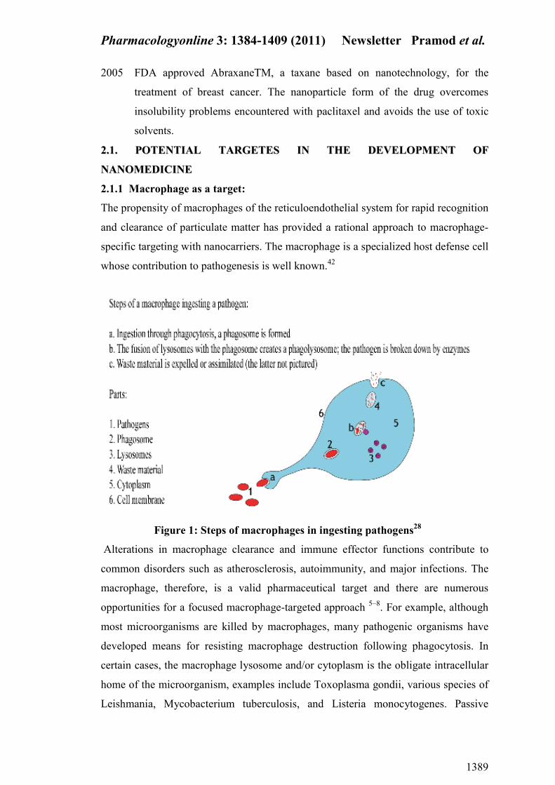
2.1.1 Macrophage as a target:
The propensity of macrophages of the reticuloendothelial system for rapid recognition
and clearance of particulate matter has provided a rational approach to macrophage-
specific targeting with nanocarriers. The macrophage is a specialized host defense cell
whose contribution to pathogenesis is well known.42
Figure 1: Steps of macrophages in ingesting pathogens28
Alterations in macrophage clearance and immune effector functions contribute to
common disorders such as atherosclerosis, autoimmunity, and major infections. The
macrophage, therefore, is a valid pharmaceutical target and there are numerous
opportunities for a focused macrophage-targeted approach 5–8
. For example, although
most microorganisms are killed by macrophages, many pathogenic organisms have
developed means for resisting macrophage destruction following phagocytosis. In
certain cases, the macrophage lysosome and/or cytoplasm is the obligate intracellular
home of the microorganism, examples include Toxoplasma gondii, various species of
Leishmania, Mycobacterium tuberculosis, and Listeria monocytogenes. Passive
Pharmacologyonline 3: 1384-1409 (2011) ewsletter Pramod et al.
1390
targeting of nanoparticulate vehicles with encapsulated antimicrobial agents to
infected macrophages is therefore a logical strategy for effective microbial killing 5, 6,
9–11. The endocytic pathway will direct the carrier to lysosomes where pathogens are
resident. Degradation of the carrier by lysosomal enzymes releases drug into the
phagosome-lysosome vesicle itself or into the cytoplasm either by diffusion or by
specific transporters, depending on the physicochemical nature of the drug molecule.
Approved formulations for human subjects are limited to lipid-based nanosytems
(100–200 nm) with entrapped amophotericin B (Amp-B), and are recommended for
treatment of visceral eishmaniasis or confirmed infections caused by specific fungal
species 5, 9, 11
. This mode of targeting has significantly reduced the required clinically
effective quantity of Amp-B for treatment, achieving therapeutic drug concentrations
in the infected macrophages. Other beneficial effects include significant reduction in
nephrotoxicity, a common side effect associated with Amp-B administration, and pro
inflammatory cytokine release 12, 13
.
2.1.2 Endothelium as a target
The concept of targeting to the blood vessels is an attractive one, particularly with the
view that the endothelium plays an important role in a number of pathological
processes including cancer (dysregulated angiogenesis), inflammation, oxidative
stress and thrombosis. Indeed, a number of studies have demonstrated a level of
control of arrest and distribution of passively targeted nanoparticles by specific
endothelial cells, and these were linked to the surface properties of the carrier. Recent
studies have shown that cationic liposomes within 1 h of entering the circulation, are
internalized into endosomes and lysosomes of endothelial cells in a characteristic
organ- and vessel-specific manner 14.
These patterns seem to bear no relationship to
the morphological characteristics of the endothelium associated with a particular site,
but probably reflect vessel-specific expression of receptors for which such particles,
or their surface-associated blood proteins, are ligands. But recent dramatic progress in
the development of a human vascular map, in particular through the application of cell
and molecular biological tools such as serial analysis of gene expression (SAGE),
subtractive proteomic mapping, and in vivo phage display, is generating yet another
level of possibilities for specific targeting of drugs and biological agents [15–18]
. For
example, SAGE examines the spectrum of mRNA species within a cell or tissue and
allows rapid comparison with other cell types or tissues. Phage, however, behaves as a
nanomachine; it can be engineered to display numerous peptides on its surface and
Pharmacologyonline 3: 1384-1409 (2011) ewsletter Pramod et al.
1391
after injection one can select peptides that make the phage home to a given target .
The authors surveyed 47,160 sequences that localized to different organs within the
patient, and determined that in many cases these sequences were similar to those of
known ligands for endothelial cell-surface proteins, thus validating his technique for
identifying novel endothelial targets in humans. This approach is now being used in a
number of ways to target therapeutic agents, particularly to the vasculature of solid
tumors. Examples include integrins alpha v beta 3, alpha v beta 5 and alpha 5 beta 1,
which are up-regulated in angiogenic endothelial cells and play a role in the process
of angiogenesis 15, 18, 19, 20
. They bind with high affinity to sequences containing a
characteristic RGD (Arg-Gly-Asp) motif, which seems to be central to anti-integrin
approaches. Indeed, in vivo phage display studies by Assa-Munt et al. have led to the
development of a cyclic nonapeptide RGD-4C, which avidly binds to the integrins
alpha v beta 3 and alpha v beta 5. Coupling of RGD-4C to doxorubicin yielded a
compound significantly more effective than doxorubicin alone, and with less side
effects to the heart and liver, the main sites of doxorubicin toxicity.43
2.1.3 Extravasation: targeting of solid cancers
The development of “stealth” technologies has provided opportunities for passive
accumulation of intravenously injected nanoparticles (20–150 nm) in pathological
sites expressing “leaky” vasculature by extravasation 6. In spite of these limitations,
there are regulatory approved formulations of long circulating liposomes with
entrapped doxorubicin for management/treatment of AIDS-related Kaposi’s sarcoma,
refractory ovarian cancer, and metastatic breast cancer 21
. These formulations exhibit
favorable pharmacokinetics when compared with the free drug, for example the area
under the curve after a dose of 50 mg/m2 doxorubicin encapsulated in stealth
liposomes is 300-fold greater than that of free doxorubicin. As a result of these
promising pharmacokinetic profiles, stealth doxorubicin containing liposomes are
currently undergoing additional early- to late-phase clinical trials.(56)
A number of
engineering issues must be considered for cancer drug delivery. First, the carrier
must have a high drug loading capacity and remain stable within the vasculature with
minimum drug loss.

Pharmacologyonline 3: 1384-1409 (2011) ewsletter Pramod et al.
1392
Here, doxorubicin is loaded actively by an ammonium sulfate gradient (as
doxorubicin sulfate) yielding highly stable liposomes with high contents of
doxorubicin aggregates 22
. Second, it has been widely established that the majority of
extravasated particulate systems, such as liposomes, do not interact with target cancer
cells [23]
. They are often distributed heterogeneously in perivascular clusters that do
not move significantly. The process of particle extravasation must be followed by the
efflux of drug from the carrier, resulting in target exposure (being tumor cells, tumor-
associated macrophages, components of tumor vasculature, or extracellular mediators
such as proangiogenic proteases) to drug molecules. Here, the drug must be released
at a rate that maintains free drug levels in the therapeutic range.44
2.1.4 anoparticles for cytoplasmic drug delivery
The endosomal membrane is particularly important for priming MHC class I-
restricted cytotoxic T lymphocyte responses, for survival of genetic materials against
nuclease degradation in the lysosomal compartment, or for those drugs that must
reach cytoplasm in sufficient quantities (as for treatment of cytoplasmic infections or
reaching nuclear receptors) after endocytic delivery with nanoparticulate carriers.
Here, there are advances in particle engineering too. For instance, nanoparticles made
from poly(DL-lactide-co-glycolide) can escape the endo-lysosomal compartment
within minutes of internalization in intact form and reach the cytoplasm 23
. The
mechanism of rapid escape is by selective reversal of the surface charge of
Figure 2: Location of Endothelium29
Pharmacologyonline 3: 1384-1409 (2011) ewsletter Pramod et al.
1393
nanoparticles from the anionic to the cationic state in endo-lysosomes, thus resulting
in a local particle-membrane interaction with subsequent cytoplasmic release. Another
impressive approach for cytoplasmic delivery of nanoparticles is their surface
manipulation with short peptides known as protein transduction domains such as
HIV-1 TAT protein transduction domain (TAT PTD), which is a short basic region
comprising residues ,or heterologous recombinant TAT-fusion peptides.The
electrostatic interaction between the cationic TAT PTD and negatively charged
cellsurface constituents, such as heparan sulfate proteoglycans and glycoproteins
containing sialic acids, is a necessary event before internalization . 57
After this ionic
interaction, cellular uptake occurs by lipid raftdependent macropinocytosis in a
receptor-independent manner; this is followed by a pH drop and destabilization of
integrity of the macropinosome vesicle lipid bilayer, which ultimately results in the
release of TAT-cargo into the cytosol . This mode of entry may further suggest the
avidity of TAT PTD for glycophosphoinositol- anchored glycoproteins, which are
present in lipid rafts, or binding to cholesterol membrane constituents that trigger
macropinocytosis. Indeed, an influenza vaccine based on the latter principle is
currently available for human use . This vaccine is administered parenterally and is
well tolerated in children, young adults and the elderly. The influenza strains chosen
are dependent on the yearly recommendations of the World Health Organization. The
delivery system is comprised of unilamellar vesicles of 150 nm in diameter, but
intercalated into their lipid bilayer are viral components, which include neuraminidase
and hemagglutinnin (HA) glycoprotein. The mode of action of these virus-like
liposomes (virosomes) is dependent on HA glycoprotein, the major antigens of the
influenza virus. The HA is composed of two subunits, HA1 and HA2. The first
subunit has high affinity for sialic acid present on the surface of antigen presenting
cells thus facilitating virosome binding. The HA2 subunit is a fusion peptide and is
activated at low pH (5.0). Hence, in late endosomes, where pH is acidic, the virosome
becomes fusion-competent; this process releases entrapped antigens into the cytosol
for subsequent processing and presentation . 45
2.2 TTYYPPEESS OOFF AA OOMMAATTEERRIIAALLSS UUSSEEDD II AA OOMMEEDDIICCII EE
2.2.1 LIPOSOME
Pharmacologyonline 3: 1384-1409 (2011) ewsletter Pramod et al.
1394
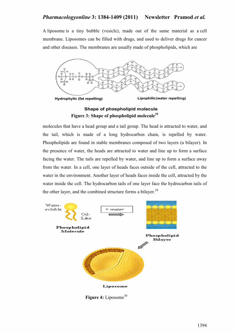
A liposome is a tiny bubble (vesicle), made out of the same material as a cell
membrane. Liposomes can be filled with drugs, and used to deliver drugs for cancer
and other diseases. The membranes are usually made of phospholipids, which are
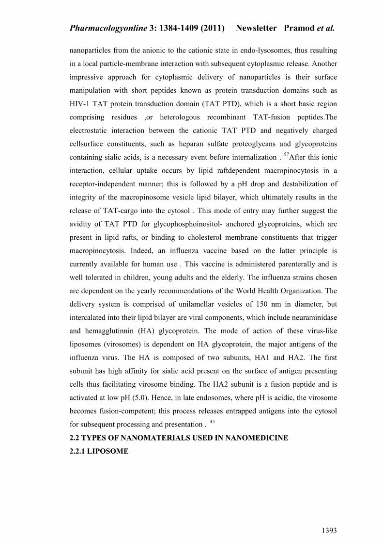
Figure 3: Shape of phospholipid molecule29
molecules that have a head group and a tail group. The head is attracted to water, and
the tail, which is made of a long hydrocarbon chain, is repelled by water.
Phospholipids are found in stable membranes composed of two layers (a bilayer). In
the presence of water, the heads are attracted to water and line up to form a surface
facing the water. The tails are repelled by water, and line up to form a surface away
from the water. In a cell, one layer of heads faces outside of the cell, attracted to the
water in the environment. Another layer of heads faces inside the cell, attracted by the
water inside the cell. The hydrocarbon tails of one layer face the hydrocarbon tails of
the other layer, and the combined structure forms a bilayer.24
Figure 4: Liposome30

Pharmacologyonline 3: 1384-1409 (2011) ewsletter Pramod et al.
1395
When membrane phospholipids are disrupted, they can reassemble themselves into
tiny spheres, smaller than a normal cell, either as bilayers or monolayers. The bilayer
structures are liposomes. The monolayer structures are called micelles. The lipids in
the plasma membrane are chiefly phospholipids like phosphatidylethanolamine and
phosphatidylcholine. Phospholipids are amphiphilic with the hydrocarbon tail of the
molecule being hydrophobic; its polar head hydrophilic. As the plasma membrane
faces watery olutions on both sides, its phospholipids accommodate this by forming a
phospholipid bilayer with the hydrophobic tails facing each other. Liposomes can be
composed of naturally-derived phospholipids with mixed lipid chains (like
egg phosphatidylethanolamine),or of pure surfactant components like DOPE (Di-
oleoyl Phosphatidyl Ethanolamine).
Application of liposome
• Liposomes are used for drug delivery due to their unique properties. A
liposome encapsulates a region on aqueous solution inside a hydrophobic
membrane; dissolved hydrophilic solutes cannot readily pass through the
lipids.
• Hydrophobic chemicals can be dissolved into the membrane, and in his way
liposome can carry both hydrophobic molecules and hydrophilic molecules to
deliver the molecules to sites of action, the lipid bilayer can fuse with other
bilayers such as the cell membrane, thus delivering the liposome contents. By
making liposomes in a solution of DNA or drugs (which would normally be
unable to diffuse through the membrane) they can be (indiscriminately)
delivered past the lipid bilayer.55
• Liposomes are used as models for artificial cells. Liposomes can also be
designed to deliver drugs in other ways. Liposomes that contain low (or
high) pH can be constructed such that dissolved aqueous drugs will
be charged in solution. As the pH naturally neutralizes within the liposome
(protons can pass through some membranes), the drug will also be neutralized,
allowing it to freely pass through a membrane. These liposomes work to
deliver drug by diffusion rather than by direct cell fusion. .
• The use of liposomes for transformation or transfection of DNA into a host
cell is known as lipofection.

Pharmacologyonline 3: 1384-1409 (2011) ewsletter Pramod et al.
1396
2.2.2 Dendrimers
Dendrimers (dendri means tree, mer means branch) are a novel class of 3-D nano
scale, core–shell structures that can be precisely synthesized for a wide range of
applications. Specialized chemistry techniques allow for precise control over the
physical and hemical properties of the dendrimers. They are constructed generation by
generation in a series of controlled steps that increase the number of small branching
molecules around a central core molecule. Up to 10 generations can be incorporated
into a single dendrimer molecule. The core, branching, and surface molecules are
chosen to give desired properties and functions.54
Figure 5: The core, branching, and surface molecules of Dendrimers 31
As a result of their unique architecture and construction, dendrimers possess
inherently valuable physical, chemical, and biological properties. These are as
follows:
• Precise architecture, size, and shape control—Dendrimers branch out in a highly
predictable fashion to form amplified 3D structures with highly ordered
architectures.
• High uniformity and purity—The proprietary stepwise synthetic process used
produces dendrimers with highly uniform sizes (monodispersity) possessing
precisely defined surface functionality and very low impurity levels.
• High loading capacity—Internal cavities intrinsic to dendrimer structures can be
used to carry and store a wide range of metals, organic, or inorganic molecules
• Low toxicity—Most dendrimer systems display very low cytotoxicity levels.
• Low immunogenicity when injected or used topically.
a )Properties of Dendrimers
The surface properties of dendrimers may be manipulated by the use of appropriate
“capping” reagents on the outermost generation. In this way dendrimers can be
readily decorated to yield a novel range of functional properties. These are as follows:

Pharmacologyonline 3: 1384-1409 (2011) ewsletter Pramod et al.
1397
• Polyvalency—The outer shell of each dendrimer can be manipulated to contain a
large number of reactive groups. Each of these reactive sites has the potential to
interact with a target entity, often resulting in polyvalent interactions.
• Flexible charge and solubility properties—Through use of appropriate capping
groups on the dendrimer exterior, the charge and solubility of dendrimers can be
readily manipulated.53
• Flexible binding properties—By using appropriate capping groups on the
dendrimer exterior, dendrimers can be designed to exhibit strong affinity for
specific targets.
b) Applications
Potential applications of dendrimers in nanomedicine are as follows:
i. Diagnostics
1. Sensors
2. Imaging contrast agents
ii. Drug delivery
1. Improved delivery of existing drugs
2. Improved solubility of existing drugs
iii. Drug development
1. Polyvalent dendrimers interacting simultaneously with
multiple drug targets
2. Development of new pharmaceuticals with novel
activities
3. Improving pharmacological activity of existing drugs
4. Improving bioavailability of existing drugs
iv. Medicine and surgery
1. Prevention of scar tissue formation after surgery
22..22..33 VVIIRROOSSOOMMEESS
A Virosome is a unilamellar phospholipid bilayer vesicle with a mean diameter of 150
nm. Essentially, virosomes represent reconstituted empty influenza virus envelopes,
devoid of the nucleocapsid including the genetic material of the source virus.[26]
Virosomes are not able to replicate but are pure fusion-active vesicles. In contrast
to liposomes, virosomes contain functional viral envelope glycoproteins: influenza
Pharmacologyonline 3: 1384-1409 (2011) ewsletter Pramod et al.
1398
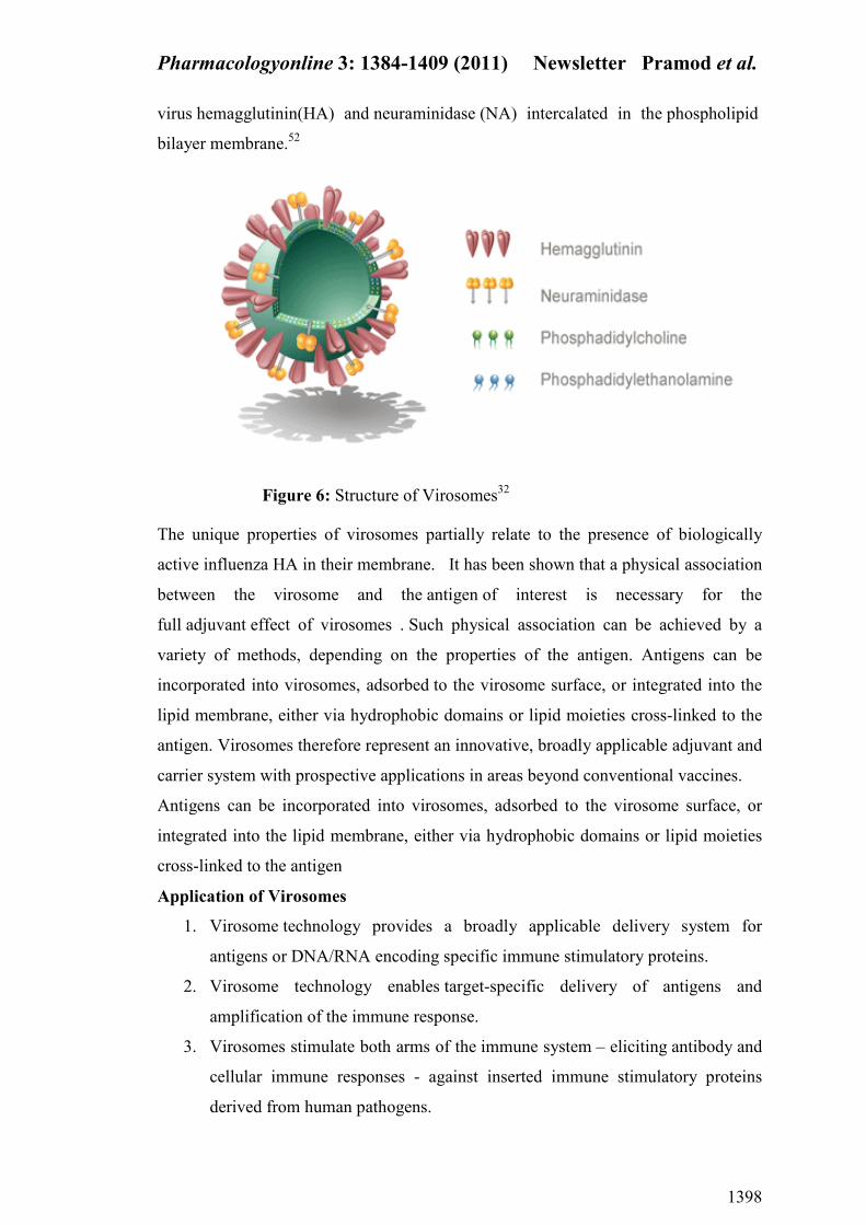
virus hemagglutinin(HA) and neuraminidase (NA) intercalated in the phospholipid
bilayer membrane.52
Figure 6: Structure of Virosomes32
The unique properties of virosomes partially relate to the presence of biologically
active influenza HA in their membrane. It has been shown that a physical association
between the virosome and the antigen of interest is necessary for the
full adjuvant effect of virosomes . Such physical association can be achieved by a
variety of methods, depending on the properties of the antigen. Antigens can be
incorporated into virosomes, adsorbed to the virosome surface, or integrated into the
lipid membrane, either via hydrophobic domains or lipid moieties cross-linked to the
antigen. Virosomes therefore represent an innovative, broadly applicable adjuvant and
carrier system with prospective applications in areas beyond conventional vaccines.
Antigens can be incorporated into virosomes, adsorbed to the virosome surface, or
integrated into the lipid membrane, either via hydrophobic domains or lipid moieties
cross-linked to the antigen
Application of Virosomes
1. Virosome technology provides a broadly applicable delivery system for
antigens or DNA/RNA encoding specific immune stimulatory proteins.
2. Virosome technology enables target-specific delivery of antigens and
amplification of the immune response.
3. Virosomes stimulate both arms of the immune system – eliciting antibody and
cellular immune responses - against inserted immune stimulatory proteins
derived from human pathogens.

Pharmacologyonline 3: 1384-1409 (2011) ewsletter Pramod et al.
1399
4. Virosomes are completely biodegradable and can exert an immune response
via different routes of administration.51
22..22..44 AAQQUUAASSOOMMEESS
Aquasomes are the nanobiopharmaceutical carrier system contains the particle core
composed of nanocrystalline calcium phosphate or ceramic diamond, and is covered
by a polyhydroxyl oligomeric film. Aquasomes are spherical 60–300 nm particles
used for drug and antigen delivery Three types of core materials are mainly used for
producing aquasomes: tin oxide, nanocrystalline carbon ceramics (diamonds) and
brushite (calcium phosphate dihydrate). The brushite is unstable and converts to
hydroxyapatite upon prolong storage. Hydroxyapatite, a better core for the preparation
of aquasomes. 50
APPLICATIO S OF AQUASOMES
1. Aquasomes as red blood cell substitutes, haemoglobin immobilized on oligomer
surface because release of oxygen by haemoglobin is conformationally
sensitive. By this toxicity is reduced, haemoglobin concentration of 80%
achieved and reported to deliver blood in non linear manner like natural blood
cells.
2. Aquasomes used as vaccines for delivery of viral antigen i.e. Epstein-Barr and
Immune deficiency virus to evoke correct antibody.
3. Aquasomes have been used for successful targeted intracellular gene therapy, a
five layered composition comprised of ceramic core, polyoxyoligomeric film,
therapeutic gene segment, additional carbohydrate film and a targeting layer of
conformationally conserved viral membrane protein.
4. Aquasomes for pharmaceuticals delivery i.e. insulin, developed because drug
activity is conformationally specific.Bioactivity preserved and activity increased
to 60% as compared to i.v. administration and toxicity not reported .
5. Aquasomes also used for delivery of enzymes like DNAase and pigments/dyes
because enzymes activity fluctuates with molecular conformation and cosmetic
properties of pigments are sensitive to molecular conformation.49
2.2.5 Quantum Dots :
QDs are nanoscale crystals of semiconductor material that glow, or fluoresce when
excited by a light source such as a laser. QD nanocrystals of cadmium selenide 200–
10,000 atoms wide, coated with zinc sulfide. Quantum dots are tiny crystals that glow
when stimulated by UV light. The wavelength or colour of light depends on size of

Pharmacologyonline 3: 1384-1409 (2011) ewsletter Pramod et al.
1400
crystals. Latex beads filled with these nanoscale semiconductor dots can be designed
to level specific DNA sequence by combining different sized quantum dots within a
single bead, we can create probes that release distincts colours and intensity of the
light. When the crystals are stimulated by UV light, each beam emits lights that serves
as spectral barcode identifying a particular region of DNA. The size of the QD
determines the frequency of light emitted when irradiated with low energy light
Eviflours are quantum dots which bind to antibodies and proteins and are used in
research assays.44
Figure 8: Multifunctional Quantum Dot Coated With Amphiphilic Polymer34
Figure 7:Structure and applications of Q-dots.33

Pharmacologyonline 3: 1384-1409 (2011) ewsletter Pramod et al.
1401
Applications of QDs
• Fluorescence detection—microscopy, biosensors, multicolor flow cytometry
• Molecular diagnostics, Ex vivo live cell imaging
• In vivo targeting of cells, tissues, and tumors with monitoring by PET and
MRI
• High-throughput screening and also used as a multifunctional quantum dot
coated
2.2.6 Carbon anotubes
Carbon nanotubes are rolled-up sheets of carbon . These nanotubes can go down to 1
nm in diameter, are stronger than any material in the universe, and can be of any
length. These can be used as probes for AFMs(atomic force microscopy) that can
image individual molecules in both wet and dry environments. The bonding in carbon
nanotubes is sp², with each atom joined to three neighbours, as in graphite. The tubes
can therefore be considered as rolled-up graphene sheets (graphene is an individual
graphite layer). 43
Types of Carbon Nanotubes
Carbon nanotubes are cylindrical sheets of carbon atoms with diameters of about 1
nanometer. Carbon nanotubes can be thought of as a rolled up sheet of graphite.
Depending on how the sheet is rolled into a tube, different nanotube structures are
produced. The image to the right shows several types of nanotubes, each with a
different atomic structure. The structures can be clearly distinguished by looking at
the cross section or along the axis of the nanotube the cathode. It is the deposit on the
cathode which contains the carbon nanotubes. Single-walled nanotubes are produced
when Co and Ni or some other metal is added to the anode. carbon nanotubes can
also be made by passing a carbon-containing gas, such as a hydrocarbon, over a
catalyst. The catalyst consists of nano-sized particles of metal, usually Fe, Co or Ni.

Pharmacologyonline 3: 1384-1409 (2011) ewsletter Pramod et al.
1402
These particles catalyse the breakdown of the gaseous molecules into carbon, and a
tube then begins to grow with a metal particle at the tip . The third important method
for making carbon nanotubes involves using a powerful laser to vaporise a metal-
graphite target. This can be used to produce single-walled tubes with high yield.
Medical Applications of anotubes:
• Cyclic peptide nanotubes can act as a new type of antibiotic against bacterial
pathogens.
• Nanoscale electromechanical systems (nanotweezers) based on carbon nanotubes
have been developed for manipulation and interrogation of nanostructures within a
cell.
• Carbon nanotubes can be used as tips for AFM.
• Nanotubes can be used in biosensors.
• Blood-compatible carbon nanotubes, with heparin immobilized on the surface, are
building blocks for in vivo nanodevices. Activated partial thromboplastin time and
thromboelastography studies prove that heparinization can significantly enhance
the blood compatibility of nanomaterials
2.2.7 Fullerenes
A molecule of 60 carbon atoms that form a hollow sphere 1 nm in diameter. The
molecule was named buckyball or fullerene. fullerenes represent a family of related
Figure 9: Types of Carbon Nanotubes35

Pharmacologyonline 3: 1384-1409 (2011) ewsletter Pramod et al.
1403
structures containing 20, 40, 60, 70, or 84 carbons. C60, however, is the most
abundant member of this family.
Fullerenes are entirely insoluble in water, but suitable functionalization makes the
molecules soluble . Upon contact with water, under a variety of conditions, C60
spontaneously forms a stable aggregate with nanoscale dimensions (25–500 nm),
termed nano-C60 that are both soluble and toxic to bacteria .
22..22..88 AA OOSSHHEELLLLSS
Nanoshells are ball-shaped, about the size of a virus or 1/20th of an RBC, and consist
of a core of nonconducting glass that is covered by a metallic shell, typically either
gold or silver. These nanoshells involve a quasiparticle called plasmon which is a
collective excitation or quantum plasma oscillation where the electrons
simultaneously oscillate with respect to all the ions. The simultaneous oscillation can
be called Plasmon hybridization where the tenability of the oscillation is associated
with mixture of the inner and outer shell where they hybridize to give a lower energy
or higher energy. This lower energy couples strongly to incident light whereas, the
higher energy is an anti-bonding and weakly combines to incident light. The
hybridization interaction is stronger for thinner shell layers, hence, the thickness of
the shell and overall particle radius determines which wavelength of light it couples
with.27
Figure 10:Fullerenes35

Pharmacologyonline 3: 1384-1409 (2011) ewsletter Pramod et al.
1404
The interaction of light and nanoparticles affects the placements of charges which
affects the coupling strength. Incident light polarized parallel to the substrate gives a
s-polarization , hence the charges are further from the substrate surface which gives a
stronger interaction between the shell and core. Otherwise, a p-polarization is formed
which gives a more strongly shifted Plasmon energy causing a weaker interaction and
coupling.
Nanoshells possess highly favorable optical and chemical properties for biomedical
imaging and therapeutic applications. These particles are also effective substrates for
surface-enhanced Raman scattering (SERS) and are easily conjugated to antibodies
and other biomolecules . the same conjugation protocols used to bind biomolecules to
gold colloid are easily modified for nanoshells. The core/shell ratio and overall size of
a gold nanoshell influence its scattering and absorption properties. Gold Nanoshells
(Spectra Biosciences, Columbia, MD, USA) possess physical properties similar to
gold colloid, in particular a strong optical absorption due to the collective electronic
response of the metal to light. The optical absorption of gold colloid yields a brilliant
red color . It is a type of spherical nanoparticle consisting of a dielectric core which
is covered by a thin metallic shell (usually gold).
Applications of anoshells :
Nanoshells possess highly favorable optical and chemical properties it is often used
for biomedical imaging, therapeutic applications, fluorescence enhancement of weak
molecular emitters, surface enhanced Raman spectroscopy and surface enhanced
infrared absorption spectroscopy.
2.2.9 Polymeric Micelles
Polymeric micelles are essentially based on amphiphilic block coplymers such as the
pluronics polyoxyethylene polyoxypropylene block co-polymers self assemble into
Figure 11: Nanoshells36

Pharmacologyonline 3: 1384-1409 (2011) ewsletter Pramod et al.
1405
polymeric micelles. Hydrophobic drugs may be solubilized within the core of micelles
or, alternatively, conjugated to the micelles forming polymers. Although micelles are
rather dynamic systems, which continuously exchanged units between the micell
structure and free units in solution, polyoxyethylene polyaspartic acid micelles are
sufficiently stable in the blood and effectively alter the pharmacokinetics of the
solubilized drugs. They thus circulate for prolonged periods and are capable of
selectively delivering more drugs to tumour tissue as compared to administration of
the drugs in solution.
2.2.10 Gold anoparticles
DNA molecules are attached to gold nanoparticles, which tangle with other specially
designed pieces of DNA into clumps that appear blue. The presence of lead causes the
connecting DNA to fall apart. That cuts loose the individual gold nanoparticles and
changes the color from blue to red. Gold nanoparticles are also used as a connecting
point to build biosensors for detection of disease. A common technique for a
diagnostic test consists of an antibody attached to a Fluorescent molecule. When the
antibody attaches to a protein associated with the disease, the fluorescent molecule
lights up under UV light.
2.2.11 Paramagnetic and Super paramagnetic anoparticles
Paramagnetic particles are important tools for cell sorting, protein separation, and
single-molecule measurements. The particles used in these applications must meet
the following requirements:
• uniform in size,
• highly paramagnetic,
• stable in physiological
• salt buffer,
• functionizable, and 100–1,000nm in size.
They have been used for the detection of model pathogens. Paramagnetic Nano-
particles, which are linked to antibodies, enable highly specific biological cell
separations. Super paramagnetic iron oxide nanoparticles : (SPIONs) with
appropriate surface chemistry have been widely used experimentally in vivo
applications such as MRI contrast enhancement, tissue repair, immunoassay,
detoxification of biological fluids, hyperthermia, drug delivery, and in cell separation.
These applications require that these nanoparticles have high magnetization values
Pharmacologyonline 3: 1384-1409 (2011) ewsletter Pramod et al.
1406
and size smaller than 100 nm with overall narrow particle size distribution, so that the
particles have uniform physical and chemical properties. In addition, these
applications need special surface coating of the magnetic particles, which not only has
to be nontoxic and biocompatible but also has to allow a targetable delivery with
particle localization in a specific area. Nature of surface coatings of the nanoparticles
not only determines the overall size of the colloid but also plays a significant role in
biokinetics and biodistribution of nanoparticles in the body .Magnetic nanoparticles
can bind to drugs, proteins, enzymes, antibodies, or nucleotides and can be directed to
an organ, tissue, or tumor using an external magnetic field
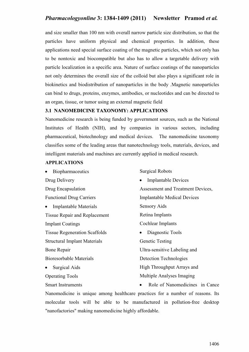
33..11 AA OOMMEEDDIICCII EE TTAAXXOO OOMMYY:: AAPPPPLLIICCAATTIIOO SS
Nanomedicine research is being funded by government sources, such as the National
Institutes of Health (NIH), and by companies in various sectors, including
pharmaceutical, biotechnology and medical devices. The nanomedicine taxonomy
classifies some of the leading areas that nanotechnology tools, materials, devices, and
intelligent materials and machines are currently applied in medical research.
APPLICATIO S
• Biopharmaceutics
Drug Delivery
Drug Encapsulation
Functional Drug Carriers
• Implantable Materials
Tissue Repair and Replacement
Implant Coatings
Tissue Regeneration Scaffolds
Structural Implant Materials
Bone Repair
Bioresorbable Materials
• Surgical Aids
Operating Tools
Smart Instruments
Surgical Robots
• Implantable Devices
Assessment and Treatment Devices,
Implantable Medical Devices
Sensory Aids
Retina Implants
Cochlear Implants
• Diagnostic Tools
Genetic Testing
Ultra-sensitive Labeling and
Detection Technologies
High Throughput Arrays and
Multiple Analyses Imaging
• Role of Nanomedicines in Cance
Nanomedicine is unique among healthcare practices for a number of reasons. Its
molecular tools will be able to be manufactured in pollution-free desktop
"nanofactories" making nanomedicine highly affordable.
Pharmacologyonline 3: 1384-1409 (2011) ewsletter Pramod et al.
1407
References
1. Jain KK. The Handbook of Nanomedicine, 1
st ed. Jain Pharma Biotech
Publications; Basel, 2008. p.1 2. Jain KK. The Handbook of Nanomedicine, 1
st ed. Jain Pharma Biotech
Publications; Basel, 2008. p. 2-4. 3. Moghimi, S. M, Hunter, A. C. and Murray, J. C. “Nanomedicine: current
Status and future prospects” FASEB J. ,2005, 19, 311–330 4. Jain K K. The Handbook of Nanomedicine, 1
st ed. Jain Pharma Biotech
Publications, Basel. 2008. p.5 5. Allen, T. M., and Cullis, P. R. “Drug delivery systems: entering the
mainstream”. Science, 2004, 303, 1818–1822 6. Moghimi, S. M., Hunter, A. C., and Murray, J. C. “Long-circulating and
target-specific nanoparticles: theory to practice” Pharmacol. Rev., 2001, 53,
283–318 7. Sahoo, S. K., and Labhasetwar, V. “Nanotech approaches to drug delivery and
imaging” Drug Discov. Today, 2003, 8, 1112–1120 8. Panyam J., and Labhasetwar, V. “Biodegradable nanoparticles for drug and
gene delivery to cells and tissues” Adv. Drug Deliv. Rev. 2004, 55, 329–347 9. Veerareddy, P. R., and Vobalabonia, V. “Lipid-based formulas of
amphotericin B” Drugs Today, 2004, 40, 133–145 10.
Lavasanifar, A., Samuel, J., Sattari, S., and Kwon, G. S. “Block copolymer
micelles for the encapsulation and delivery of amphotericin B” Pharm.
Res,2002, 19, 418–422 11.
Agrawal, A. K., and Gupta, C. M. “Tuftsin-bearing liposomes in treatment of
macrophage-based infections” Adv. Drug Deliv. 2000, Rev. 41, 135–146 12.
Miller C B, Waller, EK, Klingemann, HG, Dignani MC, Anaissie EJ,
Cagnoni, PJ, McSweeney, P Fleck, PR, Fruchtman SM, McGuirk J. et al.
“Lipid formulations of amphotericin B preserve and stabilize renal functions
in HSCT recipients.” Bone Marrow Transplant. 2004, 33, 543–548 13.
Shadkchan, Y., Keisari, Y., and Segal, E. “Cytokines in mice treated with
amphotericin B-intralipid” Med. Mycol. 2004, 42, 123–128 14.
McLean JW, Fox EA, Baluk P, Bolton PB, Haskell A, Pearlman R, Thurston
G, Umemoto EY, and McDonald DM. “Organ-specific endothelial uptake of
cationic liposome-DNA complexes in mice” Am. J. Physiol. 1997, 273,
H387– H404 15.
St Croix, B, Rago C, Velculescu V, Traverso G, Romans KE., Montgomery,
E., Lal, A., Riggins, G. J., Lengauer, C., Vogelstein, B., et al. “Genes
expressed in human tumorendothelium” Science,2000, 289, 1197–1202 16
. Arap, W., Kolonin, M. G., Trepel, M., Lahdenranta, J., Cardo-Vila, M.,
Giordano, R. J., Mintz, P. J., Ardelt, P. U., Yao, V. J.,Vidal, C. I., et al. “Steps
toward mapping the human vasculature by phage display” Nat. Med.2002. 8,
121–127 17.
Chi, J. T., Chang, H. Y., Haraldsen, G., Jahnsen, F. L., Troyanskaya,O. G.,
Chang, D. S., Wang, Z., Rockson, S., van de Rijn, M., Botstein, D., et al.
“Endothelial cell diversity revealed by global expression profiling” Proc. Natl.
Acad. Sci. 2003,USA 100, 10623–10628 18.
Pasqualini, R., Arap, W., and McDonald, D. M. “Probing the structural and
molecular diversity of tumour vasculature” Trend. Mol. Med. 2002,8, 563–571
Pharmacologyonline 3: 1384-1409 (2011) ewsletter Pramod et al.
1408
19. Eliceiri, B. P., and Cheresh, D. A. “The role of alpha v integrins during
angiogenesis: insights into potential mechanisms of action and clinical
development” J. Clin. Invest. 1999, 103, 1227–1230 20.
Kim, S., Bell, K., Mousa, S. A., and Varner, J. A. “A regulation of
angiogenesis in vivo by ligation of integrin alpha 5 beta 1with the central cell-
binding domain of fibronectin” Am. J. Pathol. 2000, 156, 1345–1362 21.
Gabizon, A., Shmeeda, H., and Barenholz, Y. “Pharmacokinetics of pegylated
liposomal doxorubicin—review of animal and human studies” Clin.
Pharmacokinetics,2003, 42, 419 –436 22.
Barenholz, Y. “Liposome application: problems and prospects” Curr. Opin.
Colloid Interface Sci.2001, 6, 66–77 23.
Drummond, D. C., Meyer, O., Hong, K., Kirpotin, D. B., and Papahajopoulos,
D. “Optimizing liposomes for delivery of chemotherapeutic agents to solid
tumours” Pharmacol. Rev.1999, 51,743 24.
Gomez-Hens, A., Fernandez-Romero, J.M. “Analytical methods for the
control of liposomal delivery systems” Trends Anal Chem ,2002, 25:167–178. 25.
A. Robert Jr. Freitas Nanomedicine, Volume I: Basic Capabilities, 1999,
ISBN 157059645X. 26.
Coombs RRH Robinson DW. Nanotechnology in Medicine and the
Biosciences, 1996, ISBN 2884490809 27.
Mozafari, M.R.(ed), Nanocarrier Technologies: Frontiers of Nanotherapy
(Chapters 1 and 2),2006. P. 10-11, 25-34 28
. National Nanotechnology Initiative: Research and Development FY ,
http://www.nano.gov/2002budget.html.2002. 29.
M.C.Roco, “National Nanotechnology Investment in the FY, Budget
Request,” AAAS Report XXVIII: Research & Development FY ;
http://www.aaas.org/spp/rd/04pch25.htm.2004. 30.
“Nanomedicine: Grounds for Optimism, and a Call for Papers,” Lancet 362
2003,673. 31.
“RB-162 Biomedical Applications of Nanoscale Devices,” Business
Communications Company, Inc., 25 Van Zant Street, Norwalk, CT06855
2003. 32.
R.A.Freitas, Jr., Nanomedicine, Volume I: Basic Capabilities, Landes
Bioscience, Georgetown, TX ,1999, http://www.nanomedicine.
com/NMI.htm. 33.
R.A.Freitas, Jr., J. Amer. Dent. Assoc. 131, 1559, 2000, http://
www.rfreitas.com/Nano/Nanodentistry.htm.
34. R.A.Freitas, Jr., Nanomedicine, Volume IIA: Biocompatibility, Landes
Bioscience, Georgetown, TX (2003);
http://www.nanomedicine.com/NMIIA.htm. 35,
D.O.W eber, Health Forum J. 42, 32 (1999). 36.
R.A.Freitas, Jr., Stud. Health Technol. Inform. 2002,80, 45. 37.
K.Bogunia-K ubik and M.Sugisaka, Biosystems, 2002, 65, 123. 38.
C.A.Haberzettl, Nanotechnology, 2002, 13, R9 . 39.
D.F .Emerich and C.G.Thanos, Expert Opin. Biol. Ther.2003, 3, 655. 40.
“NIH Roadmap: Nanomedicine,” National Institutes of Health 2003;
http://www.nihroadmap.nih.gov/nanomedicine/index.asp. 41.
“Alliance for Nanomedical Technologies,” http://www.research.
cornell.edu/anmt/.
Pharmacologyonline 3: 1384-1409 (2011) ewsletter Pramod et al.
1409
42. T.A.Desai, W.H.Chu, J.K.T u, G.M.Beattie, A.Hayek, and M.Ferrari,
Biotechnol. Bioeng.1998, 57, 118 . 43.
L.Leoni and T.A.Desai, IEEE Trans. Biomed. Eng. 2001, 48, 1335. 44.
S.L.T ao and T.A.Desai, Adv. Drug Deliv. Rev.2003, 55, 315. 45.
S.B.Lee and C.R.Martin, J. Am. Chem. Soc. 2002, 124, 11850 . 46.
M.Nishiza wa, V.P .Menon, and C.R.Martin, Science ,1995, 268, 700 . 47.
C.R.Martin and P.K ohli, Nature Rev. Drug Discov.2003, 2, 29 . 48.
E.D.Steinle, D.T .Mitchell, M.W irtz, S.B.Lee, V.Y .Y oung, and C.R.Martin,
Anal. Chem. 2002, 74, 2416. 49.
S.B.Lee, D.T .Mitchell, L.T rofin, T.K.Ne vanen, H.Soderlund, and
C.R.Martin, Science, 2002, 296, 2198. 50.
D.T .Mitchell, S.B.Lee, L.T rofin, N.Li, T.K.Ne vanen, H.Soderlund, and
C.R.Martin, J. Am. Chem. Soc. 2002, 124, 11864. 51.
Z.Siwy and A.Fulinski, Phys. Rev. Lett. 2002, 89, 198103. 52.
J.Schmidt, 11th Foresight Conference on Molecular Nanotechnology, Palo
Alto, CA 2003;http://www.foresight.org/Conferences
MNT11/Abstracts/Schmidt/index.html. 53.
B.Cornell, V.Braach-Maksvytis, L.King, P.Osman, B.Raguse, L.W ieczorek,
and R.P ace, Nature, 1997, 387, 580. 54.
L.A.Pozhar , E.P .K ontar, and M.Z.Hu, J. Nanosci. Nanotechnol.2,2002, 209. 55.
S.K.Bhatia and D.Nicholson, Phys. Rev. Lett. 2003,90, 016105. 56.
A.Meller , L.Ni von, E.Brandin, J.Golo vchenko, and D.Branton,Proc. Natl.
Acad. Sci. ,2000, (USA) 97, 1079,
http://www.pnas.org/cgi/content/full/97/3/1079.