phase evolution in an al0.5crfeconicu high entropy alloya 500 g charge of al0.5crfeconicu was melted...
TRANSCRIPT

lable at ScienceDirect
Intermetallics 71 (2016) 43e50
Contents lists avai
Intermetallics
journal homepage: www.elsevier .com/locate/ intermet
Phase evolution in an Al0.5CrFeCoNiCu High Entropy Alloy
N.G. Jones*, R. Izzo, P.M. Mignanelli, K.A. Christofidou, H.J. StoneDepartment of Materials Science and Metallurgy, University of Cambridge, 27 Charles Babbage Road, Cambridge, CB3 0FS, UK
a r t i c l e i n f o
Article history:Received 25 September 2015Received in revised form17 November 2015Accepted 9 December 2015Available online 8 January 2016
Keywords:High-Entropy AlloysPhase transformationHeat treatmentElectron microscopyScanning
* Corresponding author.E-mail address: [email protected] (N.G. Jones).
http://dx.doi.org/10.1016/j.intermet.2015.12.0010966-9795/© 2015 The Authors. Published by Elsevie
a b s t r a c t
The phase evolution of an Al0.5CrFeCoNiCu High Entropy Alloy has been characterised followingisothermal exposures between 0.1 and 1000 h at temperatures of 700, 800 and 900 �C. The NiAl based B2phase formed extremely quickly, within 0.1 h at the higher exposure temperatures, whilst the Cr-rich sphase formed more slowly. The solvus temperatures of these two phases were found to be ~975 and~875 �C respectively. Compilation of the data presented here with results previously reported in theliterature enabled the production of a time-temperature-transformation diagram, which clearly indicatesthat the diffusion kinetics of this material cannot be considered sluggish.© 2015 The Authors. Published by Elsevier Ltd. This is an open access article under the CC BY license
(http://creativecommons.org/licenses/by/4.0/).
1. Introduction
High Entropy Alloy (HEA) systems have been the subject ofextensive research since their inception just over a decade ago. Ahuge number of different alloys have already been studied andseveral beneficial properties identified, including good mechanical,wear and corrosion characteristics [1e6]. However, due to thecompositionally complex nature of these materials, the develop-ment of optimal alloy chemistries will only be realistically possiblevia high-throughput computational and experimental techniques[7e10]. Efficient use of such techniques requires an improved un-derstanding of the fundamental mechanisms behind the behaviourof these materials, which can only be achieved through systematicstudies of the compositional and processing dependence of keymaterial properties.
The multi-element basis of HEAs has been reported to give riseto a number of key benefits over their conventional, single elementbased counterparts [11e14]. These effects include; increased en-tropies of mixing, which improve the structural stability, particu-larly at elevated temperatures, suppressing the formation ofundesirable embrittling intermetallic phases [11,12]; severely dis-torted crystal lattices, which are thought to be responsible for thelow thermal and electrical conductivity of these materials, as wellas providing strengthening by impeding dislocation movement,
r Ltd. This is an open access article
and; sluggish diffusion kinetics, which gives rise to low graingrowth rates and enhanced creep resistance. However, despite theconsiderable number of research studies published to date, thevalidity of these underlying principles has not yet been conclusivelyestablished.
Slow diffusion in HEAs is thought to be a direct consequence ofthe complex arrangement of atoms of different elemental species.In a conventional, single element dominated solid solution, thebonding conditions of the local neighbourhood prior to, andfollowing a solute atom jump are essentially equivalent. The sameis not true in an HEA, where the local atomic configuration is ex-pected to vary considerably from site to site. As a result, it has beensuggested that a diffusing species may become trapped at lowerenergy sites or rejected by higher energy sites, both of which wouldretard atomic motion [14]. Calculations have shown that for amigrating Ni atom the difference in lattice potential energy be-tween sites is greater in an CrMnFeCoNi HEA than it is in FeeNieCraustenitic steels, which implies slower rates of diffusion in the HEA[15]. A series of quasi-binary diffusion couples were used todetermine the activation energies of diffusion for each elementalspecies in CrMnFeCoNi [15]. Whilst the activation energies of somespecies were slightly higher in the HEA than those previously re-ported in Fe, Ni, Co or austenitic FeeNieCr alloys, the activationenergies of other species were lower. Normalisation of these datawith the alloy's melting temperature, and extrapolation to obtainthe diffusion coefficients near melting, did indicate that diffusionwas slowest in the HEA, although none of these values can beconsidered anomalously low.
under the CC BY license (http://creativecommons.org/licenses/by/4.0/).

Table 1Target and measured composition of the material studied, with the standard devi-ation in measurements given as the error.
At.% Cr Fe Co Ni Cu Al
Target 18.2 18.2 18.2 18.2 18.2 9.0Measured 19.1 18.7 18.6 18.2 18.2 7.2Error ±0.3 ±0.1 ±0.1 ±0.1 ±0.5 ±0.2
1 Larger versions of each individual image within Fig. 2 are provided as onlinesupplementary information.
N.G. Jones et al. / Intermetallics 71 (2016) 43e5044
The nucleation and growth of a new phase requires long rangecoordinated inter-diffusion of the different elemental species and,therefore, would not be expected to occur quickly in materials withlow diffusion coefficients. However, rapid precipitation has beenreported in some HEAs, including the observation of a fine scalespinodal microstructure in Al0.5CrFeCoNiCu following rapid cooling[16,17] and evidence has been obtained that this phase separationoccurs evenwhen cooling at rates that approach 1 �C s�1 [18]. Thus,it would appear that diffusion is not universally slow in all HEAs.
Previous work has established the presence of multiple phaseswithin the microstructure of Al0.5CrFeCoNiCu following long-termheat treatments at temperatures between 700 and 1000 �C. Theseinclude a NiAl based B2 phase, the topologically close packed sphase and an Ni3Al based L12 phase, in addition to two fcc solidsolutions [19e21]. However, little is known about the speed atwhich these phases form, information that is intrinsically linked tothe diffusion kinetics of the alloy. Therefore, to address this issue,the phase evolution in Al0.5CrFeCoNiCu has been studied followinga series of different exposure times at temperatures of 700, 800 and900 �C, and the data used to construct a time-temperature-trans-formation diagram.
2. Experimental methods
A 500 g charge of Al0.5CrFeCoNiCu was melted in a vacuum in-duction furnace from elemental metals with purity �99.9%. Thematerial was held in the molten state for ~30 min before being castinto six 10 mm diameter bars, which were ~100 mm in length. Theas-cast bars were encapsulated in evacuated, Ar-backfilled glassampoules and homogenised at 1100 �C for 96 h. This temperaturewas selected based upon previous reports, which indicated that thefirst melting event observed in this alloy occurred at ~1150 �C[20,21]. At the end of the homogenisation treatment, five of thebars were water quenched, whilst the remaining bar was air-cooled. The water quenched bars were sectioned into ~10 mmlengths, which were individually encapsulated as described aboveand exposed for periods between 0.1 and 1000 h at temperatures of700, 800 and 900 �C. All samples were water quenched followingthermal exposure.
The microstructures of the homogenised and heat-treatedsamples were characterised using back-scattered electron (BSE)imaging in an FEI Nova NanoSEM 450. Bulk and phase composi-tions, as well as elemental distribution maps, were obtained in thesame instrument with a Bruker XFlash 6 solid-state energydispersive X-ray (EDX) detector. Bulk compositions were obtainedby averaging the data from several large area measurements, whilephase chemistry was determined from several point analyses.Selected local crystallographic characterisation was performed us-ing electron backscatter diffraction (EBSD) in the NanoSEM 450,whilst bulk information from all samples was collected using aBruker D8 diffractometer with Ni-filtered Cu Ka radiation. Diffrac-tion spectra were collected within the range 5e135� 2q in 0.03� 2qsteps counting for 2 s per step and between 42 and 52� 2q in 0.03�
2q steps counting for 8 s per step. Differential Scanning Calo-rimetery (DSC) was used to obtain the solvus temperatures of thesolid-state phase transformations that occurred during continuousheating from room temperature to 1400 �C. Measurements wereperformed in a Netzsch 404 calorimeter under flowing argon at aheating rate of 10 �C min�1.
3. Results
The bulk composition of the cast material, determined byaveraging large area EDX analyses at the top and bottom of each bar,is provided in Table 1. The measured composition of each element
waswithin 1 at% of the target value in all cases except Al, whichwasfound to be 1.8 at% below the nominal concentration. Al has a lowvapour pressure and losses are expected during the melting pro-cess. In the current study, these losses were greater than expected,resulting in a slightly depleted concentration of Al in the final alloy.The bulk homogeneity of thematerial was very good, as can be seenby the low error values in Table 1, which correspond to the standarddeviation between all of the different measurements.
BSE micrographs of the material following the 96 h homogeni-sation treatment at 1100 �C in both the air cooled and waterquenched states are shown in Fig. 1. In both conditions, the materialhad a dendritic microstructure, which is consistent with manyprevious studies [1,19e21]. In line with this previous work, EDXanalysis showed that the dendrites were multi-component solidsolutions, whilst the interdendritic constituent was Cu-rich. How-ever, in the air cooled material, acicular precipitates could be seenwithin the interdendritic regions. Elemental mapping indicatedthat these phases were rich in Ni and Al, and, therefore, were likelyto be the B2 NiAl phase [19e21]. Similar features were not observedin the water quenched material, suggesting that diffusion in theinterdendritic phase was sufficiently fast to allow elementalredistribution, leading to precipitation of the B2 phase during aircooling. As a result, only material that had been water quenchedfrom the homogenisation treatment was used in the remaining partof this study.
Images showing the microstructural evolution of Al0.5CrFeCo-NiCu as a function of exposure time at 700, 800 and 900 �C aregiven in Fig. 21. After exposure for 1 h at 700 �C, fine globularprecipitates were observed in the microstructure that were notpresent in either the as-quenched material or following 0.1 h at700 �C. These precipitates, labelled as a in Fig. 2, were observed topreferentially form on the grain boundaries within the dendriticmicrostructure. EDX analysis showed that the precipitates wererich in Ni and Al, consistent with the B2 phase previously reported[19,21]. In addition to the globular phase, small acicular pre-cipitates, labelled as b in Fig. 2, with similar backscattered electroncontrast were also observed within the interdendritic material. Thefine scale of these precipitates meant that accurate compositionalanalysis was challenging but elevated levels of Ni & Al weremeasured, which, in keeping with previous work [19,21], suggestedthat these precipitates were also the B2 phase. Following 10 h at700 �C, the occurrence of acicular B2 precipitates within theinterdendritic regions had become extensive, with individual pre-cipitates extending across the entire width of the interdendriticregions. Additional fine precipitates, with similar morphology andbackscattered contrast to those in the interdendritic regions, wereobserved within the dendritic phase after exposure for 50 h,labelled as c in Fig. 2. Thus, within 50 h exposure to 700 �C, threedifferent populations of the B2 phase were observed to haveformed in Al0.5CrFeCoNiCu; globular precipitates on the grainboundaries, acicular precipitates within the interdendritic phaseand acicular precipitates within the dendrites. With longer

Fig. 1. BSE micrographs of Al0.5CrFeCoNiCu after heat treatment at 1100 �C for 96 h followed by air cooling and water quenching.
N.G. Jones et al. / Intermetallics 71 (2016) 43e50 45
exposure times, all three populations of the B2 phase coarsenedand an increase in volume fraction was observed. This was partic-ularly true of the acicular precipitates within the dendritic regions,which dominated themicrostructure after 1000 h. During this time,another intermetallic precipitate was also observed to have formed.Following a 100 h exposure, regions of material that had similarbackscattered electron contrast to the original dendrites wereobserved, labelled as d in Fig. 2, but, critically, unlike the dendriticmaterial, these regions were free from any internal precipitation.EDX analysis of these areas showed significantly elevated Cr con-centration, consistent with previous reports of the s phase at thistemperature [19,21]. Phase mapping, using concurrent EDX andEBSD, was performed on material exposed for 1000 h at 700 �C andconfirmed that all of the Ni and Al rich regions had a B2 crystalstructure and that the Cr rich areaswere tetragonal, consistent withthe s phase.
At 800 �C the phase evolution of Al0.5CrFeCoNiCu with exposuretime followed the same general trend as that at 700 �C, althoughthere were some key differences. The formation of globular B2 onthe grain boundaries and the precipitation of acicular B2 within theinterdendritic material occurred more rapidly, within the first fewminutes of exposure, with both of these features evident in themicrograph following a 0.1 h exposure. Similarly, acicular B2 pre-cipitates were observed within the multi-component dendritesfollowing 1 h at 800 �C. This was considerably faster than at 700 �C,where the acicular intradendritic precipitates were observed toform between 10 and 50 h. In addition, the acicular B2 precipitatesformed within the dendrites at 800 �C appeared to be fully devel-oped following a 10 h exposure and coarsened with longer expo-sure times. The Cr-rich s phase was also found to form at shorterexposure times at 800 �C than was observed at 700 �C. Evidence ofits presence was found in the microstructure following a 10 hexposure at 800 �C, as can be seen in the EDXmaps shown in Fig. 3.Despite a lack of backscattered contrast, the elemental partitioningmaps clearly show regions with an elevated concentration of Cr,which also contain Fe and Co, next to the globular B2 precipitates.Regular arrays of fine scale bright precipitates were also seenwithin the dendrites following exposure for 10 h at 800 �C, Fig. 4.These features became more apparent as the exposure timeincreased and they coarsened. It is believed that these precipitatesare Cu rich and form as a result of the miscibility gap in this system[20] and their formation is consistent with the nano-scale spinodaldecomposition of the multi-component dendrites previouslyobserved in this alloy [16,18].
At short exposure times, up to an hour, the phase evolution at
900 �C was similar to that observed at 800 �C. However, in contrastto the material exposed at 800 �C, all three populations of the B2phase, globular grain boundary, acicular in the interdendritic con-stituent and acicular in the dendritic regions, were observedfollowing 0.1 h at 900 �C. In addition, an extremely fine scale reg-ular array of bright contrast precipitates could be seen within thedendrites after a 1 h exposure, Fig. 5. As can be seen in Fig. 2, thisthree-phase microstructure coarsened with increasing exposuretime, but at no stagewas any evidence of the s phase observed. Thisfinding is consistent with previous reports that have indicated thatthe s solvus temperature must lie between 850 �C, where s hasbeen reported following a 1000 h exposure [21], and 900 �C [19].
Full X-ray diffraction spectra, between 5 and 135� 2q, were ac-quired from material in all heat treatment conditions. However, itwas found that the reflections within the range 42 and 52� 2qprovided the most concise evidence of phase evolution. The spectrafor the different times at each exposure temperature are shown inFig. 6, along with the spectrum from the homogenised and waterquenched starting material, identified as an exposure time of 0 h. Inthe spectrum obtained from the homogenised material, only peakscorresponding to an fcc structure were observed, but, in keepingwith previous work [1,19e21], the shape of line profile of thesereflections indicated that they resulted from multiple fcc basedstructures, all with similar lattice parameters. B2 reflections wereobserved in the XRD spectra following a 1 h exposure at 700 �C and0.1 h exposures at 800 and 900 �C, consistent with the electronmicroscopy results presented in Fig. 2. The separation of the twomain fcc reflections, the multi-component solid solution and theCu-rich phase, is well captured in the XRD spectra and providessome qualitative information about the effect of temperature onthe diffusion rates in Al0.5CrFeCoNiCu. Reflections corresponding tothe s phase were also present in the XRD spectra following expo-sure at 700 and 800 �C but not at 900 �C. Their occurrence is inkeepingwith the electronmicroscopy results but the exposure timeafter which they can be discerned does not correlate so well. Fromthe XRD spectra, reflections corresponding to the s phase werepresent following a 1000 h exposure at 700 �C and after 50 h at800 �C. In contrast, microstructural evidence of this phase wasdetected in the elemental partitioning maps obtained from mate-rial following shorter exposure times, 100 h at 700 �C and 10 h at800 �C. It is believed that the localised probing possible withelectron microscopy enabled identification of the s phase at anearlier stage of its formation, corresponding to a volume fractionbelow the detection limit of the XRD equipment used.
A DSC thermogram obtained from Al0.5CrFeCoNiCu following
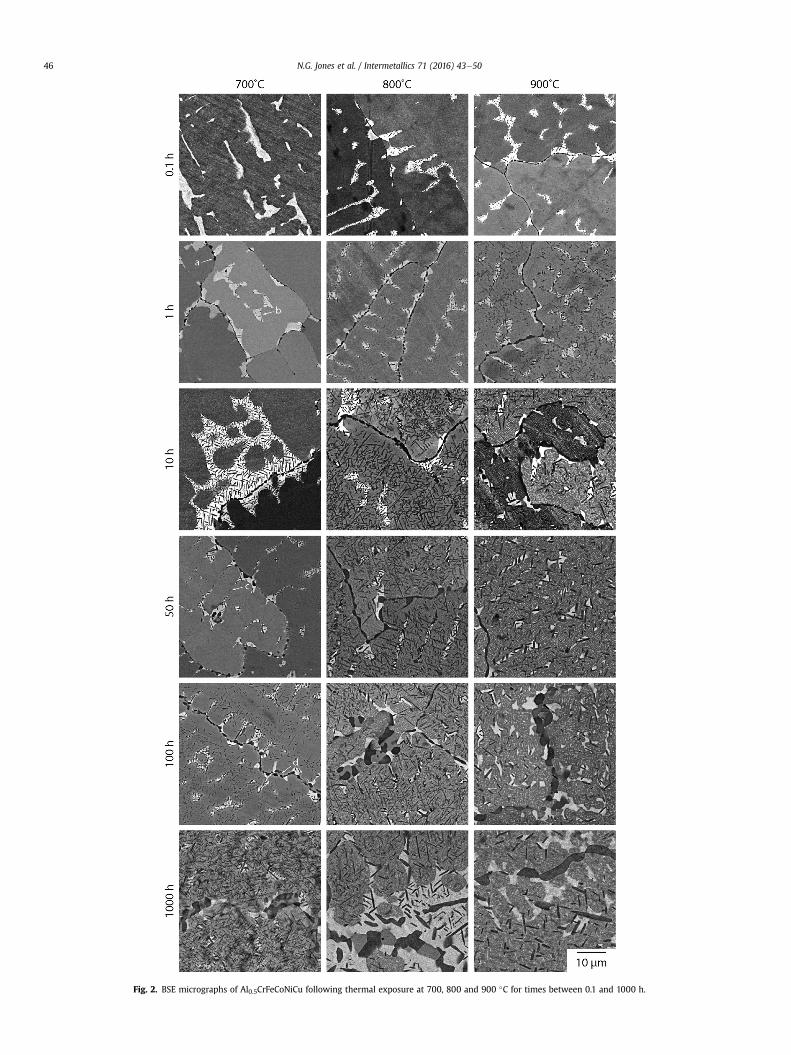
Fig. 2. BSE micrographs of Al0.5CrFeCoNiCu following thermal exposure at 700, 800 and 900 �C for times between 0.1 and 1000 h.
N.G. Jones et al. / Intermetallics 71 (2016) 43e5046

Fig. 3. BSE micrograph and corresponding EDX elemental partitioning maps showingthe detection of the s phase in Al0.5CrFeCoNiCu following heat treatment for 10 h at800 �C.
Fig. 4. High magnification BSE micrograph of Al0.5CrFeCoNiCu following 10 h at 800 �Cshowing the regular arrays of bright precipitates within the dendrites.
Fig. 5. High magnification BSE micrograph of Al0.5CrFeCoNiCu following 1 h at 900 �Cshowing the bright fine scale decomposition product within the grey multi-componentdendrites.
N.G. Jones et al. / Intermetallics 71 (2016) 43e50 47
exposure for 1000 h at 900 �C is shown in Fig. 7. Microstructuralcharacterisation and XRD analysis of this material indicated thatonly the two fcc parent phases and the B2 precipitates were pre-sent. The thermogram shows four distinct events that occur be-tween 600 and 1400 �C; two large endothermic peaks at ~1350 and~1150 �C and two sigmoidal like deviations which finish at ~975and ~800 �C respectively. Previous studies have shown that the two
highest temperature events correspond to the melting of the den-dritic and interdendritic constituent, whilst the lowest temperatureevent is related to a non-equilibrium L12 phase that forms duringcooling [18,20,21]. These studies also established that only the twofcc phases are present following 1000 h at 1000 �C and thus it isbelieved that the sigmoidal deviation finishing at ~975 �C corre-sponds to the B2 solvus temperature. This is in good agreementwith previous unpublished work, where the microstructure of arcmelted Al0.5CrFeCoNiCu, which comprised of two fcc phases and L12precipitates [16,20] in the as cast state, contained B2 precipitatesfollowing a 0.25 h exposure at 950 �C, Fig. 8.

Fig. 6. X-ray diffraction spectra from Al0.5CrFeCoNiCu in the homogenised state and following thermal exposure at 700, 800 and 900 �C for times between 0.1 and 1000 h.
Fig. 7. DSC thermogram during heating of Al0.5CrFeCoNiCu following 1000 h at 900 �C.
Fig. 8. BSE micrograph of as arc melted Al0.5CrFeCoNiCu following exposure at 950 �Cfor 0.25 h showing the presence of the B2 phase.
N.G. Jones et al. / Intermetallics 71 (2016) 43e5048
4. Discussion
Compilation of the current experimental results with thosealready published in the literature allowed the construction of atime-temperature-transformation diagram for Al0.5CrFeCoNiCu,Fig. 9. In this diagram, the filled symbols correspond to datacollected during the present work, whilst the open symbols showdata from previous studies. It should be noted that the L12, fccsuperlattice phase, which is known to form in this alloy[1,16,17,19e21], has been omitted from this figure as no evidencehas been obtained to indicate that its formation can be suppressedby rapid cooling. As such, post mortem examination of samplesrecovered from elevated temperature heat treatments cannot beused to unambiguously identify whether this phase was present atthe exposure temperature or whether elemental partitioning be-tween different phases in the evolvingmicrostructure influences itsoccurrence.
It is clear that the B2 phase can form extremely quickly in thismaterial, having been observed on prior grain boundaries and inthe interdendritic constituent following 0.1 h exposures at 900 and800 �C and after 1 h at 700 �C. However, its formation within themulti-component dendrites was slower and showed a significantdependence on the exposure temperature. Intradendritic B2 pre-cipitates were observed following 0.1 h at 900 �C, 1 h at 800 �C and50 h at 700 �C. Previously, micrographs have been published thatshow similar acicular precipitates within the dendrites following a24 h exposure at 700 �C [19]. This observation is consistent with thecurrent work, however, the result cannot be directly incorporatedinto Fig. 9 as, prior to exposure, the material had been cold rolledand annealed at 900 �C, during which time the B2 phase formed.

Fig. 9. Time-temperature-transformation diagram for Al0.5CrFeCoNiCu. The filledpoints correspond to experimental results of the present work, open symbols repre-sent data taken from Ref. [21] or previous unpublished data.
N.G. Jones et al. / Intermetallics 71 (2016) 43e50 49
The structurally complex s phase formed more slowly than theB2 phase in Al0.5CrFeCoNiCu and had a lower solvus temperature,between 850 and 900 �C. In the present work, evidence of thisphase was found after 10 h at 800 �C and following 100 h at 700 �C.Again, this observation is in slight disagreement with Ng et al. [19]who presented XRD spectra clearly containing characteristic s re-flections after only 24 h at 700 �C. As stated previously, thisinconsistency is believed to be related to the fact that the materialin Ref. [19] had been cold worked and annealed at 900 �C prior toexposure at 700 �C. The results of the present work have clearlydemonstrated that the B2 phase forms at shorter exposure timesthan the s phase. In addition, previous work [21], has shown thatthe B2 phase accommodates reasonable concentrations of Fe, Co &Cu, ~8, ~10 and ~10 at% respectively, at temperatures between 700and 850 �C, but far less Cr, ~3 at%. Thus, the formation of the B2phasewould significantly enrich the surroundingmaterial in Cr andthis appears to destabilise the fcc phase with respect to interme-tallic phase formation. This situation is exacerbated in the Cu-richinterdendritic material as Cr, Co and Fe are rejected as the phasemoves towards its equilibrium composition from its metastablestarting point, where only Ni and Al retain appreciable concentra-tions in solid solution [21]. Therefore, it is unsurprising that the sphase is commonly observed adjacent to B2 precipitates, near orwithin the inderdendritic constituent. As a result, the formation ofthe B2 phase during interpass annealing at 900 �C in the materialstudied in Ref. [19] would have created areas within the materialwith elevated Cr concentrations, prior to exposure at 700 �C. Thepresence of such regions within the material whilst it is exposed attemperatures where the s phase is stable, in addition to anyremnant dislocations, would no doubt accelerate its formation,thereby accounting for the discrepancy between reference [19] andthe present study.
As stated in the introduction, reconstructive phase trans-formations require cooperative movement of different atomicspecies over relatively large distances. The rapid formation of theB2 phase during both isothermal heat treatments, and air coolingfrom 1100 �C indicates that neither the nucleation kinetics nor thediffusional growth of this phase are sluggish in Al0.5CrFeCoNiCu.The formation of the s phase did require longer exposure times,particularly at 700 �C, but even then, these times are not dis-proportionally long when compared to other materials. For
example, in relatively compositionally simple duplex stainlesssteels with similar Cr concentrations to Al0.5CrFeCoNiCu, the sphase has been reported to form very quickly at temperatures be-tween 700 and 850 �C [22,23]. In contrast, within more composi-tionally complex materials, such as Ni-base superalloys, whichagain have comparable Cr contents, the s phase requires severalhundred hours to form at these temperatures [24,25]. Thus, whilstdiffusion in HEAs is widely reported to be sluggish, the data pre-sented here clearly shows that it is not anomalously slow inAl0.5CrFeCoNiCu.
5. Conclusions
The phase evolution of an Al0.5CrFeCoNiCu High Entropy Alloyhas been characterised following isothermal exposures between 0.1and 1000 h at temperatures of 700, 800 and 900 �C. In line withprevious reports, the starting material contained two fcc phasesand two additional intermetallic phases were observed to formduring the thermal exposures, a NiAl based B2 phase and the morecomplex Cr-rich s phase. The B2 phase, which had a solvus tem-perature of ~975 �C, formed extremely quickly in this material,within 1 h at 700 �C and in less than 0.1 h at 800& 900 �C, as well asduring air cooling from 1100 �C. The s phase, which has a solvus of~875 �C, formed more slowly than the B2, requiring between 1 &10 h at 800 �C and 50 & 100 h at 700 �C. The formation of the sphase is thought to be directly related to the destabilisation of theparent material around a B2 precipitate, as this phase has littlesolubility for Cr and so enriches the surrounding area. Combinationof these data with existing results from the literature enabled atime-temperature-transformation diagram to be proposed for thefirst time. Since the formation of a new phase requires long rangecoordinated diffusion, the experimental observation of interme-tallic phase formation within 0.1 h indicates that the diffusion ki-netics within this High Entropy Alloy are not anomalously slow.
Acknowledgements
The authors would like to thank K. Roberts and S. Rhodes fortheir assistance, and the EPSRC/Rolls-Royce Strategic Partnership(EP/M005607/1 and EP/H022309/1) for funding. The originalresearch data for this study can be found at https://www.repository.cam.ac.uk/handle/1810/252934.
Appendix A. Supplementary data
Supplementary data related to this article can be found at http://dx.doi.org/10.1016/j.intermet.2015.12.001.
References
[1] C. Tong, Y. Chen, S. Chen, J. Yeh, T. Shun, C. Tsau, S. Lin, S. Chang, Metall. AndMater. Trans. A 36A (2005) 881e893.
[2] Y.J. Zhou, Y. Zhang, Y.L. Wang, G.L. Chen, Appl. Phys. Lett. 90 (2007) 181904.[3] O.N. Senkov, C. Woodward, D.B. Miracle, JOM 66 (2014) 2030e2042.[4] C.Y. Hsu, J.W. Yeh, S.K. Chen, T.T. Shun, Metall. Mater. Trans. A 35 (2004)
1465e1469.[5] P.K. Huang, J.W. Yeh, T.T. Shun, S.K. Chen, Adv. Eng. Mater. 6 (2004) 74e78.[6] Y. Hsu, W. Chiang, J. Wu, Mater. Chem. Phys. 92 (2005) 112e117.[7] D.B. Miracle, Mater. Sci. Technol. 31 (2015) 1142e1147.[8] D. Miracle, J. Miller, O. Senkov, C. Woodward, M. Uchic, J. Tiley, Entropy 16
(2014) 494e525.[9] O.N. Senkov, J.D. Miller, D.B. Miracle, C. Woodward, Nature Communications,
6(1) 1e10.[10] F. Zhang, C. Zhang, S.L. Chen, J. Zhu, W.S. Cao, U.R. Kattner, Calphad 45 (2013)
1e10.[11] J. Yeh, S. Chen, S. Lin, J. Gan, T. Chin, T. Shun, C. Tsau, S. Chang, Adv. Eng. Mater.
6 (2004) 299e303.[12] J.-W. Yeh, Ann. de chimie Sci. des Materiaux 31 (2006) 633e648.[13] J.-W. Yeh, JOM 65 (2013) 1759e1771.

N.G. Jones et al. / Intermetallics 71 (2016) 43e5050
[14] M.-H. Tsai, J.-W. Yeh, Mater. Res. Lett. 2 (2014) 107e123.[15] K.Y. Tsai, M.H. Tsai, J.W. Yeh, Acta Mater. 61 (2013) 4887e4897.[16] E.J. Pickering, H.J. Stone, N.G. Jones, Mater. Sci. Eng. A 645 (2015) 65e71.[17] X.D. Xu, P. Liu, S. Guo, A. Hirata, T. Fujita, T.G. Nieh, C.T. Liu, M.W. Chen, Acta
Mater. 84 (2015) 145e152.[18] N.G. Jones, K.A. Christofidou, H.J. Stone, Mater. Sci. Technol. 31 (2015)
1171e1177.[19] C. Ng, S. Guo, J. Luan, S. Shi, C.T. Liu, Intermetallics 31 (2012) 165e172.[20] N.G. Jones, J.W. Aveson, A. Bhowmik, B.D. Conduit, H.J. Stone, Intermetallics 54
(2014) 148e153.[21] N.G. Jones, A. Frezza, H.J. Stone, Mater. Sci. Eng. A 615 (2014) 214e221.[22] J.W. Elmer, T.A. Palmer, E.D. Specht, Metall. Mater. Trans. A 38 (2007)
464e475.[23] R. Magnabosco, Mater. Res. 12 (2009) 321e327.[24] A. Oradei-Basile, J.F. Radavich, in: E.A. Loris (Ed.), Superalloys 718, 625 and
Various Derviatives, 1991, pp. 325e335.[25] R.C. Reed, M.P. Jackson, Y.S. Na, Metall. Mater. Trans. A 30 (1999) 521e533.