phenotypic differences between asian and african …stalinrajv,devrijfms,widagdo...
TRANSCRIPT

Phenotypic Differences between Asianand African Lineage Zika Viruses inHuman Neural Progenitor Cells
Fatih Anfasa,a,b Jurre Y. Siegers,a Mark van der Kroeg,c Noreen Mumtaz,a
V. Stalin Raj,a Femke M. S. de Vrij,c W. Widagdo,a Gülsah Gabriel,d Sara Salinas,e
Yannick Simonin,e Chantal Reusken,a Steven A. Kushner,c
Marion P. G. Koopmans,a Bart Haagmans,a Byron E. E. Martina,a,f Debby van Riela
Department of Viroscience, Erasmus MC, Rotterdam, The Netherlandsa; Faculty of Medicine, UniversitasIndonesia, Jakarta, Indonesiab; Department of Psychiatry, Erasmus MC, Rotterdam, The Netherlandsc; HeinrichPette Institute for Experimental Virology, Hamburg, Germanyd; UMR1058, Pathogenesis and Control of ChronicInfections, INSERM, Université de Montpellier, Etablissement Français Du Sang, Montpellier, Francee; ArtemisOne Health Research Foundation, Utrecht, The Netherlandsf
ABSTRACT Recent Zika virus (ZIKV) infections have been associated with a range ofneurological complications, in particular congenital microcephaly. Human neural pro-genitor cells (hNPCs) are thought to play an important role in the pathogenesis ofmicrocephaly, and experimental ZIKV infection of hNPCs has been shown to inducecell death. However, the infection efficiency and rate of cell death have varied be-tween studies, which might be related to intrinsic differences between African andAsian lineage ZIKV strains. Therefore, we determined the replication kinetics, includ-ing infection efficiency, burst size, and ability to induce cell death, of two Asian andtwo African ZIKV strains. African ZIKV strains replicated to higher titers in Vero cells,human glioblastoma (U87MG) cells, human neuroblastoma (SK-N-SH) cells, andhNPCs than Asian ZIKV strains. Furthermore, infection with Asian ZIKV strains did notresult in significant cell death early after infection, whereas infection with AfricanZIKV strains resulted in high percentages of cell death in hNPCs. The differences be-tween African and Asian lineage ZIKV strains highlight the importance of includingrelevant ZIKV strains to study the pathogenesis of congenital microcephaly and cau-tion against extrapolation of experimental data obtained using historical AfricanZIKV strains to the current outbreak. Finally, the fact that Asian ZIKV strains infectonly a minority of cells with a relatively low burst size together with the lack ofearly cell death induction might contribute to its ability to cause chronic infectionswithin the central nervous system (CNS).
IMPORTANCE The mechanism by which ZIKV causes a range of neurological compli-cations, especially congenital microcephaly, is not well understood. The fact thatcongenital microcephaly is associated with Asian lineage ZIKV strains raises the questionof why this was not discovered earlier. One possible explanation is that Asian and Afri-can ZIKV strains differ in their abilities to infect cells of the CNS and to cause neurode-velopmental problems. Here, we show that Asian ZIKV strains infect and induce celldeath in human neural progenitor cells—which are important target cells in the devel-opment of congenital microcephaly—less efficiently than African ZIKV strains. These fea-tures of Asian ZIKV strains likely contribute to their ability to cause chronic infections, of-ten observed in congenital microcephaly cases. It is therefore likely that phenotypicdifferences between ZIKV strains could be, at least in part, responsible for the ability ofAsian ZIKV strains to cause congenital microcephaly.
KEYWORDS African strains, Asian strains, growth kinetics, human neural progenitorcells, neuronal cells, one-step growth curve, pathogenesis, phenotype, Zika virus, celldeath
Received 30 June 2017 Accepted 3 July2017 Published 26 July 2017
Citation Anfasa F, Siegers JY, van der Kroeg M,Mumtaz N, Stalin Raj V, de Vrij FMS, WidagdoW, Gabriel G, Salinas S, Simonin Y, Reusken C,Kushner SA, Koopmans MPG, Haagmans B,Martina BEE, van Riel D. 2017. Phenotypicdifferences between Asian and African lineageZika viruses in human neural progenitor cells.mSphere 2:e00292-17. https://doi.org/10.1128/mSphere.00292-17.
Editor Michael J. Imperiale, University ofMichigan—Ann Arbor
Copyright © 2017 Anfasa et al. This is an open-access article distributed under the terms ofthe Creative Commons Attribution 4.0International license.
Address correspondence to Debby van Riel,[email protected].
F.A. and J.Y.S. contributed equally to this article.
RESEARCH ARTICLEHost-Microbe Biology
crossm
July/August 2017 Volume 2 Issue 4 e00292-17 msphere.asm.org 1
on April 7, 2019 by guest
http://msphere.asm
.org/D
ownloaded from

Since the emergence of Zika virus (ZIKV) in 2015 in South America, infections havecaused a wide spectrum of neurological diseases, such as Guillain-Barré syndrome,
myelitis, meningoencephalitis, and in particular congenital microcephaly (1). Eventhough ZIKV was first detected in Uganda in 1947 in a rhesus monkey and has causedrepeated outbreaks since 2007, not much was known about the pathogenesis ofdisease caused by ZIKV before the 2015 outbreak. Since then, several studies haveshown that ZIKV can infect a variety of neuronal cells, but more insight into thepathogenesis of ZIKV-induced central nervous system (CNS) diseases is needed (2).
An important question that remains is whether the emergence of ZIKV in SouthAmerica and the associated clinical findings are the result of genetic and phenotypicchanges in the emerging ZIKV strain or whether they can be attributed to theintroduction of ZIKV in a large naive population (3). Phylogenetically, two distinctlineages of ZIKV exist: the African lineage and the Asian lineage (4). The currentoutbreak strain belongs to the Asian lineage, and sequence analysis revealed that thevirus has changed significantly over the last 50 years, both in nucleotide sequences andamino acid composition (4, 5). The prototype ancestral ZIKV strain, MR766, of theAfrican lineage has been used in many initial studies (6–9), but recent in vitro and in vivostudies have shown some differences between African and Asian ZIKV strains (10–14).Whether there are also phenotypic differences between Asian ZIKV strains, caused byamino acid substitutions acquired just before the outbreak in South America, iscurrently unknown (5).
ZIKV has been shown to replicate and induce cell death in neuronal cells of fetalmice (5, 15), as well as in human neural progenitor cells and brain organoids (6, 7, 11,12), a mechanism thought to play an important role in the pathogenesis of ZIKV-induced microcephaly. A recent study has shown that an African ZIKV strain might beable to infect human neural stem cells (hNSCs) and astrocytes more efficiently thanAsian ZIKV strains (12). However, a comprehensive study on the replication kinetics andthe ability to cause cell death of different African and Asian ZIKV strains is currentlylacking (2).
To be able to detect phenotypic differences between Asian and African ZIKV strainsor between recent Asian ZIKV strains, it is important to characterize and understand thein vitro replication kinetics—including the infection efficiency, burst size, and ability tocause cell death— of these viruses. Therefore, we determined the replication kinetics oftwo Asian ZIKV strains (isolated in 2013 and 2016) and two African ZIKV strains (isolatedin 1947 and 1961) on induced pluripotent stem cell-derived human neural progenitorcells (hNPCs) and several human neural cell lines.
RESULTSPhylogenetic and amino acid variance analysis of ZIKV strains selected in this
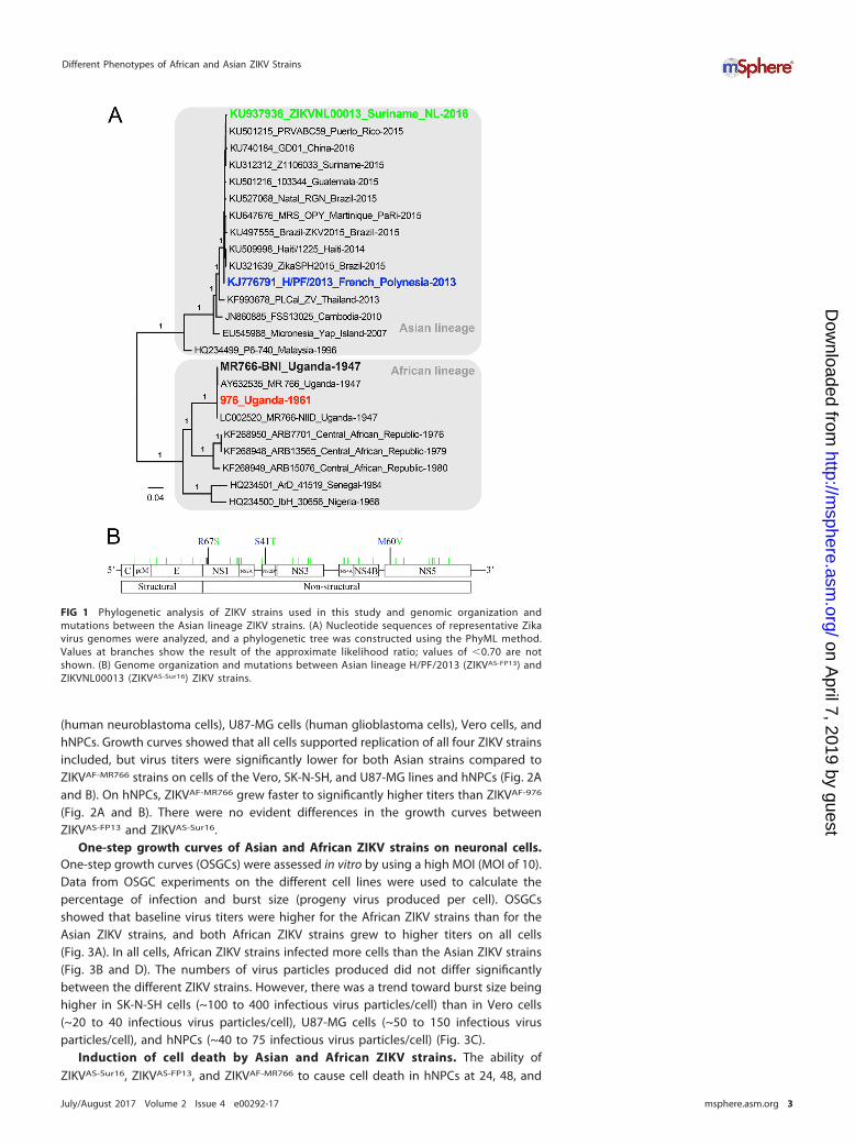
study. Four ZIKV strains were included in this study (Fig. 1A). Two African strains, ZIKVMR766 (ZIKVAF-MR766) and Uganda 976 (ZIKVAF-976), were isolated in 1947 and 1961,respectively, and passaged on mouse brain tissue and Vero cells. The two Asian ZIKVstrains included were H/PF/2013 (ZIKVAS-FP13) and ZIKVNL00013 (ZIKVAS-Sur16), whichwere isolated in 2013 and 2016, respectively, and passaged 4 times on Vero cells. Aphylogenetic analysis of the complete genome of the selected strains with other ZIKVgenomes shows their positions in the Asian or African lineages (Fig. 1A). There are over50 amino acid (aa) differences between the African and Asian ZIKV strains that havepreviously been described (5). The amino acid differences between the Asian ZIKVstrains were located in the NS1 (R67S; position 863), NS2B (S41T; position 1417), andNS5 (M60V; position 2634) proteins (Fig. 1B). Of these amino acid differences, themutation at position 2634 is only observed in viruses isolated from the recent outbreak(4, 5, 16). The amino acid difference at position 1417 of ZIKVAS-Sur16 was not present inthe original clinical isolate but was acquired during passaging on Vero cells (17).
Growth curves of Asian and African ZIKV strains on neuronal cells. Growthcurves were determined for ZIKVAS-FP13, ZIKVAS-Sur16, ZIKVAF-MR766, and ZIKVAF-976 by invitro infections using low multiplicities of infection (MOI [0.1 and 0.01]) on SK-N-SH cells
Anfasa et al.
July/August 2017 Volume 2 Issue 4 e00292-17 msphere.asm.org 2
on April 7, 2019 by guest
http://msphere.asm
.org/D
ownloaded from
(human neuroblastoma cells), U87-MG cells (human glioblastoma cells), Vero cells, andhNPCs. Growth curves showed that all cells supported replication of all four ZIKV strainsincluded, but virus titers were significantly lower for both Asian strains compared toZIKVAF-MR766 strains on cells of the Vero, SK-N-SH, and U87-MG lines and hNPCs (Fig. 2Aand B). On hNPCs, ZIKVAF-MR766 grew faster to significantly higher titers than ZIKVAF-976
(Fig. 2A and B). There were no evident differences in the growth curves betweenZIKVAS-FP13 and ZIKVAS-Sur16.
One-step growth curves of Asian and African ZIKV strains on neuronal cells.One-step growth curves (OSGCs) were assessed in vitro by using a high MOI (MOI of 10).Data from OSGC experiments on the different cell lines were used to calculate thepercentage of infection and burst size (progeny virus produced per cell). OSGCsshowed that baseline virus titers were higher for the African ZIKV strains than for theAsian ZIKV strains, and both African ZIKV strains grew to higher titers on all cells(Fig. 3A). In all cells, African ZIKV strains infected more cells than the Asian ZIKV strains(Fig. 3B and D). The numbers of virus particles produced did not differ significantlybetween the different ZIKV strains. However, there was a trend toward burst size beinghigher in SK-N-SH cells (~100 to 400 infectious virus particles/cell) than in Vero cells(~20 to 40 infectious virus particles/cell), U87-MG cells (~50 to 150 infectious virusparticles/cell), and hNPCs (~40 to 75 infectious virus particles/cell) (Fig. 3C).
Induction of cell death by Asian and African ZIKV strains. The ability ofZIKVAS-Sur16, ZIKVAS-FP13, and ZIKVAF-MR766 to cause cell death in hNPCs at 24, 48, and
FIG 1 Phylogenetic analysis of ZIKV strains used in this study and genomic organization andmutations between the Asian lineage ZIKV strains. (A) Nucleotide sequences of representative Zikavirus genomes were analyzed, and a phylogenetic tree was constructed using the PhyML method.Values at branches show the result of the approximate likelihood ratio; values of �0.70 are notshown. (B) Genome organization and mutations between Asian lineage H/PF/2013 (ZIKVAS-FP13) andZIKVNL00013 (ZIKVAS-Sur16) ZIKV strains.
Different Phenotypes of African and Asian ZIKV Strains
July/August 2017 Volume 2 Issue 4 e00292-17 msphere.asm.org 3
on April 7, 2019 by guest
http://msphere.asm
.org/D
ownloaded from

72 hours postinfection (hpi) was determined after infection with an MOI of 3. Cells werestained for either ZIKV antigen or terminal deoxynucleotidyltransferase-mediateddUTP-biotin nick end labeling (TUNEL [DNA fragmentation]) and measured by flowcytometry. Uninfected cells and �-propiolactone (BPL)-inactivated ZIKVAF-MR766 wereincluded as controls. In addition, cells were fixed at 48 hpi for immunofluorescentdouble staining for ZIKV antigen and TUNEL. A maximum of 12% TUNEL positivity wasobserved in BPL control and negative-control cells 72 hpi.
Infection with ZIKVAS-FP13 and ZIKVAS-Sur16 resulted in approximately 20% infectionat 72 hpi, and up to 9% of cells were TUNEL positive, the latter comparable to controland BPL-treated cells. Immunofluorescent staining revealed that very few TUNEL-positive cells were ZIKV infected (Fig. 4A and B). In contrast, infection with ZIKVAF-MR766
resulted in 46% infection and 30% TUNEL-positive cells at 72 hpi (Fig. 4A). Immuno-fluorescent staining revealed that in ZIKVAF-MR766-infected cells, the majority of TUNEL-positive cells were also infected, indicating that ZIKVAF-MR766 is able to induce cell deathearly after infection in hNPCs (Fig. 4B).
DISCUSSION
This study on the in vitro replication of different ZIKV strains shows that African ZIKVstrains replicated more efficiently in Vero, human glioblastoma, and human neuroblas-toma cells and hNPCs than Asian ZIKV strains. In hNPCs, which are considered animportant target cell type for the development of congenital microcephaly, AfricanZIKV strains induced cell death early after infection, which was not observed afterinfection with Asian ZIKV strains.
FIG 2 Growth curves of ZIKV strains on Vero, SK-N-SH, and U87-MG cells and hNPCs. (A and B) Growth curves of Asian lineage strainsH/PF/2013 (ZIKVAS-FP13 [blue lines]) and ZIKVNL00013 (ZIKVAS-Sur16 [green lines]) and African lineage MR766 (ZIKVAF-MR766 [black lines]) and976 Uganda (ZIKVAF-976 [red lines]) on Vero, human neuroblastoma (SK-N-SH), and human glioblastoma (U87-MG) cells and humanneuronal progenitor cells (hNPCs) at MOI of 0.1 (A) and 0.01 (B). Data are presented as means with standard deviations from at least 3independent experiments. Statistical significance was calculated using the Student t test in comparison with ZIKVAF-MR766. *, P � 0.05; **,P � 0.01; ***, P � 0.001; ****, P � 0.0001. TCID50, 50% tissue culture infectious dose.
Anfasa et al.
July/August 2017 Volume 2 Issue 4 e00292-17 msphere.asm.org 4
on April 7, 2019 by guest
http://msphere.asm
.org/D
ownloaded from

FIG 3 One-step growth curve (OSGC) kinetics of Asian and African lineage ZIKV strains. (A) OSGCs of Asian lineage strains H/PF/2013 (ZIKVAS-FP13 [blue lines])and ZIKVNL00013 (ZIKVAS-Sur16 [green lines]) and African lineage MR766 (ZIKVAF-MR766 [black lines]) and 976 Uganda (ZIKVAF-976 [red lines]) on Vero, humanneuroblastoma (SK-N-SH), and human glioblastoma (U87-MG) cells and human neuronal progenitor cells (hNPCs). (B) Percentage of ZIKV infection determinedby immunofluorescent microscopy of two Asian and two African ZIKV strains. (C) Number of infectious viruses produced per cell (burst size) for each virus inthe 4 different cell lines. (D) Representative immunofluorescent images of ZIKV-infected cells stained for ZIKV antigen (green). Magnification, �200. For panelsA and B, data are presented as means with standard deviations and nonlinear curve fit for at least 3 independent experiments. For panel C, data are presentedas means with standard errors of the means from at least 3 independent experiments. Statistical significance was calculated using a one-way ANOVA withTukey’s multiple comparisons test for panel A. For panels B and C, the Student t test was used. *, P � 0.05; **, P � 0.01; ***, P � 0.001; ****, P � 0.0001. TCID50,50% tissue culture infectious dose.
Different Phenotypes of African and Asian ZIKV Strains
July/August 2017 Volume 2 Issue 4 e00292-17 msphere.asm.org 5
on April 7, 2019 by guest
http://msphere.asm
.org/D
ownloaded from

Overall there were few phenotypic differences between ZIKVAS-FP13 and ZIKVAS-Sur16.This suggests that the mutations between these viruses, including position 2634 uniquefor ZIKV isolated from this outbreak, does not lead to large phenotypic changes, at leastnot in these cell lines. The fact that Vero and SK-N-SH cells permit efficient replicationof Asian ZIKV strains supports the usage of these cells for virus isolation from clinicalsamples (18).
The replication kinetics and ability to cause cell death in hNPCs differed substantiallybetween African and Asian ZIKV strains. Asian ZIKV strains infect and replicate lessefficiently in hNPCs than the African ZIKV strains. This “reduced” replication is not anintrinsic feature of Asian ZIKV strains, since they replicate to high titers in Vero andSK-N-SH cells. One possible explanation for the increased ability of African ZIKV strainsto infect hNPCs in this study could be that these strains have adapted to neural cellsdue to their passage history in mouse brain tissues (18) and that the 4-aa deletion inthe E protein of these viruses contributes to the observed phenotype. However, similarresults— high percentage of infection and induction of cell death in hNPCs— havebeen observed with a low-passage-number 1989 African ZIKV strain (ArB41644) (12).Upon sequencing, we did not find any deletion in the E protein (GenBank accession no.KY576904) of this low-passage-number African lineage ZIKV strain. Therefore, thesestudies together suggest that Asian ZIKV strains infect hNPCs less efficiently thanAfrican ZIKV strains, regardless of the passage history of the ZIKV strains. Both Asianlineage ZIKV strains do not seem to induce cell death early after infection, whereasZIKVAF-MR766 does. This fits with previous observations, where more apoptotic nucleiwere observed after infection with an African ZIKV strain than with an Asian ZIKV strain(12), which suggests that there are intrinsic differences between Asian and African ZIKVstrains in their ability to cause cell death in hNPCs.
The observed phenotypic characteristics of Asian lineage ZIKV strains might con-tribute to their ability to cause chronic infection in tissues of the CNS (17–21). First,Asian ZIKV strains infect relatively few hNPCs. Second, Asian ZIKV strains release less
FIG 4 Ability to cause cell death of African and Asian lineage ZIKV strains in human neural progenitor cells. (A) Percentageof human neural progenitor cells infected with African lineage ZIKV strain ZIKVAF-MR766 (black lines) and Asian lineage ZIKVstrains H/PF/2013 (ZIKVAS-FP13 [blue lines]) and ZIKVNL00013 (ZIKVAS-Sur16 [green lines]) and percentage of TUNEL-positive cellsmeasured over 72 h. The left y axis represents the percentage of cells infected with ZIKV, and the right y axis represents thepercentage of TUNEL-positive cells. Data are presented as means with standard errors of the means from at least 3independent experiments. (B) Representative immunofluorescent images of human neural progenitor cells infected withdifferent ZIKV strains 48 h postinfection and double stained for ZIKV antigen (red) and TUNEL (green). Asterisks indicatedouble-positive cells. Magnification, �200.
Anfasa et al.
July/August 2017 Volume 2 Issue 4 e00292-17 msphere.asm.org 6
on April 7, 2019 by guest
http://msphere.asm
.org/D
ownloaded from

than 40 infectious virus particles per infected hNPC, which is relatively low comparedto other viruses, such as influenza virus and simian immunodeficiency virus (SIV) (22,23). A low burst size has previously also been associated with prolonged virus replica-tion within the CNS for Japanese encephalitis virus, another flavivirus (24). Finally, AsianZIKV strains do not seem to induce cell death early after infection in neural progenitorcells, which might result in chronic infection and replication within the CNS (19, 20).This fits with a recent animal study using Stat2�/� mice, which showed that AfricanZIKV strains induce short episodes of severe neurological symptoms followed bylethality, while Asian ZIKV strains manifest prolonged signs of neuronal malfunctions.Limited mortality was also only observed in one Asian ZIKV strain (25).
Taken together, we here show that African and Asian ZIKV strains differ in theirabilities to infect and replicate in different neural cells, as well as their abilities to causecell death early after infection. This implies that caution is necessary against extrapo-lation of experimental data obtained using historical African ZIKV strains to the currentoutbreak. In addition, the fact that Asian ZIKV strains infect only a minority of cells witha relatively low burst size together with the lack of early cell death induction mightcontribute to their ability to cause chronic infections within the CNS.
MATERIALS AND METHODSCells. Human induced pluripotent stem cell (IPSC)-derived neural progenitor cells (NPCs) (Ax0015;
Axol, Cambridge, United Kingdom) were cultured in neural maintenance basal medium with supple-ments (Ax0031; Axol) according to the manufacturer’s specification. Human IPSC-derived NPCs weregrown on plates coated with 20 �g/ml laminin (L2020; Sigma-Aldrich). Human neuroblastoma SK-N-SHand human glioblastoma U87-MG cells were purchased from Sigma-Aldrich and grown in Eagle’sminimum essential medium (EMEM) with Earle’s balanced salt solution (EBSS [Lonza, Breda, The Neth-erlands) containing 10% heat-inactivated fetal bovine serum (HI-FBS [Lonza]), 100 U penicillin (Gibco LifeSciences, USA), 100 �g/ml streptomycin (Gibco), 2 mM L-glutamine (Lonza), 1% nonessential amino acids(Lonza), 1 mM sodium pyruvate (Gibco), and 1.5 mg/ml sodium bicarbonate (Lonza). Both immortalizedcell lines SK-N-SH and U87-MG were used below passage 25. Vero cells (ATCC, USA) were grown inDulbecco’s modified Eagle’s medium (DMEM) containing 10% HI-FBS, 100 �g/ml streptomycin, 100 Upenicillin, 2 mM L-glutamine, 1% sodium bicarbonate, and 1% HEPES buffer (all from Gibco). Human NPCsare primary cells, while the other cells are from immortalized cell lines. All cells used in this study testednegative for Mycoplasma sp.
Viruses. Zika virus strain Uganda 976 (ZIKVAF-976) was provided by Misa Korva (University of Ljubljana;European Virus Archive goes Global [EVAg] no. 007V-EVAg1585). Zika virus MR766 (ZIKVAF-MR766) wasprovided by Stephan Günther (Bernhard-Nocht-Istitute for Tropical Medicine). This strain has threenucleotides different (C6258T, G6273T, and G10671A) from the reference MR766 strain (GenBankaccession no. KU955594). Zika virus strain H/PF/2013 (ZIKVAS-FP13) was obtained from UMR 190-Unite DesVirus Emergents (EVAg no. 001V-EVA1545). Zika virus Suriname ZIKVNL00013 (ZIKVAS-Sur16) was isolatedfrom a patient in The Netherlands (EVAg no. 011V-01621) (17). All virus stocks used in this study weregrown in Vero cells. The following passage numbers were used: passage 6 (P6) for ZIKVAF-976, unknownfor ZIKVAF-MR766, and P4 for ZIKVAS-FP13 and ZIKVAS-Sur16. Virus titers were determined in Vero cells 5 daysafter infection by means of cytopathic effect (CPE), and the 50% tissue culture infective dose (TCID50) wascalculated using the Spearman-Kärber method (26). All virus stocks were stored at �80°C until furtheruse. A summary of the isolation history of all ZIKV strains used in this study and related information isprovided in Table 1.
Next-generation sequencing. For genomic characterization of the virus strains, RNA was isolatedfrom 140 �l of the virus stocks with the QIAmp Viral Mini RNA kit (Qiagen, Germany). Subsequently, theproduct was eluted in 40 �l double-distilled water. Viral metagenomic libraries were constructed with
TABLE 1 Source host, isolation, and passage history as well as GenBank accession numbers of the ZIKV strains used in the studya
Lineage StrainSourcehost
Yr ofisolation Location Passage history
GenBankaccession no. EVAg no.
Asian ZIKVNL00013 Human 2016 Suriname 4� on Vero cells KU937936 011V-01621H/PF/2013 Human 2013 French Polynesia 4� on Vero cells KJ776791.2 001V-EVA1545
African MR766 Monkey 1947 Uganda Unknown (multiple times on SMB and 1�on Vero cells)
KU955594 NA
Uganda 976 Monkey 1961 Uganda 2� on SMB, 3� on Vero E6 cells, 1� onVero cells
NA 007V-EVAg1585
aAbbreviations: SMB, suckling mouse brain; Vero, African green monkey kidney cells; Vero E6, African green monkey kidney clone E6 cells; EVAg, European VirusArchive goes Global; NA, not available.
Different Phenotypes of African and Asian ZIKV Strains
July/August 2017 Volume 2 Issue 4 e00292-17 msphere.asm.org 7
on April 7, 2019 by guest
http://msphere.asm
.org/D
ownloaded from

454 pyrosequencing as previously described (27), and the libraries were sequenced using a 454 GS-Juniormachine (Roche, USA) according to the manufacturer’s instructions.
Phylogenetic analysis. Nearly full-length ZIKV genomes of 4 isolates (ZIKVAF-976, ZIKVAF-MR766,ZIKVAS-FP13, and ZIKVAS-Sur16) and other reference sequences were obtained from the GenBank database.The sequences were aligned using ClustalW, and a phylogenetic tree was constructed by using thePhyML method in SeaView 4 (http://pbil.univ-lyon1.fr/software/seaview) with the approximate likelihoodratio test based on a Shimodaira-Hasegawa-like procedure which used general time reversible as asubstitution model. Nearest-neighbor interchange, subtree pruning, and regrafting-based tree searchalgorithms were used to estimate tree topologies (28). The obtained tree was visualized by using FigTreeversion 1.3.1 (http://tree.bio.ed.ac.uk/software/figtree).
Replication kinetics of Zika virus strains. Replication kinetics of ZIKV strains ZIKVAS-976, ZIKVAS-MR766,ZIKVAS-FP13, and ZIKVAS-Sur16 were studied in vitro by means of one-step growth curve (OSGC) experimentswith a multiplicity of infection (MOI) of 10 and focal experiments (growth curves) with MOI of 0.1 and0.01. Human neural progenitor cells and SK-N-SH, U87MG, and Vero cells were seeded into 96-well plates(2 � 104 cells) (Greiner, USA). After 24 h, monolayers were inoculated with the different ZIKV strains orVero cell culture medium as a control at an MOI of 10, 0.1, or 0.01 for 1 h at 37°C in 5% CO2. After 1 hof virus absorption, the inoculum was removed and cells were washed 3 times and replenished with freshmedium that contains 2% FCS (no FCS for hNPCs) and cultured for 24 or 72 h at 37°C for the OSGC andgrowth curve, respectively. For the OSGC, supernatant was collected every 2 h up to 24 h andsubsequently stored at �80°C until virus titer determination. Cells were fixed in 4% paraformaldehyde(PFA) for 20 min at room temperature, washed with phosphate-buffered saline (PBS), and permeabilizedand stored in 70% ethanol for immunofluorescent staining. For the growth curves, supernatant wascollected at time points 0, 1, 12, 24, 48, and 72 hpi and stored at �80 until use. All growth curves andOSGCs were performed 3 times (biological replicates), and each growth curve included duplicate(technical replicates) measurements from which the average was used for future analysis.
Determination of virus titers. Virus titers (TCID50) in the supernatant were determined by endpointtitrations on Vero cells. Tenfold serial dilutions were made and inoculated onto a monolayer of Vero cells.Cytopathic effect (CPE) was determined at 5 days postinfection (dpi), and virus titers were calculatedusing the Spearman-Kärber method (26). An initial 1:10 dilution of supernatant resulted in a detectionlimit of 101.5 TCID50/ml.
Immunofluorescence microscopy. Infected cells from the OSGC at the time when 50% of virusparticles are released (BT50) were fixed with 4% PFA for 20 min at room temperature, washed, andpermeabilized with 70% ethanol. Subsequently, cells were washed twice in PBS and incubated for 1 h inthe dark and at room temperature with anti-Flavivirus group antigen (MAB10216, clone D1-4G2-4-15,1:200 dilution; Millipore, Germany) or mouse IgG2a isotype control (MAB003, 1:50 dilution; R&D Systems)in PBS containing 0.1% bovine serum albumin (BSA). Afterward, the cells were washed three times withPBS– 0.1% BSA and incubated for 1 h with goat anti-mouse IgG2a conjugated with Alexa Fluor 488 (1:250dilution; Life Technologies, Inc., The Netherlands) in PBS– 0.1% BSA at room temperature and in the dark.After 1 h, cells were washed three times and mounted with ProLong Diamond Antifade mountant withDAPI (4=,6-diamidino-2-phenylindole [Life Technologies, Inc., USA]). Zika virus-infected cells were iden-tified by use of a Zeiss LSM 700 confocal laser scanning microscope fitted on an Axio observer Z1inverted microscope (Zeiss). All images were processed using Zen 2010 software (Zeiss). Per sample, 5high-power fields were photographed and scored blindly by three individuals to determine the per-centages of infected and noninfected cells.
Calculation of percentage of infection and burst size. The percentage of infection and burst sizewere calculated from the OSGC experiment. The burst size is defined as the number of progeny virusparticles produced per infected cell and was calculated as follows. The time at which half the number ofprogeny virus were released into the supernatant (50% effective concentration [EC50] for dose-responsecurve) was determined, which was calculated by using a nonlinear regression analysis (sigmoidaldose-response, variable slope) in GraphPad Prism 6.0h using the infectious virus titer data from the OSGCmeasured over 24 h (2-h increments). At this time point, infected cells were fixed and stained for ZIKV(as described above), and the number of infected cells was calculated by counting virus-infected/uninfected cells in 5 randomly chosen panels in duplicate by 3 blind assessors. The average number ofinfected cells from 5 panels was taken and corrected for the surface area of a single 96-well flat bottomplate. Next, the infectious virus titer over 24 h was calculated by subtracting time point 0 from 24 h,which then was divided by the number of infected cells, resulting in the number of progeny virusparticles produced per infected cell.
TUNEL assay. Human neural progenitor cells (hNPCs) were cultured in a 24-well plate and inoculatedwith ZIKVAS-MR766, ZIKVAS-FP13, or ZIKVAS-Sur16 at an MOI of 3. In addition, ZIKVAS-MR766 was inactivated using�-propiolactone (BPL) (1:4,000 vol/vol; Sigma-Aldrich, USA) at 4°C for 48 to 72 h. Subsequently, BPL wasinactivated for 24 h at 37°C. Both inactivated ZIKVAS-MR766 and Vero cell culture supernatant served asnegative controls. Viruses and controls were allowed to absorb for 1 h, after which hNPCs were washedthree times in hNPC medium. Subsequently the medium was replenished with fresh medium andcultured at 37°C for 24, 48, or 72 h. The numbers of dead cells were measured with a Sigma-Aldrich Insitu cell death detection kit with fluorescein (Sigma-Aldrich, USA). Briefly, the cells were first fixed with4% PFA and permeabilized with a 1:1 dilution of 1% Triton X-100 and 70% ethanol. Noninfected cellswere treated with 180 IU/ml DNase (Roche Diagnostics, Mannheim, Germany) for 15 min at roomtemperature to serve as a positive control. The In situ cell death detection kit with fluorescein was usedaccording to the manufacturer’s instructions. Cells stained only with labeling solution were used as anegative control as suggested by the manufacturer. The number of TUNEL-positive cells was measured
Anfasa et al.
July/August 2017 Volume 2 Issue 4 e00292-17 msphere.asm.org 8
on April 7, 2019 by guest
http://msphere.asm
.org/D
ownloaded from

using a BD FACSCanto II (BD Biosciences, USA). Data were analyzed using FlowJo 10 software (Ashland,OR, USA). All experiments were performed three times (biological replicates), and each experimentincluded duplicate (technical replicate) measurements from which the average was calculated and usedfor further analysis.
Flow cytometry assay. Cells were infected the same way as described for the TUNEL assay. At timepoints 24, 48, and 72 h, cells were collected, fixed, and permeabilized using BD Cytofix/Cytoperm solution(BD Biosciences, USA) according to the manufacturer’s instructions. Cells were blocked using 10% normalgoat serum (NGS [Dako, Denmark]) for 10 min on ice. Subsequently, Zika virus was detected using mousemonoclonal antibody against anti-flavivirus group antigen (MAB10216, clone D1-4G2-4-15; Millipore,Germany) at a 1:200 dilution or mouse IgG2a isotype control (MAB003; Dako, Denmark) at a 1:50 dilutionin BD Perm/Wash containing 2% NGS and incubated for 1 h on ice and in the dark. Cells were washedtwice, and goat anti-mouse IgG2a conjugated with Alexa Fluor 488 (Life Technologies, Inc., TheNetherlands) at a 1:250 dilution was incubated for 1 h in the dark and on ice. After incubation of thesecondary antibody, cells were washed twice and resuspended in fluorescence-activated cell sorter(FACS) buffer. The percentage of infected cells was measured using a BD FACSCanto II (BD Biosciences,USA). Data were analyzed using FlowJo 10 software (Ashland, OR, USA). All experiments were performedthree times (biological replicates), and each experiment included duplicate (technical replicate) mea-surements from which the average was calculated and used for further analysis.
Statistical analysis. The statistical analyses were performed using GraphPad Prism 6.0h software (LaJolla, CA) for Mac. Student’s t test was used for comparison between two groups. For comparisonbetween multiple groups, one-way analysis of variance (ANOVA) with Tukey’s multiple-comparison testwas used. P values of �0.05 were considered significant.
Accession number(s). Sequences of the E protein from African ZIKV strain ArB41644 have beensubmitted to GenBank under accession no. KY576904.
ACKNOWLEDGMENTSWe acknowledge Claudia Schapendonk for excellent technical assistance and Thijs
Kuiken and Barry Rockx for critical reading of the manuscript.D.V.R. and this study were supported by a fellowship from the Erasmus MC Foun-
dation. F.A. was supported by a Directorate of Higher Education (DIKTI) PhD grant fromthe Ministry of Research, Technology and Higher Education of the Republic of Indone-sia. Part of this work was further supported by the European Union program ZIKAlliance(contract no. 734548). Work at UMR1058 was supported by Reacting and La RégionLanguedoc-Roussillon.
REFERENCES1. Ritter JM, Martines RB, Zaki SR. 2017. Zika virus: pathology from the
pandemic. Arch Pathol Lab Med 141:49 –59. https://doi.org/10.5858/arpa.2016-0397-SA.
2. Li H, Saucedo-Cuevas L, Shresta S, Gleeson JG. 2016. The neurobiologyof Zika virus. Neuron 92:949 –958. https://doi.org/10.1016/j.neuron.2016.11.031.
3. Weaver SC. 2017. Emergence of epidemic Zika virus transmission andcongenital Zika syndrome: are recently evolved traits to blame? mBio8:e02063-16. https://doi.org/10.1128/mBio.02063-16.
4. Wang L, Valderramos SG, Wu A, Ouyang S, Li C, Brasil P, Bonaldo M,Coates T, Nielsen-Saines K, Jiang T, Aliyari R, Cheng G. 2016. Frommosquitos to humans: genetic evolution of Zika virus. Cell Host Microbe19:561–565. https://doi.org/10.1016/j.chom.2016.04.006.
5. Pettersson JH, Eldholm V, Seligman SJ, Lundkvist Å, Falconar AK, GauntMW, Musso D, Nougairède A, Charrel R, Gould EA, de Lamballerie X.2016. How did Zika virus emerge in the Pacific Islands and Latin Amer-ica? mBio 7:e01239-16. https://doi.org/10.1128/mBio.01239-16.
6. Garcez PP, Loiola EC, Madeiro da Costa R, Higa LM, Trindade P, Delvec-chio R, Nascimento JM, Brindeiro R, Tanuri A, Rehen SK. 2016. Zika virusimpairs growth in human neurospheres and brain organoids. Science352:816 – 818. https://doi.org/10.1126/science.aaf6116.
7. Tang H, Hammack C, Ogden SC, Wen Z, Qian X, Li Y, Yao B, Shin J, ZhangF, Lee EM, Christian KM, Didier RA, Jin P, Song H, Ming GL. 2016. Zikavirus infects human cortical neural progenitors and attenuates theirgrowth. Cell Stem Cell 18:587–590. https://doi.org/10.1016/j.stem.2016.02.016.
8. Dang J, Tiwari SK, Lichinchi G, Qin Y, Patil VS, Eroshkin AM, Rana TM.2016. Zika virus depletes neural progenitors in human cerebral or-ganoids through activation of the innate immune receptor TLR3. CellStem Cell 19:258 –265. https://doi.org/10.1016/j.stem.2016.04.014.
9. Qian X, Nguyen HN, Song MM, Hadiono C, Ogden SC, Hammack C, YaoB, Hamersky GR, Jacob F, Zhong C, Yoon KJ, Jeang W, Lin L, Li Y, Thakor
J, Berg DA, Zhang C, Kang E, Chickering M, Nauen D, Ho CY, Wen Z,Christian KM, Shi PY, Maher BJ, Wu H, Jin P, Tang H, Song H, Ming GL.2016. Brain-region-specific organoids using mini-bioreactors for model-ing ZIKV exposure. Cell 165:1238 –1254. https://doi.org/10.1016/j.cell.2016.04.032.
10. Lazear HM, Govero J, Smith AM, Platt DJ, Fernandez E, Miner JJ, DiamondMS. 2016. A mouse model of Zika virus pathogenesis. Cell Host Microbe19:720 –730. https://doi.org/10.1016/j.chom.2016.03.010.
11. Cugola FR, Fernandes IR, Russo FB, Freitas BC, Dias JL, Guimarães KP,Benazzato C, Almeida N, Pignatari GC, Romero S, Polonio CM, Cunha I,Freitas CL, Brandão WN, Rossato C, Andrade DG, Faria DP, Garcez AT,Buchpigel CA, Braconi CT, Mendes E, Sall AA, Zanotto PM, Peron JP,Muotri AR, Beltrão-Braga PC. 2016. The Brazilian Zika virus strain causesbirth defects in experimental models. Nature 534:267–271. https://doi.org/10.1038/nature18296.
12. Simonin Y, Loustalot F, Desmetz C, Foulongne V, Constant O, Fournier-Wirth C, Leon F, Molès JP, Goubaud A, Lemaitre JM, Maquart M, Leparc-Goffart I, Briant L, Nagot N, Van de Perre P, Salinas S. 2016. Zika virusstrains potentially display different infectious profiles in human neuralcells. EBioMedicine 12:161–169. https://doi.org/10.1016/j.ebiom.2016.09.020.
13. Meda N, Salinas S, Kagoné T, Simonin Y, Van de Perre P. 2016. Zika virusepidemic: Africa should not be neglected. Lancet 388:337–338. https://doi.org/10.1016/S0140-6736(16)31103-5.
14. Zhang F, Hammack C, Ogden SC, Cheng Y, Lee EM, Wen Z, Qian X,Nguyen HN, Li Y, Yao B, Xu M, Xu T, Chen L, Wang Z, Feng H, Huang WK,Yoon KJ, Shan C, Huang L, Qin Z, Christian KM, Shi PY, Xu M, Xia M,Zheng W, Wu H, Song H, Tang H, Ming GL, Jin P. 2016. Molecularsignatures associated with ZIKV exposure in human cortical neuralprogenitors. Nucleic Acids Res 44:8610 – 8620. https://doi.org/10.1093/nar/gkw765.
15. Miner JJ, Sene A, Richner JM, Smith AM, Santeford A, Ban N, Weger-
Different Phenotypes of African and Asian ZIKV Strains
July/August 2017 Volume 2 Issue 4 e00292-17 msphere.asm.org 9
on April 7, 2019 by guest
http://msphere.asm
.org/D
ownloaded from

Lucarelli J, Manzella F, Rückert C, Govero J, Noguchi KK, Ebel GD,Diamond MS, Apte RS. 2016. Zika virus infection in mice causes panu-veitis with shedding of virus in tears. Cell Rep 16:3208 –3218. https://doi.org/10.1016/j.celrep.2016.08.079.
16. Faria NR, Azevedo RS, Kraemer MU, Souza R, Cunha MS, Hill SC, Thézé J,Bonsall MB, Bowden TA, Rissanen I, Rocco IM, Nogueira JS, Maeda AY,Vasami FG, Macedo FL, Suzuki A, Rodrigues SG, Cruz AC, Nunes BT,Medeiros DB, Rodrigues DS, Nunes Queiroz AL, da Silva EV, HenriquesDF, Travassos da Rosa ES, de Oliveira CS, Martins LC, Vasconcelos HB,Casseb LM, Simith DB, Messina JP, Abade L, Lourenco J, Junior AlcantaraLC, de Lima MM, Giovanetti M, Hay SI, de Oliveira RS, Lemos PS, deOliveira LF, de Lima CP, da Silva SP, de Vasconcelos JM, Franco L,Cardoso JF, Vianez-Junior JL, Mir D, Bello G, Delatorre E, Khan K, CreatoreM, Coelho GE, de Oliveira WK, Tesh R, Pybus OG, Nunes MR, VasconcelosPF. 2016. Zika virus in the Americas: early epidemiological and geneticfindings. Science 352:345–349. https://doi.org/10.1126/science.aaf5036.
17. van der Eijk AA, van Genderen PJ, Verdijk RM, Reusken CB, Mögling R,van Kampen JJ, Widagdo W, Aron GI, GeurtsvanKessel CH, Pas SD, Raj VS,Haagmans BL, Koopmans MP. 2016. Miscarriage associated with Zikavirus infection. N Engl J Med 375:1002–1004. https://doi.org/10.1056/NEJMc1605898.
18. Bhatnagar J, Rabeneck DB, Martines RB, Reagan-Steiner S, Ermias Y,Estetter LB, Suzuki T, Ritter J, Keating MK, Hale G, Gary J, MuehlenbachsA, Lambert A, Lanciotti R, Oduyebo T, Meaney-Delman D, Bolaños F,Saad EA, Shieh WJ, Zaki SR. 2017. Zika virus RNA replication and persis-tence in brain and placental tissue. Emerg Infect Dis 23:405– 414. https://doi.org/10.3201/eid2303.161499.
19. Driggers RW, Ho CY, Korhonen EM, Kuivanen S, Jääskeläinen AJ, SmuraT, Rosenberg A, Hill DA, DeBiasi RL, Vezina G, Timofeev J, Rodriguez FJ,Levanov L, Razak J, Iyengar P, Hennenfent A, Kennedy R, Lanciotti R, duPlessis A, Vapalahti O. 2016. Zika virus infection with prolonged maternalviremia and fetal brain abnormalities. N Engl J Med 374:2142–2151.https://doi.org/10.1056/NEJMoa1601824.
20. Hanners NW, Eitson JL, Usui N, Richardson RB, Wexler EM, Konopka G,Schoggins JW. 2016. Western Zika virus in human fetal neural progen-
itors persists long term with partial cytopathic and limited immunogeniceffects. Cell Rep 15:2315–2322. https://doi.org/10.1016/j.celrep.2016.05.075.
21. McGrath EL, Rossi SL, Gao J, Widen SG, Grant AC, Dunn TJ, Azar SR,Roundy CM, Xiong Y, Prusak DJ, Loucas BD, Wood TG, Yu Y, Fernández-Salas I, Weaver SC, Vasilakis N, Wu P. 2017. Differential responses ofhuman fetal brain neural stem cells to Zika virus infection. Stem Cell Rep8:715–727. https://doi.org/10.1016/j.stemcr.2017.01.008.
22. Stray SJ, Air GM. 2001. Apoptosis by influenza viruses correlates withefficiency of viral mRNA synthesis. Virus Res 77:3–17.
23. Chen HY, Di Mascio M, Perelson AS, Ho DD, Zhang L. 2007. Determina-tion of virus burst size in vivo using a single-cycle SIV in rhesus ma-caques. Proc Natl Acad Sci U S A 104:19079 –19084. https://doi.org/10.1073/pnas.0707449104.
24. Chen LK, Lin YL, Liao CL, Lin CG, Huang YL, Yeh CT, Lai SC, Jan JT, ChinC. 1996. Generation and characterization of organ-tropism mutants ofJapanese encephalitis virus in vivo and in vitro. Virology 223:79 – 88.https://doi.org/10.1006/viro.1996.0457.
25. Tripathi S, Balasubramaniam VR, Brown JA, Mena I, Grant A, Bardina SV,Maringer K, Schwarz MC, Maestre AM, Sourisseau M, Albrecht RA, Kram-mer F, Evans MJ, Fernandez-Sesma A, Lim JK, García-Sastre A. 2017. Anovel Zika virus mouse model reveals strain specific differences in viruspathogenesis and host inflammatory immune responses. PLoS Pathog13:e1006258. https://doi.org/10.1371/journal.ppat.1006258.
26. Kärber G. 1931. Beitrag zur kollektiven Behandlung pharmakologischerReihenversuche. Naunyn-Schmiedebergs. Arch Exp Pathol Pharmakol162:480 – 483.
27. Smits SL, Raj VS, Oduber MD, Schapendonk CM, Bodewes R, Provacia L,Stittelaar KJ, Osterhaus AD, Haagmans BL. 2013. Metagenomic analysisof the ferret fecal viral flora. PLoS One 8:e71595. https://doi.org/10.1371/journal.pone.0071595.
28. Gouy M, Guindon S, Gascuel O. 2010. SeaView version 4: a multiplatformgraphical user interface for sequence alignment and phylogenetic treebuilding. Mol Biol Evol 27:221–224. https://doi.org/10.1093/molbev/msp259.
Anfasa et al.
July/August 2017 Volume 2 Issue 4 e00292-17 msphere.asm.org 10
on April 7, 2019 by guest
http://msphere.asm
.org/D
ownloaded from