phosphoserine and phosphohydroxypyruvic · pdf filephosphoserine andphosphohydroxypyruvic acid...
TRANSCRIPT

Plant Physiol. (1977) 60, 109-114
Phosphoserine and Phosphohydroxypyruvic AcidEVIDENCE FOR THEIR ROLE AS EARLY INTERMEDIATES IN PHOTOSYNTHESIS'
Received for publication December 14, 1976 and in revised form March 1, 1977
LARRY S. DALEY2 AND R. G. S. BIDWELLDepartment of Biology, Queens University, Kingston, Ontario, K7L 3N6, Canada
ABSTRACT
PhotoSyntetic fxtion of 14CO2 in the bean Phascolus vulgaris, cv.Pencil Pod Black Wax, resulted in the appearance of labeled compoundsthat were characterized as phosphoserine and phosphobydroxypyruvateby chromatographic separation ad by the synthesis of chemical deriva-tives. In 4CO2/12CO2 pule-chase experiments these metaboltes demon-strated the rapid pool saturation and depletion of 14C characteristic ofearly intermediates in photosynthetic carbon fiation. ITey were presentin sufficent amounts to account for about 35% of total carbon fixed in 1minute.
The three compounds, P-hydroxypyruvate,3 PGA, and P-ser-ine, constitute a chemical series of C3 compounds that vary incomposition only at the a carbon, C-2. Carbons C-1 and C-3 donot differ in this series, and are part of a carboxyl group and thephosphorylated ester of a primary alcohol, respectively. As aresult of this close similarity in structure, and because of therelatively hindered condition of the a carbon, direct chromato-graphic resolution of these compounds is difficult to the degreethat the separation of P-hydroxypyruvate from PGA is cited asimpractical (26). Partial separation has been achieved (30, 31)but the variable RF values and small RF differences were inade-quate for our needs. Therefore, we developed other methods forseparating P-hydroxypyruvate, PGA, and P-serine by chromato-graphic procedures and chemical derivatization. A more detailedanalysis of the chemistry of these and related structures will bedescribed elsewhere. This paper presents the results of l4CO212CO2 pulse-chase experiments describing the kinetics of P-hy-droxypyruvate, hydroxypyruvate, and P-serine together with themore conventional products of photosynthetic carboxylation andreduction in bean leaves.
MATERIALS AND METHODS
Plant Material. Beans (Phaseolus vulgaris, cv. Pencil PodBlack Wax) were planted in Perlite and grown in a greenhousewith supplementary nutrients. Primary leaves were used at thetime the first trifoliate initials started to expand. The tip andsome of the lateral lobes were removed from each leaf to make
1 Research supported by grants from the National Research Councilof Canada to R. G. S. B.
2 Present address: Depts. of Botany and Horticulture, University ofGeorgia, Athens Ga. 30602.
3 Abbreviations: BAW: butanol-acetic acid-water (chromatographicsolvent); EFW: ethanol-formic acid-water (chromatographic solvent);GAP: glyceraldehyde phosphate; HMP: hexose monophosphate; INH:isonicotinic acid hydrazide; OPD: o-phenylene diamine; PGA: 3-phos-phoglyceric acid; P-hydroxypyruvate: phosphohydroxypyruvate; P-ser-ine: o-phosphoserine; RuDP: ribulose diphosphate.
samples of identical size. Tests showed that this did not affect the,rates of photosynthesis/unit area of leaf.CO2 Supply. Three leaves were placed with their petioles in a
10-ml beaker containing tap water and exposed to water-filteredincandescent light (1,000 ,ueinsteins m-2 sec-'). The leaves werecovered with a 260-ml bell jar containing air with about 400 I.I/1C02, and 0.75 ,Ci '4CO2 was introduced. The leaves were leftfor the required time at 25 C. The 12C02 chase was accomplishedby removing the bell jar and allowing the 14CO2 to disperse in afume hood.
Preparation of Labeled Plant Material. When sampled, theplant material was dipped in dry ice-propanol to halt reactions.The petiole was snapped off and the rest of the leaf was crushedonto one corner of a sheet of Whatman 3MM chromatographypaper (23 x 28 cm) using a stainless steel sampling die. Thematerial was immediately killed by hot vapors from boiling 80%ethanol (2). The chromatogram was developed twice in the shortdirection with 0.8% (v/v) phenol in water (pH 5.4), and thentwice in the same direction with BAW (butanol-acetic acid-water, 12:3:5, v/v/v). As monitored by radioautography andwith phosphomolybdate and ninhydrin stains (20), this treat-ment divided the radioactive products of photosynthesis intothree classes: (a) insoluble, remaining at the origin; (b) water-soluble, phosphate-positive, migrating slowly in phenol andBAW; and (c) water-soluble, ninhydrin-positive, phosphate-negative, fast migrating in phenol and BAW. These last twofractions were separated by a convenient endogenous fluores-cent marker which was located by long wave UV irradiation (RF0.45). The UV irradiation of intermediates was minimized byrapidly scanning from the top of the chromatogram downwarduntil reaching the marker, at which time the irradiation wasdiscontinued. Each set of fractions a, b, and c (12 replicates foreach of 12 time samples) was counted individually on paper inscintillation fluid. The amount of 14C label in the parent phos-phate fractions (fractions b) was statistically analyzed (Monroe326 program 9216W). The data were found to fit best twoparabolic regression curves, one for the pulse and one for thechase, which were statistically significant at levels of probabilityof 1% or less. The standard errors found were 0.14 and 0.095 x10⁶ dpm for pulse and chase, respectively. Fraction b pool sizewas 0.7 to 0.8 x 10⁶ dpm/leaf. The recovery of "4C in individualcompounds assayed as described below was between 91% (frac-tion c) and 93% (fraction b) of 14C applied to chromatograms.
All replicates of each fraction (a) or (b) were eluted with0.8% (v/v) pH 5.4 phenol in water, pooled, and held frozen(-20 C). Dilute phenol was used to avoid bacterial contamina-tion, whenever aqueous solutions of metabolites were prepared.In each of the methods described below, multiple aliquots weretaken from each solution. The means of all analyses on eachreplicate are shown in Figures 1 and 2.
Relative Migration of Compounds Described. Relative migra-tion (RFS) are described in Table I. Fraction b was found freefrom fractions a and c by two-dimensional chromatography
109 www.plantphysiol.orgon May 22, 2018 - Published by Downloaded from
Copyright © 1977 American Society of Plant Biologists. All rights reserved.

DALEY AND BIDWELL
14o,"2,2cop
2 - (d) RuOP
I - 0
0
(c) PGA
0 4 8 12 16 0 4 8 12 16
TIME, minutesFIG. 1. Radioactivity in phosphorylated intermediates of fraction b.
(a) P-hydroxypyruvate; (b) P-serine (0): by residue on OPD treatmentand BAW development; (@): by EFW chromatography; (c) PGA (0):by OPD determination, (0): by reversible INH treatment; (d) RuDP;(e) GAP (0): by loss from RF 0.35 to 0.45 on OPD treatment, (0): byEFW chromatography; (f) HMP.
Plant Physiol. Vol. 60, 1977
Table I. Relative mobility of phosphorylated compounds,and some of their o-phenylene diamine (OPD) and iso-nicotinic hydrazide derivatives (INH) in differentsolvent systems and different papers. The term 'di-phosphate' is the traditional description of the areaof the chromatogram where RuDP is found, but whichmay contain other 14C diphosphates when 14CO2 pulseis long. The term HMP refers to unresolved mixes ofG-l-P, G-6-P, F-l-P, and F-6-P. These terms are usedto emphasize cochromatographic behavior of phosphory-lated intermediates of photosynthesis. Please notepositions of HMP and P-serine, and also Hydroxypyruvate-P and 3-PGA in solvent BAW.
BAW EFW-2
Paper (Whatman No.) 3MM 17 3MM
Compound Rf
'diphosphates' 0.07 0.09 --RuDP -- -- 0.15Fructose 1,6 diP -- -- 0.20Serine-P 0.12 0.14 0.46HMP 0.13 0.16 --Glucose-l-P -- -- 0.51Glucose-6-P -- -- 0.53Fructose-l-P -- -- 0.55Fructose-6-P -- -- 0.573-PGA 0.21 0.27 0.60Hydroxypyruvate-P 0.24 0.28 0.60Glyceraldehyde-3-P 0.31 0.38 0.66
OPD adducts of:3 PGA 0.60 0.68 --
P-hydroxypyruvate & GAP 0.68 0.77 --
INH adducts of:GAP 0.67 -- --P-hydroxypyruvate 0.68 -- --3-PGA (reversible) 0.80 -- --
'4co2 12co2
a)I
a)
a)
C-
E
0
Ena4-
I-a
(b) Glycolicocid
6
4 0
2
QA00A ID
TIME
14(
6-
4 -
2
O,:
ioLo.?0
., minutesFIG. 2. Radioactivity in nonphosphorylated
c. (a) glyceric acid; (b) glycolic acid; (c) serine
(phenol versus BAW) as detected by radic was found free from fractions a and b by
Determination of Radioactivity. The rawas determined by liquid scintillation coutoluene (4 g: 0.4 g/l) or Omnifluor-toluecorrected to dpm by use of an external stEquench curve. To avoid chemiluminescenpermitted before scintillation counting beOPD Condensation Products (Quinox
COP '2coP mg/ml) reagent was prepared in 2 N HCI. An aliquot of theeluted phosphate fraction was treated with an equal volume of
(c) Serine + the reagent. The reaction was allowed to proceed for about 2glycine weeks at room temperature in darkness. Quinoxalinols are re-
ported to be stable in the dark (17). Separation was carried outby ascending chromatography in the dark on Whatman No. 17
o paper, developed once in BAW. The migration of standard P-hydroxypyruvate and hydroxypyruvate (P-hydroxypyruvate and
/0 K hydroxypyruvate quinoxalinols) was detected by their fluores-
cence when excited by long wave UV light. The experimentalo * . sample sections were sliced from the chromatograms before UV
irradiation of standards. Exposure of OPD-treated samples to(d) Sucrose UV or strong light was avoided, since we have observed photo-
decomposition of product quinoxalinols. Variation of migrationbetween different sheets of paper was determined using
o 5,5',7,7'-indigotetrasulfonic acid (tetrapotassium salt) standardsand by observing the yellow-brown color of untreated OPD andthe pink, blue, and orange quinoxalinols formed. These spotswere easily visible in dim light. Control reaction mixtures (with-out OPD and spotted in a final concentration of 1 N HCL) wererun separate from OPD-treated samples since we observed thatcross-contamination with OPD of samples chromatographed in
4 8 12 16 the same tank was possible. The radioactivity in each section ofthe chromatogram was counted as described.
i intermediates of fraction Determination of Sucrose, Serine-Glycine, Glycerate and+ glycine; (d) sucrose. Glycolate. These compounds, located in fraction c, were sepa-
rated by standard two-dimensional chromatography (1, 13),ioautography. Fraction located by autoradiography, and counted. The serine-glycinethe same procedure. spot was eluted and rechromatographed in one direction twiceadioactivity of samples on Whatman 3MM paper by the ascending technique usingnting in PPO-POPOP- EFW-1 (95% ethanol-90% formic acid-water, 652:25:168, v/v/Me (4 g/l). Cpm were v). Serine and glycine were not separated, but a third compo-andard ratio calibrated nent, also found in standards, was removed. The reference areasice, dark adaption was were cut away and stained with ninhydrin (20). The areas of thegan. chromatograms corresponding to serine-glycine were decarbox-:alinols). The OPD (4 ylated to determine radioactivity in C-1 as described below.
110
(a.Ea}
0
cn
a)
aL)
aI--
0
x.
-a
.
0
0
ef
u 14 t
www.plantphysiol.orgon May 22, 2018 - Published by Downloaded from Copyright © 1977 American Society of Plant Biologists. All rights reserved.

PHOTOSYNTHETIC INTERMEDIATES
Hydrolysis of P-serine to Senne. Ninhydrin decarboxylationof P-serine was found to be slow and not quantitative. Thereforealiquots of fraction b containing carried P-serine were treatedunder N2 with 6 N HCl at 130 C (autoclave) for 16 hr. Serine wasresolved from the hydrolysate by two-dimensional paper chro-matography on Whatman 3MM paper using 80% (v/v) phenol(pH 5.4), and in the second direction 1-propanol-ethyl acetate-water, 7:2:4 (v/v/v) (13) run twice. Serine was located on refer-ence chromatograms by ninhydrin, and radioactive serine fromthe sample hydrolysate was located by radioautography and bycomparison with the reference chromatograms. Unhydrolyzedsamples of fraction b had no detectable serine. The serine spotswere cut out, their radioactivity determined, then they weredecarboxylated.Decarboxylation of Amino Acids. The sections of the paper
chromatograms containing the amino acids in question werewashed with toluene and dried on a pin board. To each, 0.1 mlof 0.23 M (pH 5.6) citrate (Na+) buffer which also contained 50i.g/ml of carrier amino acid was applied and the papers were air-dried. Standard specifically labeled amino acids ([U-14C]serine,[3-'4C]serine, [U-14C]glycine, [2-14C]glycine) were applied topaper and prepared in the same way. The total radioactivity ofeach sample was measured. The samples were then placed in testtubes (0.8 x 7.4 cm) and covered with 1 ml of a freshly preparedmixture of 65% (v/v) anhydrous DMSO containing 6% (w/v)ninhydrin and 35% (v/v) (pH 5.4) 0.23 M sodium citrate. Thisbuffer was made 4 mg/ml in semicarbazide hydrochloride, whichacts as a chemical trap for volatile aldehydes produced by ninhy-drin action on amino acids. The final pH of this mixture was 7.7due to the inclusion of DMSO in the mixture. Each test tube wasplaced inside a 12-ml conical centrifuge tube containing 1.5 mlof ethanolamine-methyl Cellosolve, 2:5 (v/v). The centrifugetube was closed with a serum stopper and the apparatus wasincubated at 37 C overnight. In the morning the mixture was adark purple. After incubation and cooling, 50 ,ul of 10 mg/mlINH in 1 M H2SO4 was injected carefully with a syringe throughthe serum stopper into the small test tube. The reaction wasallowed to proceed for about 30 min. Then 1 ml of 50% H2SO4was added to the small test tube by injection through the stop-per. The solution in the test tube bubbled, releasing CO2 andturning from purple to yellow. This color change was attributedto the formation of an adduct of the residual carbon skeleton ofthe amino acid with semicarbazide and/or INH. The evolvedCO2 was absorbed by the ethanolamine solution and its radioac-tivity was determined. The background was measured fromunlabeled samples quenched with ethanolamine-methyl Cello-solve. Standard amino acids of known isotopic distributionshowed quantitative and complete decarboxylation and less than0.5 contamination of the carboxyl fraction by other carbons fromthe molecules (Table II).The data from 1 to 6 min were fitted to a second degree
polynomial, with these resulting best fits for P-serine and serine,respectively: y = 66.83 - 12.18179t + 1.45536t2, and y =10.32 + 11.15179t - 1.57679t2. In these equationsy is per centlabel in C-1 and t is time in min. Both linear and quadratic termsof these equations were significant at the 5% level. Examinationof the linear and quadratic effects of both equations shows thatthe lines generated by these equations are simple mirror imagesof each other. The program used was the Statistical AnalysisSystem, designed and implemented by A. J. Barr and J. H.Goodnight (North Carolina State Univ).
Determination of P-hydroxypyruvate. Aliquots of fraction bfrom each sample plus standard PHOP were reacted with OPDas described-a second series of aliquots was treated in the sameway but OPD was omitted. The two series were divided intosubaliquots and three subaliquots/time interval in each serieswere chromatographed on Whatman No. 17 paper with BAW.All operations were carried out under dim, indirect incandescent
Table II. Decarboxylation of specifically labeled amino acids
14 14 ~~~~1C inTotal 14C Carboxyl C carboxyl
dpm %
(g_liC)Serine 67,755 24,431 36.0644,170 15,588 35.2941,446 13,075 31.55
Avs. 34.3
(3-lifC)Serine 29,883 161 0.5424,778 146 0.5957,951 209 0.36
Avs. 0.5(U-1 C)Glycine 4,048 1,968 48.62
47,598 24,646 51.78
Avs. 50.2
(2-lC)G3Ycine 117,714 100 0.08153,846 201 0.1364,515 121 0.19
Avs. 0.1
light. The area of the chromatogram corresponding to the con-densation product of P-hydroxypyruvate and OPD (P-hydroxy-pyruvate quinoxalinol), RF 0.72 to 0.81, was determined bycomparison with authentic P-hydroxypyruvate quinoxalinol. Thechromatograms were sliced to separate standards from sample P-hydroxypyruvate quinoxalinol. The location of the standard wasdetermined by fluorescence under long wavelength UV light,and the radioactivity of this section from the sample chromato-grams was measured. To determine the net radioactivity corre-sponding to P-hydroxypyruvate quinoxalinol, values for GAP-OPD condensation products and for the small amounts of labelfound without OPD treatment in this part of the chromatogramswere subtracted. The background (minus-OPD) for this part ofthe chromatograms was about 200 dpm or less, and was nevermore than 16% of that found with OPD-treated samples. Un-treated P-hydroxypyruvate co-chromatograms with PGA, butOPD-treated P-hydroxypyruvate runs ahead of PGA (Table I).
Determination of GAP. Aliquots of fractions b for each sam-ple were treated with or without OPD as above, GAP cannotform a quinoxalinol, but we found that it does form a condensa-tion product with OPD. Since the label corresponding to GAPon OPD treatment moves from a low RF (0.35-0.45) to a highRF (0.72-0.81), GAP can be determined by the loss of labelfrom the lower RF area. The background determined in the plus-OPD series (the radioactivity left at the low RF) was subtractedfrom the amount of label originally found at the low RF in theminus-OPD series. The difference is considered to be GAP.GAP was also determined by ascending paper chromatographyin EFW-1 with good agreement.
Determination of PGA. Reversibility of hydrazide reactionproducts by excess ketones, usually benzaldehyde or formalde-hyde, is a standard procedure of organic chemistry (18). Thisparticular technique is based on the reversibility of the PGA-INH complex by benzaldehyde. P-hydroxypyruvate and GAPalso form complexes with INH but these complexes are notreversible under our conditions. Aliquots (10 ml) of fraction bfor each sample and also standards of P-hydroxypyruvate, GAP,PGA, P-serine, and phosphoglycolic acid (18 ,ug of each plus 1ml of 0.8% phenol in water) were placed in separate beakers.Ethanol (95%, 53 ml) and concentrated HCl (25 ILI) were addedto each beaker. The solution was allowed to evaporate, withmild heat, reducing volume to about 1 ml, and evaporation wascompleted at room temperature. Complete desiccation at warmtemperatures was avoided since the HCl-H20 azeotrope is 5.6 Nin HCI. Another aliquot (10 ml) of each fraction b was added,the beakers containing the standards received 10 ml of water.INH (0.185 mg/ml) was added in 50 ml of 95% ethanol-95%
Plant Physiol. Vol. 60, 1977 illl
www.plantphysiol.orgon May 22, 2018 - Published by Downloaded from Copyright © 1977 American Society of Plant Biologists. All rights reserved.

DALEY AND BIDWELL
formic acid, 19:1 (v/v), and the contents of the beakers wereagain allowed to evaporate. The contents of each beaker weretransferred with ethanol and water washes to a Whatman 3MMpaper chromatogram and developed twice in BAW in the longdirection. The migration of standards was checked by rapidobservation of the weak fluorescence of INH derivatives underlong wave UV. The chromatograms were then washed twice inbenzaldehyde-hexane, 1:19 (v/v), air-dried overnight, and de-veloped once with BAW in the short direction. The chromato-grams bearing fraction b were then air-dried again and radioau-tographed. The chromatograms of the standards were dried andsprayed with molybdate phosphate spray (20) producing a stainthat was activated by short wave UV after IR treatment. Thebackground was reduced by ammonium vapors. The process wasrepeated until staining was satisfactory. Radioactivity in thePGA spot was then determined.
Determination of HMP and RuDP. These were obtained fromthe corresponding areas of chromatograms of untreated samplesdeveloped with BAW or EFW-1. HMP was corrected for P-serine onBAW chromatograms, using data from EFW-1 chro-matography.
Determination of P-serine. Aliquots of fraction b for eachsample were chromatographed on Whatman 3MM paper withEFW-2 (95% ethanol-90% formic acid-water, 625:21:168, v/v/v). Standards of o-phospho-DL-serine and PGA (40 ,g/spot)were run on each chromatogram. This solvent system was suffi-cient to resolve P-serine from the rest of the components foundin fraction b. However, resolution of P-serine from unfraction-ated samples required predevelopment in the same directionwith 80% phenol followed by BAW.
P-serine Formation as Affected by Oxygen. Fully expandedtrifoliate leaves were allowed to photosynthesize as describedearlier in a 260-ml bell jar with flowing N2 containing 350 ul/lCO2 and three levels of 02: 1%, 21%, and 48%. After severalmin the flow of gas was stopped and 5 ,uCi of "4CO2 wasintroduced into the bell jar. After 5 min the chamber wasopened and leaf discs were cut and chilled at once in liquid N2.The discs were crushed onto chromatograms and immediatelytreated with vapors of boiling 80% ethanol. Carrier P-serine (30,ug) was added to each chromatogram and a P-serine standardwas placed beside the leaf disc on each chromatogram. Thechromatograms were developed by the ascending technique,successively, in the same direction with 80% phenol, BAW,80% phenol, and EFW-2, allowing 24-hr air-drying betweendevelopment. The chromatograms were sliced into longitudinalstrips and the position of P-serine standards was determined byninhydrin dip (20). The strips of the chromatogram containingthe samples were divided into three sections: insoluble materialfound at the origin, P-serine, and other soluble materials (bal-ance). Three replicates were taken of all samples and the wholeexperiment was repeated.
RESULTS AND DISCUSSION
Details of the chemical characterization of P-hydroxypyru-vate, hydroxypyruvate, and P-serine will be presented else-where. We feel that the procedures of the chromatographicseparation of these compounds and their derivatives, as com-pared at every step with authentic compounds and their deriva-tives, provide sufficient evidence for their identification. Wher-ever two different methods were used to estimate any givencompound (e.g. PGA, GAP, P-serine) they are identified on thefigures by contrasting symbols; the two methods were almostalways in close agreement.Data for the pulse-chase kinetics of 14C in the identified
intermediates of photosynthesis are shown in Figures. 1 and 2.Fraction b contains phosphorylated compounds and includes themain intermediates of the photosynthetic cycle. P-hydroxypyru-
vate incorporated "4CO2 in the pulse and lost it during the '2CO2chase in a manner consistent with an early role of this compoundas a metabolite in photosynthetic carbon fixation (Fig. la). Allother phosphorylated intermediates, RuDP, P-serine, GAP,PGA, and HMP (Figs. 1, b, c, d, e, f), saturate at about the sametime, 4 min. We suggest that this is due to a complex PGA pool.Total cellular PGA does not saturate as fast as RuDP (19) andthere is a rapid turnover of RuDP (16).The principal components of fraction c are sucrose, serine-
glycine, glycerate and glycolate. As expected, these compoundshad pulse-chase patterns that showed later saturation in thepulse and slower disappearance of 14C in the chase than did themetabolites of fraction b (Fig. 2). The glycerate pool (Fig. 2a)filled and flushed more slowly than PGA. This indicates thatglycerate is not produced as an artifact of extraction by thedephosphorylation of PGA. Inspection of Figures 1 and 2 showsthat P-hydroxypyruvate cannot be derived from glycerate. Thekinetics of sucrose (Fig. 2d) is that of a metabolic end product.The kinetics of fraction a, which contains the insoluble prod-
ucts of photosynthesis, is not presented here. Fraction a wouldcontain mainly starch and a small amount of protein in anexperiment of this duration, and it accumulated only a smallproportion of the total label. It did not lose label during thechase, indicating that its main components were end products ofmetabolism.The kinetics of "4CO2 incorporation in these experiments is
somewhat slower than that found by others (15). We attributethis to the relatively slow rate of "4CO2 equilibration in the leafcuvette, and the rather slow metabolism of the bean leaf (8, 27).Since the pulse was quite long (8 min) this does not affect theanalysis of the pulse, and the chase was unaffected. Indeed, thisslow incorporation amplifies the sequential differences betweenmetabolite saturation curves, which accord with published datafor the established metabolites (3, 27).The data showing the results of the ninhydrin decarboxylation
of P-serine and serine-glycine are presented in Figure 3. Theserine-glycine fraction was not resolved, but was purified awayfrom an unknown contaminant. Inspection of the chromato-grams indicated that a relatively smaller proportion of glycinethan serine was present in these spots.The data presented in Figure 3 suggest three main points.
First, serine and P-serine are not closely related metabolically.Second, the data suggest that serine-glycine was derived fromthe operation of the glycolate pathway (24) since the proportionof its total radioactivity in the carboxyls was initially low but roseto nearly 40% by 8 min. The third point is that P-serine behavesas if it were related to a product of C3 carboxylation, whosenormal labeling pattern is to be largely carboxyl-labeled, ini-
14co2 12co2I00
0D~ 80800
60 \ | P- serine
o 04040 serine +
o glycine20
- O. . .
- 0 4 8 12 16TIME, min.
FIG. 3. Percentage of radioactivity in carboxyl groups of P-Serineand serine + glycine fractions.
112 Plant Physiol. Vol. 60, 1977
www.plantphysiol.orgon May 22, 2018 - Published by Downloaded from Copyright © 1977 American Society of Plant Biologists. All rights reserved.
PHOTOSYNTHETIC INTERMEDIATES
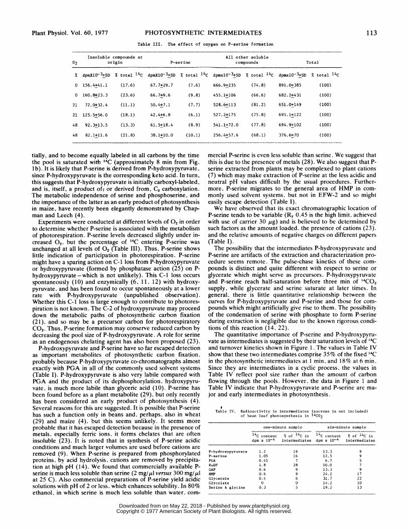
Table III. The effect of oxygen on P-serine formation
Insoluble compounds at All other soluble02 origin P-serine compounds Total
% dpmXlO-3+SD % total 14C dpmXlO-3+SD % total 14C dpmxlO03+SD % total 14C dpmxlO-3+SD % total 14C
0 156.4+41.1 (17.6) 67.7+29.7 (7.6) 666.9+235 (74.8) 891.0+385 (100)
0 160.8+23.3 (23.6) 66.7+9.6 (9.8) 455.1+106 (66.6) 682.2+431 (100)
21 72.0+32.4 (11.1) 50.4+7.1 (7.7) 528.6+113 (81.2) 651.0+149 (100)
21 125.5+56.0 (18.1) 42.4+6.8 (6.1) 527.2+175 (75.8) 695.1+122 (100)
48 92.3+13.5 (13.3) 61.5+18.4 (8.9) 541.1+72.0 (77.8) 694.9+102 (100)
48 82.1+11.6 (21.8) 38.1+10.0 (10.1) 256.4+57.6 (68.1) 376.6+70 (100)
tially, and to become equally labeled in all carbons by the timethe pool is saturated with 14C (approximately 8 min from Fig.lb). It is likely that P-serine is derived from P-hydroxypyruvate,since P-hydroxypyruvate is the corresponding keto acid. In turn,this suggests that P-hydroxypyruvate is initially carboxyl-labeled,and is, itself, a product of; or derived from, C3 carboxylation.The metabolic independence of serine and phosphoserine, andthe importance of the latter as an early product of photosynthesisin maize, have recently been elegantly demonstrated by Chap-man and Leech (4).
Experiments were conducted at different levels of 02 in orderto determine whether P-serine is associated with the metabolismof photorespiration. P-serine levels decreased slightly under in-creased 02. but the percentage of 14C entering P-serine was
unchanged at all levels of 02 (Table III). Thus. P-serine showslittle indication of participation in photorespiration. P-serinemight have a sparing action on C-I loss from P-hydroxypyruvateor hydroxypyruvate (formed by phosphatase action (25) on P-hydroxypyruvate - which is not unlikely). This C-I loss occurs
spontaneously (10) and enzymically (6. 11. 12) with hydroxy-pyruvate, and has been found to occur spontaneously at a lowerrate with P-hydroxypyruvate (unpublished observation).Whether this C-1 loss is large enough to contribute to photores-piration is not known. The C-2 of hydroxypyruvate may proceeddown the metabolic paths of photosynthetic carbon fixation(21). and so may be a precursor carbon for photorespiratoryCO2. Thus. P-serine formation may conserve reduced carbon bydecreasing the pool size of P-hydroxypyruvate. A role for serineas an endogenous chelating agent has also been proposed (23).
P-hydroxypyruvate and P-serine have so far escaped detectionas important metabolites of photosynthetic carbon fixation.probably because P-hydroxypyruvate co-chromatographs almostexactly with PGA in all of the commonly used solvent systems(Table I). P-hydroxypyruvate is also very labile compared withPGA and the product of its dephosphorylation. hydroxypyru-vate. is much more labile than glyceric acid (10). P-serine hasbeen found before as a plant metabolite (29). but only recentlyhas been considered an early product of photosynthesis (4).Several reasons for this are suggested. It is possible that P-serinehas such a function only in beans and. perhaps. also in wheat(29) and maize (4). but this seems unlikely. It seems more
probable that it has escaped detection because in the presence ofmetals. especially ferric ions, it forms chelates that are ofteninsoluble (23). It is noted that in synthesis of P-serine acidicconditions and much larger volumes are used before cations are
removed (9). When P-serine is prepared from phosphorylatedproteins, by acid hydrolysis. cations are removed by precipita-tion at high pH (14). We found that commercially available P-serine is much less soluble than serine (2 mg/,ul versus 300 mg/,ulat 25 C). Also commercial preparations of P-serine yield acidicsolutions with pH of 2 or less. which enhances solubility. In 80%ethanol, in which serine is much less soluble than water. com-
mercial P-serine is even less soluble than serine. We suggest thatthis is due to the presence of metals (28). We also suggest that P-serine extracted from plants may be complexed to plant cations(7) which may make extraction of P-serine at the less acidic andneutral pH values difficult by the usual procedures. Further-more, P-serine migrates to the general area of HMP in com-monly used solvent systems. but not in EFW-2 and so mighteasily escape detection (Table I).We have observed that its exact chromatographic location of
P-serine tends to be variable (RF 0.45 is the high limit, achievedwith use of carrier 30 ,g) and is believed to be determined bysuch factors as the amount loaded, the presence of cations (23).and the relative amounts of negative charges on different papers(Table I).The possibility that the intermediates P-hydroxypyruvate and
P-serine are artifacts of the extraction and characterization pro-cedure seems remote. The pulse-chase kinetics of these com-pounds is distinct and quite different with respect to serine orglycerate which might serve as precursors. P-hydroxypyruvateand P-serine reach half-saturation before three min of '4CO2supply, while glycerate and serine saturate at later times. Ingeneral. there is little quantitative relationship between thecurves for P-hydroxypyruvate and P-serine and those for com-pounds which might artificially give rise to them. The possibilityof the condensation of serine with phosphate to form P-serineduring extraction is negligible due to the known rigorous condi-tions of this reaction (14. 22).The quantitative importance of P-serine and P-hydroxypyru-
vate as intermediates is suggested by their saturation levels of 14Cand turnover kinetics shown in Figure 1. The values in Table IVshow that these two intermediates comprise 35% of the fixed '4Cin the photosynthetic intermediates at 1 min. and 18% at 6 min.Since they are intermediates in a cyclic process. the values inTable IV reflect pool size rather than the amount of carbonflowing through the pools. However. the data in Figure 1 andTable IV indicate that P-hydroxypyruvate and P-serine are ma-jor and early intermediates in photosynthesis.
Table IV. Radioactivity in intermediates (sucrose is not included)of bean leaf photosynthesis in 14C02
one-minute sample six-minute sample
14C content % of 14C in 14C content % of 14C indpm x 10-4 intermediates dpm x 10-4 intermediates
P-hydroxypyruvate 1.2 19 13.3 9P-serine 1.05 16 12.5 9PGA 0.45 7 6.7 5RuDP 1.8 28 10.0 7GAP 0.6 9 13.3 9HMP 0.6 9 24.2 17Glycerate 0.4 6 31.7 22Glycolate 0 0 14.2 10Serine & glycine 0.3 5 19.2 13
Plant Physiol. Vol. 60, 1977 113
www.plantphysiol.orgon May 22, 2018 - Published by Downloaded from Copyright © 1977 American Society of Plant Biologists. All rights reserved.
DALEY AND BIDWELL
Figure 1 indicates that P-hydroxypyruvate may be formedsimultaneously with or independently from PGA, rather thanbeing derived from it. The data in Figure 3 suggest that P-serine(and, presumably, its keto acid P-hydroxypyruvate) are formedas a result of a carboxylation reaction. This is in accord with theearlier proposal that P-hydroxypyruvate arises as the result of acarboxylation of RuDP (5).A substantial amount of photorespiratory CO2 might arise
from the spontaneous or enzymic decarboxylation of P-hydroxy-pyruvate or hydroxypyruvate. While the data presented here areconsistent with such a hypothesis, the proof must await addi-tional evidence. However, these data strongly support an impor-tant role of P-hydroxypyruvate and P-seine in the early metabo-lism of carbon fixation in the primary leaf of the bean.
Acknowledgments -The authors are grateful to G. Couvillon and G. Ware for assistancewith the statistical analyses, and to H. M. Vines for technical advice and support at the Universityof Georgia, Athens.
LITERATURE CITED
1. BENSON AA, JA BASSHAM, M CALVIN, TC GOODALE, VA HAAS, W STEPICA 1950 The pathof carbon in photosynthesis. V. J. Am Chem Soc 72: 1710-1718
2. BIDWELL RGS 1962 Direct paper chromatography of soluble compounds in small samples oftissue adhering to paper. Can J Biochem Physiol 40: 757-761
3. CALVIN M 1962 The path of carbon in photosynthesis. Science 135: 879-8894. CHAPMAN DJ, RM LEECH 1976 Photoserine as an early product of photosynthesis in
isolated chloroplasts and leaves of Zea mays seedlings. FEBS Lett 68: 160-1645. DALuY L 1975 Light activation of ribulose diphosphate carboxylase: conditions and prod-
ucts of the reaction. PhD dissertation. Univ Calif, Davis6. DE LA HABA G, IG LEDNER, E RACKER 1955 Crystalline transketolase from bakers' yeast:
Isolation and properties. J Biol Chem 214: 409-4267. EPSTEIN E 1972 Mineral Nutrition of Plants: Principles and Perspectives. John Wiley &
Sons, New York p 638. FRASER DE, RGS BIDWELL 1974 Photosynthesis during the ontogeny of the bean plant. Can
J Bot 52: 2561-25709. FoLSCH G, 0 MELLANDER 1957 0-phosphoryl serine. Acta Chem Scand 11: 1232-1239
10. HEDRICK JL, HJ SALLACH 1961 The metabolism of hydroxypyruvate. I. The nonenzymicdecarboxylation and autoxidation of hydroxypyruvate. J Biol Chem 236: 1867-1871
11. HEDRICK JL, HJ SALLACH 1961 The metabolism of hydroxypyruvate. II. The enzymatic
oxidation and decarboxylation of hydroxypyruvate. J Biol Chem 236: 1872-187512. HEDRCK JL, HJ SALLACH 1961 The nonoxidative decarboxylation of hydroxypyruvate in
mammalian systems. Arch Biochem Biophys 105: 261-26913. HELLEDUSr JA, RGS BIDWELL 1963 Protein turnover in wheat and snapdragon leaves. Can
J Bot 41: 179-18214. KENNEDY EP, SW SmrsH 1954 The isolation of radioactive phosphoserine from "phospho-
protein" of Ehrlich ascites tumor. J Biol Chem 207: 153-16315. KORTSCHAK HP, CE HARrM, GO Buas 1965 Carbon dioxide fixation in sugarcane leaves.
Plant Physiol 40: 209-21316. LABER UJ, E LATZKO, M GiBBs 1974 Photosynthetic path of carbon dioxide in spinach and
com leaves. J Biol Chem 249: 3436-344117. LANGENBECK U, H-U M6HRING, K-P DLECD(ANN 1975 Gas chromatography of a-keto
acids as their o-trimethylsilylquinoxalinol derivatives. J Chromatogr 15: 65-7018. LEHMAN G, H TEICHMANN 1972 In G Hilgetag, A Martini, eds, Preparative Organic
Chemistry. John Wiley & Sons, New York pp 508-51319. MAHON JD, H FOCK, DT CANVTN 1974 Changes in specific radioactivities of sunflower leaf
metabolites during photosynthesis in "4CO2 and '2C02 at normal and low oxygen. Planta120: 125-134
20. MERcK E 1971 Dyeing Reagents for Thin Layer and Paper Chromatography. E Merck,Darmstadt Germany
21. MILHAUD G JR, AA BENSON, M CALVIN 1956 Metabolism of pyruvic acid-2-C'4 andhydroxypyruvic acid-2-C14 in algae J Biol Chem 218: 599-606
22. NEUHAUS FC, S KoKES 1958 Phosphoserine In CS Vesting, ed, Biochemical PreparationsVol 6. John Wiley & Sons, New York pp 75-79
23. OSTERBERG R 1957 Metal and hydrogen-ion binding properties of 0-phosphoserine. Nature179: 176-77
24. RABSON R, NE TOLBERT, PC KEARNEY 1962 Formation of serine and glyceric acid by theglycolate pathway. Arch Biochem Biophys 98: 154-163
25. RANDALL DD, NE TOLBERT, D GREMEL 1971 3-Phosphoglycerate phosphatase in plants.II. Distribution, physiological considerations, and comparison with P-glycolate phospha-tase. Plant Physiol 48: 480-487
26. SIEGEL MI, MD LANE 1973 Chemical and enzymatic evidence for the participation of a 2-carboxy-3-ketoribitol-1,5-diphosphate intermediate in the carboxylation of ribulose 1,5-diphosphate. J Biol Chem 248: 5486-5498
27. TAMAS IA, RGS BIDWELL 1970 "4CO2 fixation in leaf discs of Phaseolus vulgaris. Can J Bot48: 1259-1263
28. WALBOT V 1977 Heavy metal impurities impair the spectrophotometric assay of ribulosebisphosphate carboxylase activity. Plant Physiol 59: 107-110
29. WANG D, RH BumIs 1963 Carbon metabolism of '4C-labeled amino acids in wheat leaves.11. Serine and its role in glycine metabolism. Plant Physiol 38: 430-439
30. WOOD T 1961 A procedure for the analysis of acid soluble phosphorous compounds andrelated substances in muscle and other tissues. J Chromatogr 6: 142-151
31. WOOD T 1968 The detection and identification of intermediates of the pentose phosphatecycle and related compounds. J Chromatogr 35: 352-361
114 Plant Physiol. Vol. 60, 1977
www.plantphysiol.orgon May 22, 2018 - Published by Downloaded from Copyright © 1977 American Society of Plant Biologists. All rights reserved.