photo-induced heterodisulfide metathesis for reagent-free ... · photo-induced heterodisulfide...
TRANSCRIPT
S1
Supporting Information for:
Photo-induced Heterodisulfide Metathesis for Reagent-free
Synthesis of Polymer Nanoparticles
Longyu Li, Cunfeng Song, Matthew Jennings and S. Thayumanavan*
Department of Chemistry, University of Massachusetts, 710 N. Pleasant Street, Amherst, MA
01003-9336, USA
* Corresponding author: [email protected]
Fig. S1 GPC traces for the polymers
Fig. S2 NMR Spectra of the polymers
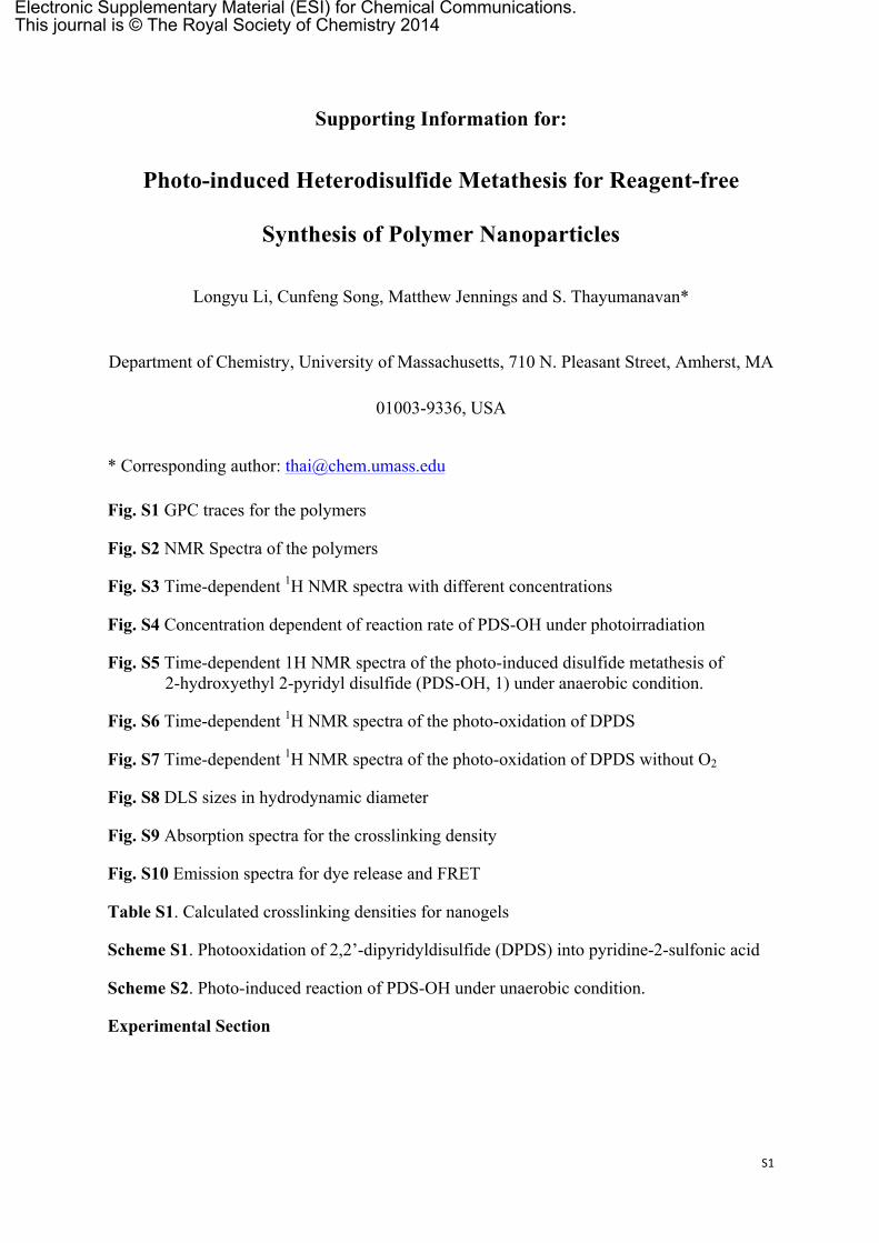
Fig. S3 Time-dependent 1H NMR spectra with different concentrations
Fig. S4 Concentration dependent of reaction rate of PDS-OH under photoirradiation
Fig. S5 Time-dependent 1H NMR spectra of the photo-induced disulfide metathesis of 2-hydroxyethyl 2-pyridyl disulfide (PDS-OH, 1) under anaerobic condition.
Fig. S6 Time-dependent 1H NMR spectra of the photo-oxidation of DPDS
Fig. S7 Time-dependent 1H NMR spectra of the photo-oxidation of DPDS without O2
Fig. S8 DLS sizes in hydrodynamic diameter
Fig. S9 Absorption spectra for the crosslinking density
Fig. S10 Emission spectra for dye release and FRET
Table S1. Calculated crosslinking densities for nanogels
Scheme S1. Photooxidation of 2,2’-dipyridyldisulfide (DPDS) into pyridine-2-sulfonic acid
Scheme S2. Photo-induced reaction of PDS-OH under unaerobic condition.
Experimental Section
Electronic Supplementary Material (ESI) for Chemical Communications.This journal is © The Royal Society of Chemistry 2014

S2
469 969 1469Time/sec
Mn=9746Mw=11685PDI=1.20
Fig. S1 Evolution of GPC traces for polymer
Fig. S2 NMR Spectra of the polymers
S3

S4

S5
Fig. S3 Time-dependent 1H NMR spectra of PDS and DPDS with different concentrations.

S6
Fig. S4 Concentration dependent of reaction rate of PDS under photoirradiation.
Fig. S5 Time-dependent 1H NMR spectra of the photo-induced disulfide metathesis of 2-hydroxyethyl 2-pyridyl disulfide (PDS-OH, 1) under anaerobic condition. Control spectra of bis(2-hydroxyethyl) disulfide (2), 2, 2’-dipyridyl disulfide (DPDS, 3) and 2-thiopyridone were also included. The concentration was 2.5 mg/mL.
Fig. S6 Time-dependent 1H NMR spectra of the photooxidation of 2, 2’-dipyridyl disulfide (DPDS).

S7
Control spectrum of 2-thiopyridone was also included.
Fig. S7 Time-dependent 1H NMR spectra of DPDS under the photoirradiation without O2
Fig. S8 DLS sizes in hydrodynamic diameter, a) micelle aggregates prepared with polymer solutions with different concentrations, b) nanogels prepared with polymer solutions with different concentrations, the photoirradiation time was 2 h for all samples.

S8
300 350 400 450 5000
1
Abs
Wavelength (nm)
1mg-0 min 1mg-DTT-0 min 1mg-1 h 1mg-DTT-1 h 1mg-2 h 1mg-DTT-2 h 1mg-3 h 1mg-DTT-3 h 1mg-1-4 h 1mg-DTT-4 h
300 350 400 450 5000
1
Abs
Wavelength (nm)
2mg-0 min 2mg-DTT-0 min 2mg-1 h 2mg-DTT-1 h 2mg-2 h 2mg-DTT-2 h 2mg-3 h 2mg-DTT-3 h 2mg-4 h 2mg-DTT-4 h
300 350 400 450 5000
1
Abs
Wavelength (nm)
5mg-0 min 5mg-DTT-0 min 5mg-1 h 5mg-DTT-1 h 5mg-2 h 5mg-DTT-2 h 5mg-3 h 5mg-DTT-3 h 5mg-4 h 5mg-DTT-4 h
300 350 400 450 500
0
1A
bs
Wavelength (nm)
10mg-0 min 10mg-DTT-0 min 10mg-1 h 10mg-DTT-1 h 10mg-2 h 10mg-DTT-2 h 10mg-3 h 10mg-DTT-3 h 10mg-4 h 10mg-DTT-4 h
Fig. S9 Absorption spectra for the crosslinking density
Fig. S10 Dye release from the nanogels prepared via photoirradiation for 2 h in response to varied GSH concentrations, a) 0 mM, b) 10 mM. Sample concentration was 0.1 mg/mL. FRET experiment based DiI/DiO exchange, c) micelle aggregates, d) nanogels prepared via photoirradiation for 1 h, e) nanogels prepared via photoirradiation for 2 h. Sample concentration was 0.1 mg/mL.

S9
Table S1. Calculated crosslinking densities for nanogels
Crosslinking density/% concentration 1 h 2 h 3 h 4 h
1 mg/mL 16 46 76 91 2 mg/mL 13 33 52 69 5 mg/mL 10 29 36 48
10 mg/mL 7 23 30 38
Scheme S1. Photooxidation of 2,2’-dipyridyldisulfide (DPDS) into pyridine-2-sulfonic acid.
Scheme S2. Photo-induced reaction of PDS-OH under unaerobic condition.

S10
Experimental Section General Methods.
2,2-Dithiodipyridine, 2-mercaptoethanol, polyethyleneglycol monomethylether methacrylate (PEGMA, MW 450), D,L-dithiothreitol (DTT), 1,1'-dioctadecyl-3,3,3',3'-tetramethylindocarbocyanine perchlorate (DiI) and 3,3'-dioctadecyloxacarbocyanine perchlorate (DiO), 4-Cyano-4-(phenylcarbonothioylthio)-pentanoic acid and other conventional reagents were obtained from commercial sources and were used as received without further purification. The 2,2’-azobisisobutyronitrile (AIBN) was purified by recrystallization from ethanol. 1H-NMR spectra were recorded on a 400 MHz Bruker NMR spectrometer using the residual proton resonance of the solvent as the internal standard. Chemical shifts are reported in parts per million (ppm). Molecular weights of the polymers were estimated by gel permeation chromatography (GPC) using PMMA standard with a refractive index detector. Dynamic light scattering (DLS) measurements were performed using a Malvern Nanozetasizer. UV-visible absorption spectra were recorded on a Varian (model EL 01125047) spectrophotometer. The fluorescence spectra were obtained from a JASCO FP-6500 spectrofluorimeter.
Synthesis of 2-hydroxyethyl 2-pyridyl disulfide (PDS-OH) and 2-thiopyridone
PDS-OH was synthesized according to the previous report.[1] 2, 2’-dipyridyl disulfide (DPDS) (10.3 g, 0.047 mol) was dissolved in 100 mL of methanol. Then 1.2 mL of glacial acetic acid, used as catalyst, was added. To this mixture, a solution of mercaptoethanol (4.4 g, 0.056 mol, 4 mL) in 100 mL methanol was added drop-wise at room temperature with continuous stirring. Once the addition was over, the reaction mixture was stirred at room temperature overnight. The stirring was stopped; solvent was evaporated to get the crude product as yellow oil which was purified by flash column chromatography using silica gel as stationary phase and mixture of ethyl acetate/hexane as eluent. When the polarity of the eluent was increased to 40% ethyl acetate/hexane, the desired product came out as colorless oil. Yield : 80 % 1H NMR: (CD3OD, 400 MHz), δ (ppm): 8.40 (m, 1H, aromatic proton ortho-N), 7.84 (m, 1H, aromatic proton meta-N), 7.80 (m, 1H, aromatic proton para-N), 7.22 (m, 1H, aromatic proton, ortho-disulfide linkage), 3.77(t, 2H, -CH2OH), 2.94 (t, 2H, -SS-CH2-). 13C-NMR (CD3OD, 100MHz) δ(ppm): 160.01, 148.93, 137.69, 121.03, 120.11, 59.19, 41.20.
2-thiopyridone can also be prepared at the same time by continually increasing the polarity of the eluent up to 55% ethyl acetate/hexane to get the desired product. After concentrating the solution, light yellow Needle-shaped crystals were got. 1H NMR: (CD3OD, 400 MHz), δ (ppm): 7.67 (m, 1H), 7.47 (m, 2H), 6.85 (m, 1H). 13C-NMR (CD3OD, 100MHz ) δ(ppm): 177.40, 138.00, 137.25, 133.08, 113.57.
Synthesis of PDS monomer

S11
PDS monomer was also synthesized according to the previous report.[1] To a solution of PDS-OH (12 g, 64.1 mmol) in 50 mL of dry dichloromethane was added 7.8 g (76.9 mmol) of triethylamine. The mixture was firstly cooled in an ice-bath. To this cold mixture, a solution of methacryloyl chloride (6.7 g, 64.1 mmol) in 25 mL dichloromethane was then added drop-wise with continuous stirring. After the addition was over the reaction mixture was stirred at room temperature overnight. The stirring was stopped and the reaction mixture was washed with 3x100 mL distilled water and then with 100 mL of brine. The organic layer was collected, dried over anhydrous Na2SO4 and concentrated to get the crude product as yellow oil. It was purified by column chromatography using silica gel as stationary phase and mixture of ethyl acetate/hexane as eluent. The pure product was collected at 30 % ethylacetate /hexane. Yield: 80 % 1H NMR: (CDCl3, 400 MHz), δ (ppm): 8.52 (m, 1H, aromatic proton ortho-N), 7.62-7.83 (m, 2H, aromatic proton meta-N and para-N), 7.15 (m, 1H, aromatic proton, orthodisulfide linkage), 6.23 (d, 1H, vinylic proton, cis-ester), 5.65(d, 1H, vinylic proton, trans-ester) 4.41 (t, 2H, -S-S-CH2CH2O-), 3.13 (t, 2H, -S-S-CH2CH2O-), 1.91(s, 3H, methyl proton of the methacryloyl group). 13C-NMR (CD3OD, 100MHz ) δ(ppm): 167.06, 159.76, 149.73, 137.05, 135.96, , 126.05, 120.85, 119.77, 62.41, 37.44, 18.28.
Synthesis of random copolymer containing PDS groups
Random copolymers were prepared by reversible addition−fragmentation chain transfer (RAFT) polymerization.[2] A mixture of 4-cyano-4-(phenylcarbonothioylthio)pentanoic acid (24 mg, 0.086 mmol), poly(ethylene glycol) monomethyl ether methacrylate (0.4 g, 0.84 mmol), PDS (0.537 g, 2.1 mmol), and 2,2′-azobis(isobutyronitrile) (AIBN; 2.8 mg, 0.017 mmol) was dissolved in tetrahydrofuran (THF) (2 mL) and degassed by performing three freeze−pump−thaw cycles. The reaction mixture was sealed and then transferred into a preheated oil bath at 65 °C for 10 h. To remove unreactive monomers, the resultant mixture was precipitated in cold ethyl ether (20 mL) to yield the random copolymer as a waxy liquid. GPC (THF) Mw, 12 kDa; polydispersity, 1.2. 1H NMR (400 MHz, CDCl3) δ 8.46, 7.68, 7.11, 4.35−4.09, 3.94−3.37, 3.03, 2.04−1.64, 1.43−0.87. The molar ratio between two blocks was determined by integrating the methoxy proton in the poly-(ethylene glycol) unit and the aromatic proton in the pyridine and was found to be 1:2.70 (PEO:PDS).
Time-dependent 1H NMR measurement
In 5mm NMR tubes, 1 mL of samples with different concentrations was added. 1H-NMR spectra were recorded on a 400 MHz Bruker NMR spectrometer using the residual proton resonance of the solvent as the internal standard. These tubes were put inside UV chamber (SYLVANIA F15T8/350/BL, 15w). After each 1 hour’s photoirradiation, another NMR spectrum for each sample was taken with the same parameters. The integrations of peak at 2.94 ppm and peak at 2.83 ppm were used to determine the percent of remaining PDS-OH.
Nanoparticles preparation

S12
1 mL of polymer solution of different concentrations was added to a 7 mL vial. These vials were then put inside an UV chamber (SYLVANIA F15T8/350/BL, 15w). After photoirradiation for certain times, nanoparticles with different sizes were formed in solutions. The crosslinking density could be determined by taking some volume of polymer solution from the above solutions into cuvettes for UV-vis measurement. The concentration of these samples was all 0.1 mg/mL. Absorption spectra were taken first. Then, excess DTT were added into these samples and then the solution was observed using absorption spectroscopy again after 3 hours, during which all PDS functionalities could be changed to 2-thiopyridine by DTT.
Determination of crosslinking density
In 7mL small glass vial, 1 mL of polymer solution with different concentrations was added. From them, certain volume of polymer solution was taken and then added into cuvettes for UV-vis measurement. The volume was made to 1 mL by adding water. The concentration of these samples was all 0.1 mg/mL. Absorption spectra were taken. Excess DTT were added into these samples, one more absorption spectra were taken after 3 hours, during which all PDS functionalities could be changed to 2-thiopyridine by DTT.
Then these vials were put inside UV chamber (SYLVANIA F15T8/350/BL, 15w). After each 1 hour’s photoirradiation, certain volume of polymer solution was taken from the above solutions and then added into cuvettes for UV-vis measurement. The volume was made to 1 mL by adding water. The concentration of these samples was all 0.1 mg/mL. Absorption spectra were taken. Excess DTT were added into these samples, one more absorption spectra were taken after 3 hours, during which all PDS functionalities could be changed to 2-thiopyridine by DTT.
Preparation of nanogels contaning DiI/DiO
In 7mL small glass vial, polymer was dissolved in 1 mL water. Then calculated volume of DiI in acetone stock solution (5 mg/mL) or DiO in acetone stock solution (2.5 mg/mL) was added into the polymer solution. The final dye concentration was about 2 wt%. The mixed solution was stirred overnight at room temperature, open to the atmosphere allowing the organic solvent to evaporate. Then these vials were put inside UV chamber (SYLVANIA F15T8/350/BL, 15w). After 2 hour’s photoirradiation, insoluble DiI/DiO was removed by filtration and the byproduct was removed from the nanogel solution by extensive dialysis using a membrane with a molecular weight cut-off of 7,000 g/mol. Finally, nanogels stock solutions with concentration of 1 mg/mL were prepared for further studies. Redox-responsive guest release experiment
A solution of nanogel containing DiI (100 µL) was mixed with water (800 µL) in a cuvette, and then GSH stock solution (100 µL) was added. The fluorescence spectra were recorded using the excitation wavelength of 530 nm. For control experiment, A solution of nanogel containing DiI (100 µL) was mixed with water (900 µL) in a cuvette. The fluorescence spectra were recorded using the excitation wavelength of 530 nm. FRET experiment

S13
A solution of nanogel containing DiI (100 µL) was mixed with a solution of nanogel containing DiO (100 µL) in a cuvette, and then milliQ water (800 µL) was added to adjust the volume. The fluorescence spectra were recorded using the excitation wavelength of 450 nm. Reference [1] S. Ghosh, S. Basu, S. Thayumanavan, Macromolecules 2006, 39, 5595-5597.
[2] L. Li, J.-H. Ryu, S. Thayumanavan, Langmuir 2013, 29, 50-55.