photoluminescence of silicon-vacancy defects in ... general physics institute ras, vavilov street...
TRANSCRIPT
Photoluminescence of silicon-vacancy defects in nanodiamonds of
different chondrites
A. A. SHIRYAEV1, 2, #
, A. V. FISENKO3, L. F. SEMJONOVA
3, A. A. KHOMICH
4,
and I. I. VLASOV4,5
1 Institute of Physical Chemistry and Electrochemistry RAS, Leninsky pr. 31, korp. 4,
119071, Moscow, Russia
2 Institute of Ore Deposits, Petrography, Geochemistry and Mineralogy RAS, Staromonetny
per. 35, 119071, Moscow, Russia
3 Vernadsky Institute of Geochemistry and Analytical Chemistry RAS, Kosygin Street 19,
Moscow, Russia
4 General Physics Institute RAS, Vavilov Street 38, 119991 Moscow, Russia
5 National Research Nuclear University MEPhI, Kashirskoe Avenue 31, Moscow 115409,
Russia
# Corresponding author email: [email protected] AND [email protected]

Abstract
Photoluminescence spectra show that silicon impurity is present in lattice of some
nanodiamond grains (ND) of various chondrites as a silicon-vacancy (SiV) defect. The
relative intensity of the SiV band in the diamond-rich separates depends on chemical
composition of meteorites and on size of ND grains. The strongest signal is found for the size
separates enriched in small grains; thus confirming our earlier conclusion that the SiV defects
preferentially reside in the smallest (≤ 2 nm) grains. The difference in relative intensities of
the SiV luminescence in the diamond-rich separates of individual meteorites are due to
variable conditions of thermal metamorphism of their parent bodies and/or uneven sampling
of nanodiamonds populations. Annealing of separates in air eliminates surface sp2-carbon,
consequently, the SiV luminescence is enhanced. Strong and well-defined luminescence and
absorption of the SiV defect is a promising feature to locate cold (< 250 °C) nanodiamonds in
space.
INTRODUCTION
Nanodiamonds present in meteorites (meteoritic nanodiamonds – MND) remain an
enigmatic substance. Their matrix normalized abundance may reach 2000 ppm (Huss and
Lewis, 1995). Since their discovery (Lewis et al., 1987) the formation process(es) and
astrophysical source(s) of MND remain highly debatable despite significant efforts (see
extensive review by Daulton (2005)). Whereas isotopic composition of noble gases and, in
particular, of Xe indicates that some of nanodiamonds might be related to supernovae
explosions (e.g., Lewis et al., 1987, Jorgenson 1988; Ott et al. 2012), isotopic compositions of
bulk carbon and of the principal chemical impurity – nitrogen – in the diamond-rich separates
are less conclusive and may support hypothesis of that at least a fraction of MND was formed
in the Solar system (Dai et al., 2002). Processes of MND formation are also debatable, but
combined analysis of information about structure and chemical impurities (Shiryaev et al.,
2011) suggests that the growth process of (at least) N-containing grains should be very fast.
The CVD-like (Chemical Vapour Deposition) process (Daulton et al., 1996), possibly
triggered by a shock wave(s) (Shiryaev et al., 2011), looks plausible, but other processes
cannot be excluded (e.g., Blake et al., 1987; Byakov et al., 1990; Duley and Grishko, 2001;
Kouchi et al., 2005; Stroud et al., 2011).
Analysis of isotopic composition of different elements is indispensable for
identification of nanodiamonds source(s). Eventual detection of nanodiamonds in
astrophysical spectra would provide additional information. Detailed knowledge of
spectroscopic properties of meteoritic nanodiamonds is thus of considerable importance.
Attempts to observe nanodiamond features in astrophysical spectra are rather numerous, but
very few of them can be (relatively) unambiguously assigned to diamonds. The most reliable
observations are those by Guillois et al. (1999) and van Kerkhoven et al. (2002) who reported
observation of infra-red features at 3.43 and 3.53 microns in spectra of several Herbig Ae/Be
stars. Perfect match of these bands to peculiar configuration of C-H bonds on surfaces of
“large” nanodiamonds (about 50 nm) makes the assignment of the observed bands to heated
nanodiamonds plausible. Subsequent spatially resolved studies (Habart et al., 2004; Goto et
al., 2009) showed that the diamond-related emission originates from the inner region (<15
AU) of the circumstellar dust disk, whereas PAH emission extends towards the outer region.
The observed spatial heterogeneity may partly reflect temperature and UV flux distribution in
the dust disk. However, the “diamond” bands are observed in less than 4% of the studied
Herbing stars (Acke and van den Ancker, 2006). It is interesting to note a discrepancy
between the sizes of tentatively observed nanodiamonds derived from radiative energy budget
(1-10 nm, van Kerkhoven et al. (2002)) and the fact that the required well-resolved IR bands
are observed only when the nanodiamond grains become sufficiently large to possess crystal
faces resembling those of macroscopic diamond. Investigation of diamond grains smaller than
30 nm shows that the corresponding IR features are blurred and shifted (Sheu et al., 2002;
Maturilli et al., 2014). The discrepancy might be resolved if the hydrogen surface coverage is
incomplete in contrast to the assumption in van Kerkhoven et al. (2002). Chang et al. (2006)
ascribed so-called Extended Red Emission – a broad luminescence-related emission from
some astrophysical objects (see Witt, 2013 for review) - to the photoluminescence of
nitrogen-vacancy (NV) complexes in nanodiamond particles with sizes approx. 100 nm or
larger. Note that the NV defects are extremely difficult to detect in diamond grains smaller
than 30-50 nm (e.g., Bradac et al., 2009; Vlasov et al., 2010) and they are not observed in the
nanodiamonds extracted from meteorites (Shiryaev et al., 2011).
If correct, these assignments imply existence of large (tens-hundreds of nanometers)
nanodiamonds in space. In the same time, the meteoritic nanodiamonds are characterized by
considerably smaller sizes (median size ~2.6 nm) as shown independently by TEM (Fraundorf
et al., 1989; Lewis et al., 1989; Daulton et al., 1996) and MALDI (Lyon et al. 2005, Maul et
al., 2005). Recently published work with atom-probe tomography also showed presence of
nanodiamonds in the small size range 2-3 nm (Heck et al. 2014). Spectroscopic properties of
nanodiamonds are strongly size-dependent and up to now no reliable astrophysical
observations of features resembling spectra of nanodiamonds similar to those from meteorites
are known.
Recently we have reported observation of an important point defect – the silicon-
vacancy complex (SiV) – in nanodiamond-rich separates from Efremovka (CV3) and Orgueil
(CI) chondrites (Shiryaev et al., 2011). Subsequent studies demonstrated that the SiV
luminescence appears to be confined to the smallest diamond grains with sizes below 2 nm
(Vlasov et al., 2014). We report here on examination of the SiV defects in diamond-rich
separates extracted from meteorites of various chemical classes and groups. Implications of
these observations for astrospectroscopy are discussed.
SAMPLES AND METHODS
The diamond-rich separates were extracted from Orgueil (CI), Boriskino (CM2),
Efremovka (CV3), Kainsaz (CO3), and Krymka (LL3) chondrites. The extraction was
performed according to the standard protocol involving dissolution of the meteorite piece by
HCl, HCl+HF, KOH, H2O2, K2Cr2O7, HClO4 at temperatures up to 220 °С (Tang et al., 1988).
The separates possess a translucent pale yellow color, typical for meteoritic colloidal
diamond. Colloidal ammonia solutions of the Orgueil, Boriskino and Krymka separates were
further separated into grain size fractions by centrifugation at various accelerations and
duration. Efficiency of the employed separation method is confirmed by the differences in
isotopic composition of C, N and noble gases between the different size fractions (e.g.,
Verchovsky et al., 1998, 2006; Fisenko et al., 2004; Fisenko and Semjonova, 2006).
Photoluminescence (PL) spectra were measured at room temperature for the following
samples of nanodiamond-rich separates: bulk separate (OD7) and fine-grain (OD13) fraction
(supernatant after 13500 g (50 h) centrifugation of the bulk separate) of Orgueil; BD5 and
BD9 - fine- and coarse-grain fractions (supernatant and sediment, respectively) after
centrifugation (105 g, 4h) of Boriskino separate; bulk separate of Kainsaz; DE1 and DE2 –
bulk separates of Efremovka, but the sample DE1 was additionally subjected to heavy liquid
sedimentation (ρ=2 g/cm3), which depleted the specimen in the grains with density below
2 g/cm3; bulk separate (DKr) and coarse grain (DKr4) fraction (sediments after 10
5 g (4h)
centrifugation of the bulk separate) of Krymka.
The PL measurements were performed using a LABRAM HR spectrometer with an
Ar+ laser (488 nm). To avoid eventual heating of the samples by probing laser beam a minute
amount of the sample was mechanically pressed into pure oxygen-free copper foil. This
method allows production of very thin nanodiamond sample. Since its “fluffiness” is greatly
reduced, good thermal contact with thermal sink is achieved. The laser power was limited to 1
mW and even prolonged laser irradiation had virtually no influence on recorded spectra,
justifying the applied procedure. The accuracy of the determination of the positions of the
peaks in the PL spectra was ±0.1 nm. The spectra were corrected for wavelength-dependent
sensitivity of CCD detector. For the bulk and coarse-size (DKr and DKr4) fractions of the
Krymka separates the measurements were also performed in temperature range from 25 to
450 °C in water-cooled TS 1500 Linkam stage in air. The duration of our heating experiment
(30-45 min for the total cycle; the exposure to high T’s was much shorter) was clearly
insufficient for noticeable modification of defects structure of the nanodiamonds.
RESULTS AND DISCUSSION
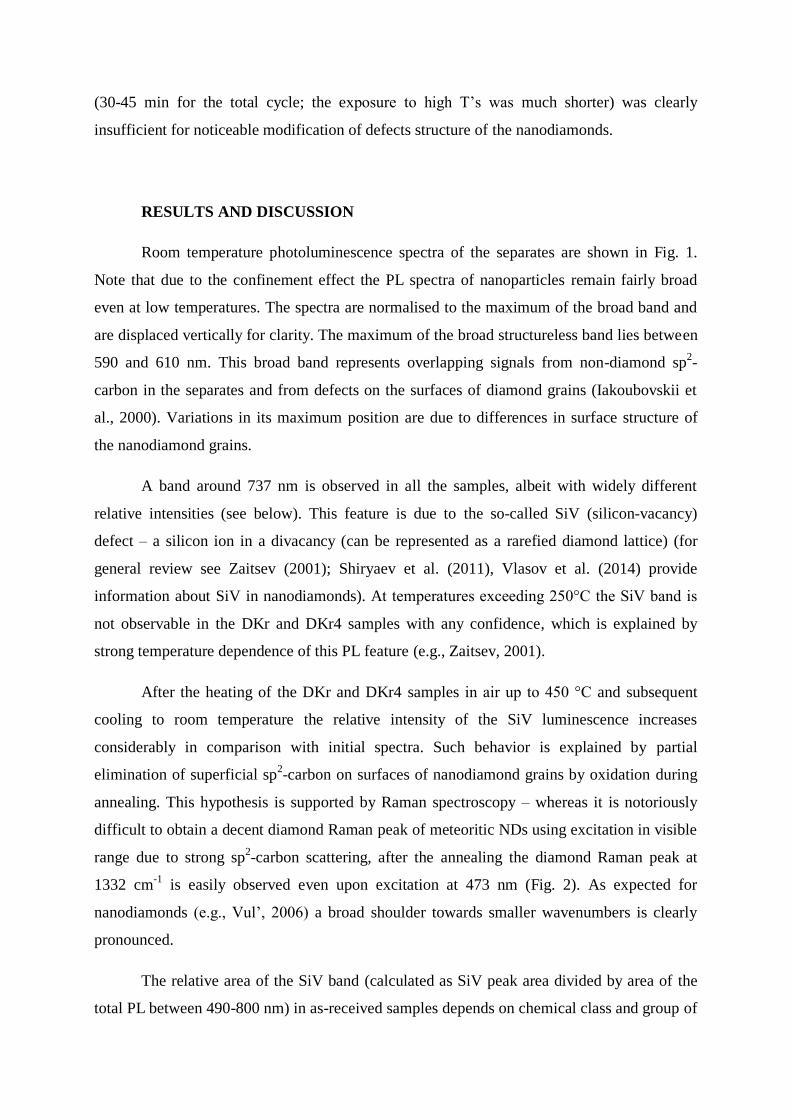
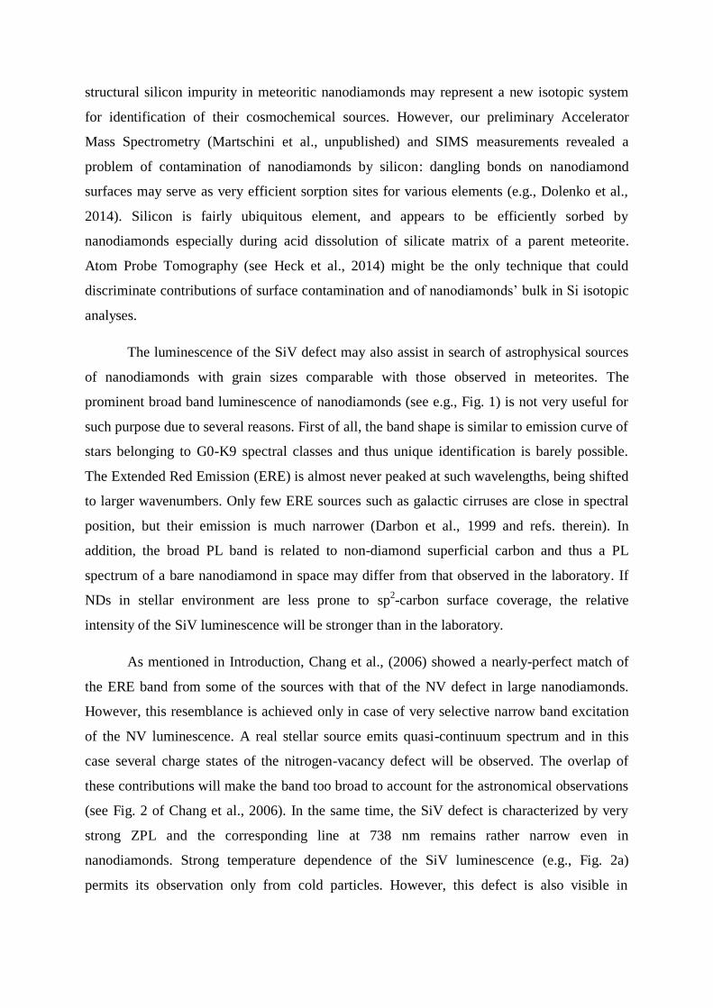
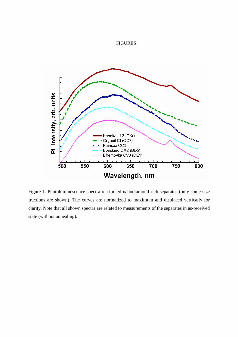
Room temperature photoluminescence spectra of the separates are shown in Fig. 1.
Note that due to the confinement effect the PL spectra of nanoparticles remain fairly broad
even at low temperatures. The spectra are normalised to the maximum of the broad band and
are displaced vertically for clarity. The maximum of the broad structureless band lies between
590 and 610 nm. This broad band represents overlapping signals from non-diamond sp2-
carbon in the separates and from defects on the surfaces of diamond grains (Iakoubovskii et
al., 2000). Variations in its maximum position are due to differences in surface structure of
the nanodiamond grains.
A band around 737 nm is observed in all the samples, albeit with widely different
relative intensities (see below). This feature is due to the so-called SiV (silicon-vacancy)
defect – a silicon ion in a divacancy (can be represented as a rarefied diamond lattice) (for
general review see Zaitsev (2001); Shiryaev et al. (2011), Vlasov et al. (2014) provide
information about SiV in nanodiamonds). At temperatures exceeding 250°C the SiV band is
not observable in the DKr and DKr4 samples with any confidence, which is explained by
strong temperature dependence of this PL feature (e.g., Zaitsev, 2001).
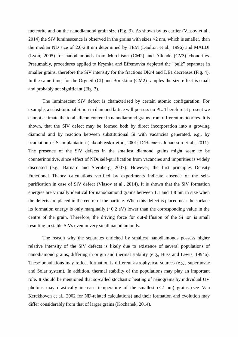
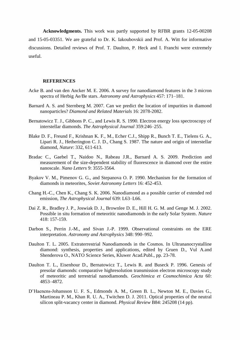
After the heating of the DKr and DKr4 samples in air up to 450 °C and subsequent
cooling to room temperature the relative intensity of the SiV luminescence increases
considerably in comparison with initial spectra. Such behavior is explained by partial
elimination of superficial sp2-carbon on surfaces of nanodiamond grains by oxidation during
annealing. This hypothesis is supported by Raman spectroscopy – whereas it is notoriously
difficult to obtain a decent diamond Raman peak of meteoritic NDs using excitation in visible
range due to strong sp2-carbon scattering, after the annealing the diamond Raman peak at
1332 cm-1
is easily observed even upon excitation at 473 nm (Fig. 2). As expected for
nanodiamonds (e.g., Vul’, 2006) a broad shoulder towards smaller wavenumbers is clearly
pronounced.
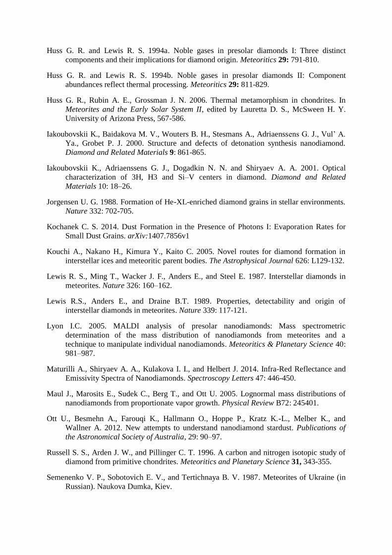
The relative area of the SiV band (calculated as SiV peak area divided by area of the
total PL between 490-800 nm) in as-received samples depends on chemical class and group of
meteorite and on the nanodiamond grain size (Fig. 3). As shown by us earlier (Vlasov et al.,
2014) the SiV luminescence is observed in the grains with sizes ≤2 nm, which is smaller, than
the median ND size of 2.6-2.8 nm determined by TEM (Daulton et al., 1996) and MALDI
(Lyon, 2005) for nanodiamonds from Murchison (CM2) and Allende (CV3) chondrites.
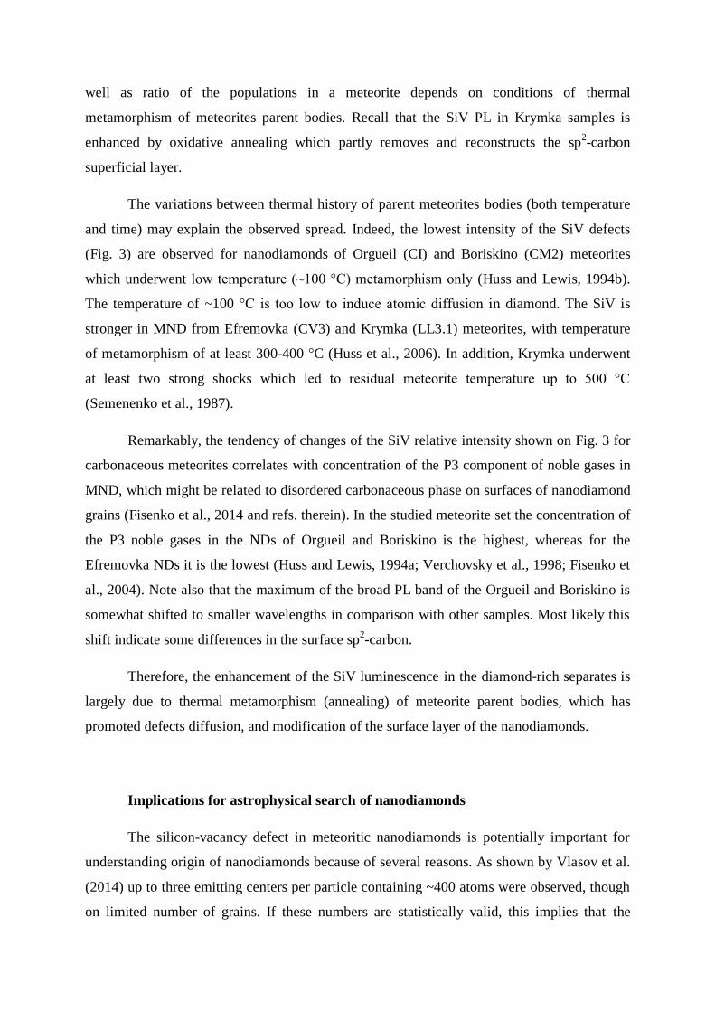
Presumably, procedures applied to Krymka and Efremovka depleted the “bulk” separates in
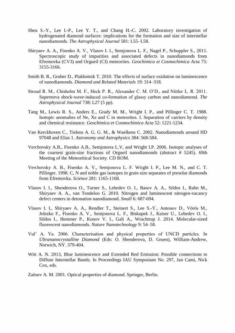
smaller grains, therefore the SiV intensity for the fractions DKr4 and DE1 decreases (Fig. 4).
In the same time, for the Orgueil (CI) and Boriskino (CM2) samples the size effect is small
and probably not significant (Fig. 3).
The luminescent SiV defect is characterised by certain atomic configuration. For
example, a substitutional Si ion in diamond lattice will possess no PL. Therefore at present we
cannot estimate the total silicon content in nanodiamond grains from different meteorites. It is
shown, that the SiV defect may be formed both by direct incorporation into a growing
diamond and by reaction between substitutional Si with vacancies generated, e.g., by
irradiation or Si implantation (Iakoubovskii et al, 2001; D’Haenens-Johansson et al., 2011).
The presence of the SiV defects in the smallest diamond grains might seem to be
counterintuitive, since effect of NDs self-purification from vacancies and impurities is widely
discussed (e.g., Barnard and Sternberg, 2007). However, the first principles Density
Functional Theory calculations verified by experiments indicate absence of the self-
purification in case of SiV defect (Vlasov et al., 2014). It is shown that the SiV formation
energies are virtually identical for nanodiamond grains between 1.1 and 1.8 nm in size when
the defects are placed in the centre of the particle. When this defect is placed near the surface
its formation energy is only marginally (~0.2 eV) lower than the corresponding value in the
centre of the grain. Therefore, the driving force for out-diffusion of the Si ion is small
resulting in stable SiVs even in very small nanodiamonds.
The reason why the separates enriched by smallest nanodiamonds possess higher
relative intensity of the SiV defects is likely due to existence of several populations of
nanodiamond grains, differing in origin and thermal stability (e.g., Huss and Lewis, 1994a).
These populations may reflect formation is different astrophysical sources (e.g., supernovae
and Solar system). In addition, thermal stability of the populations may play an important
role. It should be mentioned that so-called stochastic heating of nanograins by individual UV
photons may drastically increase temperature of the smallest (<2 nm) grains (see Van
Kerckhoven et al., 2002 for ND-related calculations) and their formation and evolution may
differ considerably from that of larger grains (Kochanek, 2014).
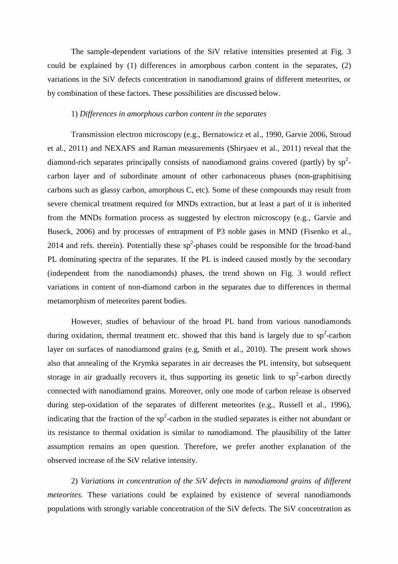
The sample-dependent variations of the SiV relative intensities presented at Fig. 3
could be explained by (1) differences in amorphous carbon content in the separates, (2)
variations in the SiV defects concentration in nanodiamond grains of different meteorites, or
by combination of these factors. These possibilities are discussed below.
1) Differences in amorphous carbon content in the separates
Transmission electron microscopy (e.g., Bernatowicz et al., 1990, Garvie 2006, Stroud
et al., 2011) and NEXAFS and Raman measurements (Shiryaev et al., 2011) reveal that the
diamond-rich separates principally consists of nanodiamond grains covered (partly) by sp2-
carbon layer and of subordinate amount of other carbonaceous phases (non-graphitising
carbons such as glassy carbon, amorphous C, etc). Some of these compounds may result from
severe chemical treatment required for MNDs extraction, but at least a part of it is inherited
from the MNDs formation process as suggested by electron microscopy (e.g., Garvie and
Buseck, 2006) and by processes of entrapment of P3 noble gases in MND (Fisenko et al.,
2014 and refs. therein). Potentially these sp2-phases could be responsible for the broad-band
PL dominating spectra of the separates. If the PL is indeed caused mostly by the secondary
(independent from the nanodiamonds) phases, the trend shown on Fig. 3 would reflect
variations in content of non-diamond carbon in the separates due to differences in thermal
metamorphism of meteorites parent bodies.
However, studies of behaviour of the broad PL band from various nanodiamonds
during oxidation, thermal treatment etc. showed that this band is largely due to sp2-carbon
layer on surfaces of nanodiamond grains (e.g, Smith et al., 2010). The present work shows
also that annealing of the Krymka separates in air decreases the PL intensity, but subsequent
storage in air gradually recovers it, thus supporting its genetic link to sp2-carbon directly
connected with nanodiamond grains. Moreover, only one mode of carbon release is observed
during step-oxidation of the separates of different meteorites (e.g., Russell et al., 1996),
indicating that the fraction of the sp2-carbon in the studied separates is either not abundant or
its resistance to thermal oxidation is similar to nanodiamond. The plausibility of the latter
assumption remains an open question. Therefore, we prefer another explanation of the
observed increase of the SiV relative intensity.
2) Variations in concentration of the SiV defects in nanodiamond grains of different
meteorites. These variations could be explained by existence of several nanodiamonds
populations with strongly variable concentration of the SiV defects. The SiV concentration as
well as ratio of the populations in a meteorite depends on conditions of thermal
metamorphism of meteorites parent bodies. Recall that the SiV PL in Krymka samples is
enhanced by oxidative annealing which partly removes and reconstructs the sp2-carbon
superficial layer.
The variations between thermal history of parent meteorites bodies (both temperature
and time) may explain the observed spread. Indeed, the lowest intensity of the SiV defects
(Fig. 3) are observed for nanodiamonds of Orgueil (CI) and Boriskino (CM2) meteorites
which underwent low temperature (~100 °C) metamorphism only (Huss and Lewis, 1994b).
The temperature of ~100 °C is too low to induce atomic diffusion in diamond. The SiV is
stronger in MND from Efremovka (CV3) and Krymka (LL3.1) meteorites, with temperature
of metamorphism of at least 300-400 °C (Huss et al., 2006). In addition, Krymka underwent
at least two strong shocks which led to residual meteorite temperature up to 500 °C
(Semenenko et al., 1987).
Remarkably, the tendency of changes of the SiV relative intensity shown on Fig. 3 for
carbonaceous meteorites correlates with concentration of the P3 component of noble gases in
MND, which might be related to disordered carbonaceous phase on surfaces of nanodiamond
grains (Fisenko et al., 2014 and refs. therein). In the studied meteorite set the concentration of
the P3 noble gases in the NDs of Orgueil and Boriskino is the highest, whereas for the
Efremovka NDs it is the lowest (Huss and Lewis, 1994a; Verchovsky et al., 1998; Fisenko et
al., 2004). Note also that the maximum of the broad PL band of the Orgueil and Boriskino is
somewhat shifted to smaller wavelengths in comparison with other samples. Most likely this
shift indicate some differences in the surface sp2-carbon.
Therefore, the enhancement of the SiV luminescence in the diamond-rich separates is
largely due to thermal metamorphism (annealing) of meteorite parent bodies, which has
promoted defects diffusion, and modification of the surface layer of the nanodiamonds.
Implications for astrophysical search of nanodiamonds
The silicon-vacancy defect in meteoritic nanodiamonds is potentially important for
understanding origin of nanodiamonds because of several reasons. As shown by Vlasov et al.
(2014) up to three emitting centers per particle containing ~400 atoms were observed, though
on limited number of grains. If these numbers are statistically valid, this implies that the
structural silicon impurity in meteoritic nanodiamonds may represent a new isotopic system
for identification of their cosmochemical sources. However, our preliminary Accelerator
Mass Spectrometry (Martschini et al., unpublished) and SIMS measurements revealed a
problem of contamination of nanodiamonds by silicon: dangling bonds on nanodiamond
surfaces may serve as very efficient sorption sites for various elements (e.g., Dolenko et al.,
2014). Silicon is fairly ubiquitous element, and appears to be efficiently sorbed by
nanodiamonds especially during acid dissolution of silicate matrix of a parent meteorite.
Atom Probe Tomography (see Heck et al., 2014) might be the only technique that could
discriminate contributions of surface contamination and of nanodiamonds’ bulk in Si isotopic
analyses.
The luminescence of the SiV defect may also assist in search of astrophysical sources
of nanodiamonds with grain sizes comparable with those observed in meteorites. The
prominent broad band luminescence of nanodiamonds (see e.g., Fig. 1) is not very useful for
such purpose due to several reasons. First of all, the band shape is similar to emission curve of
stars belonging to G0-K9 spectral classes and thus unique identification is barely possible.
The Extended Red Emission (ERE) is almost never peaked at such wavelengths, being shifted
to larger wavenumbers. Only few ERE sources such as galactic cirruses are close in spectral
position, but their emission is much narrower (Darbon et al., 1999 and refs. therein). In
addition, the broad PL band is related to non-diamond superficial carbon and thus a PL
spectrum of a bare nanodiamond in space may differ from that observed in the laboratory. If
NDs in stellar environment are less prone to sp2-carbon surface coverage, the relative
intensity of the SiV luminescence will be stronger than in the laboratory.
As mentioned in Introduction, Chang et al., (2006) showed a nearly-perfect match of
the ERE band from some of the sources with that of the NV defect in large nanodiamonds.
However, this resemblance is achieved only in case of very selective narrow band excitation
of the NV luminescence. A real stellar source emits quasi-continuum spectrum and in this
case several charge states of the nitrogen-vacancy defect will be observed. The overlap of
these contributions will make the band too broad to account for the astronomical observations
(see Fig. 2 of Chang et al., 2006). In the same time, the SiV defect is characterized by very
strong ZPL and the corresponding line at 738 nm remains rather narrow even in
nanodiamonds. Strong temperature dependence of the SiV luminescence (e.g., Fig. 2a)
permits its observation only from cold particles. However, this defect is also visible in

absorption, thus complementing IR observations of C-H bands pronounced in IR emission
spectra of hot particles (>400-500 °C).
The search for the SiV luminescence from astrophysical sources is not trivial due to
expected weakness of the feature and necessary corrections for sky brightness. One may note
a perfect correspondence of the SiV band position with some of Diffuse Interstellar bands
(DIBs, Galazutdinov et al., 2000). Unfortunately the DIBs are far too narrow to be positively
identified as the SiV luminescence. Bands at similar wavelengths are not uncommon in
spectra of supernovae (e.g. Filippenko, 1997), they are mostly due to Doppler-shifted ions
emission (such as O[I], Ca[I]) and reliable identification of the SiV requires careful analysis
of extended spectral region. A dedicated search for the SiV luminescence and absorption from
astrophysical sources is currently underway.
CONCLUSIONS
A band of the silicon-vacancy (SiV) defect in diamond lattice is observed in
photoluminescence spectra of different grain-size fractions of nanodiamonds (ND) extracted
from chondrites of various groups. At present our statistics includes the following classes and
groups: CI, CM2, CO3, CV3, and LL3. The concentration of silicon in NDs lattice may reach
hundreds of atomic ppm, which makes this element important for identification of
astrophysical sources and formation processes of meteoritic nanodiamonds.
The variations in relative intensities of the SiV luminescence between the
nanodiamond-rich separates extracted from the meteorites of different classes are due to
variations of temperature of thermal metamorphism of their parent bodies and/or uneven
sampling of nanodiamonds populations. The silicon impurity is preferentially incorporated
into the smallest grains (less than 2 nm). The thermal history of parent meteorite bodies is
important for enhancement of the luminescence of trapped Si atoms promoting formation of
specific lattice defect and by modification of nanodiamonds surfaces. The strong and well-
defined luminescence and absorption of the SiV defect is a promising feature to locate cold
nanodiamonds in space.
Acknowledgments. This work was partly supported by RFBR grants 12-05-00208
and 15-05-03351. We are grateful to Dr. K. Iakoubovskii and Prof. A. Witt for informative
discussions. Detailed reviews of Prof. T. Daulton, P. Heck and I. Franchi were extremely
useful.
REFERENCES
Acke B. and van den Ancker M. E. 2006. A survey for nanodiamond features in the 3 micron
spectra of Herbig Ae/Be stars. Astronomy and Astrophysics 457: 171–181.
Barnard A. S. and Sternberg M. 2007. Can we predict the location of impurities in diamond
nanoparticles? Diamond and Related Materials 16: 2078-2082.
Bernatowicz T. J., Gibbons P. C., and Lewis R. S. 1990. Electron energy loss spectroscopy of
interstellar diamonds. The Astrophysical Journal 359:246–255.
Blake D. F., Freund F., Krishnan K. F., M., Echer C.J., Shipp R., Bunch T. E., Tielens G. A.,
Lipari R. J., Hetherington C. J. D., Chang S. 1987. The nature and origin of interstellar
diamond, Nature: 332, 611-613.
Bradac C., Gaebel T., Naidoo N., Rabeau J.R., Barnard A. S. 2009. Prediction and
measurement of the size-dependent stability of fluorescence in diamond over the entire
nanoscale. Nano Letters 9: 3555-3564.
Byakov V. M., Pimenov G. G., and Stepanova O. P. 1990. Mechanism for the formation of
diamonds in meteorites, Soviet Astronomy Letters 16: 452-453.
Chang H.-C., Chen K., Chang S. K. 2006. Nanodiamond as a possible carrier of extended red
emission, The Astrophysical Journal 639: L63–L66.
Dai Z. R., Bradley J. P., Joswiak D. J., Brownlee D. E., Hill H. G. M. and Genge M. J. 2002.
Possible in situ formation of meteoritic nanodiamonds in the early Solar System. Nature
418: 157-159.
Darbon S., Perrin J.-M., and Sivan J.-P. 1999. Observational constraints on the ERE
interpretation. Astronomy and Astrophysics 348: 990–992.
Daulton T. L. 2005. Extraterrestrial Nanodiamonds in the Cosmos. In Ultrananocrystalline
diamond: synthesis, properties and applications, edited by Gruen D., Vul A.and
Shenderova O., NATO Science Series, Kluwer Acad.Publ., pp. 23-78.
Daulton T. L., Eisenhour D., Bernatowicz T., Lewis R. and Buseck P. 1996. Genesis of
presolar diamonds: comparative highresolution transmission electron microscopy study
of meteoritic and terrestrial nanodiamonds. Geochimica et Cosmochimica Acta 60:
4853–4872.
D’Haenens-Johansson U. F. S., Edmonds A. M., Green B. L., Newton M. E., Davies G.,
Martineau P. M., Khan R. U. A., Twitchen D. J. 2011. Optical properties of the neutral
silicon split-vacancy center in diamond. Physical Review B84: 245208 (14 pp).

Dolenko T.A., Burikov S.A., Laptinskiy K.A., Laptinskaya T.V., Rosenholm J.M., Shiryaev
A.A., Sabirov A.R., Vlasov I.I. 2014. Study of adsorption properties of functionalized
nanodiamonds in aqueous solutions of metal salts using optical spectroscopy. Journal of
Alloys and Compounds 586: S436–S439.
Duley W. W. and Grishko V. I. 2001. Evolution of carbon dust in aromatic infrared emission
sources: formation of nanodiamonds. The Astrophysical Journal 554:L209–L212.
Filippenko A. V. 1997. Optical spectra of supernovae. Annual Review of Astronomy and
Astrophysics. 35:309–355.
Fisenko A.V. and Semjonova L.V. 2006. Populations of nanodiamonds in meteorites from
data on isotopic composition and content of nitrogen. Solar System Research 40: 485-
499.
Fisenko A.V., Verchovsky A.B., Semjonova L.V., and Pillinger C.T. 2004. Noble gases in the
grain-size fractions of presolar diamond from the Boriskino CM2 meteorite.
Geochemistry International 42: 408-419.
Fisenko A.V., Verchovsky A.B., and Semjonova L.V. 2014. Kinetics of Xe-P3 release during
pyrolysis of the coarse-grained fractions of Orgueil (CI) meteorite nanodiamonds.
Meteoritics & Planetary Science 49: 611-620.
Fraundorf P., Fraundorf G., Bernatowicz T. J., Lewis R. S., and Ming T. 1989. Stardust in the
TEM. Ultramicroscopy 27:401–411.
Galazutdinov G. A., Musaev F. A., Krełowski, J., Walker, G. A. H. 2000. Narrow Diffuse
Interstellar Bands: a survey with precise wavelengths. The Publications of the
Astronomical Society of the Pacific 112(771): 648-690.
(http://www.sao.ru/hq/coude/DIBwavelength.htm).
Garvie L. A. J. 2006. Surface electronic states of meteoritic nanodiamonds. Meteoritics &
Planetary Science 41: 667–672.
Garvie L. A. J. and Buseck P. R. 2006. Carbonaceous materials in the acid residue from the
Orgueil carbonaceous chondrite meteorite. Meteoritics & Planetary Science 41:633–
642.
Goto M., Henning Th., Kouchi A., Takami H., Hayano Y., Usuda T., Takato N., Terada H.,
Oya S., Jäger C. 2009. Spatially resolved 3 μm spectroscopy of Elias 1: origin of
diamonds in protoplanetary disks. The Astrophysical Journal 693: 610-616.
Guillois O., Ledoux G. & Reynaud C. 1999. Diamond infrared emission bands in
circumstellar media. The Astrophysical Journal 521: L133-L136
Habart E., Testi L., Natta A., and Carbillet M. 2004. Diamonds in HD 97048: a closer look.
The Astrophysical Journal 614: L129–L132.
Heck P R., Stadermann F. J., Isheim D., Auciello O., Daulton T. L., Davis A. M., Elam J. W.,
Floss C., Hiller J., Larson D. J., Lewis J. B., Mane A., Pellin M. J., Savina M. R.,
Seidman D. N. and Stephan T. 2014. Atom-probe analyses of nanodiamonds from
Allende. Meteoritics & Planetary Science 49: 453–467.
Huss G. R. and Lewis R. S. 1994a. Noble gases in presolar diamonds I: Three distinct
components and their implications for diamond origin. Meteoritics 29: 791-810.
Huss G. R. and Lewis R. S. 1994b. Noble gases in presolar diamonds II: Component
abundances reflect thermal processing. Meteoritics 29: 811-829.
Huss G. R., Rubin A. E., Grossman J. N. 2006. Thermal metamorphism in chondrites. In
Meteorites and the Early Solar System II, edited by Lauretta D. S., McSween H. Y.
University of Arizona Press, 567-586.
Iakoubovskii K., Baidakova M. V., Wouters B. H., Stesmans A., Adriaenssens G. J., Vul’ A.
Ya., Grobet P. J. 2000. Structure and defects of detonation synthesis nanodiamond.
Diamond and Related Materials 9: 861-865.
Iakoubovskii K., Adriaenssens G. J., Dogadkin N. N. and Shiryaev A. A. 2001. Optical
characterization of 3H, H3 and Si–V centers in diamond. Diamond and Related
Materials 10: 18–26.
Jorgensen U. G. 1988. Formation of He-XL-enriched diamond grains in stellar environments.
Nature 332: 702-705.
Kochanek C. S. 2014. Dust Formation in the Presence of Photons I: Evaporation Rates for
Small Dust Grains. arXiv:1407.7856v1
Kouchi A., Nakano H., Kimura Y., Kaito C. 2005. Novel routes for diamond formation in
interstellar ices and meteoritic parent bodies. The Astrophysical Journal 626: L129-132.
Lewis R. S., Ming T., Wacker J. F., Anders E., and Steel E. 1987. Interstellar diamonds in
meteorites. Nature 326: 160–162.
Lewis R.S., Anders E., and Draine B.T. 1989. Properties, detectability and origin of
interstellar diamonds in meteorites. Nature 339: 117-121.
Lyon I.C. 2005. MALDI analysis of presolar nanodiamonds: Mass spectrometric
determination of the mass distribution of nanodiamonds from meteorites and a
technique to manipulate individual nanodiamonds. Meteoritics & Planetary Science 40:
981–987.
Maturilli A., Shiryaev A. A., Kulakova I. I., and Helbert J. 2014. Infra-Red Reflectance and
Emissivity Spectra of Nanodiamonds. Spectroscopy Letters 47: 446-450.
Maul J., Marosits E., Sudek C., Berg T., and Ott U. 2005. Lognormal mass distributions of
nanodiamonds from proportionate vapor growth. Physical Review B72: 245401.
Ott U., Besmehn A., Farouqi K., Hallmann O., Hoppe P., Kratz K.-L., Melber K., and
Wallner A. 2012. New attempts to understand nanodiamond stardust. Publications of
the Astronomical Society of Australia, 29: 90–97.
Russell S. S., Arden J. W., and Pillinger C. T. 1996. A carbon and nitrogen isotopic study of
diamond from primitive chondrites. Meteoritics and Planetary Science 31, 343-355.
Semenenko V. P., Sobotovich E. V., and Tertichnaya B. V. 1987. Meteorites of Ukraine (in
Russian). Naukova Dumka, Kiev.
Sheu S.-Y., Lee I.-P., Lee Y. T., and Chang H.-C. 2002. Laboratory investigation of
hydrogenated diamond surfaces: implications for the formation and size of interstellar
nanodiamonds. The Astrophysical Journal 581: L55–L58.
Shiryaev A. A., Fisenko A. V., Vlasov I. I., Semjonova L. F., Nagel P., Schuppler S., 2011.
Spectroscopic study of impurities and associated defects in nanodiamonds from
Efremovka (CV3) and Orgueil (CI) meteorites. Geochimica et Cosmochimica Acta 75:
3155-3166.
Smith B. R., Gruber D., Plakhotnik T. 2010. The effects of surface oxidation on luminescence
of nanodiamonds. Diamond and Related Materials 19: 314–318.
Stroud R. M., Chisholm M. F., Heck P. R., Alexander C. M. O’D., and Nittler L. R. 2011.
Supernova shock‐wave‐induced coformation of glassy carbon and nanodiamond. The
Astrophysical Journal 738: L27 (5 pp).
Tang M., Lewis R. S., Anders E., Grady M. M., Wright I. P., and Pillinger C. T. 1988.
Isotopic anomalies of Ne, Xe and C in meteorites. I. Separation of carriers by density
and chemical resistance. Geochimica et Cosmochimica Acta 52: 1221-1234.
Van Kerckhoven C., Tielens A. G. G. M., & Waelkens C. 2002. Nanodiamonds around HD
97048 and Elias 1. Astronomy and Astrophysics 384: 568-584.
Verchovsky A.B., Fisenko A.B., Semjonova L.V, and Wright I.P. 2006. Isotopic analyses of
the coarsest grain-size fractions of Orgueil nanodiamonds (abstract # 5245). 69th
Meeting of the Meteoritical Society. CD ROM.
Verchovsky A. B., Fisenko A. V., Semjonova L. F. Wright I. P., Lee M. N., and C. T.
Pillinger. 1998. C, N and noble gas isotopes in grain size separates of presolar diamonds
from Efremovka. Science 281: 1165-1168.
Vlasov I. I., Shenderova O., Turner S., Lebedev O. I., Basov A. A., Sildos I., Rahn M.,
Shiryaev A. A., van Tendeloo G. 2010. Nitrogen and luminescent nitrogen-vacancy
defect centers in detonation nanodiamond. Small 6: 687-694.
Vlasov I. I., Shiryaev A. A., Rendler T., Steinert S., Lee S.-Y., Antonov D., Vörös M.,
Jelezko F., Fisenko A. V., Semjonova L. F., Biskupek J., Kaiser U., Lebedev O. I.,
Sildos I., Hemmer P., Konov V. I., Gali A., Wrachtrup J. 2014. Molecular-sized
fluorescent nanodiamonds. Nature Nanotechnology 9: 54–58.
Vul’ A. Ya. 2006. Characterisation and physical properties of UNCD particles. In
Ultrananocrystalline Diamond (Eds: O. Shenderova, D. Gruen), William-Andrew,
Norwich, NY. 379-404.
Witt A. N. 2013, Blue luminescence and Extended Red Emission: Possible connections to
Diffuse Interstellar Bands; In Proceedings IAU Symposium No. 297, Jan Cami, Nick
Cox, eds.
Zaitsev A. M. 2001. Optical properties of diamond. Springer, Berlin.
FIGURES
Figure 1. Photoluminescence spectra of studied nanodiamond-rich separates (only some size
fractions are shown). The curves are normalized to maximum and displaced vertically for
clarity. Note that all shown spectra are related to measurements of the separates in as-received
state (without annealing).
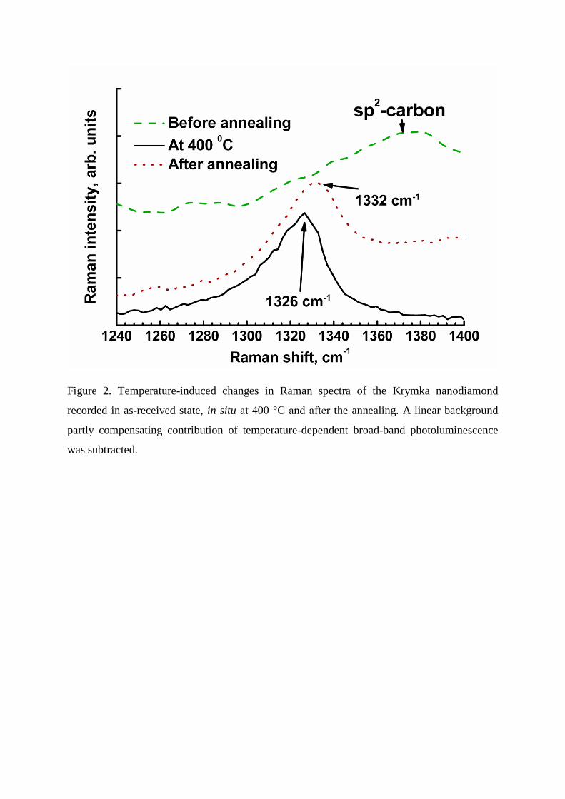
Figure 2. Temperature-induced changes in Raman spectra of the Krymka nanodiamond
recorded in as-received state, in situ at 400 °C and after the annealing. A linear background
partly compensating contribution of temperature-dependent broad-band photoluminescence
was subtracted.

Figure 3. Relative area of the SiV band in dependence of chemical composition of parent
meteorite. The total area was calculated between 490 and 800 nm; the SiV band area was
calculated after linear background subtraction between 720 and 760 nm. For all samples
except Kainsaz points corresponding to size fractions are shown.
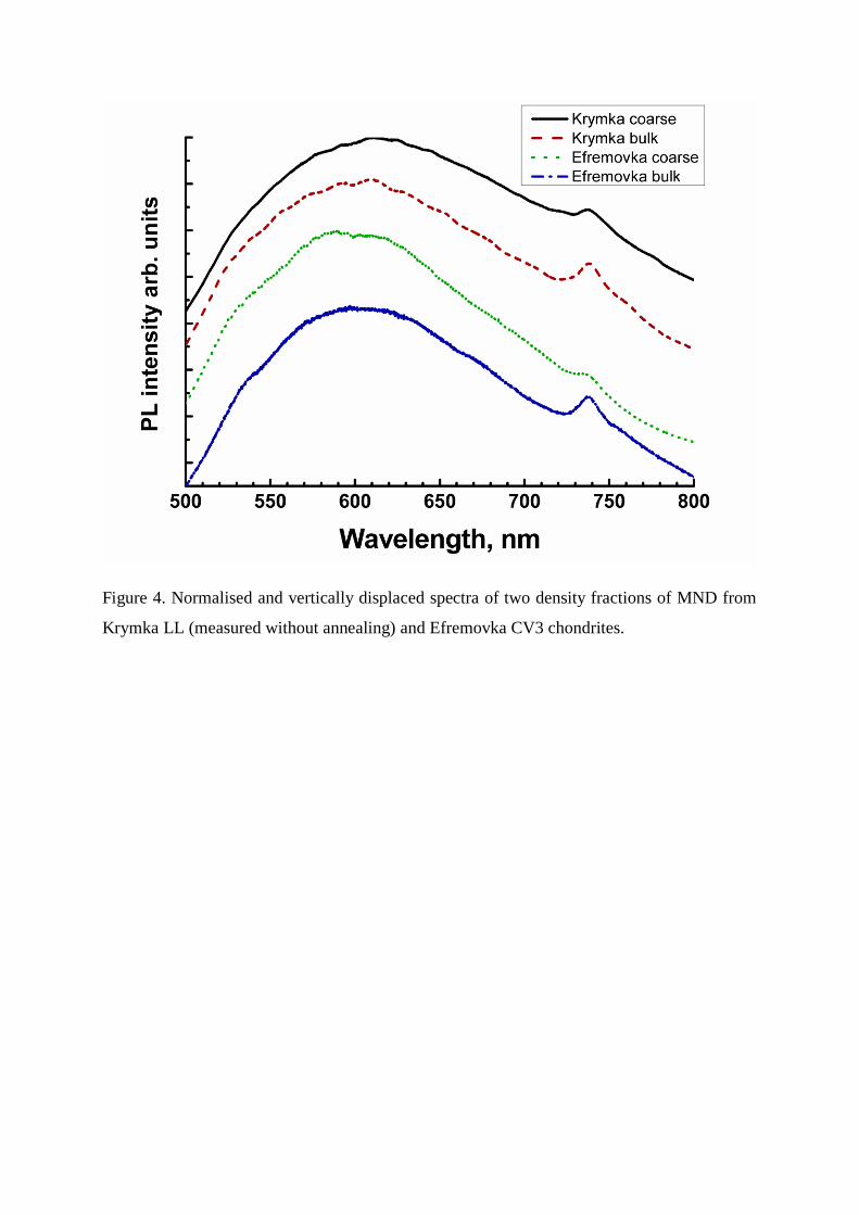
Figure 4. Normalised and vertically displaced spectra of two density fractions of MND from
Krymka LL (measured without annealing) and Efremovka CV3 chondrites.