photosynthetic limitations and volatile and non-volatile isoprenoids in the poikilochlorophyllous...
TRANSCRIPT

Photosynthetic limitations and volatile and non-volatileisoprenoids in the poikilochlorophyllous resurrection plantXerophyta humilis during dehydration and rehydrationpce_2536 1..14
MEGAN BECKETT1, FRANCESCO LORETO2, VIOLETA VELIKOVA2,3, CECILIA BRUNETTI2,4,MARTINA DI FERDINANDO4, MASSIMILIANO TATTINI2, CARLO CALFAPIETRA5 & JILL M. FARRANT1
1Department of Molecular and Cell Biology, University of Cape Town, Private Bag, Rondebosch 7701, South Africa,2Institute for Plant Protection, National Research Council, Via Madonna del Piano 10, 50019 Sesto Fiorentino, Florence,Italy, 3Institute for Plant Physiology and Genetics, Bulgarian Academy of Sciences, Sofia 1113, Bulgaria, 4Department ofHorticulture, University of Florence, 50019 Sesto Fiorentino, Florence, Italy and 5Institute of Agroenvironmental and ForestBiology, National Research Council, Via Salaria Km 29300, 00015 Monterorotondo Scalo, Rome, Italy
ABSTRACT
We investigated the photosynthetic limitations occurringduring dehydration and rehydration of Xerophyta humilis,a poikilochlorophyllous resurrection plant, and whethervolatile and non-volatile isoprenoids might be involved indesiccation tolerance. Photosynthesis declined rapidly afterdehydration below 85% relative water content (RWC).Raising intercellular CO2 concentrations during desiccationsuggest that the main photosynthetic limitation was photo-chemical, affecting energy-dependent RuBP regeneration.Imaging fluorescence confirmed that both the number ofphotosystem II (PSII) functional reaction centres and theirefficiency were impaired under progressive dehydration,and revealed the occurrence of heterogeneous photosyn-thesis during desiccation, being the basal leaf area moreresistant to the stress. Full recovery in photosyntheticparameters occurred on rehydration, confirming that pho-tosynthetic limitations were fully reversible and that nopermanent damage occurred. During desiccation, zeaxan-thin and lutein increased only when photosynthesis hadceased, implying that these isoprenoids do not directlyscavenge reactive oxygen species, but rather protect photo-synthetic membranes from damage and consequent dena-turation. X. humilis was found to emit isoprene, a volatileisoprenoid that acts as a membrane strengthener in plants.Isoprene emission was stimulated by drought and peaked at80% RWC. We surmise that isoprene and non-volatile iso-prenoids cooperate in reducing membrane damage inX. humilis, isoprene being effective when desiccation ismoderate while non-volatile isoprenoids operate whenwater deficit is more extreme.
Key-words: carotenoids; desiccation tolerance; imagingfluorescence; isoprene; photosynthesis; volatile organiccompounds.
INTRODUCTION
Desiccation tolerance is defined as the ability of an organ-ism to dry to equilibrium with the air dry state and toresume full metabolic function on rehydration. Althoughnear complete measurable water loss is a common occur-rence in the development of seeds and pollen (reviewed inBerjak, Farrant & Pammenter 2007) and in a large numberof lichen and bryophyte species, there are only a few higherorder plants that are able to survive extreme water deficit intheir vegetative tissues (reviewed in Farrant 2007; Oliver2007; Moore et al. 2009). Desiccation tolerance has onlybeen observed in about 350 angiosperm species, termed‘resurrection plants’, the majority of which are found intropical and subtropical zones in southern Africa.
In vegetative tissues, severe water stress results in a dis-ruption of electron transport and therefore disequilibriumbetween reactive oxygen species (ROS) production andscavenging ensues (Halliwell 1987; Halliwell & Gutteridge1999;Apel & Hirt 2004). Not only there is an excess of ROSproduced from respiratory metabolism, but also from a dis-ruption of photosynthesis and consequent inefficient use oflight-generated photosynthetic electron transport. Excessenergy from excited chlorophyll molecules is transferred tooxygen causing a rapid production of free radical species.While desiccation-sensitive plants are unable to adequatelydeal with the surge in ROS production, which ultimatelyleads to cell death (Smirnoff 1993; Kranner & Birtic 2005),resurrection plants have many mechanisms in place tofirstly reduce ROS formation and, secondly, quench theiractivity (reviewed in Farrant 2007; Farrant, Cooper & Nell2012).
Photosynthetic ROS production is minimized in resurrec-tion plants at high relative water contents (RWCs), thisbeing accomplished by one of two mechanisms termedpoikilochlorophylly and homoiochlorophylly (Gaff 1989;Sherwin & Farrant 1998; Tuba, Proctor & Csintalan 1998;Farrant 2000, 2007). Poikilochlorophylly involves the break-down of chlorophyll and partial dismantling of thylakoidmembranes during dehydration, resulting in a cessation ofCorrespondence: J. M. Farrant. e-mail: [email protected]
Plant, Cell and Environment (2012) doi: 10.1111/j.1365-3040.2012.02536.x
bs_bs_banner
© 2012 Blackwell Publishing Ltd 1

photosynthesis at water contents between 80 and 65% inspecies tested to date. Homoiochlorophyllous resurrectionplants retain chlorophyll and thylakoid membranes in thedesiccated state but light–chlorophyll interactions that ini-tiate photosynthesis and thus ROS production are mini-mized by a number of adaptive mechanisms. These includeleaf folding to reduce the surface area exposed to light andheat, the presence of reflective hairs and/or waxes, and theaccumulation of anthocyanins or other pigments that maskchlorophyll and act as antioxidants (reviewed in Farrant2007; Farrant et al. 2012).
Clearly, there is a very controlled regulation of physicaland metabolic processes in resurrection plants that enablesminimization of the stresses associated with desiccation andthat allows full recovery when water becomes available.These adaptations separate resurrection plants fromdesiccation-sensitive plants.
Non-volatile isoprenoids, such as tocopherols, zeaxanthinand b-carotene, form an integral part of the non-enzymaticoxidative defence system in all plants (Demmig-Adams &Adams III 1996; Munné-Bosch & Alegre 2002;Telfer 2002).Recently, carotenoids, particularly zeaxanthin, have beenreported to enhance drought tolerance (Davison, Hunter &Horton 2002; Du et al. 2010), not only by quenching thesinglet oxygen-excited state of chlorophyll or throughnon-photochemical quenching (NPQ), but also exerting aspecific role in preserving thylakoid membranes from per-oxidation (Havaux & Niyogi 1999; Havaux, Dall’Osto &Bassi 2007), a function that cannot be served by other non-volatile isoprenoids, such as a-tocopherol and b-carotene(Havaux et al. 2007).
There are various other molecules that function duringdrought stress in some desiccation-sensitive plants, whichhave not yet been investigated in resurrection plants.Among these are volatile organic compounds (VOCs). Iso-prene (C5H8, 2-methyl-1,3-butadiene) is a natural productof many organisms and is the most abundant VOC emittedby terrestrial plants (Guenther et al. 1995). During environ-mental stress conditions such as drought stress or elevatedtemperature, the amount of carbon lost due to isopreneemission (regularly limited to 1–2% of photosynthesis) canincrease dramatically (Sharkey & Loreto 1993). Manyplants from a broad range of taxonomic groups emit iso-prene, such as mosses, ferns, gymnosperms and angiosperms(Tingey et al. 1987; Hanson et al. 1999; Sharkey et al. 2005).However, there are also many members from these groupsthat do not emit isoprene. As the energy cost of isopreneemission is significant, especially under stress conditions(Sharkey, Chen & Yeh 2001; Sharkey, Wiberley & Donohue2008), it is probable that benefit outweighs the cost in theplants in which isoprene emission occurs. Researchers havetherefore been intent on determining and investigating thebenefits that isoprene confers on emitting plants.
To date, the best characterized roles of isoprene havebeen in thermotolerance (Sharkey & Yeh 2001; Sharkeyet al. 2008; Velikova et al. 2011) and ozone stress (Loretoet al. 2001; Loreto & Velikova 2001). Isoprene has beenshown to protect the photosynthetic apparatus against
ozone damage, quench ozone products, such as hydrogenperoxide, and help reduce lipid peroxidation of membranes.While isoprene has been proposed to operate a protectiveaction also under drought stress conditions (Sharkey &Loreto 1993; Fortunati et al. 2008), there have been noreports on its role in plants that tolerate extreme waterdeficit stress.
Volatile isoprenoids, such as isoprene, may form part ofan additional protective system against oxidative stress,which is not conserved among all plants. While, as discussedabove, many plants emit isoprene, this work has mainlybeen reported for desiccation-sensitive woody plants(Vickers et al. 2009), and it is not known whether resurrec-tion plants emit volatile isoprenoids. As such, emissionseems to convey an adaptive advantage to plants to survivein adverse environmental conditions, investigating VOCs inresurrection plants could uncover an additional mechanismin desiccation tolerance.
The aim of this study was firstly to investigatephotosynthetic regulation and some of the known con-served antioxidant processes during dehydration ofthe poikilochlorophyllous resurrection plant Xerophytahumilis, and, thereafter, to investigate whether this speciesemits VOCs and to characterize their role in acquisition ofdesiccation tolerance.
MATERIALS AND METHODS
Plant material
X. humilis plants were collected in the Pilanesberg NatureReserve, South Africa, and maintained in a glasshouse aspreviously described (Sherwin & Farrant 1996). Plants werethen dehydrated and transported to Italy in the desiccatedstate. On arrival, they were rehydrated and maintained in aclimatized glasshouse and allowed to acclimatize for 3weeks at a temperature maintained above 25 °C and vari-able during the experiment between 25 and 31 °C, and alight intensity reaching 700 mmol m-2 s-1 during sunny daysprior to conducting the experiments described below. Trays(15 ¥ 20 cm with a soil depth of about 5 cm) containing upto 15 plants were used for the procedures described below.
RWC determination
Trays were well watered to ensure plants were fullyhydrated at commencement of an experiment. Thereafter,whole plants were dehydrated by withholding water, andallowing plants to dry naturally. Soil was watered to fieldcapacity to allow for rehydration.
The water content was obtained gravimetrically on a dryweight (DW) basis by oven drying at 70 °C for 48 h. RWCwas measured using the standard formula: RWC = watercontent/water content at full turgor and was expressedas a percentage. Full turgor was achieved as previouslydescribed (Mowla et al. 2002). Three leaf sections randomlychosen from plants within each tray were cut for RWCdetermination.
2 M. Beckett et al.
© 2012 Blackwell Publishing Ltd, Plant, Cell and Environment

Measurement of chlorophyll fluorescence
A Maxi-Imaging-PAM-fluorometer (Heinz Walz GmbH,Effeltrich, Germany) was used for chlorophyll a fluores-cence measurements.The MAXI version of the IMAGING-PAM M-Series employs a very compact and powerful300W LED array for homogeneous illumination of up to10 ¥ 13 cm areas with pulse-modulated excitation, actiniclight and saturation pulses. The charge-coupled device(CCD) camera has a resolution of 640 ¥ 480 pixels. Pixelvalue images of the fluorescence parameters were displayedusing a false colour code ranging from black (0.000) to red,yellow, green, blue and pink (1.000).
Plants were dark adapted for at least 20–35 min prior tothe determination of Fo and Fm (minimum and maximumfluorescence, respectively). Longer dark-adaptation timecourses were used as plants underwent dehydration tocompensate for a slightly delayed relaxation of the reac-tion centres of photosystem II (PSII). The maximumquantum yield of PSII photochemistry (Fv/Fm) was deter-mined as (Fm - Fo)/Fm. Leaves were adapted to the specificlight level and a saturating pulse of 0.8 s with 6000 mmolphotons m-2 s-1 was applied in order to determine themaximum fluorescence (F′m) and the steady-state fluores-cence (Fs) during the actinic illumination. It was testedalong the dehydration that the light intensity was sufficientto saturate PSII and that the length of the pulse did notinduce photoinhibition. The quantum efficiency of PSIIphotochemistry, FPSII, was calculated using the formula:(F′m - Fs)/F′m (Genty, Briantais & Baker 1989). The coef-ficient of photochemical quenching, qP, is a measurementof the fraction of open centres calculated as (F′m - Fs)/(F′m - F′o) (Schreiber, Schliwa & Bilger 1986). The valueof F′o was estimated using the approximation: F′o = Fo/(Fv/Fm + Fo/F′m) (Oxborough & Baker 1997). Calculation ofquenching due to non-photochemical dissipation ofabsorbed light energy (NPQ) was determined using theequation NPQ = (Fm - F′m)/F′m (Bilger & Björkman 1991).Chlorophyll fluorescence determinations were obtainedfrom n = 7 leaves, selected on the images of whole plantparts that were reached by an incident light intensity of610 mmol m-2 s-1, as this was the light intensity closest togrowth light intensity (see below).
Pigment analyses
Individual carotenoids were identified and quantified asreported in García-Plazaola & Becerril (1999). Fresh leafmaterial (120–150 mg) was extracted with 2 ¥ 4 mL acetone(added with 0.5 g L-1 CaCO3) and 15 mL aliquots wereinjected in a Perkin Elmer Flexar chromatograph equippedwith a quaternary 200Q/410 pump and LC 200 diode arraydetector (DAD) (all from Perkin Elmer, Bradford, CT,USA).
Photosynthetic pigments were separated by a250 ¥ 4.6 mm Waters Spherisorb ODS1 (5 mm) columnoperating at 30 °C, eluted with a linear gradient solventsystem, at a flow rate of 1.2 mL min-1 consisting of CH3CN/
MeOH/H2O (8.4/0.8/0.7, A) and MeOH/ethyl acetate (6.8/3.2, B) during an 18 min run: 0–12 min from 100 to 0% A;12–18 min at 0% A.
Violaxanthin cycle pigments, lutein and a-, b-carotenewere identified using visible spectral characteristics andretention times. The compounds were calibrated as such:neoxanthin, violaxanthin and antheraxanthin with thecalibration curve of lutein at lower concentration points;lutein with the calibration curve of lutein; zeaxanthinwith the calibration curve of zeaxanthin; and a-caroteneand b-carotene with the calibration curve of b-carotene(all from Extrasynthese, Lyon-Nord, Genay, France).Chlorophyll a and b were quantified by spectrophoto-metric analysis (Lichtenthaler & Buschmann 2001). Thedehydration-rehydration experiment was repeated twiceand at least three biological replicates were obtained foreach RWC point plotted.
Photosynthesis and VOC measurements
Gas exchange and VOC emission measurements were per-formed simultaneously on leaves of X. humilis grown insoil. Plants were transferred to the laboratory for the dura-tion of the measurement and kept under a light source withan intensity of 700 mmol photons m-2 s-1, equivalent to themean light intensity in the greenhouse during daylight.Leaves were marked with small adhesive labels and thesame leaves were followed during dehydration and rehy-dration. The experiment was repeated three times and atleast four biological repeats (different leaves from differentplants) were obtained for each RWC plotted.
Rates of photosynthesis (A) and photosynthesis responseto intercellular CO2 concentration (A/Ci responses) weremeasured on a 2 cm2 leaf piece flattened in the cuvette of aLI-6400 (Li-Cor Biosciences Inc., NE, USA) infrared gasanalyser (IRGA) allowing simultaneous measurementsof gas exchange and florescence on the same leaf area.The LI-6400 gas-exchange system also allowed the controlof temperature (30 � 0.2 °C), light intensity (610 mmolm-2 s-1), relative humidity (40–50%) and CO2 concentration(50–1500 ppm during A/Ci responses, otherwise fixed at 400ppm) during measurements.The equations used to calculatephotosynthesis were those previously derived by von Cae-mmerer & Farquhar (1981). Readings were taken fromthree to four leaves selected randomly from three trays,each day of dehydration and rehydration, between 3 and 4 hafter dawn. Readings were taken over a time period of5 min after reaching steady-state gas exchange, and theaverage of the technical repeats (at least three on threedifferent leaves) for each measurement was calculated.A/Ci
responses were analysed with the mechanistic model of CO2
assimilation proposed by Farquhar, von Caemmerer &Berry (1980), and the relevant kinetic parameters, maximalvelocity of carboxylation (Vcmax) and maximal electrontransport rate (Jmax) were estimated.
To measure VOC emissions, the outlet of the cuvette wasdisconnected from the LI-6400 system and the flow was
Photosynthesis, isoprenoids and drought 3
© 2012 Blackwell Publishing Ltd, Plant, Cell and Environment

diverted into a silcosteel cartridge packed with 200 mg ofTenax (Agilent, Cernusco sul Naviglio, Italy). A volumeof 8 L of air was pumped through the trap at a rateof 150 mL min-1. The cartridge was analysed by gaschromatography-mass spectrometry (GC-MS). GC-MSanalyses were performed with an Agilent 6850 gas chro-matograph coupled to an Agilent 5975C Mass SelectiveDetector (Agilent Technologies, Wilmington, DE, USA) orwith a Perkin Elmer Clarus 580 gas chromatograph coupledto a Perkin Elmer Clarus 560S Mass Selective Detector(Perkin Elmer, Inc., Waltham, MA, USA). The Agilent GCwas supplied with a thermal desorber UNITY (MarkesInternational Limited, Llantrisant, UK) whereas the PerkinElmer GC was coupled to a thermal desorber TurboMatrix300 (Perkin Elmer, Inc.). Both GCs were equipped with asplitless injector and a HP-5MS capillary column (30 m inlength, 250 mm in diameter and 0.25 mm film thickness).Thecolumn oven temperature was kept at 40 °C for the first5 min, then increased by 5 °C min-1 to 250 °C, and main-tained at 250 °C for 2 min. Helium was used as carrier gas.The concentration of each volatile was calculated by com-parison with the peak area of a gaseous standard. TheGC-MS was calibrated weekly using cylinders with stan-dard mixtures of the main isoprenoids generally emitted byplants (isoprene, a-pinene and limonene) at an averageconcentration of 60 ppb (Rivoira, Milan, Italy). Compoundswere identified using the NIST library provided with theGC/MS ChemStation software on both Agilent and PerkinElmer measurements. GC peak retention time was substan-tiated by analysis of parent ions and main fragments of thespectra.
Isoprene emission was also measured online, by divertingthe air at the exit of the gas-exchange cuvette into a ProtonTransfer Reaction-Mass Spectrometer (PTR-MS; Ionicon,Innsbruck, Austria), which allowed fast detection of iso-prene. Once the exit of the gas exchange system had beenconnected to the PTR-MS and a steady state in isopreneemission had been reached, the cuvette containing the leafwas covered with a black cloth until the emission reachedthe lowest possible level (approximately 400 s), and thecloth was then removed again. The PTR-MS was operatedin a single-ion mode to detect isoprene (protonatedm/z = 69) down to a threshold of 1 ppt. Calibrations usingan isoprene gaseous standard (60 ppt) were performeddaily before measurements. Details on isoprene analysis byPTR-MS can be found in Tholl et al. (2006). VOC measure-ments were repeated at least six times on different leaves ofdifferent plants at each single RWC.
Statistical analyses
Means and standard errors were calculated with GraphPad Prism (Version 5; GraphPad Software, Inc., La Jolla,CA, USA). Biological replications varied by experimentand are indicated in the appropriate figure legend. The sig-nificance of differences between means at different RWCswas analysed using Student’s t-test.
RESULTS
The changes in photosynthesis during dehydration of X.humilis are shown in Fig. 1a. Net photosynthesis decreasedrapidly below 80% RWC and had ceased by 57% RWC,when chlorophyll content had decreased about 50%(Fig. 1b). Recovery of photosynthesis was initiated uponrehydration above 60% RWC (Fig. 1a). This coincided withan increase in chlorophyll content (Fig. 1b) and the likelyregeneration and reassembly of thylakoid membranes.
GC-MS analyses revealed that isoprene emission occursfrom X. humilis plants. Isoprene emission was light depen-dent, as in all other isoprene emitters (data not shown)suggesting that emission is dependent on photosynthesis.Thus, the changes in emission of isoprene during dehydra-tion and rehydration were assessed in relation to photosyn-thetic changes. There was a significant increase (P < 0.001)in isoprene emission, from 1 to more than 4 nmol m-2 s-1, as
(a)
(b)
(c)
(d)
Figure 1. Change in photosynthesis (a) (mean � SEM, n = 5),total chlorophyll content (b) (mean � SEM, n = 3), isopreneemission (c) (mean � SEM, n = 6), and hexanal (d)(mean � SEM, n = 3) during dehydration (left panels) andrehydration (right panels) of Xerophyta humilis plants. DW, dryweight; RWC, relative water content.
4 M. Beckett et al.
© 2012 Blackwell Publishing Ltd, Plant, Cell and Environment

the leaf RWC decreased from 100 to 82% (Fig. 1c). Iso-prene emission then declined progressively until a RWC of50%, after which no further emission was recorded. Photo-synthesis in these leaves started to decline below a RWC of80%, when isoprene emission was at its maximum, andhad decreased once plant tissues had a recorded RWC of62%. As the ratio of isoprene emission to photosynthesisincreased in response to initial dehydration, the amount ofcarbon lost as isoprene increased exponentially as RWCdecreased from 100 to 62%.At around 60% RWC, this ratioreached as much as 26% of the total carbon assimilated byphotosynthesis (data not shown). During rehydration, pho-tosynthesis recovered to prestress levels. However, isopreneemission recovered at a slower rate than photosynthesis.Upon full rehydration to 100% RWC, isoprene emissionreached levels of 3.6–5.2 nmol m-2 s-1, this being equivalentto the maximum emission upon dehydration, and muchhigher than it was prior to dehydration in X. humilis.
Analysis of the volatile samples collected during dehydra-tion and rehydration using GC-MS revealed the emission ofthe oxygenated C6 VOC hexanal (Fig. 1d). The emission ofhexanal, which is a sensitive marker of membrane denatur-ation (Loreto et al. 2006; Capitani et al. 2009), increased inresponse to dehydration and peaked at 35% RWC, beforedecreasing rapidly during the very late stages of dehydra-tion. During rehydration, there was no emission of hexanaldetected from X. humilis, showing that lipoxygenase (LOX)activity is not stimulated during the recovery process.
The photosynthetic response to intercellular CO2 (Ci)was already affected in leaves that had dehydrated to 85%RWC, but the effect was especially evident at CO2 concen-trations higher than ambient (Fig. 2). In rehydrated leaves,
the photosynthetic response to Ci attained the prestresslevel indicating a complete recovery of photosynthesis andno permanent limitations caused by dehydration.
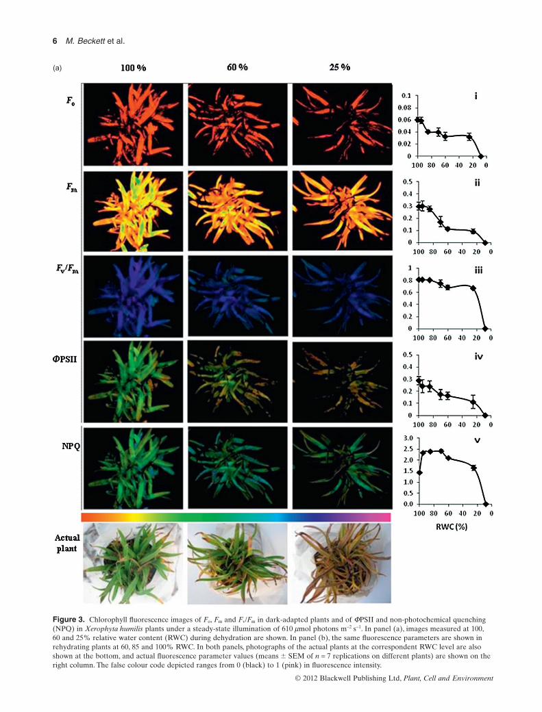
The change in chlorophyll fluorescence in leaves ofX. humilis during dehydration is shown in Fig. 3a, withrepresentative images of chlorophyll fluorescence taken ata whole plant level, and at 100, 60 and 25% RWC, respec-tively. On the side of each image, the actual average(� SEM) of the values for the given parameter is shownat various RWCs during dehydration. The initial fluores-cence Fo, measured when the plant had been dark adaptedand all reaction centres are assumed to be open, decreasedslightly at the onset of dehydration but thereafterremained constant during further drying (Fig. 3a-i). Themaximal fluorescence Fm, measured during the high-intensity rapid flash of light, which causes all reactioncentres of darkened plants to close, is reached in theabsence of photochemical and NPQ. As can be seen in theimages of Fig. 3a-ii, Fm followed a similar trend to Fo
during early stages of dehydration, but declined substan-tially also between 80 and 60% RWC.
The maximum efficiency of PSII in dark-adapted leaves(Fv/Fm) remained relatively high during the initial stages ofdehydration and started to decline from about 80% until60% RWC with a rapid decline from 20% RWC onwards(Fig. 3a-iii).As can be seen in the images for Fv/Fm, the falsecolour remained blue-purple even until 25% RWC, indicat-ing at this stage that the efficiency of PSII, if all reactioncentres were open, was still relatively high. The responsecurve in this study corresponds to that reported to date forresurrection plants.
Figure 3a-iv shows the actual quantum yield, for example,the true efficiency of photosystem II (FPSII) during dehydra-tion. FPSII measures the rate of linear electron transport ratedriving photosynthesis and photorespiration when leavesare illuminated and photosynthesis is activated. The imagesfor FPSII clearly show that the efficiency of PSII rapidlydeclined, and that the electron transport rate was very het-erogeneous within each leaf during dehydration. The latterobservation was quantitatively confirmed by separatelyassessing the fluorescence values measured in the basalthird and in the apical third of the leaves (Table 1). Hetero-geneities developed during dehydration in both maximaland actual quantum yield of fluorescence (Fv/Fm and FPSII,respectively), being the apical part more sensitive to thestress. Because a lower electron transport rate producesphotochemical limitations of photosynthesis, this indicatedthat the photochemical apparatus of the basal part of theleaves was more preserved from dehydration stress.
The NPQ of fluorescence, the mechanism whereby plantsconvert excess energy to heat and thus minimize sub-cellular damage especially in high light conditions, initiallyincreased as the plants dehydrated from 100 to 80% RWC.However, this value declined below RWCs of 60% (Fig. 3a-v), this coinciding with the decrease in photosynthesis aschlorophyll was broken down. Moreover, no heterogeneityof NPQ of fluorescence was observed during the develop-ment of dehydration (Table 1).
Figure 2. Response of photosynthesis to variations ofintercellular CO2, Ci, before dehydration [100% relative watercontent (RWC), circles], during early dehydration (85% RWC,triangles) and after rehydration (100% RWC, squares) ofXerophyta humilis plants (mean � SEM, n = 3]). The curves werefitted using Farquhar, von Caemmerer & Berry (1980) model.Continuous lines fit the data points before dehydration and afterrehydration at RWC = 100%; the dashed line fits data collected atRWC = 85%. The maximal electron transport rate (Jmax) valuescalculated by Farquhar’s model are shown in the text.
Photosynthesis, isoprenoids and drought 5
© 2012 Blackwell Publishing Ltd, Plant, Cell and Environment
(a)
Figure 3. Chlorophyll fluorescence images of Fo, Fm and Fv/Fm in dark-adapted plants and of FPSII and non-photochemical quenching(NPQ) in Xerophyta humilis plants under a steady-state illumination of 610 mmol photons m-2 s-1. In panel (a), images measured at 100,60 and 25% relative water content (RWC) during dehydration are shown. In panel (b), the same fluorescence parameters are shown inrehydrating plants at 60, 85 and 100% RWC. In both panels, photographs of the actual plants at the correspondent RWC level are alsoshown at the bottom, and actual fluorescence parameter values (means � SEM of n = 7 replications on different plants) are shown on theright column. The false colour code depicted ranges from 0 (black) to 1 (pink) in fluorescence intensity.
6 M. Beckett et al.
© 2012 Blackwell Publishing Ltd, Plant, Cell and Environment

(b)
Figure 3. Continued
Photosynthesis, isoprenoids and drought 7
© 2012 Blackwell Publishing Ltd, Plant, Cell and Environment

During rehydration, there was full recovery of all chloro-phyll fluorescence parameters, this process being initiatedupon rehydration above 60% RWC, coincident with pro-duction of chlorophyll and initiation of photosynthesis,and being complete, with maximal fluorescence yieldagain achieved upon rehydration to 100% RWC (Fig. 3b).However, Fo and the maximal quantum yield of fluores-cence appeared to fully recover already at 85% RWC,indicating that the photochemical apparatus was fullyreconstituted at this rehydration stage (Fig. 3b-i andiii, respectively). On the other hand, the actual quantumyield of fluorescence (FPSII, Fig. 3b-iv) only reached themaximum at 100% RWC and the NPQ (Fig. 3b-v) reacheda maximum at 85% RWC but then dropped. These findingssuggest that photochemical energy was safely dissipated asheat at 85% RWC whereas it became again used to pro-duced photosynthetic electron transport rate only when theRWC recovery was complete, for example, that photosyn-thesis was still limited by processes that are not directlyrelated to PSII structure at RWC = 85% during rehydra-tion. Independent of the residual photosynthetic limitationduring rehydration, the overall complete recovery (see alsoFigs 1–2) suggests that chloroplasts did not suffer from per-manent damage upon dehydration, concurring previousreports for this and other Xerophyta species (Sherwin &Farrant 1998; Tuba et al. 1998; Collett et al. 2003; Ingle et al.2008). Again, rehydration affected heterogeneously leaffluorescence properties, the recovery being more rapid atthe base of the leaves, whereas the leaf tips often did notrehydrate (Fig. 3b and Table 1).
During dehydration of X. humilis, there was asteady decline in violaxanthin from 0.13 mmol g-1 DW at100% RWC to 0.005 mmol g-1 DW in dry leaf tissue(Fig. 4a). Zeaxanthin content increased significantly from0.012 mmol g-1 DW in control leaves (100% RWC) to
0.082 mmol g-1 DW in the desiccated leaf tissue (10%RWC) (Fig. 4a).
During rehydration, the reverse reactions took place. Ascan be seen in Fig. 4a, zeaxanthin levels remained constantduring the initial stages of rehydration and then decreasedrapidly upon rehydration above 60% RWC, with violaxan-thin increasing at this stage. At the end of rehydration,violaxanthin content was higher than it was prior to dehy-dration. Zeaxanthin remained high until 60% RWC.
Table 1. Variations in fluorescenceparameters within single leaves at differentrelative water contents (RWCs) duringdehydration and rehydration ofXerophyta humilis plants
RWC Leaf part Fv/Fm FPSII NPQ
Dehydration100% Apical 0.754 � 0.034a,b 0.214 � 0.012b 1.96 � 0.40a,b
Basal 0.791 � 0.005a 0.220 � 0.050b,c 2.21 � 0.26a
60% Apical 0.685 � 0.032c,d 0.167 � 0.004c 1.70 � 0.41a,b
Basal 0.748 � 0.004b 0.209 � 0.002b 1.88 � 0.19a,b
25% Apical 0.633 � 0.024d 0.086 � 0.002d 1.30 � 0.27b
Basal 0.705 � 0.003c 0.171 � 0.010c 1.53 � 0.03b
Rehydration60% Apical 0.711 � 0.022b,c 0.139 � 0.019c 2.51 � 0.29a
Basal 0.708 � 0.006c 0.230 � 0.033b 1.64 � 0.07b
85% Apical 0.755 � 0.013b 0.159 � 0.010c 2.23 � 0.23a
Basal 0.752 � 0.014b 0.219 � 0.046bc 1.59 � 0.13b
100% Apical 0.749 � 0.017b 0.163 � 0.005c 2.15 � 0.29a
Basal 0.801 � 0.011a 0.349 � 0.011a 1.56 � 0.05b
Maximal and actual quantum yield of fluorescence (Fv/Fm and FPSII, respectively) and non-photochemical quenching of fluorescence (NPQ) were measured in the apical and basalparts of the leaves. Means � SEM (n = 4) are shown. Leaves were put at the same distancefrom the fluorescence camera to avoid artefacts due to position. Differences of values ofeach parameter at varying RWC were assessed by Tukey’s and are shown by different letterswhen significantly different at the 5% (P < 0.05)
(b)
(a)
Figure 4. Change in zeaxanthin and violaxanthin (a) and othercarotenoid pigments (b) during dehydration and rehydration ofXerophyta humilis plants. Values are calculated relative to dryweight (DW) (mean � SEM, n = 3). RWC, relative water content.
8 M. Beckett et al.
© 2012 Blackwell Publishing Ltd, Plant, Cell and Environment

Activation of the epoxidation of zeaxanthin back to violax-anthin thus seems to occur at 60% RWC during rehydra-tion. Antheraxanthin levels did not change significantly.
The changes in the remaining carotenoids, which do notform part of the xanthophyll cycle, are shown in Fig. 4b.Neoxanthin decreased during dehydration, and upon rehy-dration it increased again and reached a level comparableto what it was before dehydration. The concentration oflutein initially decreased as the plants dehydrated from0.292 mmol g-1 DW at 100% RWC to 0.164 mmol g-1 DW at82% RWC, but increased during the latter stages of dehy-dration to levels slightly higher than the pre-desiccatedcondition. During dehydration, b-carotene progressivelydecreased.
GC-MS analyses revealed that isoprene emission occursfrom X. humilis plants. Isoprene emission was light depen-dent, as in all other isoprene emitters (data not shown)suggesting that emission is dependent on photosynthesis.Thus, the changes in emission of isoprene during dehydra-tion and rehydration were assessed in relation to photosyn-thetic changes in a set of measurements different fromthose shown in Fig. 1.
DISCUSSION
Regulation of photosynthesis
Photosynthesis in X. humilis was not particularly resistantto dehydration, and it started to be inhibited at relativelyhigh RWCs. The response of photosynthesis to intercellularCO2 concentration (A/Ci response) provides a rapid andreliable method to identify in vivo photosynthetic limita-tions (Farquhar & Sharkey 1982). Before introducing ourdiscussion on A/Ci results, it should be said that, whenstomata close in response to strong dehydration, an increas-ing contribution of (1) stomatal patchiness; (2) cuticularconductance, is often observed. This may produce an over-estimation of Ci (Meyer & Genty 1998) and consequentlyimpair the analysis of A/Ci responses. While the readershould be aware that this could also affect our A/Ci analysis,we observe that the impact of patchiness, as revealed alsoby our imaging fluorescence measurements, was minimizedby selecting for gas-exchange measurements leaf areas thatshowed homogeneous fluorescence.We also reason that theimpact of cuticular transpiration should be higher at low Ci
(Meyer & Genty 1998), whereas in the present study, pho-tosynthesis of dehydrated leaves was lower than in controls,especially at CO2 concentrations higher than ambient, forexample, when photosynthesis does not increase linearlywith increasing Ci. This indicates photosynthesis limitationsdue to RuBP regeneration in plants undergoing mild dehy-dration (85% RWC). Because RuBP regeneration dependson efficient electron transport rate supplying sufficientreducing power (NADPH) and chemical energy (ATP), ourdata indicate a possible photochemical limitation of photo-synthesis in mildly dehydrated X. humilis leaves. Indeed,the maximum electron transport rapacity (Jmax) estimatedafter removing diffusive limitations according to Sharkey
et al. (2007) from A/Ci best fitting (Fig. 2) dropped signifi-cantly from 90.5 � 9.5 mmol m-2 s-1 before dehydration to51.7 � 4.6 mmol m-2 s-1 at 85% RWC. The possibility thatlight reactions limit photosynthesis in dehydrating leaves ofX. humilis was further investigated by chlorophyll fluores-cence analysis, as shown below. Interestingly, such a photo-chemical limitation developed simultaneously with theonset of chlorophyll degradation in dehydrating leaves.Thus, the reduction in photochemical activity might be dueto chlorophyll degradation and/or, as has been proposed forhomoiochlorophyllous resurrection plants, due to chloro-phyll shading and masking by anthocyanins (Farrant 2000;Farrant et al. 2003). Chlorophyll had been entirely degradedby the time the plants desiccated, as has been reported inother studies on Xerophyta species (Tuba et al. 1996, 1998;Farrant 2000).
During rehydration, recovery of photosynthesis was ini-tiated upon rehydration above 60% RWC (Fig. 1a). Thiscoincided with the production of chlorophyll (Fig. 1b) andthe regeneration and reassembly of thylakoid membranes.It has been proposed that this delay is required for otherprotection mechanisms, notably regeneration or activationof antioxidant activity in order to minimize the potentialdamage associated with photosynthetically produced ROSupon rehydration (Farrant 2007). Interestingly, the rehy-drated leaves did not show any residual impact of the stresson the photosynthetic apparatus, as indicated by A/Ci
responses of rehydrated leaves overlaying those obtainedbefore dehydration. The calculated Jmax after full rehydra-tion was 89.6 � 17.5 mmol m-2 s-1, for example, very similarto that calculated in the same leaves before dehydration.This is interpreted to indicate total de novo synthesis ofchlorophylls and to the consequent generation of a func-tional photochemical apparatus.
Chlorophyll fluorescence
The photochemical limitations of leaves of X. humilisduring dehydration are clearly seen when using imagingchlorophyll fluorescence. Fluorescence analyses can giveinsights into a plant’s capacity to withstand environmentalstresses and also give a measure of the amount of damage astress has caused to the photosynthetic apparatus (Maxwell& Johnson 2000). Imaging fluorescence adds to this capac-ity, the possibility to study spatial heterogeneities duringstress development, for example, caused by leaf ontogeny oranatomy, or different sensitivity to stress.
Fluorescence analysis revealed that the maximumquantum yield of PSII (Fv/Fm), a specific indicator of photo-inhibition (Maxwell & Johnson 2000), was not affecteduntil considerable water loss (to less than 20% RWC) hadoccurred. Thus, photochemical limitations during the earlyphases of stress do not imply photo-inhibition. Rather, therapid decrease of the minimum fluorescence (Fo) and of thephotochemical quenching (qP) of fluorescence (data notshown, but mirroring FPSII of which qP is the most impor-tant component, Genty et al. 1989) indicates that thenumber of photochemical reaction centres able to operate
Photosynthesis, isoprenoids and drought 9
© 2012 Blackwell Publishing Ltd, Plant, Cell and Environment

basal fluorescence and photosynthetic electron transport(Genty et al. 1989) was somehow impaired already at theearly stages of dehydration. Thus, it may be speculated thatphotochemical impairment in dehydrating leaves is due tothe loss of active PSII centres rather to a reduced efficiencyof their photochemical function. Imaging fluorescenceclearly revealed that the fluorescence signal, hence the pho-tochemical efficiency, was not homogeneous across theleaves. A main difference could be observed between thebasal part of the leaves that continued to have a high pho-tochemical efficiency until the tissues were dehydrated, andthe apical parts in which such efficiency, and more specifi-cally the actual quantum yield of PSII (FPSII) which isindicative of the electron transport rate driving photosyn-thetic carbon reduction and carbon oxygenation cycles(Genty et al. 1989), was largely impaired during early stagesof dehydration. Thus, it may be surmised that the inactivefraction of PSII in dehydrating leaves resides in the apicalpart of the leaves. Because apical tissues are older than thatat basal regions, it is likely that the loss of PSII activity in theformer is associated with the onset of senescence processes(Kikuzawa & Lechowicz 2011) possibly exacerbated by fre-quent cycles of drying and rehydration. This hypothesis issupported by the observation that often the leaf tips ofmonocot resurrection plants, and older leaves of dicotspecies, do not rehydrate (Farrant 2000, 2007; VanderWilligen et al. 2001).
During rehydration, there was full recovery of all chloro-phyll fluorescence parameters. Such recovery correlateswith the results from A/Ci response curves and suggests thatchloroplasts became fully functional upon rehydration, con-curring with previous reported for this and other Xerophytaspecies (Tuba et al. 1993a, b; Sherwin & Farrant 1998; Ingleet al. 2008).While NPQ followed the same initial trend, withvalues increasing to 2.4 upon rehydration to 85% RWC, fullrehydration resulted in a subsequent decline of this param-eter to values equivalent of those prior to dehydration(compare Fig. 3a-v and 3b-v). These data are of interest, asthey indicate the capacity to rapidly and efficiently regulatethe dissipation of excess light non-radiatively in this resur-rection plant. They also suggest that at 85% RWC, despitePSII not being fully operational, sufficient protection hadaccrued in rehydrating tissues such that dissipation ofexcess energy as heat could be reduced.
Pigment analyses
Carotenoids play a significant role in photoprotection asthey can quench ROS, thereby reducing permanent damageassociated with excess excitation energy under stress situa-tions including drought (Demmig-Adams & Adams III1996; Munné-Bosch & Alegre 2000). There was a steadydecline in violaxanthin from the start of dehydration and aconsiderable increase in zeaxanthin content once the RWChad declined below 40%.The reverse was observed in rehy-drating leaves, but interestingly, zeaxanthin did not revertinto violaxanthin until the leaf RWC was greater than 60%.These changes are likely to be due to the well-known
pathway of de-epoxidation of violaxanthin into zeaxanthinvia the intermediate antheraxanthin, with concomitant dis-sipation of excess solar radiation heat, a phenomenonreported as ‘the xanthophyll cycle’, with resulting antioxi-dant action and photoprotection (Demmig et al. 1988;Demmig-Adams & Adams III 1996). This mechanism isgenerally activated by dark-to-light transitions, but here weshow that the same mechanism may be induced by dehy-dration in X. humilis. Although X. humilis is poikilochloro-phyllous, photosynthetic-generated ROS are likely to occurduring the early stages of dehydration when chlorophyll hasnot been completely degraded. However, changes in xan-thophylls de-epoxidation only occurred when plants wereseverely dehydrated and there was no physiological activity.Thus, it is unlikely that in X. humilis, the xanthophyll cycleserves primarily to quench ROS formation during dehydra-tion. The increase in zeaxanthin biosynthesis paralleledthe considerable reductions in chlorophyll concentration,and presumably originated by the action of violaxanthinde-epoxidase and/or b-carotene hydroxylase. b-carotenehydroxylase synthesizes zeaxanthin from b-carotene.Increased zeaxanthin was found to confer drought toler-ance in Arabidopsis (Davison et al. 2002) and rice plants(independent of zea-induced ABA biosynthesis, Du et al.2010). Zeaxanthin concentration accounted for 185 mmol-mol-1 total chlorophyll at 35% RWC, and increased steeply
to reach 650 mmol mol-1 total chlorophyll at 10% RWC.This high concentration in zeaxanthin is unlikely to bebound to light-harvesting chlorophyll-protein complexes(Havaux & Niyogi 1999). Zeaxanthin may therefore residein other parts of the thylakoids. Zeaxanthin, but notb-carotene, may increase rigidity of thylakoid membranes,and reduce peroxidative damage under drought stress(Havaux et al. 1996, 2007), and we surmise that this is thepredominant role of zeaxanthin in this resurrection plant, incoordination with isoprene biosynthesis and emission (seebelow). This confirms that photosynthetic membranes,while dismantled (Sherwin & Farrant 1998; Farrant 2000),are not completely destroyed in dry leaves of X. humilis(Dace et al. 1998; Ingle et al. 2008).
Among the remaining carotenoids, the changes in luteinseem particularly interesting. Indeed, the concentration oflutein rapidly decreased, but then increased again duringthe latter stages of dehydration, to levels slightly higherthan the pre-desiccated condition. The sequence of luteinstimulation is similar to that observed for zeaxanthin and islikely to reflect a similar function of these compoundsunder dehydration. Lutein is also suggested to reduce oxi-dative damage during dehydration (Demmig-Adams &Adams III 2002) and plays an important role in NPQquenching (Niyogi, Björkman & Grossman 1997). In addi-tion to this potential role, we propose that lutein, likezeaxanthin, might act as a membrane strengthener duringdehydration in X. humilis.
Contrary to lutein, b-carotene concentration progres-sively decreased during dehydration. It is probable that thedecline in b-carotene was due to its expenditure as an anti-oxidant and/or its absorption of excess light and therefore
10 M. Beckett et al.
© 2012 Blackwell Publishing Ltd, Plant, Cell and Environment

protecting the plant from oxidative damage (Telfer 2002).b-carotene reduction might also occur if the same pool ofcarbon that generates isoprenoids is temporarily allocatedto more effective defensive molecules such as zeaxanthin,lutein and isoprene (see below). b-carotene was re-synthesized again only during the late stages of rehydrationpossibly effecting some protection as photosynthesis is onceagain initiated. However, because it is also an accessorypigment to photosynthesis, it is likely to be involved in theinduction of photosynthesis occurring at this stage. Thesetrends in b-carotene are comparable with those observed inother resurrection plants (Kranner et al. 2002).
Volatile emissions
Isoprene, one of the most abundant biogenic VOCs (Loreto& Schnitzler 2010), is mainly emitted by hydrophilic plants(Vickers et al. 2009). This is the first report that isoprene ispossibly emitted by a resurrection plant that is adapted torecurrent dehydration events. The discovery that isopreneemission may be occurring in such plant species has animportant ecological value.
In light of the hypothesized roles of isoprene in providingprotection against lipid peroxidation and oxidative damageand in facilitating membrane stabilization during abioticstress (Loreto et al. 2001; Sharkey & Yeh 2001; Velikova,Edreva & Loreto 2004; Peñuelas & Munné-Bosch 2005;Vickers et al. 2009; Velikova et al. 2011), it is possible thatisoprene also contributes to the protection mechanismsduring desiccation of X. humilis. Indeed, more than 20% ofthe photosynthetic carbon is allocated into isoprene undersevere dehydration, suggesting an important function ofthis molecule in dehydrating leaves. As in previous studieson desiccation-sensitive plants, which have been subjectedto a drought stress (Loreto & Sharkey 1993; Fang, Monson& Cowling 1996; Pegoraro et al. 2004; Brilli et al. 2007),isoprene emission is firstly stimulated and then respondsmore slowly in time to drought than photosynthesis. Thereason for the transient increase in isoprene emissionduring drought stress is still not known. Among the mostlikely explanations are that: (1) transiently higher foliartemperatures in plants recovering from stress, possibly inturn due to lower latent heat dissipation due to stomatalclosure. The temperature dependence of isoprene emissionis a well-reported phenomenon (Loreto & Sharkey 1990;Niinemets, Loreto & Reichstgein 2004); or (2) lower inter-cellular CO2 concentration, again a consequence ofdrought-induced stomatal closure. Low intercellular CO2
has been recently been reported to positively affect iso-prene emission in a range of cultivated poplars (Guidolotti,Calfapietra & Loreto 2011).
During rehydration of X. humilis, isoprene emissionrecovered at a slower rate than photosynthesis. This couldbe related to the differences in severity of water deficitexperienced (95% of total cellular water) prior to recoveryby resurrection plants compared with the relatively mildwater deficits imposed on desiccation-sensitive plantsin similar experiments. Recovery of photosynthesis is
delayed in poikilochlorophyllous resurrection plants, asre-establishment of this metabolism requires re-synthesisof chlorophyll and reassembly of thylakoid membranes(Farrant 2000; Ingle et al. 2008).We propose the slow recov-ery of isoprene emission, and the absence of the typicalpost-drought burst of emission, might be consequences ofthe need to reassemble the photosynthetic machinerybefore carbon could be assimilated for the production ofisoprene in X. humilis.
Isoprene drops to values below those measured in fullyhydrated leaves at the same RWC at which zeaxanthin bio-synthesis started to be stimulated during dehydration. Simi-larly, isoprene is again emitted when zeaxanthin and luteinbiosynthesis drop in rehydrating leaves. The inverse tempo-ral correlation between isoprene emission and zeaxanthinconcentration in our experiment appears of particular inter-est, as both isoprenoids may confer thermo-tolerance tothylakoid membranes (Havaux et al. 1996; Loreto & Schnit-zler 2010). Isoprene might function in early stages ofdehydration, when the electron transport rate can stilldrive photosynthesis and photosynthesis-dependent carbonfixation into volatile isoprenoids, whereas zeaxanthinmight preserve thylakoid membranes from more severeoxidative damage/disruption (Havaux et al. 2007).This well-coordinated mechanism of membrane defence againstdehydration damage, driven by non-volatile and volatileisoprenoids, may be an integral part of the resistance andrevival mechanism in poikilochlorophyllous resurrectionplants.
In addition to isoprene, VOC analysis revealed the emis-sion of the oxygenated C6 VOC hexanal during dehydration(Fig. 1d).The biochemical pathway leading to the formationof this and other oxygenated VOCs has been well docu-mented (Croft, Juttner & Slusarenko 1993; Hatanaka 1993).LOXs catalyse the addition of oxygen to polyunsaturatedfatty acids to produce an unsaturated fatty acid hydroper-oxide. In plants, the substrates for LOX are linoleic andlinolenic acid, which are common constituents of the plantmembranes (Croft et al. 1993). LOX enzymes are reportedto preferentially act on free fatty acids, which are generatedfrom cell membranes in response to ROS accumulationunder stressful conditions (Porta & Rocha-Sosa 2002;Beauchamp et al. 2005). LOX activity produces 9- or13-hydroperoxylinoleic or -linolenic acid, or a mixture, andthe degradation of the hydroperoxides leads to the forma-tion of volatile C6 compounds (Heiden et al. 2003).
As can be seen in Fig. 1d, the emission of hexanalincreased in response to dehydration and peaked at 35%RWC, before decreasing rapidly during the very late stagesof dehydration. LOX-generated C6-oxygenated VOCs maybe used as a proxy of membrane denaturation (Capitaniet al. 2009). Thus, confirming previous reports of thylakoiddisassembly into membranous vesicles (Farrant 2000), itmay be suggested that this process started at around 35%RWC during dehydration of X. humilis leaves. Because noemission of hexenal was observed in rehydrating leaves,it may be inferred that rehydration does not affectfurther membrane structure, or that the capacity to emit
Photosynthesis, isoprenoids and drought 11
© 2012 Blackwell Publishing Ltd, Plant, Cell and Environment

C6-oxygenated VOCs has ceased. A study conducted inclover also found that the aldehyde (Z)-3-hexenal and thealcohol (Z)-3-hexenol were produced during the dryingprocess (De Gouw et al. 1999) and LOX activity has beenmonitored in olive trees and shown to increase during theprogression of water deficit (Sofo et al. 2004).Those authorsproposed that production of LOX-generated C6 VOCscould serve as secondary messengers serving inter alia astranscription factors that in turn activate drought stress-associated genes.
As for volatile and non-volatile isoprenoids, the sequenceat which different compounds related to membrane integ-rity appear is of particular interest. Hexanal emissiononly increased once isoprene emission started to decrease,peaking when isoprene emission had been completelyinhibited and zeaxanthin is synthesized at the highest ratein dehydrating leaves. One of the proposed roles of iso-prene is that, as a small lipophilic molecule, it mightenhance hydrophobic interactions within membranes orprotein complexes (Singsaas et al. 1997; Sharkey & Yeh2001). Indeed, there is accumulating evidence for thehypothesized role of stabilizing chloroplast membranes,especially during high temperature and ozone stress(Velikova & Loreto 2005;Velikova, Fares & Loreto 2008). Itcould be hypothesized that, in X. humilis, C6 LOX accumu-lation and emission at RWC lower than 60% reveal damageto membrane structures that are specifically protected byisoprene, for example, the ordered arrays of light-harvestingcomplex PSII in the stacked region of the thylakoid grana(Velikova et al. 2011). However, when plants are severelydehydrated, membrane properties associated with isopreneprotection appear not to be present and nor is there therelease this class of volatile compound.
CONCLUSIONS
We have shown that photosynthesis is limited by light-dependent RuBP regeneration during early dehydrationstages in the poikilochlorophyllous resurrection plantX. humilis. To cope with light stresses, all photosynthesizingorganisms make use of conserved mechanisms of photo-protection. To reduce photo-oxidative damage duringdehydration, resurrection plants may operate alternative,non-ubiquitous strategies. Among these, isoprene mayhave an important role for protection of photosyntheticmembranes during early stages of dehydration, possibly incooperation with other non-volatile isoprenoids.
ACKNOWLEDGMENTS
We thank Borakalalo National Parks for allowing plantcollection. The work was supported by a grant to J.M.F.from the Harry Oppenheimer Memorial Trust Foundationand by the University of Cape Town Climate Change Stra-tegic thrust initiative. M.B. received scholarships from theUniversity of Cape Town and the National Research Foun-dation. Prof Francesco Ferrini is gratefully acknowledgedfor allowing the use of laboratory space, instrumentation
and growth facilities at the University of Firenze. Dr Fab-rizio Pietrini is gratefully acknowledged for expert advice inimaging-fluorescence measurements at CNR-Rome.
REFERENCES
Apel K. & Hirt H. (2004) Reactive oxygen species: metabolism,oxidative stress, and signal transduction. Annual Review of PlantBiology 55, 373–399.
Beauchamp J., Wisthaler A., Hansel A., Kleist E., Miebrach M.,Niinemets Ü., Schurr U. & Wildt J. (2005) Ozone induced emis-sions of biogenic VOC from tobacco: relationships betweenozone uptake and emission of LOX products. Plant, Cell & Envi-ronment 28, 1334–1343.
Berjak P., Farrant J.M. & Pammenter N.W. (2007) Seed desiccation-tolerance mechanisms. In Plant Desiccation Tolerances (edsM.A. Jenks & A.J. Wood), pp. 151–192. Blackwell Publishing,Oxford.
Bilger W. & Björkman O. (1991) Temperature dependence of vio-laxanthin de-epoxidation and non-photochemical fluorescencequenching in intact leaves of Gossypium hirsutum L. and Malvaparviflora L. Planta 184, 226–234.
Brilli F., Barta C., Fortunati A., Lerdau M., Loreto F. & CentrittoM. (2007) Response of isoprene emission and carbon metabo-lism to drought in white poplar (Populus alba) saplings. NewPhytologist 175, 244–254.
Capitani D., Brilli F., Mannina L., Proietti N. & Loreto F. (2009) Insitu investigation of leaf water status by portable unilateralNMR. Plant Physiology 149, 1638–1647.
Collett H., Butowt R., Smith J., Farrant J.M. & Illing N. (2003)Photosynthetic genes are differentially transcribed during thedehydration-rehydration cycle in the resurrection plant, Xero-phyta humilis. Journal of Experimental Botany 54, 2593–2595.
Croft K., Juttner F. & Slusarenko A.J. (1993) Volatile products ofthe lipoxygenase pathway evolved from Phaseolus vulgaris (L.)leaves inoculated with Pseudomonas syringae pv phaseolicola.Plant Physiology 101, 13–24.
Dace H., Sherwin H.W., Illing N. & Farrant J.M. (1998) Use ofmetabolic inhibitors to elucidate mechanisms of recovery fromdesiccation stress in the resurrection plant Xerophyta humilis.Plant Growth Regulation 24, 171–177.
Davison P.A., Hunter C.N. & Horton P. (2002) Overexpression ofb-carotene hydroxylase enhances stress tolerance in Arabidop-sis. Nature 418, 203–206.
De Gouw J., Howard C., Custer T. & Fall R. (1999) Emissions ofvolatile organic compounds from cut grass and clover areenhanced during the drying process. Geophysical ResearchLetters 26, 811–814.
Demmig B., Winter K., Kruger A. & Czygan F.-C. (1988) Zeaxan-thin and the heat dissipation of excess light energy in neriumoleander exposed to a combination of high light and water stress.Plant Physiology 87, 17–24.
Demmig-Adams B. & Adams W.W. III (1996) The role of xantho-phyll cycle carotenoids in the protection of photosynthesis.Trends in Plant Science 1, 21–26.
Demmig-Adams B. & Adams W.W. III (2002) Antioxidants inphotosynthesis and human nutrition. Science 298, 2149–2153.
Du H., Wang N., Cui F., Li X., Xiao J. & Xiong L. (2010) Charac-terization of the b-carotene hydroxylase gene DSM2 conferringdrought and oxidative stress resistance by increasing xantho-phylls and abscisic acid synthesis in rice. Plant Physiology 154,1304–1318.
Fang C., Monson R.K. & Cowling E.B. (1996) Isoprene emission,photosynthesis, and growth in sweetgum (Liquidambar styraci-flua) seedlings exposed to short- and long-term drying cycles.Tree Physiology 16, 441–446.
12 M. Beckett et al.
© 2012 Blackwell Publishing Ltd, Plant, Cell and Environment

Farquhar G.D. & Sharkey T.D. (1982) Stomatal conductance andphotosynthesis. Annual Review of Plant Physiology 33, 317–345.
Farquhar G.D., von Caemmerer S. & Berry J.A. (1980) A biochemi-cal model of photosynthetic CO2 assimilation in leaves of C3species. Planta 149, 78–90.
Farrant J.M. (2000) A comparison of mechanisms of desiccationtolerance among three angiosperm resurrection plant species.Plant Ecology 151, 29–39.
Farrant J.M. (2007) Mechanisms of desiccation tolerance inangiosperm resurrection plants. In Plant Desiccation Tolerances(eds M.A. Jenks & A.J. Wood), pp. 51–90. Blackwell Publishing,Oxford.
Farrant J.M., Vander Willigen C., Loffell D.A., Bartsch S. & Whit-taker A. (2003) An investigation into the role of light duringdesiccation of three angiosperm resurrection plants. Plant, Cell& Environment 26, 1275–1286.
Farrant J.M., Cooper K. & Nell J.S. (2012) Desiccation tolerance.In Plant Stress Physiology (ed. S. Shabala), pp. 238–265. CABIInternational, Wallingford, UK.
Fortunati A., Barta C., Brilli F., Centritto M., Zimmer I., SchnitzlerJ. & Lorteto F. (2008) Isoprene emission is not tempera-ture-dependent during and after severe drought-stress: aphysiological and biochemical analysis. The Plant Journal 55,687–697.
Gaff D.F. (1989) Responses of desiccation tolerant ‘resurrection’plants to water deficit. In Adaptation of Plants to Water and HighTemperature Stress (eds K.H. Kreeb, H. Richter & T.M.Hinckley), pp. 207–230. Academic Publishing, The Hague.
García-Plazaola J.I. & Becerril J.M. (1999) A rapid HPLC methodto measure lipophylic antioxidants in stressed plants: simulta-neous determination of carotenoids and tocopherols. Phy-tochemical Analysis 10, 307–313.
Genty B., Briantais J.-M. & Baker N.R. (1989) The relationshipbetween the quantum yield of photosynthetic electron transportand quenching of chlorophyll fluorescence. Biochimica et Bio-physica Acta (BBA) – General Subjects 990, 87–92.
Guenther A., Hewit C.N., Erickson D., et al. (1995) A global modelof natural volatile organic compound emissions. Journal of Geo-physical Research 100, 8873–8892.
Guidolotti G., Calfapietra C. & Loreto F. (2011) The relationshipbetween isoprene emission, CO2 assimilation and water use effi-ciency across a range of poplar genotypes. Physiologia Plan-tarum 142, 297–304.
Halliwell B. (1987) Oxidative damage, lipid peroxidation and anti-oxidant protection in chloroplasts. Chemistry and Physics ofLipids 44, 327–340.
Halliwell B. & Gutteridge J.M.C. (1999) Free Radicals in Biologyand Medicine 3rd edn, Oxford University Press, Oxford.
Hanson D.T., Swanson S., Graham L.E. & Sharkey T.D. (1999)Evolutionary significance of isoprene emission from mosses.American Journal of Botany 86, 634–639.
Hatanaka A. (1993) The biogeneration of green odour by greenleaves. Phytochemistry 34, 1201–1218.
Havaux M. & Niyogi K. (1999) The violaxanthin cycle protectsplant from photooxidative damage by more than one mecha-nisms. Proceedings of the National Academy of Sciences of theUnited States of America 96, 8762–8767.
Havaux M., Tardt F., Ravenel I., Chamu D. & Parot P. (1996)Thylakoid membrane stability to heat stress studied by flashspectroscopic measurements of the electronic shift in intactpotato leaves: influence of the xanthophyll content. Plant, Cell &Environment 19, 1359–1368.
Havaux M., Dall’Osto L. & Bassi R. (2007) Zeaxanthin hasenhanced antioxidant capacity with respect to all other xantho-phylls in Arabidopsis leaves and functions independent ofbinding to PSII antennae. Plant Physiology 145, 1506–1520.
Heiden A.C., Kobel K., Langebartels C., Schuh-Thomas G. & WildtJ. (2003) Emissions of oxygenated volatile organic compoundsfrom plants. Part I: emissions from lipoxygenase activity. Journalof Atmospheric Chemistry 45, 143–172.
Ingle R.A., Collett H., Cooper K., Takahashi Y., Farrant J.M. &Illing N. (2008) Chloroplast biogenesis during rehydration of theresurrection plant Xerophyta humilis: parallels to the etioplast-chloroplast transition. Plant, Cell & Environment 31, 1813–1824.
Kikuzawa K. & Lechowicz M.J. (2011) Ecology of Leaf Longevity.Ecological Research Monographs, DOI 10.1007/978-4-431-53918-6, © Springer, Dordrecht.
Kranner I. & Birtic S. (2005) A modulating role for antioxidants indesiccation tolerance. Integrative and Comparative Biology 45,734–740.
Kranner I., Beckett R.P., Wornik S., Zorn M. & Pfeifhofer H.W.(2002) Revival of a resurrection plant correlates with its antioxi-dant status. The Plant Journal 31, 13–24.
Lichtenthaler H.K. & Buschmann C. (2001) Chlorophylls and caro-tenoids: measurement and characterization by UV-VIS spectros-copy. In Current Protocols in Food Analytical Chemistry, pp.F4.3.1–F4.3.8. John Wiley & Sons, Inc, New York, NY, USA.
Loreto F. & Schnitzler J.P. (2010) Abiotic stresses and inducedBVOCs. Trends in Plant Science 15, 154–166.
Loreto F. & Sharkey T.D. (1990) A gas exchange study of photo-synthesis and isoprene emission in red oak (Quercus rubra L.).Planta 182, 523–531.
Loreto F. & Sharkey T.D. (1993) On the relationship betweenisoprene emission and photosynthetic metabolites under differ-ent environmental conditions. Planta 189, 420–424.
Loreto F. & Velikova V. (2001) Isoprene produced by leavesprotects the photosynthetic apparatus against ozone damage,quenches ozone products and reduces lipid peroxidation ofcellular membranes. Plant Physiology 127, 1781–1787.
Loreto F., Mannozzi M., Maris C., Nascetti P., Ferranti F. & Pas-qualini S. (2001) Ozone quenching properties of isoprene and itsantioxidant role in leaves. Plant Physiology 126, 993–1000.
Loreto F., Barta C., Brilli F. & Norues I. (2006) On the induction ofvolatile organic compound emissions by plants as consequenceof wounding or fluctuations of light and temperature. Plant, Cell& Environment 29, 1820–1828.
Maxwell K. & Johnson G.N. (2000) Chlorophyll fluorescence – apractical guide. Journal of Experimental Botany 51, 659–668.
Meyer S. & Genty B. (1998) Mapping intercellular CO2 mole frac-tion (Ci) in Rosa rubiginosa leaves fed with abscisic acid by usingchlorophyll fluorescence imaging. Plant Physiology 116, 947–957.
Moore J.P., Le T.L., Brandt W.F., Driouich A. & Farrant J.M. (2009)Towards a systems-based understanding of plant desiccation tol-erance. Trends in Plant Science 14, 110–117.
Mowla S.B., Thomson J.A., Farrant J.M. & Mundree S.G. (2002) Anovel stress-inducible antioxidant enzyme identified from theresurrection plant Xerophyta viscosa. Planta 215, 716–726.
Munné-Bosch S. & Alegre L. (2000) Changes in carotenoids, toco-pherols and diterpenes during drought and recovery, and thebiological significance of chlorophyll loss in Rosmarinus offici-nalis plants. Planta 210, 925–931.
Munné-Bosch S. & Alegre L. (2002) The function of tocopherolsand tocotrienols in plants. Critical Reviews in Plant Science 21,31–57.
Niinemets U., Loreto F. & Reichstgein M. (2004) Physiological andphysico-chemical controls on foliar volatile organic compoundemissions. Trends in Plant Science 9, 180–186.
Niyogi K.K., Björkman O. & Grossman A.R. (1997) The roles ofspecific xanthophylls in photoprotection. Proceedings of theNational Academy of Sciences of the United States of America 94,14162–14167.
Photosynthesis, isoprenoids and drought 13
© 2012 Blackwell Publishing Ltd, Plant, Cell and Environment

Oliver M.J. (2007) Lessons on dehydration tolerance from desicca-tion tolerant plants. In Plant Desiccation Tolerances (eds M.A.Jenks & A.J. Wood), pp. 11–50. Blackwell Publishing, Oxford.
Oxborough K. & Baker N.R. (1997) Resolving chlorophyll a fluo-rescence images of photosynthetic efficiency into photochemicaland non-photochemical components – calculation of qP andFv’/Fm’ without measuring Fo’. Photosynthesis Research 54,135–142.
Pegoraro E., Rey A., Greenberg J., Harley P., Grace J., Malhi Y. &Guenther A. (2004) Effect of drought on isoprene emission ratesfrom leaves of Quercus virginiana Mill. Atmospheric Environ-ment 38, 6149–6156.
Peñuelas J. & Munné-Bosch S. (2005) Isoprenoids: an evolution-ary pool for photoprotection. Trends in Plant Science 10, 166–169.
Porta H. & Rocha-Sosa M. (2002) Plant lipoxygenases.Physiological and molecular features. Plant Physiology 130,15–21.
Schreiber U., Schliwa U. & Bilger W. (1986) Continuous recordingof photochemical and non-photochemical chlorophyll fluores-cence quenching with a new type of modulation fluorometer.Photosynthesis Research 10, 51–62.
Sharkey T.D. & Loreto F. (1993) Water stress, temperature, andlight effects on the capacity for isoprene emission and photosyn-thesis of kudzu leaves. Oecologia 95, 328–333.
Sharkey T.D. & Yeh S. (2001) Isoprene emission from plants.Annual Review of Plant Physiology and Plant Molecular Biology52, 407–436.
Sharkey T.D., Chen X. & Yeh S. (2001) Isoprene increases thermo-tolerance of fosmidomycin-fed leaves. Plant Physiology 125,2001–2006.
Sharkey T.D., Yeh S., Wiberley A.E., Falbel T.G., Gong D. &Fernandez D.E. (2005) Evolution of the isoprene biosyntheticpathway in kudzu. Plant Physiology 137, 700–712.
Sharkey T.D., Bernacchi C.J., Farquhar G.D. & Singsaas E.L. (2007)Fitting photosynthetic carbon dioxide response curves for C3leaves. Plant, Cell & Environment 30, 1035–1040.
Sharkey T.D., Wiberley A.E. & Donohue A.R. (2008) Isopreneemission from plants: why and how. Annals of Botany 101, 5–18.
Sherwin H. & Farrant J.M. (1996) Differences in rehydration ofthree desiccation-tolerant angiosperm species. Annals of Botany78, 703–710.
Sherwin H. & Farrant J.M. (1998) Protection mechanisms againstlight in the resurrection plants Craterostigma wilmsii and Xero-phyta viscosa. Plant Growth Regulation 24, 203–210.
Singsaas E.L., Lerdau M., Winter K. & Sharkey T.D. (1997) Iso-prene increases thermotolerance of isoprene-emitting species.Plant Physiology 115, 1413–1420.
Smirnoff N. (1993) The role of active oxygen in the response ofplants to water deficit and desiccation. New Phytologist 125,27–58.
Sofo A., Dichio B., Xiloyannis C. & Masia A. (2004) Lipoxygenaseactivity and proline accumulation in leaves and roots of olivetrees in response to drought stress. Physiologia Plantarum 121,58–65.
Telfer A. (2002) What is b-carotene doing in the photosystem IIreaction centre? Philosophical Transactions of the Royal Societyof London. Series B: Biological Sciences 357, 1431–1440.
Tholl D., Boland W., Hansel A., Loreto F., Röse U. & Schnitzler J.-P.(2006) Practical approaches to plant volatile analysis. The PlantJournal 45, 540–560.
Tingey D.T., Evans R.C., Bates E.H. & Gumpertz M.L. (1987)Isoprene emissions and photosynthesis in three ferns – theinfluence of light and temperature. Physiologia Plantarum 69,609–616.
Tuba Z., Lichtenthaler H.K., Csintalan Z.S. & Pocs T. (1993a)Regreening of the desiccated leaves of the poikilochlorophyllousXerophyta scabrida upon rehydration. Journal of Plant Physiol-ogy 142, 103–108.
Tuba Z., Lichtenthaler H.K., Maroti I. & Csintalan Z.S. (1993b)Resynthesis of thylakoids and functional chloroplasts in the des-iccated leaves of the poikilochlorophyllous Xerophyta scabridaupon rehydration. Journal of Plant Physiology 142, 742–748.
Tuba Z., Lichtenthaler H.K., Csintalan Z., Nagy Z. & Szente K.(1996) Loss of chlorophylls, cessation of photosynthetic CO2
assimilation and respiration in the poikilochlorophyllous plantXerophyta scabrida during desiccation. Physiologia Plantarum96, 383–388.
Tuba Z., Proctor C.F. & Csintalan Z. (1998) Ecophysiologicalresponses of homoiochlorophyllous and poikilochlorophyllousdesiccation tolerant plants: a comparison and an ecological per-spective. Plant Growth Regulation 24, 211–217.
Vander Willigen C., Pammenter N.W., Mundree S.G. & Farrant J.M.(2001) Some physiological comparisons between the resurrec-tion grass, Eragrostis nindensis, and the related desiccation-sensitive species, Eragrostis curvula. Plant Growth Regulation 35,121–129.
Velikova V. & Loreto F. (2005) On the relationship between iso-prene emission and thermotolerance in Phragmites australisleaves exposed to high temperatures and during the recoveryfrom a heat stress. Plant, Cell & Environment 28, 318–327.
Velikova V., Edreva A. & Loreto F. (2004) Endogenous isopreneprotects Phragmites australis leaves against singlet oxygen.Physiologia Plantarum 122, 219–225.
Velikova V., Fares S. & Loreto F. (2008) Isoprene and nitric oxidereduce damages in leaves exposed to oxidative stress. Plant, Cell& Environment 31, 1882–1894.
Velikova V., Várkonyi Z., Szabó M., et al. (2011) Increased thermo-stability of thylakoid membranes in isoprene-emitting leavesprobed with three biophysical techniques. Plant Physiology 157,905–916.
Vickers C.E., Gershenzon J., Lerdau M.T. & Loreto F. (2009) Aunified mechanism of action for volatile isoprenoids in plantabiotic stress. Nature Chemical Biology 5, 283–291.
Von Caemmerer S. & Farquhar G.D. (1981) Some relationshipsbetween the biochemistry of photosynthesis and the gasexchange of leaves. Planta 153, 376–387.
Received 31 January 2012; received in revised form 24 April 2012;accepted for publication 25 April 2012
14 M. Beckett et al.
© 2012 Blackwell Publishing Ltd, Plant, Cell and Environment