phthalates and baby boys · pdf filephthalates and baby boys ... and mercury levels because...
TRANSCRIPT
A 542 VOLUME 113 | NUMBER 8 | August 2005 • Environmental Health Perspectives
Environews Science Selections
Phot
odisc
Phthalates and Baby BoysPotential Disruption of Human Genital DevelopmentEpidemiologic research has revealed widespreadhuman exposure to phthalates, a class of chemicalsthat appear in products as diverse as flexible plastics,industrial solvents, and personal care products.Rodent studies indicate that prenatal exposure tosome phthalates can disrupt normal male reproductivetract development, causing effects such as reducedanogenital (anus-to-penis) distance, undescended testi-cles, and testicular abnormalities affecting function.Although the phthalate concentrations that cause theseeffects are quite high, changes in gene expression havebeen seen at much lower levels, and concern exists thathuman males might be similarly affected. The currentstudy, the first to investigate an association betweenprenatal phthalate exposure and altered genital forma-tion in humans, finds that these chemicals may indeedcontribute to such changes in boys exposed in utero[EHP 113:1056–1061].
The research team gathered physical data on 134boys aged 2–36 months who were enrolled with theirparents in the Study for Future Families II, a multicenterstudy to investigate pre- and postnatal phthalate expo-sure and potential related effects on development. Theresearchers examined each boy’s testes and scrotum,placement and size of each testis, penis size, and anogeni-tal distance; none of the boys had obvious disease ormalformation. Urine samples collected during pregnancywere available for 85 of the boys’ mothers. Sample analy-sis quantified concentrations of nine phthalate metabo-lites, which served as biomarkers of prenatal phthalate exposure.
To investigate correlation between prenatal phthalate exposureand genital development, the researchers calculated an anogenitalindex (AGI) by dividing each boy’s anogenital distance by hisweight. After considering how AGI varied as a function of age,they calculated expected values and 25th and 75th percentiles.Each boy’s AGI was categorized as either smaller than expected orat least as large as expected. Based on the age-adjusted percentilesfor AGI, the boys were further categorized as having a short,intermediate, or long AGI. The researchers also determined theproportion of boys in these three groups who had normal testicu-lar descent, scrotal size, and scrotal appearance.
More than 90% of the mothers had evidence of some phthalateexposure. Urinary metabolite concentrations were categorized as indi-cating low, intermediate, or high phthalate exposure. The researcherstested whether a boy’s exposure level correlated with his odds of hav-ing a short AGI while controlling for various confounding factorssuch as maternal smoking and timing of urine sample collection.
They found that four metabolites—monoethyl phthalate,mono-n-butyl phthalate, monobenzyl phthalate, and monoisobutylphthalate—were significantly associated with short AGI. The asso-ciation was stronger when high levels of all four metabolites wereseen. Phthalate metabolite levels among the mothers of boys classi-fied as having a short AGI were comparable to those measured inone-quarter of the U.S. female population based on data from theNational Health and Nutrition Examination Survey published in2004. The researchers also found that short AGI was associatedwith incomplete descent of one or both testicles.
Although this study was small, the researchers conclude that,consistent with animal studies, these data provide support for a
link between prenatal phthalate exposure and health effects inhumans. The researchers suggest that commonly used phthalatesmay adversely affect male reproductive development, and indicatethat this possibility needs to be investigated more thoroughly in alarger, more diverse population. –Julia R. Barrett
The Ups and Downs of Thyroid HormonePCBs May Reduce Levels in PregnancyMaintaining adequate levels of thyroid hormone (TH) duringpregnancy is critical for proper placental and fetal development.Environmental contaminants including polychlorinatedbiphenyls (PCBs), chlorinated pesticides, and mercury have beenshown to disrupt the endocrine system in both humans and ani-mals, and experimental studies have shown that these chemicalsmay decrease circulating TH levels during pregnancy. Now anepidemiologic study by a team of Canadian researchers hasrevealed that even low-level exposure to some of these chemicalscan alter TH status in expectant mothers, with unknown effects[EHP 113:1039–1045].
PCBs and other persistent organohalogens are structurallysimilar to TH, and are known to have a high affinity fortransthyretin, a TH carrier protein. Interference with maternalTH may be one mechanism behind the observed learning andbehavioral deficits in children exposed to PCBs in the womb.Most PCB congeners are transferred through the placenta to thefetus such that fetal levels are 30–50% of maternal levels.
What’s up down there? New research shows an intriguing correlation between prenatalexposure to phthalates and effects on anogenital distance in baby boys. Although similarresults have been shown in mice, this is the first such study in humans.
The researchers checked the blood of 149 pregnant women fora range of PCB congeners, several organochlorine pesticides, andmercury. The researchers also measured levels of the major formsof TH—T4 (the most common circulating form of TH) and T3(the form of TH that regulates cellular metabolism)—as well asthyroid-stimulating hormone (TSH), which is released from thepituitary gland and stimulates production of TH. Cord blood sam-ples were collected at birth and analyzed for the same hormonesand contaminants to estimate fetal exposure.
Results showed that total maternal T3 decreased with increas-ing levels of three PCB congeners, the pesticide p,p´-DDE (a per-sistent metabolite of DDT), the fungicide hexachlorobenzene, andinorganic mercury. No association was found with methylmercury,an organic form of mercury associated with neurologic deficits. Incord blood, the only negative correlation was between free T4 andinorganic mercury.
The researchers had expected the women to have high PCBand mercury levels because they lived in the polluted St. LawrenceRiver basin and were likely to have eaten high levels of potentiallycontaminated fish. But actual serum levels were 3–45 times lowerthan those previously reported. The authors say this suggests thatpregnant women may be more sensitive than the general popula-tion to chemicals that appear to reduce TH levels.
Recent epidemiologic research into PCBs’ effects onhuman TH function has been inconsistent, and some stud-ies have found no effect at exposure levels higher than thosein this study. But the current finding of a relationship atsuch low levels indicates that more investigations are neededin pregnant women, including monitoring of even subtleenvironmental exposures that can disturb maternal and/orfetal thyroid status. For this purpose, the biomarkersshould include not only TSH—which currently isthe only element of the thyroid system routinelymonitored in pregnant women—but all forms of TH.–Valerie J. Brown
Thimerosal and Animal BrainsNew Data for Assessing HumanEthylmercury RiskSince the 1930s, vaccines have containedthimerosal, a mercury-based preservativethat breaks down to ethylmercury and thio-salicylate in the body. By some calculations,children given the usual schedule of vaccinescontaining thimerosal receive ethylmercuryin doses exceeding the U.S. EnvironmentalProtection Agency’s guidelines for methyl-mercury, a known neurotoxicant. Because ofthe lack of pharmacokinetic and toxicity datafor ethylmercury, methylmercury has beenused as a reference for ethylmercury toxicitybased on the assumption that the two com-pounds share similar toxicokinetic profiles.However, a new animal study shows thatmethylmercury is an inadequate reference forethylmercury due to significant differences intissue distribution, clearance rates, and ratiosof organic to inorganic mercury in the brain[EHP 113:1015–1021].
During their first two years, children in the United States mayreceive more than 20 routine vaccinations. The rise in childhoodautism has sparked concerns that thimerosal-derived ethylmercurymay be at least partly to blame for some of these cases—concernsthat are largely driven by awareness of methylmercury’s neurotox-icity. Beginning in 1999 thimerosal-free versions of routine vac-cines for children under age 6 started becoming available.However, as of winter 2005, the flu vaccine still containedthimerosal, and the preservative continues to be used in vaccinesin other countries.
In the current study, researchers assigned 41 newborn mon-keys to one of three exposure groups. Seventeen of the monkeyswere injected with vaccines spiked with thimerosal for a totalmercury dose of 20 micrograms per kilogram (µg/kg) at ages 0, 7,14, and 21 days, mimicking the typical schedule of vaccines forhuman infants. At the same ages, another 17 monkeys received 20µg/kg methylmercury by stomach tube to mimic typicalmethylmercury exposure. A third group of 7 monkeys served asunexposed controls.
The researchers drew blood from all monkeys prior to anyexposure and at other points prior to sacrifice, which occurred 2,4, 7, or 28 days after the last dosing on day 21. Total mercury
concentrations were measured in blood samples, and total andinorganic mercury concentrations were measured in brain
samples. Organic mercury concentrations were calcu-lated from those values.
The initial absorption rate andtissue distribution of mercury was simi-lar in both exposed groups. However,total mercury progressively accumu-lated in the blood of methylmercury-exposed monkeys and remained
detectable 28 days after the lastdose. Among thimerosal-
exposed monkeys, total mer-cury in blood declined rapidlybetween doses, and theresearchers estimated clear-ance to be 5.4-fold higher
than in the methyl-mercury group. In thethimerosal group, thehalf-life of total mer-cury in blood was 6.9days, compared to 19.1
days for the methyl-mercury group.
Brain concentrationsof total mercury were
approximately 3–4 timeslower in the thimerosa l
group than in the methyl-mercury group, and total mer-
cury cleared more rapidly in thethimerosal group (with a half-life
of 24.2 days versus 59.5 days).However, the proportion of inorganic
mercury in the brain was much higherin the thimerosal group (21–86%
Science Selections
Environmental Health Perspectives • VOLUME 113 | NUMBER 8 | August 2005 A 543
Luca
DiC
ecco
/Ala
my
A sticky situation. New data show thatusing methylmercury as a reference forcalculating risk from ethylmercury in vac-cines may be fraught with problems.
of total mercury) compared to the methylmercury group (6–10%).Brain concentrations of inorganic mercury were approximatelytwice as high in the thimerosal group compared to the methylmer-cury group. Inorganic mercury remains in the brain much longerthan organic mercury, with an estimated half-life of more than ayear. It’s not currently known whether inorganic mercury presentsany risk to the developing brain.
Given these findings, the researchers caution that risk assess-ments for thimerosal based on studies using blood mercury mea-surements may not be valid, depending on the design of the study.Further, the observed differences in distribution and breakdown ofmercury compounds between exposed groups indicate thatmethylmercury is not a suitable model for thimerosal toxicity.
The researchers emphasize, however, that the risks associatedwith low-level exposures to inorganic mercury in the developingbrain are unknown, and they describe other research linking per-sistent inorganic mercury exposure with increased activation ofmicroglia in the brain, an effect recently reported in children withautism. They recommend further research focused specifically onthe biotransformation of thimerosal and its neurotoxic potential.–Julia R. Barrett
Pancreatic Effects of EDCs Low Doses Can Impair Glucagon SecretionEndocrine-disrupting chemicals (EDCs) mimic naturally occur-ring hormones such as estrogen by occupying hormone receptorsand triggering a reaction in the body. Interactions of EDCs withthe classical (nuclear) estrogen receptors ER-α and ER-β havebeen well characterized, and there is also growing knowledgeregarding interactions with nonclassical receptors (found else-where, as on the cell membrane). Both classical and nonclassicalestrogen receptors occur throughout the body, reflecting the manyroles played by estrogen in regulatingthe body’s functions. This widespreadpresence also translates to myriad waysthat EDCs could potentially interferewith health. New research now suggeststhat pancreatic cells are affected byEDC exposure, with potential healthconsequences [EHP 113:969–977].
Although the pancreas might seeman unlikely estrogen target, it bearsclassical and nonclassical receptors onboth α- and β-cells in the islet of Lan-gerhans. These cells secrete glucagonand insulin, respectively, hormonesthat regulate blood glucose levels,among other functions. In α-cells, lowblood glucose causes increased calciumoscillations—or fluctuations in intra-cellular calcium concentrations—via atransmembrane channel; these oscilla-tions trigger glucagon secretion.
Glucagon regulates functions in fattissue and in the liver, brain, kidney,intestine, and pancreas. The primaryrole of glucagon is to enhance glucosesynthesis and release in the l iver.Secondary roles include increased fattyacid release from fat cells and appetitecontrol in the central nervous system.These responses to low glucose can be
suppressed by the endogenous estrogen 17β-estradiol and, asdemonstrated in the current report, by EDCs as well.
The research team focused on two EDCs: bisphenol A, a compo-nent of such products as polycarbonate plastic and dental sealants,and diethylstilbestrol, a synthetic estrogen used from the 1940s to the1970s to prevent miscarriage. Based on previous research, theresearchers hypothesized that 17β-estradiol and the EDCs wouldbind to nonclassical estrogen receptors on the membrane ofglucagon-producing α-cells and activate a sequence of secondarymessengers within the cell, leading to control of the transmembranecalcium channel and related calcium oscillations.
To test the hypothesis, they examined freshly isolated mouse pan-creatic islets and subjected samples to physiological assays.Competitive binding assays indicated that 17β-estradiol, bisphenolA, and diethylstilbestrol shared a common membrane-binding site.This binding was unaffected in competitive assays using the pureantiestrogen ICI182,780, which inhibits only classical ER–mediatedeffects. This result indicated that the common binding site was anonclassical membrane estrogen receptor. Immunocytochemicalassays confirmed that 17β-estradiol and the EDCs bound toglucagon-producing α-cells.
Further assays of bisphenol A alone used compounds known toinhibit steps along the suspected pathway. These assays provided evi-dence that 17β-estradiol and bisphenol A affected the sequence ofcellular reactions that ultimately regulates calcium oscillations. Theseoscillations were tracked by laser scanning confocal microscopy.
The researchers note that there is some debate regarding theEDC concentrations necessary to produce biological effects inhumans and animals. Their study indicates that EDC doses in thenanomolar range are sufficient to suppress calcium oscillations,potentially affecting secretion of glucagon. The possible conse-quences of this suppression could include changes in glucose andlipid metabolism and reduced use of stored glucose and fat, whichcould contribute to obesity. –Julia R. Barrett
Science Selections
A 544 VOLUME 113 | NUMBER 8 | August 2005 • Environmental Health Perspectives
Phot
odisc
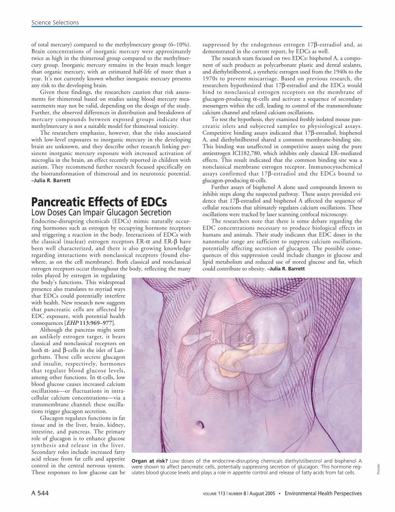
Organ at risk? Low doses of the endocrine-disrupting chemicals diethylstilbestrol and bisphenol Awere shown to affect pancreatic cells, potentially suppressing secretion of glucagon. This hormone reg-ulates blood glucose levels and plays a role in appetite control and release of fatty acids from fat cells.