phylogenesis and biological characterization of a new
TRANSCRIPT
RESEARCH ARTICLE
Phylogenesis and Biological Characterizationof a New Glucose Transporter in the Chicken(Gallus gallus), GLUT12Edouard Coudert1, Géraldine Pascal2, Joëlle Dupont3, Jean Simon1, Estelle Cailleau-Audouin1, Sabine Crochet1, Michel Jacques Duclos1, Sophie Tesseraud1, Sonia Métayer-Coustard1*
1 UR83 Recherches Avicoles, Institut National de la Recherche Agronomique, Nouzilly, France,2 Bioinformatique—Genomique Comparative et Evolutive, Institut National de la Recherche Agronomique,Castanet Tolosan, France, 3 UMR 7247 INRA-CNRS-Université de Tours-Haras Nationaux, Institut Nationalde la Recherche Agronomique, Nouzilly, France
AbstractIn mammals, insulin-sensitive GLUTs, including GLUT4, are recruited to the plasma mem-
brane of adipose and muscle tissues in response to insulin. The GLUT4 gene is absent
from the chicken genome, and no functional insulin-sensitive GLUTs have been character-
ized in chicken tissues to date. A nucleotide sequence is predicted to encode a chicken
GLUT12 ortholog and, interestingly, GLUT12 has been described to act as an insulin-sensi-
tive GLUT in mammals. It encodes a 596 amino acid protein exhibiting 71% identity with
human GLUT12. First, we present the results of a phylogenetic study showing the stability
of this gene during evolution of vertebrates. Second, tissue distribution of chicken SLC2A12mRNA was characterized by RT-PCR. It was predominantly expressed in skeletal muscle
and heart. Protein distribution was analysed by Western blotting using an anti-human
GLUT12 antibody directed against a highly conserved region (87% of identity). An immuno-
reactive band of the expected size (75kDa) was detected in the same tissues. Third a physi-
ological characterization was performed: SLC2A12mRNA levels were significantly lowered
in fed chickens subjected to insulin immuno-neutralization. Finally, recruitment of immuno-
reactive GLUT12 to the muscle plasma membrane was increased following 1h of intraperi-
toneal insulin administration (compared to a control fasted state). Thus insulin administra-
tion elicited membrane GLUT12 recruitment. In conclusion, these results suggest that the
facilitative glucose transporter protein GLUT12 could act in chicken muscle as an insulin-
sensitive transporter that is qualitatively similar to GLUT4 in mammals.
IntroductionIn mammals, facilitated transport of glucose into cells is mediated by a family of facilitative glu-cose transporter (GLUT) proteins. Fourteen isoforms have been described in the human todate: the 12 facilitative glucose transporters GLUT1-12, HMIT (H+coupled myo-inositol
PLOSONE | DOI:10.1371/journal.pone.0139517 October 2, 2015 1 / 18
OPEN ACCESS
Citation: Coudert E, Pascal G, Dupont J, Simon J,Cailleau-Audouin E, Crochet S, et al. (2015)Phylogenesis and Biological Characterization of aNew Glucose Transporter in the Chicken (Gallusgallus), GLUT12. PLoS ONE 10(10): e0139517.doi:10.1371/journal.pone.0139517
Editor: Makoto Kanzaki, Tohoku University, JAPAN
Received: June 4, 2015
Accepted: September 13, 2015
Published: October 2, 2015
Copyright: © 2015 Coudert et al. This is an openaccess article distributed under the terms of theCreative Commons Attribution License, which permitsunrestricted use, distribution, and reproduction in anymedium, provided the original author and source arecredited.
Data Availability Statement: All relevant data arewithin the paper and its Supporting Information files.
Funding: EC is a PhD student supported by a grantfrom INRA (Institut National de la RechercheAgronomique) and Région Centre. This study wasfunded by INRA.
Competing Interests: The authors have declaredthat no competing interests exist.

transporter or GLUT13) and GLUT14. All of them present common sugar transporter features:12 membrane-spanning helices, a N-linked glycosylation site and intracellular NH2 and COOHtermini [1–2]. Based on primary sequence comparisons, the GLUT family is divided into 3 clas-ses: Class I (GLUT1-4 and GLUT14); Class II (GLUT5, 7, 9 and 11) and Class III (GLUT6, 8, 10,12 and HMIT). GLUT proteins have specific roles in whole body glucose homeostasis because oftheir substrate specificity, tissue distribution, cellular location and regulation mechanisms.
Also in mammals, some GLUTs, called insulin-sensitive GLUTs, are acutely recruited to theplasma membrane in response to insulin. GLUT4, which is expressed in insulin-sensitive tis-sues (i.e. skeletal muscle, the heart, adipose tissue), is the major member of this family and oneof the most intensively studied glucose transporters [3] because it is responsible for the insulin-mediated increase in glucose uptake that occurs in response to elevated plasma glucose andinsulin levels in the post-prandial state. Recent findings suggest that, in addition to GLUT4,GLUT12 might also contribute to insulin-stimulated glucose uptake in skeletal muscle and adi-pose tissue [4–7]. Indeed, insulin has been reported to stimulate the translocation of GLUT12from intracellular membrane compartments to the plasma membrane in different models (e.g.MCF-7 breast cancer cells [4], human skeletal muscle [5]). Moreover, transgenic overexpres-sion of this protein enhances insulin sensitivity in mice [6]. These studies suggest that GLUT12may be a second insulin-sensitive glucose-transporter.
Birds and especially chickens exhibit particular features for glucose metabolism. For exam-ple, despite the presence of insulin circulating at “normal” concentrations, chickens present ahigh level of glycemia (2 g/l), and a low sensitivity to exogenous [8–10]. High doses of exoge-nous insulin are required to induce hypoglycemia and chickens resist huge doses of exogenousinsulin, which are lethal in mammals [11]. However, chickens and ducks are not totally insensi-tive to exogenous insulin, which enhances the uptake of glucose in several skeletal muscles[12–13]. Moreover, immuno-neutralization of insulin in young chickens rapidly induces con-siderable increases in plasma levels of glucose [14]. Insulin induces a rapid although modestincrease in glucose uptake by chicken myotubes, an uptake inhibited by phloretin, an inhibitorof glucose transporters [15]. Inhibitory effect of phloretin on glucose uptake has been alsodescribed in isolated muscles from English sparrows (Passer domesticus) [16]. These findingssupport the existence of functional glucose transporters in avian muscle. Nevertheless, themechanism of control of plasma glucose in chickens remains to be elucidated as immuno-reac-tive GLUT1 but not GLUT4 has been detected in chicken tissues [15].
The chicken genome database contains several sequences that have been suggested toencode glucose transporter-like proteins, but none encoding a GLUT4 ortholog. In chickens,only GLUT 1, -2, -3, -8 and -9 have been partially described and characterized [17–20]. One ofthe nucleotide sequences is annotated as encoding a chicken GLUT12 ortholog (SLC2A12gene; ENSGALG00000013980). Though not yet described or characterized in chickens, itmight act as an insulin-sensitive transporter in this species, similarly to GLUT4 in mammals.Phylogeny and synteny analyses were first used to confirm the lack of GLUT4 in chickens andsecondly to demonstrate the existence of a SLC2A12 gene and its stability within vertebratesduring evolution. To further characterize GLUT12 in chickens, we analysed its tissue distribu-tion and finally evaluated its potential sensitivity to insulin at mRNA and protein levels.
Materials and Methods
Phylogenetic and syntenic analysesAll predicted and annoted members of the GLUT family in Gallus gallus were obtained fromthe Ensembl (http://www.ensembl.org/index.html) database (Ensembl release 75). Protein IDsand chromosome location are summarized in Table 1. Analysis of avian GLUTs was performed
GLUT12 Characterization in Chickens
PLOS ONE | DOI:10.1371/journal.pone.0139517 October 2, 2015 2 / 18

on the Phylogeny.fr platform (http://www.phylogeny.fr [21, 22]) using Ensembl protein IDs.Sequences were aligned with MUSCLE (v3.7) configured for highest accuracy (MUSCLE withdefault settings). After alignment, ambiguous regions (i.e. containing gaps and/or poorly aligned)were removed with Gblocks (v0.91b) using the following parameters: minimum length of a blockafter gap cleaning equal to 10; no gap positions allowed in the final alignment; rejection of all seg-ments with contiguous non-conserved positions larger than 8; minimum number of sequences fora flank position equal to 85%. The phylogenetic tree was reconstructed using the maximum likeli-hood method implemented in the PhyML program (v3.0 aLRT). The default substitution modelwas selected. Reliability of internal branches was assessed using the aLRT test (SH-Like). Graphicalrepresentation and edition of the phylogenetic tree were performed with TreeDyn (v198.3).
Chicken GLUT12 was compared with GLUT12s from other species contained in theEnsembl database. Accession numbers are summarized in Table 2. Analyses of the orthologousrelationships between different species were carried out by combining the phylogenetic
Table 1. Predicted and annoted GLUTs from chicken (Gallus gallus) Ensembl database.
Protein Protein ID (Ensembl) Chromosome location
GLUT1 ENSGALP00000007795 21
GLUT2 ENSGALP00000015129 9
GLUT3 ENSGALP00000036432 1
GLUT5 ENSGALP00000003884 21
GLUT6 ENSGALP00000004710 17
GLUT8 ENSGALP00000001249 17
GLUT9 ENSGALP00000031420 4
GLUT10 ENSGALP00000007155 20
GLUT11 ENSGALP00000009743 15
GLUT12 ENSGALP00000022613 3
doi:10.1371/journal.pone.0139517.t001
Table 2. Predicted and annotated GLUT12s from various species in the Ensembl database.
Species Protein ID Amino acid number
Wallaby (Macropus eugenii) ENSMEUP00000005529 614
Opossum (Monodelphis domestica) ENSMODP00000021849 627
Mouse (Mus musculus) ENSMUSP00000043962 622
Rat (Rattus norvegicus) ENSRNOP00000015566 621
Rabbit (Oryctolagus cuniculus) ENSOCUP00000006133 610
Tarsier (Tarsius syrichta) ENSTSYP00000011636 435
Orangutan (Pongo pygmaeus) ENSPPYP00000019063 576
Human (Homo sapiens) ENSP00000275230 617
Macaque (Macaca mulatta) ENSMMUP00000030186 579
Pig (Sus scrofa) ENSSSCP00000004506 621
Cow (Bos taurus) ENSBTAP00000025291 621
Turkey (Meleagris gallopavo) ENSMGAP00000013845 560
Chicken (Gallus gallus) ENSGALP00000022613 596
Zebra finch (Taeniopygia guttata) ENSTGUP00000011894 558
Anole lizard (Anolis carolinensis) ENSACAP00000010115 552
Frog (Xenopus tropicalis) ENSXETP00000000236 527
Zebrafish (Danio rerio) ENSDARP00000053530 610
Fugu (Takifugu rubripes) ENSTRUP00000007853 529
Tetraodon (Tetraodon nigroviridis) ENSTNIP00000019417 522
doi:10.1371/journal.pone.0139517.t002
GLUT12 Characterization in Chickens
PLOS ONE | DOI:10.1371/journal.pone.0139517 October 2, 2015 3 / 18

reconstruction of the gene family with the syntenic comparison. The GLUT12 proteins identi-fied were then analysed by the PhyleasProg web server (Phylogenetic Analysis Programs v2.7[23]) with fine computation and results on orthologs and paralogs. The phylogenetic tree ofthe SLC2A12 gene was displayed by Archaeopteryx. Selection pressures were acted using thePALMmethod of PhyleasProg [24] on the avian GLUT12 sequence. Gene arrangement of thegenomic region encompassing GLUT12 was performed with the genome browser Genomicusv79.01 –PhyloView (http://www.genomicus.biologie.ens.fr) [25].
ChemicalsDNAse I was purchased from Roche Applied Science (Meylan, France).The pre-made poly-acrylamide solution was obtained from Euromedex (Souffelweyersheim, France). Nitrocellu-lose membrane and protein standards were purchased from Bio-Rad Laboratories (Hercules,CA, USA). Odyssey blocking buffer was purchased from Li-cor (LI-COR Inc. Biotechnology,Lincoln, NE). The Membrane Protein extraction kit was purchased from PromoKine (Promo-Cell, Heidelberg, GER).
An anti-human GLUT12 antibody was purchased from Abcam (ab100993, Cambridge,UK). This rabbit polyclonal antibody is directed against residues 250–350 of Human GLUT12.Identity between human and predicted chicken sequences in this region is high (87%). Themonoclonal anti-human vinculin antibody was from Sigma (V9131, Sigma Chemical Com-pany). Secondary antibodies (anti-rabbit and anti-mouse IgG (H+L)) were conjugated toDyLight1 680 fluorescent dye and purchased from Cell signaling (#5366 and #5470, Cell sig-naling-Ozyme, Saint Quentin Yvelines, FR).
AnimalsThis study was conducted in strict accordance with the European Union Guidelines for animalcare. All investigators were certified by the French government to carry out animal experimentsand this study was conducted under authorization 37–085 delivered to J. Dupont by the FrenchMinistry of Agriculture. Tissues were issued from our earlier study [14], where experimentalconditions are described in detail. All procedures were approved by the French AgriculturalAgency and the Scientific Research Agency and conducted in accordance with the guidelinesfor Care and Use of Agricultural Animals in Agricultural Research and Teaching. Panels of tis-sues were issued from chickens reared conventionally in an environmentally controlled poultryhouse. All the chickens were sacrificed by decapitation and different tissues were removed,quickly frozen and stored at -80°C. All efforts were made to minimize suffering.
Expression of SLC2A12 mRNA and protein distribution of GLUT12 inchicken tissuesAt 6 weeks of age, male broiler chickens (N = 6) were slaughtered and different skeletal musclesas well as several tissues such as brain, liver, adipose tissue and heart were removed and snapfrozen in liquid nitrogen.
Ontogenesis study was restricted to the Pectoralis major and the leg muscles, collected at 19days during embryogenesis, hatch and 5 days post-hatch. Muscles were removed and snap fro-zen in liquid nitrogen.
Insulin immune-neutralization modelMale broiler chickens were housed in an environment-controlled room and they were fed adlibitum with a conventional balanced diet based on corn, wheat, peas, soy bean, meal, corn
GLUT12 Characterization in Chickens
PLOS ONE | DOI:10.1371/journal.pone.0139517 October 2, 2015 4 / 18
gluten and rapeseed oil [14]. As described in this article, at 16–17 days of age, two groups ofchickens of similar body weights were constituted (N = 7 per group). The fed immuno-neutral-ized group received three i.v. injections of anti-porcine insulin guinea pig serum (1.5 ml/kg)(PromoCell, Heidelberg, Germany) at times 0, 2 and 4 h. Guinea pig serum was used as thevehicle for the anti-porcine insulin. The fed control group received three i.v. injections deliver-ing only normal guinea pig serum (1.5 ml/kg) at times 0, 2 and 4 h. At 5h post-injection, chick-ens were slaughtered and 2 different types of muscle, a fast-twitch glycolytic muscle (Pectoralismajor) and a mixed type muscle (leg muscle) were sampled, snap frozen in liquid nitrogen andstored at -80°C until analysis.
GLUT12 translocation experimentMale broiler chickens were fasted overnight or fed and injected with insulin (1U/kg) by intra-peritoneal injection or not injected (N = 3 per group). Broiler chickens were slaughtered onehour after insulin injection and leg muscles were sampled and snap frozen in liquid nitrogen.
RNA isolation and RT-PCRTotal RNA was extracted from 100 mg tissue samples (200 mg for adipose tissue samples)using RNA Now (Biogentec, Seabrook, TX, USA) according to the manufacturer’s recommen-dations. After RNase-Free DNase treatment (Ambion, Clinisciences, Montrouge, France),RNA was reverse-transcribed using Super Script II RNase H Reverse Transcriptase (Invitrogen,Carlsbad, CA, USA) with Random Primers (Promega, Charbonnieres-les-Bains, France). Thespecifically designed and validated sequences of primers used for identification of chickenSLC2A12 were: (Forward) 5’ AGA-GAG-TGG-GGA-GGT-TCC-C 3’; (Reverse) 5’ TCA-G-GA-CGA-GCC-AAG-ACA 3’. SLC2A12mRNA was detected by reverse transcriptase-poly-merase chain reaction (RT-PCR) in various chicken tissues.
cDNA samples were subsequently amplified in triplicate by real time PCR using Sybr GreenI Master kit (Roche, Mannheim, Germany) and a LighterCycler1 480 II apparatus (Roche,Meylan, France). β-actin and cytochrome (Cyt b) messengers were measured as referencesusing primers previously validated for β-actin [14, 26] or primers specifically designed and vali-dated for chicken cytochrome b (Cyt b) ((Forward) 5’ CGG- ACG-AGG-CCT-ATA-CTA-CG3’; (Reverse) 5’ GGG-AGA-ACA-TAG-CCC-ACA-AA 3’). Gene expression levels were esti-mated on the basis of PCR efficiency and threshold cycle (Ct) deviation of an unknown sampleversus a control, as previously described [27].
Western blottingTissue lysates were prepared as previously described [28] to analyse the tissue distribution ofGLUT12. Lysates were centrifuged at 1000 g for 30 min at 10°C and supernatants were thencentrifuged at 31000 g for 45 min at 10°C. Solubilized lysates (80 μg of protein) were subjectedto SDS-PAGE andWestern blotting using anti-GLUT12, then anti-vinculin antibodies (afterstripping). GLUT12 is a polyclonal antibody. After washing, membranes were incubated with aDyLight1 680 fluorescent antibody. Bands were visualized by Infrared Fluorescence using theOdyssey1 Imaging System (LI-COR Inc. Biotechnology, Lincoln, NE) and quantified by Odys-sey infrared imaging system software (Application software, version 1.2).
For GLUT12 translocation studies, membrane and cytosolic proteins were extracted withthe Promokine commercial kit (Promocell) according to the manufacturer’s recommendationsusing 750 mg muscle samples. The plasma membrane proteins extraction procedure using thiscommercial kit has been validated through different publications in the literature [29, 30] andin chickens using antibodies directed against markers specific of plasma membranes (i.e NaK
GLUT12 Characterization in Chickens
PLOS ONE | DOI:10.1371/journal.pone.0139517 October 2, 2015 5 / 18

ATPase and CD56). Moreover, we checked NaK ATPase enrichment in the membrane afterinsulin stimulation [31]. GLUT12 was detected and quantified as above.
Statistical analysisValues are presented as means ± SEM. Data were processed using the Statview Software pro-gram, version 5 (SAS Institute, Cary, NC, USA). Data were subjected to analysis of variance(ANOVA) to detect significant intergroup differences (Tissue comparison; Fed vs Ins immuno-neutralised). The means were compared by Fisher’s least significant difference test in the caseof a significant effect. P<0.05 was considered statistically significant.
Results
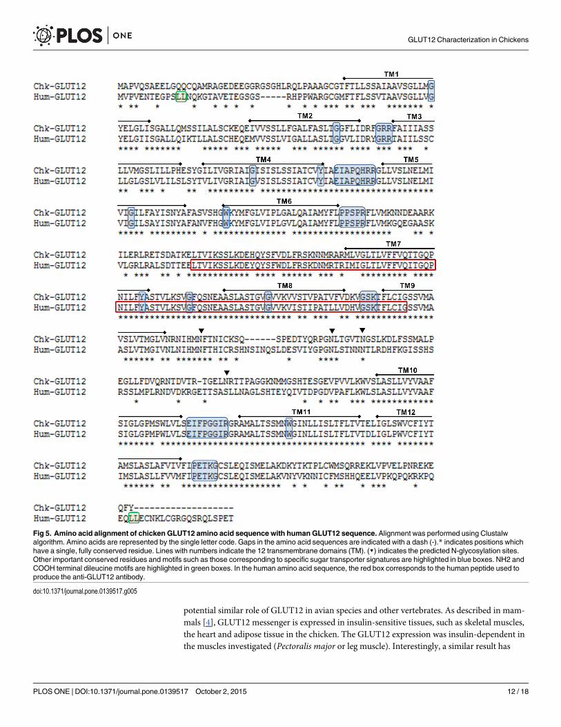
Further evidence of the absence of GLUT4 in the chicken genomeUp to ten predicted and annoted GLUTs were found in the Ensembl chicken database (Ensemblrelease 75; Table 1). According to the phylogenetic analysis of these chicken GLUTs (Fig 1), wediscriminated three main branches corresponding to the three classes of GLUTs described inmammals: Class I (GLUT1, 2, and 3), Class II (GLUT5, 9 and 11) and Class III (GLUT6, 8, 10and 12). The different chicken GLUTs showed 24 to 69% of identity between the different para-logs, highest percentage of identity being found between GLUT1 and GLUT3 (Table 3).
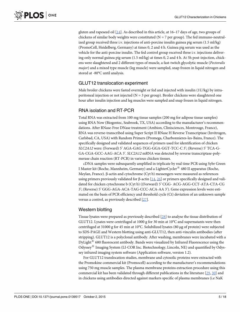
The chicken genome sequencing available to date has not provided any evidence regardingthe presence of a chicken GLUT4 although a GLUT4 ortholog or SLC2A4 gene has been foundin various species of fish (Cod, Stickleback, Fugu, Tetraedon, Platyfish, Medaka and Tilapia), aswell as in Sauropsida (Chinese softshell turtle) and Reptilia (Anole lizard) classes (data notshown). No sequence encoding a SLC2A4 ortholog has been found in other birds (Zebra finch,Turkey or Duck), the Frog or Zebrafish. The human SLC2A4 gene is located on chromosome17 in a synteny block that is well conserved within vertebrates (data not shown). The region inwhich the human SLC2A4 gene is located is almost entirely missing from the current chickensequence assembly (Fig 2).
Fig 1. Phylogenetic tree of multiple alignments of all known chicken GLUT protein sequences. The analysis of chicken GLUTs was performed on thePhylogeny.fr platform (http://www.phylogeny.fr; [20, 21]) using Ensembl protein IDs. The tree was constructed as described in Materials and Methods.Reliability of internal branches was assessed using the aLRT test. The scale represents the substitution rate.
doi:10.1371/journal.pone.0139517.g001
GLUT12 Characterization in Chickens
PLOS ONE | DOI:10.1371/journal.pone.0139517 October 2, 2015 6 / 18
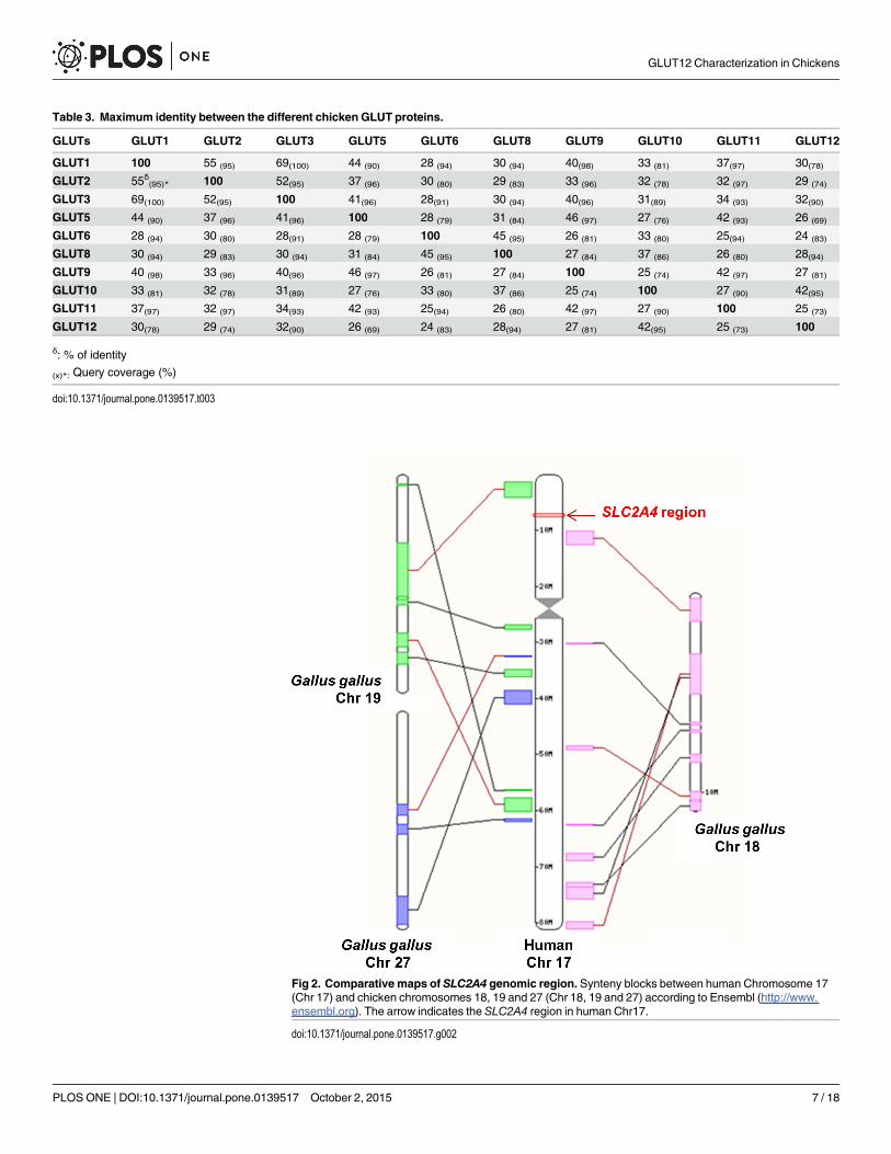
Fig 2. Comparative maps of SLC2A4 genomic region. Synteny blocks between human Chromosome 17(Chr 17) and chicken chromosomes 18, 19 and 27 (Chr 18, 19 and 27) according to Ensembl (http://www.ensembl.org). The arrow indicates the SLC2A4 region in human Chr17.
doi:10.1371/journal.pone.0139517.g002
Table 3. Maximum identity between the different chicken GLUT proteins.
GLUTs GLUT1 GLUT2 GLUT3 GLUT5 GLUT6 GLUT8 GLUT9 GLUT10 GLUT11 GLUT12
GLUT1 100 55 (95) 69(100) 44 (90) 28 (94) 30 (94) 40(98) 33 (81) 37(97) 30(78)GLUT2 55δ(95)* 100 52(95) 37 (96) 30 (80) 29 (83) 33 (96) 32 (78) 32 (97) 29 (74)
GLUT3 69(100) 52(95) 100 41(96) 28(91) 30 (94) 40(96) 31(89) 34 (93) 32(90)GLUT5 44 (90) 37 (96) 41(96) 100 28 (79) 31 (84) 46 (97) 27 (76) 42 (93) 26 (69)
GLUT6 28 (94) 30 (80) 28(91) 28 (79) 100 45 (95) 26 (81) 33 (80) 25(94) 24 (83)
GLUT8 30 (94) 29 (83) 30 (94) 31 (84) 45 (95) 100 27 (84) 37 (86) 26 (80) 28(94)GLUT9 40 (98) 33 (96) 40(96) 46 (97) 26 (81) 27 (84) 100 25 (74) 42 (97) 27 (81)
GLUT10 33 (81) 32 (78) 31(89) 27 (76) 33 (80) 37 (86) 25 (74) 100 27 (90) 42(95)GLUT11 37(97) 32 (97) 34(93) 42 (93) 25(94) 26 (80) 42 (97) 27 (90) 100 25 (73)
GLUT12 30(78) 29 (74) 32(90) 26 (69) 24 (83) 28(94) 27 (81) 42(95) 25 (73) 100
δ: % of identity
(x)*: Query coverage (%)
doi:10.1371/journal.pone.0139517.t003
GLUT12 Characterization in Chickens
PLOS ONE | DOI:10.1371/journal.pone.0139517 October 2, 2015 7 / 18

Is there another insulin-sensitive GLUT in the chicken?Phylogenetic analysis of GLUT12. The analysis of chicken GLUTs (Table 1) showed the
existence of a GLUT12 ortholog (corresponding to the SLC2A12 gene) in the chicken. TheSLC2A12 gene is located on chromosome 3, and comprises 5 exons and encodes a putative 596amino acid protein. The phylogenetic analysis of GLUT12 amino acid sequences in differentspecies (Table 2) is presented in Fig 3. We decided to determine whether selection pressuresacted on the avian GLUT12 sequence. No positive selection pressure was evidenced in these 19
Fig 3. Phylogenetic tree of protein sequence of chicken GLUT12 and GLUT12 from other vertebrates (see Table 2). The tree was constructed asdescribed in Materials and Methods using PhyleasProg analysis web server (Phylogenetic Analysis Programs v2.7).
doi:10.1371/journal.pone.0139517.g003
GLUT12 Characterization in Chickens
PLOS ONE | DOI:10.1371/journal.pone.0139517 October 2, 2015 8 / 18

species. Synteny analysis and comprehensive genome location searches (Fig 4A and 4B)showed a similar arrangement of the SLC2A12 gene and its neighbouring genes upstream anddownstream in the chicken and other species.
Sequence analysis of avian GLUT12 protein. The chicken SLC2A12 gene encodes a pro-tein which exhibits 71% sequence identity to human GLUT12. Alignment of the deducedamino acid sequence of chicken and human GLUT12 proteins showed that chicken GLUT12possesses all the features essential for sugar transport: 12 membrane-spanning helices, intracel-lular NH2 and COOH termini and conserved amino acid residues and motifs important forsugar transport activity (i.e., seven glycine residues, tryptophan in helices 6 and 11, tyrosine inhelices 4 and 7) (Fig 5).The amino acid sequence of GLUT12 presented at least four putativeextracellular N-linked glycosylation sites in the larger loop 9 at amino acid residues 376, 395,401 and 434, as predicted using the GlycoEP webserver [32]. The di-leucine motifs (LL) presentat the NH2 and COOH termini of human GLUT12, which are considered to mediate internali-zation, were not found in chicken GLUT12 (Fig 5).
Tissue expression of SLC2A12 mRNA and protein distribution of GLUT12 in chickentissues. The tissue expression pattern of the SLC2A12messenger RNA was analysed in vari-ous tissues from fed chickens by real-time PCR. SLC2A12mRNA was detected in all tissuesamples, but was the most abundant in skeletal (Pectoralis major and leg muscles) muscles, i.e.insulin-sensitive tissues (Fig 6A).
Tissue distribution of GLUT12 protein was analysed by Western blotting using an antibodyagainst human GLUT12 (Fig 6B). Immuno-reactive bands around the expected size (75 kDa)were recognized in insulin-sensitive tissues such as skeletal muscles, adipose tissue and heart.Another signal, that was stronger than that at 75 kDa, was also detected at around 50 kDa inskeletal muscle lysates but not in other tissues. This band at 50 kDa is detected only in chickenmuscle lysates from animals after hatch and not in muscles lysates from embryo (E19) (Fig6C). At E19, the 75 kDa form was highly detected and then decreased strongly at hatch. Con-versely, the 50 kDa form appeared at hatch and then increased strongly at Day 5 and is stilldetected at 6 weeks of age.
Insulin-sensitivity of chicken SLC2A12 /GLUT12. We next analysed the effects of insulinon SLC2A12 gene expression in skeletal muscles using an insulin immuno-neutralizationmodel previously described and characterized [14]. SLC2A12 gene expression was measured 5hpost-injection by qRT-PCR in two different types of muscle: a glycolytic muscle (Pectoralismajor) and a mixed type muscle (leg muscle). SLC2A12mRNA expression was significantlylower in the insulin-immuno-neutralized condition compared to controls in both muscle types(Pectoralis major and leg muscles) (Fig 7).
The last issue addressed in the present study was the existence of potential regulation ofGLUT12 at the protein level. More extreme experimental models were used: fasted vs. fed statesand acute insulin injection (1U/kg).
Membrane and cytosolic proteins were prepared from leg muscles and analysed by Westernblotting (Fig 8). The 75 kDa immuno-reactive band was observed in membrane fractions (Fig8, upper part) from fed animals in the absence of insulin injection, but not in fasted animals.Membrane GLUT12 protein significantly increased in fasted and fed conditions in response toinsulin injection. In the cytosolic fraction (Fig 8, lower part), the immuno-reactive GLUT12band was detected at identical levels, whatever the experimental treatment. These observationsdemonstrate that GLUT12 translocates to the plasma membrane in response to insulin inchicken skeletal muscle.
GLUT12 Characterization in Chickens
PLOS ONE | DOI:10.1371/journal.pone.0139517 October 2, 2015 9 / 18

GLUT12 Characterization in Chickens
PLOS ONE | DOI:10.1371/journal.pone.0139517 October 2, 2015 10 / 18

DiscussionIn mammals, GLUT4 is responsible for glucose uptake by insulin-sensitive tissues such as skel-etal muscle, heart and adipose tissue. Up to now, the chicken genome database contains severalsequences that are suggested as encoding glucose transporter-like proteins, but no one encod-ing a GLUT4 ortholog. Phylogeny and synteny analyses confirmed the lack of GLUT4 in chick-ens although a GLUT4 ortholog or SLC2A4 gene has been found in various species of fish, aswell as in Sauropsida (Chinese softshell turtle) and Reptilia (Anole lizard) classes. Moreover,the region in which the human SLC2A4 gene is located is almost entirely missing from the cur-rent chicken sequence assembly, suggesting that GLUT4 may have been lost in bird lineages.The absence of GLUT4 in chicken could explain partially the particular features of glucosemetabolism exhibited by the avian species. Nevertheless chickens are not totally insensitive toexogenous insulin and the existence of a functional insulin-sensitive glucose transport in avianmuscle has been demonstrated even though it remains lower compared to mammals [12, 14–16]. Identity of the GLUT involved is controversial. In one study conducted on ducks, animmuno-reactive band has been detected at the expected size for GLUT4 using an anti-ratGLUT4 antibody and shown to translocate to the plasma membrane in response to insulinadministration and in parallel to glucose use by the leg [13]. Nevertheless, this band has neverbeen sequenced to confirm that it is really a GLUT4 ortholog and not another GLUT showinga high sequence homology. Moreover, no sequence encoding a SLC2A4 ortholog has beenfound in ducks. GLUT1 has also been suggested, but no change in GLUT1 quantity was shownusing membranes prepared from myotubes incubated under conditions where glucose trans-port was increased [15].
Mice that have a complete ablation of GLUT4 are growth retarded and have a significantcardiac hyperthophy, reduced body weight and adiposity levels and are less sensitive to insulin[33]. Whereas specific deletion of muscle GLUT4 does not affect glucose disposal and glucosetolerance suggesting that compensation from the transporters may contribute to this unalteredhomeostasis of glucose and that another GLUT may also be involved in the regulation of wholebody glucose homeostasis [34]. Recent findings suggest that GLUT12 might contribute to insu-lin-stimulated glucose uptake in insulin-sensitive tissues, in addition to GLUT4 [4–7]. Indeed,transgenic overexpression of GLUT12 in mice enhances insulin sensitivity with no change inGLUT4 content [6] and a compensatory increase in the expression of GLUT12 was observed ingenetically altered mice lacking GLUT4 [35]. At the protein level, GLUT12 was also signifi-cantly elevated in the myocardium of GLUT4 null mice compared to wild type controls [36]. Asequence encodes a chicken GLUT12 ortholog. The phylogenetic analyses demonstrated thatno specific changes in SLC2A12 gene occurred during evolution and thus strongly supported asimilar evolution of the SLC2A12 gene in avian species and other vertebrates, suggesting a
Fig 4. Comparative maps of SLC2A12 genomic region. (A) The genomic location of SLC2A12. syntenyblocks between human Chromosome 6 (Chr 6) and chicken chromosomes 1, 2, 3 and 26 (Chr 1, Chr 2, Chr3and Chr 26) according to Ensembl (http://www.ensembl.org). The arrows indicate SLC2A12 regions. (B)Gene arrangement of the genomic region encompassing SLC2A12 according to the genome browserGenomicus (http://www.genomicus.biologie.ens.fr, Genomicus—database version: 79.01 / Web-codeversion: 2014-09-19). Schematic gene maps of the conserved syntenic regions of SLC2A12 inGallus gallusand in other vertebrates.The gene initially placed in the centre of the display and aligned over a vertical blackline is the gene that was used as query (reference gene = SLC2A12). Coloured genes over a light bluebackground corresponds to extant or ancestral genes that are orthologous to genes from the species used inthe query that show the same colour. The coding direction of the genes is indicated by the pointed end. Chr:denotes the chromosome. Neighbouring genes upstream and downstream in the chicken and other species:SGK1, serum/glucocorticoid regulated kinase 1; ALDH8A1, aldehyde dehydrogenase 8 family, member A1;HBS1L, uncharacterized protein; TBPL1, TBP-like 1; TCF21, transcription factor 21; EYA4, eyes absenthomolog 4; RPS12, ribosomal protein S12.
doi:10.1371/journal.pone.0139517.g004
GLUT12 Characterization in Chickens
PLOS ONE | DOI:10.1371/journal.pone.0139517 October 2, 2015 11 / 18
potential similar role of GLUT12 in avian species and other vertebrates. As described in mam-mals [4], GLUT12 messenger is expressed in insulin-sensitive tissues, such as skeletal muscles,the heart and adipose tissue in the chicken. The GLUT12 expression was insulin-dependent inthe muscles investigated (Pectoralis major or leg muscle). Interestingly, a similar result has
Fig 5. Amino acid alignment of chicken GLUT12 amino acid sequence with human GLUT12 sequence. Alignment was performed using Clustalwalgorithm. Amino acids are represented by the single letter code. Gaps in the amino acid sequences are indicated with a dash (-).* indicates positions whichhave a single, fully conserved residue. Lines with numbers indicate the 12 transmembrane domains (TM). (▼) indicates the predicted N-glycosylation sites.Other important conserved residues and motifs such as those corresponding to specific sugar transporter signatures are highlighted in blue boxes. NH2 andCOOH terminal dileucine motifs are highlighted in green boxes. In the human amino acid sequence, the red box corresponds to the human peptide used toproduce the anti-GLUT12 antibody.
doi:10.1371/journal.pone.0139517.g005
GLUT12 Characterization in Chickens
PLOS ONE | DOI:10.1371/journal.pone.0139517 October 2, 2015 12 / 18
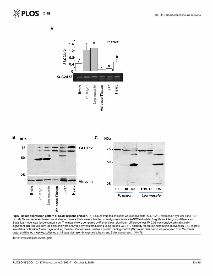
Fig 6. Tissue expression pattern of GLUT12 in the chicken. (A) Tissues from fed chickens were analysed for SLC12A12 expression by Real-Time PCR(N = 6). Values represent means and standard errors. Data were subjected to analysis of variance (ANOVA) to detect significant intergroup differences.Statistical model was tissue comparison. The means were compared by Fisher’s least significant difference test. P<0.05 was considered statisticallysignificant. (B) Tissues from fed chickens were analysed byWestern blotting using an anti-GLUT12 antibody for protein distribution analyses (N = 5). In grey:skeletal muscles (Pectoralis major and leg muscle). Vinculin was used as a protein loading control. (C) Protein distribution was analysed from Pectoralismajor and the leg muscles, collected at 19 days during embryogenesis, hatch and 5 days post-hatch. (N = 7).
doi:10.1371/journal.pone.0139517.g006
GLUT12 Characterization in Chickens
PLOS ONE | DOI:10.1371/journal.pone.0139517 October 2, 2015 13 / 18

been described in zebrafish [37], a species lacking GLUT4 [38]. GLUT12 protein is expressedin the skeletal muscles (Pectoralis major and leg muscles), heart and adipose tissue under differ-ent forms: two high molecular forms at around 75kDa (particularly clear in the heart and theadipose tissue), the expected size according to the amino acid sequence, and a low molecularform at 50 kDa exclusively in skeletal muscles. The different bands detected around 75 kDacould correspond to different glycosylated forms as suggested by the four putative extracellularN-linked glycosylation sites and preliminary deglycosylation experiments (S1 Fig). These find-ings are however still to be clearly demonstrated. The higher bands around 75 kDa form areenriched at the cell membrane of fed chickens when plasma insulin levels are typically high orunder insulin stimulation (compared to fasted chickens). Therefore insulin administered invivo stimulates the translocation of GLUT12 to the plasma membrane in chicken skeletal mus-cles. In mammals, GLUT12 translocation seems to depend on the tissues and/or the presenceof GLUT4. Indeed, there is still some controversy whether in the heart GLUT12 might be regu-lated by insulin [36]. In a healthy myocardium, insulin stimulation did not increase transloca-tion of GLUT12, whereas it increased translocation of GLUT4. Cell membrane GLUT12content was only increased in the diabetic myocardium, potentially as a compensatory mecha-nism for the observed down regulation of GLUT4.
Some study described the importance of post-translational modifications of GLUT4 for itssubcellular location and translocation [39]. Among potential post-translational modifications,a conserved N-glycosylation consensus site in GLUT4 is positioned in the first extracellularloop. Critical roles for this N-glycan chain in intracellular trafficking and stability of GLUT4have been shown and confirmed using an inhibitor of endoplasmic reticulum (ER) / Golgi α-mannosidase I (kifunensine, KIF) or a GLUT4 mutant lacking the N-glycosylation site [39–41]. The amino acid sequence of chicken GLUT12 presented at least four putative extracellularN-linked glycosylation sites in the larger loop 9. These N-glycosylation sites are a characteristicof Class III facilitated glucose transporters. A similar N-linked glycosylation motif was found
Fig 7. Effects of insulin on SLC2A12 gene expression in skeletal muscles.mRNA levels of SLC2A12were measured by Real-Time PCR in Pectoralis major and leg muscles of fed chicken controls (Fed group)and fed chickens injected with an anti-insulin antibody (Ins immuno-neutalized group) (N = 7). Cytochrome b(Cytb) and β-actin were used as housekeeping genes.Values represent means and standard errors. Datawere subjected to analysis of variance (ANOVA) to detect significant intergroup differences (Fed vs Insimmuno-neutralized). The means were compared by Fisher’s least significant difference test. P<0.05 wasconsidered statistically significant.
doi:10.1371/journal.pone.0139517.g007
GLUT12 Characterization in Chickens
PLOS ONE | DOI:10.1371/journal.pone.0139517 October 2, 2015 14 / 18

in K+Cl- cotransporters (KCCs) of the SLC12 family, and site-directed mutagenesis approacheshave demonstrated that its glycosylation is essential for regulating cell surface expression, sta-bility and activity of KCC4 proteins [42].
The GLUT12 form of lower molecular weight (50 kDa) was exclusively detected in chickenskeletal muscles after hatch. Similar signals at around 50 kDa have been reported for GLUT12in bovines and goats [43–44]. A strong resistance to the effect of insulin on glucose metabolismhas been reported in ruminant animals [45]. The authors suggest that the low sensitivity toinsulin may be related to the cytoplasmic distribution of ruminant GLUT12 lacking the dileu-cine motif (LL) [43–44]. The dileucine motif typical for internalization has been described inhuman GLUT12 sequence but is not retrieved in chicken GLUT12 nor in species considered asinsulin-resistant such as goat or bovine [43–44]. This di-leucine motif in C-term has been alsodescribed for another insulin-sensitive transporter GLUT4 and it was not retrieved in fishGLUT4 sequence [46]. Despite the lack of this motif and differences in the GLUT4 traffic char-acteristics, fish glucose transporter GLUT4 can translocate to the cell surface in response toinsulin in skeletal muscle cells. The chicken GLUT12 as well as the fish GLUT4 presented someparticular features that could result in differences in protein trafficking and stability on the cellsurface and might partially explain their atypical glucose metabolism and their natural insulinresistance [47–48]. We are conscious that an extensive characterization of their traffic is now
Fig 8. GLUT12 translocation after insulin stimulation. In the upper part, a representative Western blotshows GLUT12 content in membrane fractions prepared from leg muscle of fasted and fed animals withoutinsulin injection or 1hr after insulin injection (1U/kg). In the lower part, a representative Western blot showsGLUT12 content in the corresponding cytosolic proteins used as a control of the cytosolic GLUT12 content.Vinculin was used as a loading control for the two fractions.
doi:10.1371/journal.pone.0139517.g008
GLUT12 Characterization in Chickens
PLOS ONE | DOI:10.1371/journal.pone.0139517 October 2, 2015 15 / 18

required. Its purification and characterization are necessary to better know the nature of thisform and determine if the detected signal corresponds to an immature form or a truncatedform of GLUT12 and its role in glucose metabolism.
In the chicken, therefore, GLUT12 may act as an insulin-sensitive transporter because it isexpressed in insulin-sensitive tissues and it can be recruited to the plasma membranes in fedconditions or following insulin injection. Nevertheless, despite considerable similarity of pro-tein sequence with mammalian GLUT12, the chicken GLUT12 presented some particular fea-tures that could result in differences in protein trafficking and stability on the cell surfacemembrane in the chicken. The consequences might partially explain the low sensitivity ofchickens to exogenous insulin. Extensive characterization of the GLUT12 trafficking as well asits regulation in the chicken is now required.
Supporting InformationS1 Fig. Deglycosylation of GLUT12 from chicken leg muscle and adipose tissue. Leg muscleand adipose tissue lysates were prepared using Enzymatic DeGlycoMx Kit from QA bio andincubated for 3 hours at 37°C according to the manufacturer’s recommendations to analyse theglycosylations of GLUT12. Representative immunoblot of no deglycosylated samples (Ctl) anddeglycosylated samples (DeGly: deglycosylated). Membrane was probed with the anti-GLUT12antibody, and then anti-vinculin antibodies (after stripping).(PDF)
AcknowledgmentsThe authors thank the staff of the poultry breeding facilities (INRA, UE 1295 Pôle d’Experi-mentation Avicole de Tours, Nouzilly, France) for animal care, the staff of Avian ResearchUnit (INRA, UR83 Recherches Avicoles, Nouzilly, France) for technical assistance and D.Raine (Surrey, UK) for revision of the English language.
Author ContributionsConceived and designed the experiments: EC ST JD JS MJD SMC. Performed the experiments:EC GP ECA SC SMC. Analyzed the data: EC GP ECA SC SMC. Contributed reagents/materi-als/analysis tools: EC GP JD JS ECA SCMJD ST SMC. Wrote the paper: EC ST SMC. Paperrevision: GP JD JS ECA SCMJD ST.
References1. Augustin R. The protein family of glucose transport facilitators: It's not only about glucose after all.
IUBMB Life. 2010; 62(5): 315–333. doi: 10.1002/iub.315 PMID: 20209635
2. Mueckler M and Thorens B. The SLC2 (GLUT) family of membrane transporters. Mol Aspects Med.2013; 34(2–3):121–138. doi: 10.1016/j.mam.2012.07.001 PMID: 23506862
3. Huang S and Czech MP. The GLUT4 glucose transporter. Cell Metab.2007; 5(4):237–252 PMID:17403369
4. Rogers S, MachedaML, Docherty SE, Carty MD, Henderson MA, Soeller WC, et al. Identification of anovel glucose transporter-like protein-GLUT-12. Am J Physiol Endocrinol Metab. 2002; 282(3):E733–738. PMID: 11832379
5. Stuart CA, Howell ME, Zhang Y and Yin D. Insulin-stimulated translocation of glucose transporter(GLUT) 12 parallels that of GLUT4 in normal muscle. J Clin Endocrinol Metab. 2009; 94(9):3535–3542.doi: 10.1210/jc.2009-0162 PMID: 19549745
6. Purcell SH, Aerni-Flessner LB, Willcockson AR, Diggs-Andrews KA, Fisher SJ and Moley KH.Improved insulin sensitivity by GLUT12 overexpression in mice. Diabetes. 2011; 60(5): 1478–1482.doi: 10.2337/db11-0033 PMID: 21441439
GLUT12 Characterization in Chickens
PLOS ONE | DOI:10.1371/journal.pone.0139517 October 2, 2015 16 / 18
7. Pujol-Giménez J, Barrenetxe J, González-Muniesa P and Lostao MP. The facilitative glucose trans-porter GLUT12: what do we know and what would we like to know? J Physiol Biochem. 2013; 69(2):325–333. doi: 10.1007/s13105-012-0213-8 PMID: 23385668
8. Simon J. Chicken as a useful species for the comprehension of insulin action. Critical reviews in PoultryBiology. 1989; 2:121–148.
9. Akiba Y, Chida Y, Takahashi T, Ohtomo Y, Sato K and Takahashi K. Persistent hypoglycemia inducedby continuous insulin infusion in broiler chickens. Br Poult Sci. 1999; 40(5): 701–705. PMID: 10670686
10. Braun EJ and Sweazea KL. Glucose regulation in birds. Comp Biochem Physiol B BiochemMol Biol.2008; 151(1):1–9. doi: 10.1016/j.cbpb.2008.05.007 PMID: 18571448
11. GibsonWR, Bourne AR and Sernia C. D-Xylose transport in isolated skeletal muscle of chickens:effects of insulin and tolbutamide. Comp Biochem Physiol C. 1980; 67C(1):41–47. PMID: 6107208
12. Tokushima Y, Takahashi K, Sato K and Akiba Y. Glucose uptake in vivo in skeletal muscles of insulin-injected chicks. Comp Biochem Physiol B BiochemMol Biol; 2005; 141(1):43–48. PMID: 15820133
13. Thomas-Delloye V, Marmonier F, Duchamp C, Pichon-Georges B, Lachuer J, Barré H, et al. Biochemi-cal and functional evidences for a GLUT-4 homologous protein in avian skeletal muscle. Am J Physiol.1999; 277 6(Pt2): R1733–40.
14. Dupont J, Tesseraud S, Derouet M, Collin A, Rideau N, Crochet S, et al. Insulin immuno-neutralizationin chicken: effects on insulin signaling and gene expression in liver and muscle. J Endocrinol. 2008;197(3):531–542. doi: 10.1677/JOE-08-0055 PMID: 18492818
15. Duclos MJ, Chevalier B, Le Marchand-Brustel Y, Tanti JF, Goddard C and Simon J. Insulin-like growthfactor-I-stimulated glucose transport in myotubes derived from chicken muscle satellite cells. J Endocri-nol. 1993; 137(3):465–472. PMID: 8371077
16. Sweazea KL and Braun EJ. Glucose transport by English sparrow (Passer domesticus) skeletal mus-cle: have we been chirping up the wrong tree? J Exp Zool A Comp Exp Biol. 2005; 303(2):143–153.PMID: 15662664
17. Seki Y, Sato K, Kono T, Abe H and Akiba Y. Broiler chickens (Ross strain) lack insulin-responsive glu-cose transporter GLUT4 and have GLUT8 cDNA. Gen Comp Endocrinol. 2003; 133(1):80–87. PMID:12899849
18. Kono T, Nishida M, Nishiki Y, Seki Y, Sato K and Akiba Y. Characterisation of glucose transporter(GLUT) gene expression in broiler chickens. Br Poult Sci. 2005; 46(4):510–515. PMID: 16268111
19. Zhao JP, Bao J, Wang XJ, Jiao HC, Song ZG and Lin H. Altered gene and protein expression of glu-cose transporter1 underlies dexamethasone inhibition of insulin-stimulated glucose uptake in chickenmuscles. J Anim Sci. 2012; 90(12):4337–4345. doi: 10.2527/jas.2012-5100 PMID: 22859751
20. ZhangW, Sumners LH, Siegel PB, Cline MA and Gilbert ER. Quantity of glucose transporter and appe-tite-associated factor mRNA in various tissues after insulin injection in chickens selected for low or highbody weight. Physiol Genomics. 2013; 45(22):1084–1094. doi: 10.1152/physiolgenomics.00102.2013PMID: 24046279
21. Dereeper A, Guignon V, Blanc G, Audic S, Buffet S, Chevenet F, et al. Phylogeny.fr: robust phyloge-netic analysis for the non-specialist.Nucleic. Acids Res. 2008; 36 (Web Server issue): W465–9. doi: 10.1093/nar/gkn180 PMID: 18424797
22. Dereeper A, Audic S, Claverie JM and Blanc G. BLAST-EXPLORER helps you building datasets forphylogenetic analysis. BMC Evol Biol. 2010; 108.
23. Busset J, Cabau C, Meslin C and Pascal G. PhyleasProg: a user-oriented web server for wide evolu-tionary analyses. Nucleic Acids Res. 2011; 39 (Web Server issue):W479–85. doi: 10.1093/nar/gkr243PMID: 21531699
24. Yang Z. PAML 4: phylogenetic analysis by maximum likelihood. Mol Biol Evol. 2007; 24(8):1586–1591.PMID: 17483113
25. Louis A, Muffato M and Crollius HR. Genomicus: five genome browsers for comparative genomics ineukaryota. Nucleic Acids Research. 2013; 41:D700–D705. doi: 10.1093/nar/gks1156 PMID: 23193262
26. Joubert R, MétayerCoustard S, Swennen Q, Sibut V, Crochet S, Cailleau-Audouin E, et al. The beta-adrenergic system is involved in the regulation of the expression of avian uncoupling protein in thechicken. Domest Anim Endocrinol. 2010; 38(2):115–125. doi: 10.1016/j.domaniend.2009.08.002PMID: 19782502
27. Pfaffl MW. A newmathematical model for relative quantification in real-time RT-PCR. Nucleic AcidsRes. 2001; 29(9):e45. PMID: 11328886
28. Duchêne S, Métayer S, Audouin E, Bigot K, Dupont J and Tesseraud S. Refeeding and insulin activatethe AKT/p70S6 kinase pathway without affecting IRS1 tyrosine phosphorylation in chicken muscle.Domest Anim Endocrinol. 2008; 34(1):1–13. PMID: 17029674
GLUT12 Characterization in Chickens
PLOS ONE | DOI:10.1371/journal.pone.0139517 October 2, 2015 17 / 18

29. Girmatsion Z, Biliczki P, Takac I, Schwerthelm C, Hohnloser SH, Ehrlich JR. N-terminal arginines mod-ulate plasma-membrane localization of Kv7.1/KCNE1 channel complexes. PLoS One. 2011; 6(11):e26967. doi: 10.1371/journal.pone.0026967 PMID: 22073228
30. Cohen M, Ribaux P, Epiney M, Irion O. Role of prostate apoptosis response 4 in translocation ofGRP78 from the endoplasmic reticulum to the cell surface of trophoblastic cells. PLoS One. 2013; 8(11):e80231. doi: 10.1371/journal.pone.0080231 PMID: 24282526
31. Al-Khalili L, Yu M, Chibalin AV. Na+,K+-ATPase trafficking in skeletal muscle: insulin stimulates translo-cation of both alpha 1- and alpha 2-subunit isoforms. FEBS Lett. 2003; 536(1–3):198–202. PMID:12586363
32. Chauhan JS, Rao A and Raghava GPS. In silico Platform for Prediction of N-, O- and C-Glycosites inEukaryotic Protein Sequences. PLoS ONE. 2013; 8(6):e67008. doi: 10.1371/journal.pone.0067008PMID: 23840574
33. Katz EB, Stenbit AE, Hatton K, DePinho R, Charron MJ. Cardiac and adipose tissue abnormalities butnot diabetes in mice deficient in GLUT4. Nature. 1995; 377 (6545):51–5.
34. Fam BC, Rose LJ, Sgambellone R, Ruan Z, Proietto J, Andrikopoulos S. Normal muscle glucoseuptake in mice deficient in muscle GLUT4. J Endocrinol. 2012; 214(3):313–27. doi: 10.1530/JOE-12-0032 PMID: 22736482
35. Aerni-Flessner L, Abi-Jaoude M, Koenig A, Payne M, Hruz PW. GLUT4, GLUT1, and GLUT8 are thedominant GLUT transcripts expressed in the murine left ventricle. Cardiovasc Diabetol. 2012; 11:63doi: 10.1186/1475-2840-11-63 PMID: 22681646
36. Waller AP, George M, Kalyanasundaram A, Kang C, PeriasamyM, Hu K, et al. GLUT12 functions as abasal and insulin-independent glucose transporter in the heart. Biochim Biophys Acta. 2013; 1832(1):121–7. doi: 10.1016/j.bbadis.2012.09.013 PMID: 23041416
37. Jiménez-Amilburu V, Jong-Raadsen S, Bakkers J, Spaink HP, Marín-Juez R. GLUT12 deficiency dur-ing early development results in heart failure and a diabetic phenotype in zebrafish. J Endocrinol. 2015;224(1):1–15. doi: 10.1530/JOE-14-0539 PMID: 25326603
38. Tseng YC, Chen RD, Lee JR, Liu ST, Lee SJ, Hwang PP. Specific expression and regulation of glucosetransporters in zebrafish ionocytes. Am J Physiol Regul Integr Comp Physiol. 2009; 297(2):R275–90.doi: 10.1152/ajpregu.00180.2009 PMID: 19458281
39. Sadler JB, Bryant NJ, Gould GW andWelburn CR. Posttranslational Modifications of GLUT4 Affect ItsSubcellular Localization and Translocation. Int J Mol Sci. 2013; 14(5): 9963–9978. doi: 10.3390/ijms14059963 PMID: 23665900
40. Haga Y, Ishii K and Suzuki T. N-glycosylation is critical for the stability and intracellular trafficking of glu-cose transporter GLUT4. J Biol Chem. 2011; 286(36):31320–31327. doi: 10.1074/jbc.M111.253955PMID: 21757715
41. Zaarour N, Berenguer M, Le Marchand-Brustel Y and Govers R. Deciphering the role of GLUT4 N-gly-cosylation in adipocyte and muscle cell models. Biochem J. 2012; 445(2):265–273. doi: 10.1042/BJ20120232 PMID: 22545627
42. Weng TY, Chiu WT, Liu HS, Cheng HC, Shen MR, Mount DB, et al. Glycosylation regulates the functionand membrane localization of KCC4. Biochim Biophys Acta. 2013; 1833(5):1133–1146. doi: 10.1016/j.bbamcr.2013.01.018 PMID: 23376777
43. Miller PJ, Finucane KA, Hughes M and Zhao FQ. Cloning and expression of bovine glucose transporterGLUT12. MammGenome. 2005; 16(11):873–883. PMID: 16284803
44. Yu Q, Zhu L, Lin J, Zhang Q, Tian Q, HuW, et al. Functional analyse of GLUT1 and GLUT12 in glucoseuptake in goat mammary gland epithelial cells. PLoS One. 2013; 8(5): e65013. doi: 10.1371/journal.pone.0065013 PMID: 23724114
45. Sasaki S. Mechanism of insulin resistance in the post receptor events in sheep: 3-O-methylglucosetransport in ovine adipocytes. HormMetab Res. 1990; 22(9):457–461. PMID: 2258133
46. Díaz M, Antonescu CN, Capilla E, Klip A, Planas JV. Fish glucose transporter (GLUT)-4 differs from ratGLUT4 in its traffic characteristics but can translocate to the cell surface in response to insulin in skele-tal muscle cells. Endocrinology. 2007; 148(11):5248–57. PMID: 17702851
47. Hall JR, Short CE, Driedzic WR. Sequence of Atlantic cod (Gadus morhua) GLUT4, GLUT2 andGPDH: Developmental stage expression, tissue expression and relationship to starvation-inducedchanges in blood glucose. J Exp Biol. 2006; 209(Pt 22):4490–502.
48. Capilla E, Díaz M, Albalat A, Navarro I, Pessin JE, Keller K, et al. Functional characterization of an insu-lin-responsive glucose transporter (GLUT4) from fish adipose tissue. Am J Physiol Endocrinol Metab.2004; 287(2):E348–57. PMID: 15113704
GLUT12 Characterization in Chickens
PLOS ONE | DOI:10.1371/journal.pone.0139517 October 2, 2015 18 / 18