physeal growth arrest by excessive compression ... · mo, usa) and then incubated in an ionic...
TRANSCRIPT
Copyright © 2011 by Th e Korean Orthopaedic AssociationTh is is an Open Access article distributed under the terms of the Creative Commons Attribution Non-Commercial License (http://creativecommons.org/licenses/by-nc/3.0)
which permits unrestricted non-commercial use, distribution, and reproduction in any medium, provided the original work is properly cited.Clinics in Orthopedic Surgery • pISSN 2005-291X eISSN 2005-4408
Physeal Growth Arrest by Excessive Compression: Histological, Biochemical, and Micro-CT
Observations in RabbitsWon Joon Yoo, MD, Jung-Eun Cheon, MD*, Hye Ran Lee, MS, Tae-Joon Cho, MD, In Ho Choi, MD
Departments of Orthopedic Surgery and *Radiology, Seoul National University Children’s Hospital, Seoul National University College of Medicine, Seoul, Korea
Original Article Clinics in Orthopedic Surgery 2011;3:309-314 • http://dx.doi.org/10.4055/cios.2011.3.4.309
Received February 5, 2011; Accepted May 17, 2011Correspondence to: In Ho Choi, MDDepartment of Orthopedic Surgery, Seoul National University Children’s Hospital, 101 Daehak-ro, Jongno-gu, Seoul 110-744, KoreaTel: +82-2-2072-3640, Fax: +82-2-745-3367E-mail: [email protected]
Plain radiography is the standard imaging modality for assessing limb shortening and malalignment caused by physeal growth disturbance. Computed tomography (CT) and magnetic resonance imaging (MRI) have been used to assess the location and extent of physeal growth arrest. However, these diagnostic tools have focused mainly on the established complications of growth disturbance and on the bone bridge, the latter of which becomes evident
Background: Compressive force across the growth plate may cause retardation and even arrest of physeal growth. The purpose of this study was to investigate histologic changes, metabolic changes in terms of glycosaminoglycan (GAG) concentration, and contrast-enhanced micro-computed tomography (CEMCT) fi ndings of physeal cartilage in a rabbit model of physeal damage caused by excessive compression.Methods: Compressive forces were applied via external fi xators for two weeks to the growth plates of distal femurs and proximal tibiae of right hind-legs in 8-week-old rabbits. Left hind-legs remained intact and were used as controls. Forty-four bone speci-mens containing growth plates of distal femurs or proximal tibiae were harvested one week (n = 12) and four weeks (n = 32) after surgery, and examined for histologic fi ndings (H&E staining) and GAGs quantifi cation in physeal cartilage. After incubation in an ionic contrast material for 48 hours, specimens were scanned by CEMCT, and the pixel values of physeal cartilage were measured.Results: CEMCT showed a thin, highly attenuated line parallel to the growth plate in compressed specimens harvested at four weeks after surgery, which was found to be transversely connected trabecular bone. In these specimens, GAG content in physeal cartilage was signifi cantly lower, and CEMCT pixel values of physeal cartilage were signifi cantly higher than in the specimens from the contralateral control side.Conclusions: Excessive compressive force applied to growth plates produces altered histologic features and metabolic function in terms of decreased GAG content in physeal cartilage, changes that can be demonstrated by CEMCT.Keywords: Contrast-enhanced micro-CT, Growth plate, Growth arrest, Rabbit, Glycosaminoglycan
usually more than six months after injury.1) Currently, no diagnostic tools are available to assess altered physeal function before its clinical and radiological manifestations.
Contrast-enhanced micro-computed tomography (CEMCT) is based on the interaction between negatively charged glycosaminoglycans (GAGs) in the extracellular matrix of cartilage and ionic contrast material. Th is imag-ing tool has been applied successfully to the research of degenerative arthritis given that decreased GAG content in degenerative articular cartilage allows easier entrance of negatively charged ionic contrast material into the carti-lage matrix than in normal articular cartilage, and hence, increases X-ray attenuation.2-4)
Because growth plates are highly diff erentiated carti-

310
Yoo et al. Physeal Growth Arrest by Excessive CompressionClinics in Orthopedic Surgery • Vol. 3, No. 4, 2011 • www.ecios.org
lage tissue in which active metabolism of chondrocytes oc-curs, we postulated that GAG content in physeal cartilage might change when physeal chondrocytes are damaged by excessive compressive stress, and that this change could be detected by CEMCT. Th e purpose of this study was to in-vestigate histologic changes, metabolic changes in terms of the GAG concentration, and CEMCT fi ndings of physeal cartilage in a rabbit model of physeal damage caused by static compression.
METHODS
The study had Institutional Animal Care and Use Com-mittee approval. Eight-week-old male New Zealand white rabbits (n = 11) were anesthetized with a subcutaneous injection of ketamine (50 mg/kg) and xylazine (20 mg/kg). After sterile skin preparation, two pins each were trans-fi xed in the distal femur and in the proximal tibia of the right hind-leg under fl uoroscopic guidance. Th e pins were connected to a set of custom-made external fixators ap-plied at both sides of the leg with the knee extended (Fig. 1). Immediate compression was applied across the knee joint as tight as possible and maintained for two weeks, and then the external fixators were removed. Left hind-legs remained intact and served as controls. Postoperatively, animals were given 30 mg/kg of Cefazolin and housed in individual cages with no restrictions on diet or exercise.
Animals were euthanized one (n = 3) and four weeks (n = 8) aft er surgery by injecting excessive amount of anesthetics. Aft er removing soft tissues, 44 bone speci-
mens containing epiphysis, physis, and metaphysis were obtained from distal femurs (n = 22) and proximal tibiae (n = 22). Each specimen was cut into two pieces in the coronal plane; the anterior half was used for GAGs quanti-fi cation, and the posterior half was washed twice in phos-phate-buffered saline containing 1% protease inhibitors (Protease Inhibitor Cocktail, Sigma-Aldrich Co., St. Louis, MO, USA) and then incubated in an ionic contrast agent (Telebrix 30-meglumine ioxitalamate, Guerbet, Aulnay-sous-Bois, France) containing 1% protease inhibitors for 48 hours at 37.5oC. Specimens were then CEMCT scanned and examined histologically (H&E staining).
CEMCT ImagingBone specimens were scanned using a micro-CT unit (Sky-Scan 1076; SkyScan, Aartselaar, Belgium). Samples were rotated through 360o at a rotation step of 0.5o. The X-ray settings were standardized to 75 kV and 100 μA with an exposure time of 0.3 sec/frame, and a 0.5-mm-thick aluminum fi lter and 100% beam-hardening correc-tion were used to minimize beam-hardening artifacts. A cone-beam algorithm was used to reconstruct 8-bit cross-sectional images, with each pixel representing a 17.6 mm3 voxel. A stack of two-dimensional sections was recon-structed for each sample and stored in bitmap format with indexed gray levels ranging from 0 (black) to 255 (white).
Six representative CT images per hind-leg (3 from the femur and 3 from the tibia) were selected. Pixel values were measured from fi ve regions of interest that were set on physeal cartilage in each CT image using imaging soft -ware (Sante DICOM viewer, Santesoft, Athens, Greece). Measured pixel values were normalized versus image background pixel values: Normalized pixel value (%) = pixel value of physeal cartilage / pixel value of background image × 100.
GAGs Quantifi cationPhyseal cartilage was removed from specimens by manual dissection and weighed. A 1,9-dimethylmethylene blue (DMMB) colorimetric assay5) was used to quantify sulfated GAGs. In brief, physeal cartilage explants were incubated overnight at 65oC in 20 μL/mL of papain digest solution (5 mM EDTA, 0.1 M sodium formate and 5 mM cysteine; pH 3.0). Using a multi-pipettor, 250 μL of DMMB solu-tion was then dispensed into the wells of a 96-well plate containing 50 μL of papain digested sample per well. A linear calibration curve was generated using chondroitin-6 sulfate (Sigma-Aldrich Co.) at concentrations of 0-500 μg/mL. Th e samples were read on a microplate reader at 530 nm and 590 nm.
Fig. 1. Two custom-made external fixators were used to apply com-pressive force to the growth plate of the distal femur and proximal tibia across the knee joint in the right hind-leg.

311
Yoo et al. Physeal Growth Arrest by Excessive CompressionClinics in Orthopedic Surgery • Vol. 3, No. 4, 2011 • www.ecios.org
Data AnalysisNormalized pixel values of physeal cartilages in com-pressed and contralateral control sides were compared using the Student t-test. GAG levels per mg of cartilage were compared using the Mann-Whitney U-test between compressed and control sides. Th e p-values of < 0.05 were considered signifi cant.
RESULTS
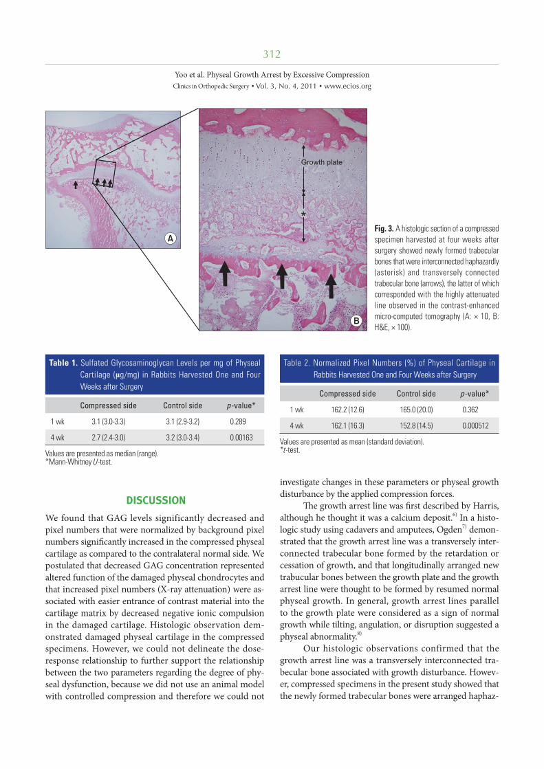
CEMCT visualized a thin, highly attenuated line parallel to the growth plate in compressed specimens harvested four weeks after surgery (Fig. 2). Histologic observa-tion revealed that the highly attenuated line in CEMCT was transversely connected trabecular bone, which cor-responded with the growth arrest line of Harris.6) Newly formed trabecular bones between the growth plate and the Harris growth arrest line were interconnected with each other, whereas all trabecular bones near the growth plate
in the control side were oriented in a uniform longitudi-nal manner (Fig. 3). Disorganized columnar architecture and decreased cellularity in proliferative and hypertrophic zones were observed in 13 compressed specimens har-vested four weeks after surgery (81%, 13/16), but not in compressed specimens harvested one week after surgery or in control specimens.
Glucosaminoglycans levels per mg of physeal carti-lage in specimens harvested one week aft er surgery were similar in compressed and control sides (p = 0.289). How-ever, at four4 weeks aft er surgery, GAG levels were signifi -cantly lower in compressed specimens (p = 0.00163) than in controls (Table 1).
Normalized pixel values of physeal cartilages mea-sured on CEMCT images were similar in compressed and control sides at one week aft er surgery (p = 0.362). Howev-er, they were signifi cantly higher in compressed specimens harvested at four weeks aft er surgery than in correspond-ing controls (p = 0.00512) (Table 2).
Fig. 2. Contrast-enhanced micro-computed tomography images showed that a thin, highly attenuated line parallel to the growth plate (arrows) was formed in compressed specimens (RF and RT) harvested at four weeks after surgery. RF: right femur, RT: right tibia, LF: left femur, LT: left tibia.
312
Yoo et al. Physeal Growth Arrest by Excessive CompressionClinics in Orthopedic Surgery • Vol. 3, No. 4, 2011 • www.ecios.org
DISCUSSION
We found that GAG levels significantly decreased and pixel numbers that were normalized by background pixel numbers signifi cantly increased in the compressed physeal cartilage as compared to the contralateral normal side. We postulated that decreased GAG concentration represented altered function of the damaged physeal chondrocytes and that increased pixel numbers (X-ray attenuation) were as-sociated with easier entrance of contrast material into the cartilage matrix by decreased negative ionic compulsion in the damaged cartilage. Histologic observation dem-onstrated damaged physeal cartilage in the compressed specimens. However, we could not delineate the dose-response relationship to further support the relationship between the two parameters regarding the degree of phy-seal dysfunction, because we did not use an animal model with controlled compression and therefore we could not
investigate changes in these parameters or physeal growth disturbance by the applied compression forces.
Th e growth arrest line was fi rst described by Harris, although he thought it was a calcium deposit.6) In a histo-logic study using cadavers and amputees, Ogden7) demon-strated that the growth arrest line was a transversely inter-connected trabecular bone formed by the retardation or cessation of growth, and that longitudinally arranged new trabucular bones between the growth plate and the growth arrest line were thought to be formed by resumed normal physeal growth. In general, growth arrest lines parallel to the growth plate were considered as a sign of normal growth while tilting, angulation, or disruption suggested a physeal abnormality.8)
Our histologic observations confirmed that the growth arrest line was a transversely interconnected tra-becular bone associated with growth disturbance. Howev-er, compressed specimens in the present study showed that the newly formed trabecular bones were arranged haphaz-
Fig. 3. A histologic section of a compressed specimen harvested at four weeks after surgery showed newly formed trabecular bones that were interconnected haphazardly (asterisk) and transversely connected trabecular bone (arrows), the latter of which corresponded with the highly attenuated line observed in the contrast-enhanced micro-computed tomography (A: × 10, B: H&E, × 100).
Table 1. Sulfated Glycosaminoglycan Levels per mg of Physeal Cartilage (μg/mg) in Rabbits Harvested One and Four Weeks after Surgery
Compressed side Control side p-value*
1 wk 3.1 (3.0-3.3) 3.1 (2.9-3.2) 0.289
4 wk 2.7 (2.4-3.0) 3.2 (3.0-3.4) 0.00163
Values are presented as median (range). *Mann-Whitney U-test.
Table 2. Normalized Pixel Numbers (%) of Physeal Cartilage in Rabbits Harvested One and Four Weeks after Surgery
Compressed side Control side p-value*
1 wk 162.2 (12.6) 165.0 (20.0) 0.362
4 wk 162.1 (16.3) 152.8 (14.5) 0.000512
Values are presented as mean (standard deviation). *t-test.

313
Yoo et al. Physeal Growth Arrest by Excessive CompressionClinics in Orthopedic Surgery • Vol. 3, No. 4, 2011 • www.ecios.org
ardly instead of the normal longitudinal arrangement, sug-gesting persistent physeal growth disturbance. Decreased GAG contents in these specimens also suggested altered metabolism in the physeal cartilage. We believe that these findings corresponded with the clinical observation in which the presence of a growth arrest line parallel to the growth plate does not always mean a normal resumption of physeal growth and that some of the physes close earlier than other normal physes.
Micro-CT has been used to evaluate subchondral bone plate morphology, trabecular patterns of the epiphy-sis, and osteophyte formation in a small animal model of osteoarthritis;4) however, its application to cartilage tis-sue has been limited due to poor soft tissue contrast. A technique of equilibrium partitioning of anionic contrast agent (Hexabrix 320) before CT scanning3) and a similar technique using gadolinium as a contrast material,2) sub-stantively overcame this poor soft tissue contrast issue and enabled quantitative measurements of GAGs in diseased articular cartilage in vitro, ex vivo, and in vivo.2-4) Recently this technique was used to monitor cartilage repair.9) Th e present study applied this technique to the assessment of growth arrest and associated physeal cartilage dysfunction. Th e applicability of CEMCT in the assessment of physeal dysfunction has depended on whether GAG content in physeal cartilage changes by compressive stress or not. While aggrecan loss in the articular cartilage of patients with osteoarthritis has been widely studied to elucidate the abnormal underlying metabolism, such as, the proteolytic cleavage of core protein, the inhibition of biosynthesis and/or the inhibition of proteoglycan retention due to hyaluronan defi cits,10,11) only one previous study has inves-tigated proteoglycan expression in physeal cartilage aft er mechanical compression.12) In this previous study, which was conducted using a rat model, proteoglycan expression assessed by Safranin-O staining and aggrecan mRNA pro-duction in physeal cartilage was found not to be altered by mechanical stress which contradicted our results. In this previous study, the authors applied controlled com-pression (0.2 MPa) that was suffi cient to retard, but not to cease physeal growth in rat vertebra, whereas histologic evidence of growth cessation and physeal cartilage damage were observed in our uncontrolled maximal compression model. Th erefore, it is likely that the two studies diff er sub-stantially in terms of the load eff ect. Bonnel et al.13) showed that distal femoral growth rates in rabbits decreased in proportion to compressive force, and that forces greater than 30 N caused cell damage and halted physeal growth.
Our findings indicate that CEMCT appears to be suitable for the assessment of physeal damage that is suf-
ficient to result in growth cessation, but further studies encompassing CEMCT imaging and GAG quantifi cation with diff erent compressive stresses on physeal cartilage are necessary to determine whether CEMCT can detect mild and reversible physeal damage as well.
Current treatment options for physeal damage have focused mainly on the management of disabilities associ-ated with growth disturbances such as limb shortening, angular deformity, and joint incongruity. However, surgi-cal complications have not been uncommon during mul-tiple limb lengthening procedures in young children, and incongruent joints have barely been restored by surgery. Although interposition of fat and other inert or biological materials aft er resection of a focal bone bridge can be per-formed before deformity worsens, this procedure has been reserved only for some selected patients with a small bone bridge and substantial growth remaining,14) and surgical outcomes oft en have been unpredictable.15) In this current situation, new therapeutic approaches may target early in-tervention before clinical and radiological manifestations of growth disturbance.
An early resection of damaged physeal cartilage be-fore bone bridge formation has been reported.16) However, this strategy may be limited by the risk of overtreatment in the absence of a reliable method of assessing physeal function before surgery. We believe that early assessment of physeal growth arrest would be necessary for selecting patients who require a closer follow-up and when consid-ering new potential early treatments designed to prevent complications of growth disturbances.
Certain limitations of this study require consider-ation. First, statistical power may not have been strong due to the small number of animals used. Estimation of an adequate number of animals was diffi cult because, to our knowledge, it was the first attempt to investigate abnor-mal physeal function following compression injury using CEMCT, and there have been no similar reports in litera-ture. We believe controlled physeal compression in a larger number of animals may delineate the relationship between compressive stress and abnormal physeal metabolism, and further determine the role of CEMCT in physeal growth arrest. Second, changes in X-ray attenuation represented by pixel numbers were not readily differentiated by the naked eye. Third, early detection of physeal dysfunction and prediction of prognosis have not yet been clinically feasible. In fact, the current technique doesn't seem to be applicable even to an in vivo animal study presumably because of limited blood supply to growth plates and high concentration-related toxicity of the contrast material. Only ex vivo animal experiments seem to be feasible for

314
Yoo et al. Physeal Growth Arrest by Excessive CompressionClinics in Orthopedic Surgery • Vol. 3, No. 4, 2011 • www.ecios.org
the evaluation of physeal cartilage function at the present time. However, we expect that this type of research using CEMCT may give some implications to clinical practice because this type of research may reveal a favorable or un-favorable eff ect of some drugs or surgical interventions on physeal cartilage function in an animal model of physeal dysfunction.
We conclude that excessive compressive force ap-plied to growth plates produced altered histologic features and metabolic function in terms of decreased GAG con-tents in the physeal cartilage, and that these changes could
be demonstrated by CEMCT.
CONFLICT OF INTEREST
No potential confl ict of interest relevant to this article was reported.
ACKNOWLEDGEMENTS
Th is study was supported by a Seoul National University Hospital Research Fund (Grant No. 04-2008-061).
REFERENCES
555.
9. Moyer HR, Wang Y, Farooque T, et al. A new animal model for assessing cartilage repair and regeneration at a nonar-ticular site. Tissue Eng Part A. 2010;16(7):2321-30.
10. Knudson CB, Knudson W. Cartilage proteoglycans. Semin Cell Dev Biol. 2001;12(2):69-78.
11. Roughley PJ. Th e structure and function of cartilage proteo-glycans. Eur Cell Mater. 2006;12:92-101.
12. Cancel M, Grimard G, Thuillard-Crisinel D, Moldovan F, Villemure I. Eff ects of in vivo static compressive loading on aggrecan and type II and X collagens in the rat growth plate extracellular matrix. Bone. 2009;44(2):306-15.
13. Bonnel F, Peruchon E, Baldet P, Dimeglio A, Rabischong P. Eff ects of compression on growth plates in the rabbit. Acta Orthop Scand. 1983;54(5):730-3.
14. Langenskiold A. Traumatic premature closure of the distal tibial epiphyseal plate. Acta Orthop Scand. 1967;38(4):520-31.
15. Hasler CC, Foster BK. Secondary tethers aft er physeal bar resection: a common source of failure? Clin Orthop Relat Res. 2002;(405):242-9.
16. Foster BK, John B, Hasler C. Free fat interpositional graft in acute physeal injuries: the anticipatory Langenskiold proce-dure. J Pediatr Orthop. 2000;20(3):282-5.
1. Futami T, Foster BK, Morris LL, LeQuesne GW. Magnetic resonance imaging of growth plate injuries: the effi cacy and indications for surgical procedures. Arch Orthop Trauma Surg. 2000;120(7-8):390-6.
2. Cockman MD, Blanton CA, Chmielewski PA, et al. Quan-titative imaging of proteoglycan in cartilage using a gado-linium probe and microCT. Osteoarthritis Cartilage. 2006; 14(3):210-4.
3. Palmer AW, Guldberg RE, Levenston ME. Analysis of car-tilage matrix fixed charge density and three-dimensional morphology via contrast-enhanced microcomputed tomog-raphy. Proc Natl Acad Sci U S A. 2006;103(51):19255-60.
4. Piscaer TM, van Osch GJ, Verhaar JA, Weinans H. Imag-ing of experimental osteoarthritis in small animal models. Biorheology. 2008;45(3-4):355-64.
5. Farndale RW, Buttle DJ, Barrett AJ. Improved quantita-tion and discrimination of sulphated glycosaminoglycans by use of dimethylmethylene blue. Biochim Biophys Acta. 1986;883(2):173-7.
6. Harris HA. Bone growth in health and disease. London: Ox-ford University Press; 1933.
7. Ogden JA. Th e evaluation and treatment of partial physeal arrest. J Bone Joint Surg Am. 1987;69(8):1297-302.
8. Shapiro F. Pediatric orthopedic deformities: basic science, diagnosis, and treatment. San Diego: Academic Press; 2001.