physical exercise preserves adult visual plasticity … · physical exercise preserves adult visual...
TRANSCRIPT
ORIGINAL RESEARCHpublished: 21 September 2016doi: 10.3389/fnagi.2016.00212
Physical Exercise Preserves AdultVisual Plasticity in Mice and Restoresit after a Stroke in theSomatosensory CortexEvgenia Kalogeraki 1,2* †, Justyna Pielecka-Fortuna 1†, Janika M. Hüppe 1
and Siegrid Löwel 1
1 Department of Systems Neuroscience, JFB Institut für Zoologie und Anthropologie, Georg-August Universität Göttingen,Göttingen, Germany, 2 Göttingen Graduate School for Neurosciences, Biophysics, and Molecular Biosciences (GGNB),Göttingen, Germany
Edited by:Orly Lazarov,
University of Illinois at Chicago, USA
Reviewed by:Rubem C. A. Guedes,
Federal University of Pernambuco,Brazil
Christiane Charriaut-Marlangue,French Institute of Health and
Medical Research (INSERM), France
*Correspondence:Evgenia [email protected]
†These authors have contributedequally to this work.
Received: 14 July 2016Accepted: 22 August 2016
Published: 21 September 2016
Citation:Kalogeraki E, Pielecka-Fortuna J,Hüppe JM and Löwel S (2016)
Physical Exercise Preserves AdultVisual Plasticity in Mice and Restores
it after a Stroke in theSomatosensory Cortex.
Front. Aging Neurosci. 8:212.doi: 10.3389/fnagi.2016.00212
The primary visual cortex (V1) is widely used to study brain plasticity, which is not onlycrucial for normal brain function, such as learning and memory, but also for recoveryafter brain injuries such as stroke. In standard cage (SC) raised mice, experience-dependent ocular dominance (OD) plasticity in V1 declines with age and is compromisedby a lesion in adjacent and distant cortical regions. In contrast, mice raised in anenriched environment (EE), exhibit lifelong OD plasticity and are protected from losingOD plasticity after a stroke-lesion in the somatosensory cortex. Since SC mice with anaccess to a running wheel (RW) displayed preserved OD plasticity during aging, weinvestigated whether physical exercise might also provide a plasticity promoting effectafter a cortical stroke. To this end, we tested if adult RW-raised mice preserved ODplasticity after stroke and also if short-term running after stroke restored OD plasticityto SC mice. Indeed, unlike mice without a RW, adult RW mice continued to show ODplasticity even after stroke, and a 2 weeks RW experience after stroke already restoredlost OD plasticity. Additionally, the experience-enabled increase of the spatial frequencyand contrast threshold of the optomotor reflex of the open eye, normally lost after astroke, was restored in both groups of RW mice. Our data suggest that physical exercisealone can not only preserve visual plasticity into old age, but also restore it after a corticalstroke.
Keywords: adult plasticity, visual cortex, stroke, physical exercise, ocular dominance
INTRODUCTION
Neuronal plasticity in the adult brain is indispensable to allow adaptive changes during agingand after lesions. A well-known model system to study experience-dependent circuit changes isocular dominance (OD) plasticity in the primary visual cortex (V1): occluding one eye of visionfor a couple of days induces an OD shift towards the open eye. This phenomenon was firstdescribed in kittens (Wiesel and Hubel, 1963) and later in mice (Dräger, 1978). OD-plasticity isage-dependent: in mice, it is most pronounced in juvenile animals (postnatal day (P) 28), reducedin young adults (P90) and absent beyond P110 if mice are raised in standard cages (SCs; Lehmannand Löwel, 2008; Espinosa and Stryker, 2012). Interestingly, raising or exposing aged rodents toan enriched environment (EE), with increased physical (running wheels (RWs)), social (bigger
Frontiers in Aging Neuroscience | www.frontiersin.org 1 September 2016 | Volume 8 | Article 212
Kalogeraki et al. Running Rescues Visual Plasticity after Stroke
housing groups) and cognitive (regularly changed labyrinths ortoys) stimulation, can overcome plasticity limitations (Sale et al.,2007; Baroncelli et al., 2010; Scali et al., 2012) and preservea lifelong OD-plasticity in mice (Greifzu et al., 2014, 2016a).Surprisingly, just one of these components has also been recentlyshown to preserve plasticity into older age in mice: adult miceraised in SCs with a RW continued to show OD-plasticitybeyond P134, while age-matched SC-mice (without a RW) didnot exhibit this plasticity (Kalogeraki et al., 2014). Furthermore,even short-term running, just during the 7-day MD period alsorestored OD-plasticity to adult SC-raised mice.
Plasticity is not only important for normal brain functionand during aging but also for recovery after brain injurieslike stroke; understanding plasticity mechanism is thereforeessential for further development of adequate therapies. Wehave previously shown that a small stroke lesion in the primarysomatosensory cortex (S1) preventedOD-plasticity in V1 of adultSC-raised mice (Greifzu et al., 2011). Remarkably, raising micein EE protected adult mice from lesion-induced impairmentsof visual plasticity, similarly as young mice were protected:EE-mice continued to display OD-plasticity even after stroke,suggesting that EE preserves amore juvenile brain into adulthood(Greifzu et al., 2014). These observations implicated that youngand/or physically active animals in a cognitively and sociallychallenging environment are less affected by a distant brainlesion and preserved their visual cortical plasticity even inadulthood. Whether all of these components are necessary fora plasticity promoting effect after stroke or just one of themhas not yet been tested. Because both long-term and short-term voluntary physical exercise promoted OD-plasticity inaging mice (Kalogeraki et al., 2014), here we tested whetherrunning alone might already provide a beneficial effect on visualplasticity after a cortical lesion in S1. We focused on runningbecause locomotion has recently been shown to massivelyincrease pyramidal cell firing in V1 (Niell and Stryker, 2010;Fu et al., 2014) and promote activity-dependent changes inV1 circuitry (Kaneko and Stryker, 2014). Do adult mice withaccess to a RW (in an otherwise SC) continue to exhibit OD-plasticity in V1 even after an S1-stroke? And if so, is long-term running required for preserving OD-plasticity or is itpossible to restore OD-plasticity in adult SC-raised mice evenafter stroke by putting them in cages with a RW? Our presentdata clearly show that voluntary running was effective in bothexperimental conditions: running preserved OD-plasticity afterstroke both in adult mice with access to a RW for all their lifeand in animals that started running only after their stroke lesion.Thus, voluntary physical exercise may help to prevent plasticityimpairments after a cortical lesion and in addition, might be auseful noninvasive therapy to support rehabilitation in an earlypost-stroke period.
MATERIALS AND METHODS
Animals and Rearing ConditionsAll mice were from the mouse colony of the central animalfacility, University Medical Center Göttingen, and housed with a
12 h light/dark cycle, with food andwater available ad libitum. Allexperimental procedures were approved by the local government(Niedersächsisches Landesamt für Verbraucherschutz undLebensmittelsicherheit, registration number 33.9-42502-04-10/0326). The experimental procedures comply with NationalInstitutes of Health guidelines for the use of animals. Thecages were translucent with an open grid cover and wood chipbedding.
Long-Term Running Groups (RW-mice)Pregnant C57BL/6J females were put into slightly larger thannormal SCs (27 × 43 × 19 cm) equipped with a RW (RW-cage)6–11 days before delivery. The offsprings were separated intofemale and male groups at postnatal day (P) 28. A total numberof 17 male and female mice between P149 and P222 were usedfor this study. Male and female mice were mixed in these groupsbecause previous publications indicated that there is no statisticaldifference between the values of female andmalemice of a similarage for all of the measured parameters: ocular dominance index(ODI), V1-activation, spatial frequency and contrast sensitivitythresholds (e.g., Kalogeraki et al., 2014). Mice were randomlyassigned to experimental groups and were housed in a group of3–5 animals per cage.
Short-Term Running Groups (14dRW-mice)Mice were raised in SCs (26 × 20 × 14 cm) without a RWuntil at least P119, and transferred to RW-cages (size see above)after induction of the lesion. Eighteen male mice born and raisedin SCs were used (age range: P119–258). Since most of theanimals of this group were from different litters, mice had to behoused alone in the RW-cages to avoid fighting. The number ofRW-turns for every mouse was counted daily.
After monocular deprivation (MD)/noMD, spatial vision waschecked daily by optomotry for 7 days. Finally, we visualizedV1-activation after stimulation of the ipsi- and contralateral eyeusing intrinsic signal optical imaging (Cang et al., 2005).
Induction of a Photothrombotic (PT) StrokeAll animals were at least P119 before PT/sham-treatment.The model of photothrombosis (PT) was used to induce acortical lesion in the left S1 using the Rose Bengal technique(Watson et al., 1985) as described previously (Greifzu et al.,2011). PT is a minimally invasive technique to induce localizedand small lesions restricted to the cortex. Briefly, mice werebox-anesthetized with 2% isoflurane in a mixture of O2/N2O(1:1), the body temperature was maintained at 37◦C via aheating pad and the animals’ head was placed in a stereotaxicframe. The skin above the skull was incised and an optic fiberbundle (aperture: 1.0 mm), mounted on a cold light source(Schott KL 1500, Germany), was positioned 2 mm lateral tothe midline and 1 mm posterior to the bregma. Hundredmicroliters Rose Bengal dye (0.1% in saline) was injectedintravenously into the tail vein. After 5 min of waiting time,the illumination of the cortex started for 15 min. Finally theskin was sutured and animals returned to their home cagesfor recovery. Lesion size was determined at the end of thebehavioral and optical imaging experiments by Glia Fibrillary
Frontiers in Aging Neuroscience | www.frontiersin.org 2 September 2016 | Volume 8 | Article 212
Kalogeraki et al. Running Rescues Visual Plasticity after Stroke
Acidic Protein (GFAP) staining. For the controls, the exact sameprocedure was followedwithout turning on the light source (RW-sham).
Monocular Deprivation (MD)The right eye of the mice was deprived for 7 days, according topublished protocols (Gordon and Stryker, 1996; Cang et al., 2005;Lehmann and Löwel, 2008). Briefly, mice were box anesthetizedwith 2% isoflurane in O2/N2O (1:1), eyelids were trimmed andclosed with two sutures. Mice were returned to their home cagesfor recovery and checked daily to ensure that the eye remainedclosed. In case of an open MD-eye, mice were excluded fromfurther experiments.
Virtual-Reality Optomotor SetupBoth the spatial frequency threshold (‘‘visual acuity’’) and thecontrast threshold (‘‘contrast sensitivity’’) of the optomotorreflex of all mice (animals with and without MD) were measuredusing the virtual-reality optomotor system (Prusky et al., 2004).Briefly, freely moving mice were positioned on a small platformsurrounded by four computer monitors (33.5 × 26.5 cm)forming a box. Mirrors were placed at the bottom and thetop of the box, giving the impression to the animal that itwas sitting in an endless cylinder. A rotating virtual cylinder,composed of a vertical sine wave grating, was projected onthe screens. Parameters like spatial frequency, contrast andspeed of the moving sine wave grating could be varied by theexperimenter. In case the mouse could detect the stimulus, itwas reflexively tracking the grating by moving the head in thedirection of rotation. Spatial frequency thresholds at full contrastand contrast thresholds at six different spatial frequencies [0.031,0.064, 0.092, 0.103, 0.192, 0.272 cycles/degree (cyc/deg)] weremeasured daily (after MD/no MD) for this study.
Optical Imaging of Intrinsic Signals andVisual StimuliAfter completion of all behavioral vision tests, visual corticalresponses were recorded and analyzed as described previously(Greifzu et al., 2014; Kalogeraki et al., 2014).
SurgeryBriefly, mice were box-anesthetized with 2% halothane inO2/N2O (1:1) and injected with atropine (0.1 mg/mouse s.c.;Franz Köhler), dexamethasone (0.2 mg/mouse s.c.; Ratiopharm),and chlorprothixene (0.2 mg/mouse i.m.; Sigma). After placingmice in a stereotaxic frame, anesthesia was maintained with 0.8%halothane in a 1:1 mixture of O2/N2O. An incision of the skinwas made over the visual cortex and low-melting point agarose(2.5% in 0.9% NaCl) and a glass coverslip were placed over theexposed area.
Data Acquisition and Visual StimulationMouse V1-responses were recorded through the skull usingthe Fourier imaging technique (Kalatsky and Stryker, 2003),optimized for the assessment of OD-plasticity (Cang et al., 2005).V1-signals were visualized with a CCD-camera (Dalsar 1 M30)
using a 135 × 50 mm tandem lens configuration (Nikon), withred illumination light (610± 10 nm). Active brain regions absorbmore of the red light and appear darker in the images. Frameswere acquired at a rate of 30 Hz, temporally binned to 7.5 Hzand stored as 512 × 512 pixel images after spatial binning of thecamera image.
Visual stimuli were presented on a high refresh rate monitor(Hitachi, ACCUVUE, HM-4921-D, 21′′) positioned 25 cm fromthe eyes. Stimuli consisted of white drifting horizontal bars(2◦ wide). The distance between two bars was 70◦ and they werepresented at a temporal frequency of 0.125 Hz. To calculate OD,the visual stimulus was restricted to the binocular visual field ofthe left V1 (−5◦ to +15◦ azimuth, 0◦ azimuth corresponding tofrontal direction) and animals were stimulated through either theleft or the right eye in alternation.
Data AnalysisVisual cortical maps were calculated from the acquired framesby Fourier analysis to extract the signal at the stimulationfrequency using custom software (Kalatsky and Stryker, 2003).While the phase component of the signal is used for thecalculation of retinotopy, the amplitude component representsthe intensity of neuronal activation (expressed as the fractionalchange in reflectance ×10−4) and was used to calculate OD.At least 3 maps per animal were averaged to compute the ODIas follows: (C − I)/(C + I), with C and I representing theresponse magnitudes of each pixel to visual stimulation of thecontralateral and ipsilateral eye. The ODI ranges from −1 to +1,with negative values representing ipsilateral and positive valuesrepresenting contralateral dominance. For comparison, we usedpreviously published data of PT-lesioned C57Bl6/J (WT) micewithout access to a RW (presented in Figures 3, 7): ODI-dataof PT-lesioned (S1) late MD WT mice (MD started 1 weekafter PT = SC-PT (1w)) are from Greifzu et al. (2011), butnewly analyzed as described above (‘‘average map analysis’’). Inaddition, SC-PT groups (noMD andMD) are taken fromGreifzuet al. (2016b).
Lesion AnalysisTo determine the size and location of the cortical PT-lesions,coronal brain sections were cut at 40 µm, and immunostainingwith an antibody against GFAP was performed. GFAP is acommon marker used to reveal brain lesions (Lai et al., 2014)by staining the astrocytes that are accumulating at the lesion site.Lesions were visible in all mice that received a PT-stroke. Sectionswere washed for 10 min with 0.1 M PB and incubated for 10 minwith 0.1M PB-Triton-X-100 (0.3%), followed by 30min blockingin 10% normal donkey serum in PB-Triton-X-100 (0.3%) atroom temperature. The sections were incubated with the primarypolyclonal rabbit-anti-GFAP antibody (Immunological Science)1:1000, diluted in 0.1 M PB-Triton-X-100 (0.3%) over nightat 4◦C. The following day the sections were washed 3 timesfor 5 min with 0.1 M PB. Incubation with the secondaryCy3-goat-anti-rabbit antibody (Jackson ImmunoResearch Inc.)was for 2 h at room temperature in dark (1:1000 dilutedin PB-Triton-X-100 (0.3%)), followed by three washes for5 min with 0.1 M PB. Sections were then transferred onto
Frontiers in Aging Neuroscience | www.frontiersin.org 3 September 2016 | Volume 8 | Article 212
Kalogeraki et al. Running Rescues Visual Plasticity after Stroke
microscope slides and mounted with Fluoromount-G with4′6-diamidin-2-phenylindol (DAPI; Jackson ImmunoResearchInc.). For quantitative lesion analyses, every third section wasanalyzed under the microscope (Axioskop, Carl Zeiss) usingAxioVision (AxioVs 40 4.5.0.0.). To quantify the position ofeach lesion, the distance from the center of the lesion to themidline and also to the anterior border of V1 were measured.In addition, we determined lesion length (anterior-posterior),diameter (medial-lateral), depth and calculated the total volumeof each lesion.
Statistical AnalysesAll intra- and intergroup comparisons were analyzed either bya two-tailed t-test or one-way analysis of variance (ANOVA)followed by multiple comparisons Bonferroni correction. Theintergroup comparison of the enhancement of the spatialfrequency and contrast sensitivity thresholds was analyzed by a
two-wayANOVA. The levels of significance were set as ∗p< 0.05;∗∗p < 0.01; ∗∗∗p < 0.001. Data are represented as means± SEM.
RESULTS
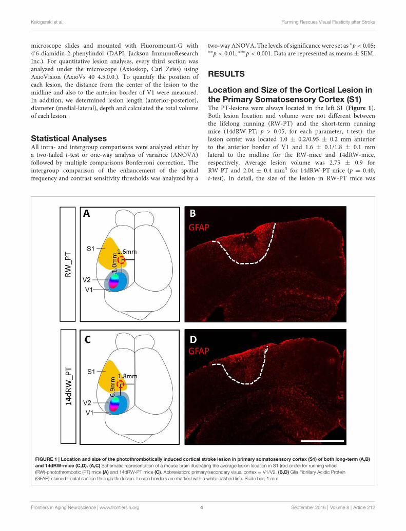
Location and Size of the Cortical Lesion inthe Primary Somatosensory Cortex (S1)The PT-lesions were always located in the left S1 (Figure 1).Both lesion location and volume were not different betweenthe lifelong running (RW-PT) and the short-term runningmice (14dRW-PT; p > 0.05, for each parameter, t-test): thelesion center was located 1.0 ± 0.2/0.95 ± 0.2 mm anteriorto the anterior border of V1 and 1.6 ± 0.1/1.8 ± 0.1 mmlateral to the midline for the RW-mice and 14dRW-mice,respectively. Average lesion volume was 2.75 ± 0.9 forRW-PT and 2.04 ± 0.4 mm3 for 14dRW-PT-mice (p = 0.40,t-test). In detail, the size of the lesion in RW-PT mice was
FIGURE 1 | Location and size of the photothrombotically induced cortical stroke lesion in primary somatosensory cortex (S1) of both long-term (A,B)and 14dRW-mice (C,D). (A,C) Schematic representation of a mouse brain illustrating the average lesion location in S1 (red circle) for running wheel(RW)-photothrombotic (PT) mice (A) and 14dRW-PT mice (C). Abbreviation: primary/secondary visual cortex = V1/V2. (B,D) Glia Fibrillary Acidic Protein(GFAP)-stained frontal section through the lesion. Lesion borders are marked with a white dashed line. Scale bar: 1 mm.
Frontiers in Aging Neuroscience | www.frontiersin.org 4 September 2016 | Volume 8 | Article 212
Kalogeraki et al. Running Rescues Visual Plasticity after Stroke
on average 1.2 ± 0.2 mm in medio-lateral, 1.4 ± 0.2 mmin anterio-posterior direction and extended vertically untillayers 5/6 (n = 7). For the 14dRW-PT mice, the lesion wason average 0.8 ± 0.1 mm in medio-lateral, 1.2 ± 0.1 mmin anterio-posterior direction and also extended untillayers 5/6 (n = 12). Notably, volume and location of thepresent PT-lesions were not different from the PT-lesionsof SC-mice without RWs (Greifzu et al., 2011; p > 0.05 forall measured parameters) which showed abolished visualplasticity.
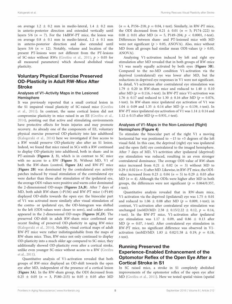
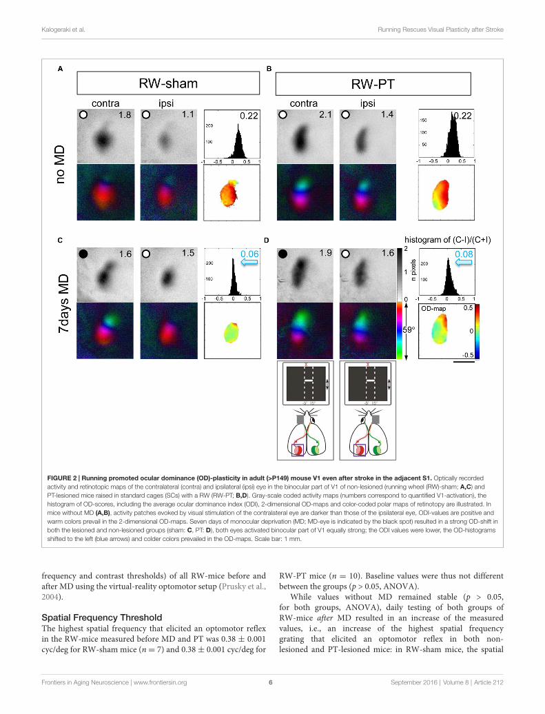
Voluntary Physical Exercise PreservedOD-Plasticity in Adult RW-Mice AfterStrokeAnalyses of V1-Activity Maps in the LesionedHemisphereIt was previously reported that a small cortical lesion inthe S1 impaired visual plasticity of SC-raised mice (Greifzuet al., 2011). In contrast, the same cortical lesion did notcompromise plasticity in mice raised in an EE (Greifzu et al.,2014), pointing out that active and stimulating environmentshave protective effects for brain injuries and may promoterecovery. As already one of the components of EE, voluntaryphysical exercise preserved OD-plasticity into late adulthood(Kalogeraki et al., 2014) here we investigated if free access toa RW would preserve OD-plasticity also after an S1 lesion.Indeed, we found that mice raised in SCs with a RW continuedto display OD-plasticity into adulthood, both in sham- and inPT-animals (Figures 2, 3), which is in contrast to SC micewith no access to a RW (Figure 3). Without MD, V1 ofboth the RW-sham (control, Figure 2A) and RW-PT mice(Figure 2B) was dominated by the contralateral eye: activitypatches induced by visual stimulation of the contralateral eyewere darker than those after stimulation of the ipsilateral eye,the average ODI-values were positive and warm color dominatedthe 2-dimensioned OD-maps (Figures 2A,B). After 7 days ofMD, both adult RW-sham (>P156) and RW-PT mice (>P149)displayed OD-shifts towards the open eye: the binocular partof V1 was activated more similarly after visual stimulation ofthe contra- or ipsilateral eye, the OD-histogram was shiftedto the left (ODI-values were closer to zero), and colder colorsappeared in the 2-dimensional OD-maps (Figures 2C,D). Thepreserved OD-shift in adult RW-sham mice confirmed ourrecent finding of preserved OD-plasticity in aging RW-mice(Kalogeraki et al., 2014). Notably, visual cortical maps of adultRW-PT mice were rather indistinguishable from the maps ofRW-sham mice. Thus, RW-mice not only continued to displayOD-plasticity into a much older age compared to SC-mice, theyadditionally showed OD-plasticity even after a cortical stroke,unlike even younger SC-mice without access to a RW (Greifzuet al., 2011).
Quantitative analysis of V1-activation revealed that bothgroups of RW-mice displayed an OD-shift towards the openeye after MD, independent of the presence of a cortical lesion(Figure 3A). In the RW-sham group, the ODI decreased from0.25 ± 0.05 (n = 3, P180–215) to 0.05 ± 0.05 after MD
(n = 4, P156–218; p = 0.04, t-test). Similarly, in RW-PT mice,the ODI decreased from 0.21 ± 0.01 (n = 5; P174–222) to0.08 ± 0.01 after MD (n = 5, P149–204; p < 0.0001, t-test).Differences between sham- and PT-lesioned mice with MDwere not significant (p > 0.05, ANOVA). Also, mice withoutMD from all groups had similar mean ODI-values (p > 0.05,ANOVA).
Analyzing V1-activation induced by left and right eyestimulation after MD revealed that in both groups of RW-miceV1 was nearly equally activated by both eyes (Figure 3B).Compared to the no-MD condition V1-activation via thedeprived (contralateral) eye was lower after MD, but thereductions in deprived eye responses in V1 were not significant.In detail, V1-activation after contralateral eye stimulation was1.79 ± 0.20 in RW-sham mice and reduced to 1.40 ± 0.10after MD (p = 0.116, t-test). In RW-PT mice V1-activation was1.76 ± 0.17 and reduced to 1.30 ± 0.16 after MD (p = 0.076,t-test). In RW-sham mice ipsilateral eye activation of V1 was1.04 ± 0.09 and 1.35 ± 0.14 after MD (p = 0.139, t-test). InRW-PT mice ipsilateral eye activation of V1 was 1.14± 0.14 and1.12± 0.15 after MD (p= 0.931, t-test).
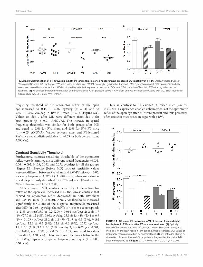
Analyses of V1-Maps in the Non-Lesioned (Right)Hemisphere (Figure 4)To stimulate the binocular part of the right V1 a movinghorizontal bar was positioned to −15 to +5 degrees of the leftvisual field. In this case, the deprived (right) eye was ipsilateraland the open (left) eye contralateral to the imaged hemisphere.After 7 days of MD, V1-activition after ipsilateral (deprived)eye stimulation was reduced, resulting in an even strongercontralateral dominance. The average ODI-value of RW-shammice increased from 0.21 ± 0.02 (n = 3) without MD to0.29± 0.02 (n= 3) afterMD. Likewise, in RW-PTmice, the ODI-value increased from 0.23 ± 0.04 (n = 5) to 0.29 ± 0.03 afterMD (n = 4). Although the ODIs were higher after MD in bothgroups, the differences were not significant (p = 0.064/0.357,t-test).
Quantitative analysis revealed that in RW-sham mice,V1-activation via the deprived (ipsilateral) eye was 1.75 ± 0.01and reduced to 1.06 ± 0.08 after MD (p = 0.009, t-test); incontrast, V1-activation after contralateral eye stimulation wasunchanged (noMD/MD: 2.58 ± 0.15/2.22 ± 0.12, p = 0.14,t-test). In the RW-PT mice, V1-activation after ipsilateraleye stimulation was 1.17 ± 0.09, and 0.86 ± 0.13 afterMD (p = 0.07, t-test). After contralateral eye stimulation inRW-PT mice, no significant difference was observed in V1-activation (noMD/MD: 1.83 ± 0.02/1.58 ± 0.19; p = 0.18,t-test).
Running Preserved theExperience-Enabled Enhancement of theOptomotor Reflex of the Open Eye After aCortical Stroke in S1In SC raised mice, a stroke in S1 completely abolishedimprovements of the optomotor reflex of the open eye afterMD (Greifzu et al., 2011). Here we tested spatial vision (spatial
Frontiers in Aging Neuroscience | www.frontiersin.org 5 September 2016 | Volume 8 | Article 212
Kalogeraki et al. Running Rescues Visual Plasticity after Stroke
FIGURE 2 | Running promoted ocular dominance (OD)-plasticity in adult (>P149) mouse V1 even after stroke in the adjacent S1. Optically recordedactivity and retinotopic maps of the contralateral (contra) and ipsilateral (ipsi) eye in the binocular part of V1 of non-lesioned (running wheel (RW)-sham; A,C) andPT-lesioned mice raised in standard cages (SCs) with a RW (RW-PT; B,D). Gray-scale coded activity maps (numbers correspond to quantified V1-activation), thehistogram of OD-scores, including the average ocular dominance index (ODI), 2-dimensional OD-maps and color-coded polar maps of retinotopy are illustrated. Inmice without MD (A,B), activity patches evoked by visual stimulation of the contralateral eye are darker than those of the ipsilateral eye, ODI-values are positive andwarm colors prevail in the 2-dimensional OD-maps. Seven days of monocular deprivation (MD; MD-eye is indicated by the black spot) resulted in a strong OD-shift inboth the lesioned and non-lesioned groups (sham: C, PT: D), both eyes activated binocular part of V1 equally strong; the ODI values were lower, the OD-histogramsshifted to the left (blue arrows) and colder colors prevailed in the OD-maps. Scale bar: 1 mm.
frequency and contrast thresholds) of all RW-mice before andafter MD using the virtual-reality optomotor setup (Prusky et al.,2004).
Spatial Frequency ThresholdThe highest spatial frequency that elicited an optomotor reflexin the RW-mice measured before MD and PT was 0.38 ± 0.001cyc/deg for RW-sham mice (n= 7) and 0.38± 0.001 cyc/deg for
RW-PT mice (n = 10). Baseline values were thus not differentbetween the groups (p > 0.05, ANOVA).
While values without MD remained stable (p > 0.05,for both groups, ANOVA), daily testing of both groups ofRW-mice after MD resulted in an increase of the measuredvalues, i.e., an increase of the highest spatial frequencygrating that elicited an optomotor reflex in both non-lesioned and PT-lesioned mice: in RW-sham mice, the spatial
Frontiers in Aging Neuroscience | www.frontiersin.org 6 September 2016 | Volume 8 | Article 212
Kalogeraki et al. Running Rescues Visual Plasticity after Stroke
FIGURE 3 | Quantification of V1-activation in both PT- and sham-lesioned mice: running preserved OD-plasticity in V1. (A) Optically imaged ODIs ofPT-lesioned SC-mice (left, light gray), RW-sham (middle, white) and RW-PT mice (right, gray) without and with MD. Symbols represent ODI-values of individuals;means are marked by horizontal lines. MD is indicated by half-black squares. In contrast to SC-mice, MD induced an OD-shift in RW-mice regardless of thetreatment. (B) V1-activation elicited by stimulation of the contralateral (C) or ipsilateral (I) eye in RW-sham and RW-PT mice without and with MD. Black filled circleindicates MD eye. ∗p < 0.05, ∗∗∗p < 0.001.
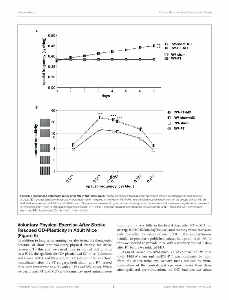
frequency threshold of the optomotor reflex of the openeye increased to 0.45 ± 0.002 cyc/deg (n = 4) and to0.45 ± 0.002 cyc/deg in RW-PT mice (n = 5; Figure 5A).Values on day 7 after MD were different from day 0 forboth groups (p < 0.01, ANOVA). The increase in spatialfrequency thresholds was similar for both groups after MDand equal to 23% for RW-sham and 23% for RW-PT mice(p > 0.05, ANOVA). Values between non- and PT-lesionedRW-mice were indistinguishable (p > 0.05 for both comparisons;ANOVA).
Contrast Sensitivity ThresholdFurthermore, contrast sensitivity thresholds of the optomotorreflex were determined at six different spatial frequencies (0.031,0.064, 0.092, 0.103, 0.192 and 0.272 cyc/deg) for all the groups(Figure 5B). Baseline (before MD) contrast sensitivity valueswere not different between RW-sham and RW-PTmice (p > 0.05;for every frequency, ANOVA). Additionally, values were similarto values previously described for C57BL/6J mice (Prusky et al.,2004; Lehmann and Löwel, 2008).
After 7 days of MD, contrast sensitivity of the optomotorreflex of the open eye increased (i.e., the lowest contrast thatelicited an optomotor reflex decreased) in both RW-shamand RW-PT mice (p < 0.001, ANOVA): thresholds increasedsignificantly for 3 out of the 6 spatial frequencies measuredafter MD (at 0.031 cyc/deg: sham/PT: to 4.8 ± 0.1 (correspondsto 21% contrast)/5.0 ± 0.2 (20%), 0.064 cyc/deg: 26.2 ± 1.7(4%)/27.0 ± 1.2 (4%), 0.092 cyc/deg: 23.1 ± 1.4 (4%)/23.4 ± 0.9(4%), 0.103 cyc/deg: 21.2 ± 1.2 (5%)/21.3 ± 0.5 (5%), 0.192cyc/deg: 12.6 ± 0.5 (8%)/ 12.9 ± 0.6 (8%), 0.272 cyc/deg:4.8 ± 0.1 (21%)/4.7 ± 0.1 (21%) on day 7; p > 0.05, p < 0.001,p < 0.001, p < 0.001, p > 0.05, p > 0.05, compared to valuesfrom day 0, ANOVA). There were no differences between thetwo RW-groups at any spatial frequency on day 7 (p > 0.05,ANOVA).
Thus, in contrast to PT-lesioned SC-raised mice (Greifzuet al., 2011), experience-enabled enhancements of the optomotorreflex of the open eye after MD were present and thus preservedafter stroke in mice raised in cages with a RW.
FIGURE 4 | ODIs and V1-activation in V1 of the non-lesioned righthemisphere in RW-mice after PT or sham treatment. (A) Opticallyimaged ODIs without and with MD of sham treated (RW-sham, white) andPT-mice (RW-PT, gray) raised in RW-cages. Symbols represent ODI-values ofindividuals; means are marked by horizontal lines. (B) V1-activation elicited bystimulation of the contralateral (C) or ipsilateral (I) eye without and after MD.Data are displayed as in Figure 3. ∗p < 0.05, ∗∗p < 0.01, ∗∗∗p < 0.001.
Frontiers in Aging Neuroscience | www.frontiersin.org 7 September 2016 | Volume 8 | Article 212
Kalogeraki et al. Running Rescues Visual Plasticity after Stroke
FIGURE 5 | Enhanced optomotor reflex after MD in RW-mice. (A)The spatial frequency threshold of the optomotor reflex in cyc/deg plotted as a functionof days. (B) Contrast sensitivity thresholds of optomotor reflex measured on 7th day of MD/noMD in six different spatial frequencies. All the groups without MD areillustrated as boxes and with MD as half-filled boxes. PT-groups are presented in gray color and sham groups in white. Note that there was a significant improvementof thresholds after 7 days of MD regardless of the induction of a lesion. There was no significant difference between sham- and PT-mice after MD, nor betweensham- and PT-mice without MD. ∗∗p < 0.01, ∗∗∗p < 0.001.
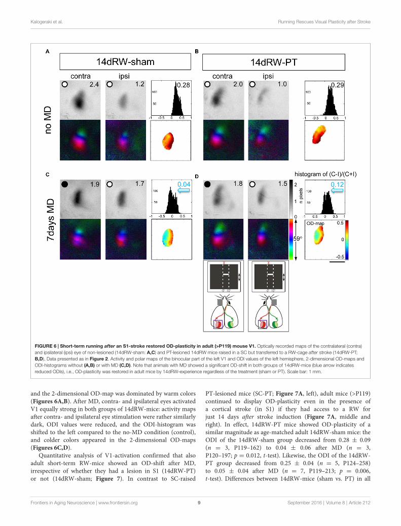
Voluntary Physical Exercise After StrokeRescued OD-Plasticity in Adult Mice(Figure 6)In addition to long-term running, we also tested the therapeuticpotential of short-term voluntary physical exercise for strokerecovery. To this end, we raised mice in normal SCs until atleast P119, the age limit for OD-plasticity of SC-mice (Lehmannand Löwel, 2008), and then induced a PT-lesion in S1 as before.Immediately after the PT-surgery, both sham- and PT-treatedmice were transferred to a SC with a RW (14d RW-mice). Whenwe performed PT and MD on the same day most animals were
running only very little in the first 4 days after PT + MD (onaverage 0.4± 0.02 km/day/mouse), and running values increasedonly thereafter to values of about 2.0 ± 0.4 km/day/mouse(similar to previously published values; Kalogeraki et al., 2014);thus we decided to provide mice with a recovery time of 7 daysafter PT before we initiated MD.
As in SC-raised C57Bl/6J mice, V1 of control 14dRW-mice(both 14dRW-sham and 14dRW-PT) was dominated by inputfrom the contralateral eye: activity maps induced by visualstimulation of the contralateral eye were darker than thoseafter ipsilateral eye stimulation, the ODI had positive values
Frontiers in Aging Neuroscience | www.frontiersin.org 8 September 2016 | Volume 8 | Article 212
Kalogeraki et al. Running Rescues Visual Plasticity after Stroke
FIGURE 6 | Short-term running after an S1-stroke restored OD-plasticity in adult (>P119) mouse V1. Optically recorded maps of the contralateral (contra)and ipsilateral (ipsi) eye of non-lesioned (14dRW-sham: A,C) and PT-lesioned 14dRW mice raised in a SC but transferred to a RW-cage after stroke (14dRW-PT;B,D), Data presented as in Figure 2. Activity and polar maps of the binocular part of the left V1 and ODI values of the left hemisphere, 2-dimensional OD-maps andODI-histograms without (A,B) or with MD (C,D). Note that animals with MD showed a significant OD-shift in both groups of 14dRW-mice (blue arrow indicatesreduced ODIs), i.e., OD-plasticity was restored in adult mice by 14dRW-experience regardless of the treatment (sham or PT). Scale bar: 1 mm.
and the 2-dimensional OD-map was dominated by warm colors(Figures 6A,B). After MD, contra- and ipsilateral eyes activatedV1 equally strong in both groups of 14dRW-mice: activity mapsafter contra- and ipsilateral eye stimulation were rather similarlydark, ODI values were reduced, and the ODI-histogram wasshifted to the left compared to the no-MD condition (control),and colder colors appeared in the 2-dimensional OD-maps(Figures 6C,D).
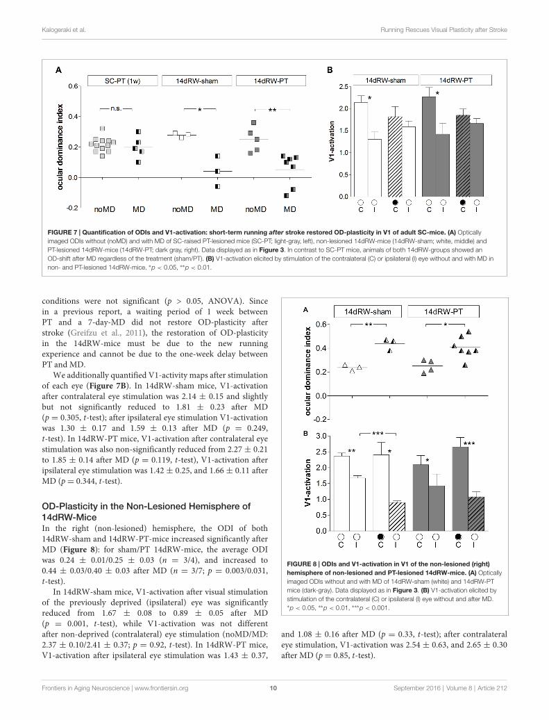
Quantitative analysis of V1-activation confirmed that alsoadult short-term RW-mice showed an OD-shift after MD,irrespective of whether they had a lesion in S1 (14dRW-PT)or not (14dRW-sham; Figure 7). In contrast to SC-raised
PT-lesioned mice (SC-PT; Figure 7A, left), adult mice (>P119)continued to display OD-plasticity even in the presence ofa cortical stroke (in S1) if they had access to a RW forjust 14 days after stroke induction (Figure 7A, middle andright). In effect, 14dRW-PT mice showed OD-plasticity of asimilar magnitude as age-matched adult 14dRW-sham mice: theODI of the 14dRW-sham group decreased from 0.28 ± 0.09(n = 3, P119–162) to 0.04 ± 0.06 after MD (n = 3,P120–197; p = 0.012, t-test). Likewise, the ODI of the 14dRW-PT group decreased from 0.25 ± 0.04 (n = 5, P124–258)to 0.05 ± 0.04 after MD (n = 7, P119–213; p = 0.006,t-test). Differences between 14dRW-mice (sham vs. PT) in all
Frontiers in Aging Neuroscience | www.frontiersin.org 9 September 2016 | Volume 8 | Article 212
Kalogeraki et al. Running Rescues Visual Plasticity after Stroke
FIGURE 7 | Quantification of ODIs and V1-activation: short-term running after stroke restored OD-plasticity in V1 of adult SC-mice. (A) Opticallyimaged ODIs without (noMD) and with MD of SC-raised PT-lesioned mice (SC-PT; light-gray, left), non-lesioned 14dRW-mice (14dRW-sham; white, middle) andPT-lesioned 14dRW-mice (14dRW-PT; dark gray, right). Data displayed as in Figure 3. In contrast to SC-PT mice, animals of both 14dRW-groups showed anOD-shift after MD regardless of the treatment (sham/PT). (B) V1-activation elicited by stimulation of the contralateral (C) or ipsilateral (I) eye without and with MD innon- and PT-lesioned 14dRW-mice. ∗p < 0.05, ∗∗p < 0.01.
conditions were not significant (p > 0.05, ANOVA). Sincein a previous report, a waiting period of 1 week betweenPT and a 7-day-MD did not restore OD-plasticity afterstroke (Greifzu et al., 2011), the restoration of OD-plasticityin the 14dRW-mice must be due to the new runningexperience and cannot be due to the one-week delay betweenPT and MD.
We additionally quantified V1-activity maps after stimulationof each eye (Figure 7B). In 14dRW-sham mice, V1-activationafter contralateral eye stimulation was 2.14 ± 0.15 and slightlybut not significantly reduced to 1.81 ± 0.23 after MD(p = 0.305, t-test); after ipsilateral eye stimulation V1-activationwas 1.30 ± 0.17 and 1.59 ± 0.13 after MD (p = 0.249,t-test). In 14dRW-PT mice, V1-activation after contralateral eyestimulation was also non-significantly reduced from 2.27 ± 0.21to 1.85 ± 0.14 after MD (p = 0.119, t-test), V1-activation afteripsilateral eye stimulation was 1.42 ± 0.25, and 1.66 ± 0.11 afterMD (p= 0.344, t-test).
OD-Plasticity in the Non-Lesioned Hemisphere of14dRW-MiceIn the right (non-lesioned) hemisphere, the ODI of both14dRW-sham and 14dRW-PT-mice increased significantly afterMD (Figure 8): for sham/PT 14dRW-mice, the average ODIwas 0.24 ± 0.01/0.25 ± 0.03 (n = 3/4), and increased to0.44 ± 0.03/0.40 ± 0.03 after MD (n = 3/7; p = 0.003/0.031,t-test).
In 14dRW-sham mice, V1-activation after visual stimulationof the previously deprived (ipsilateral) eye was significantlyreduced from 1.67 ± 0.08 to 0.89 ± 0.05 after MD(p = 0.001, t-test), while V1-activation was not differentafter non-deprived (contralateral) eye stimulation (noMD/MD:2.37 ± 0.10/2.41 ± 0.37; p = 0.92, t-test). In 14dRW-PT mice,V1-activation after ipsilateral eye stimulation was 1.43 ± 0.37,
FIGURE 8 | ODIs and V1-activation in V1 of the non-lesioned (right)hemisphere of non-lesioned and PT-lesioned 14dRW-mice. (A) Opticallyimaged ODIs without and with MD of 14dRW-sham (white) and 14dRW-PTmice (dark-gray). Data displayed as in Figure 3. (B) V1-activation elicited bystimulation of the contralateral (C) or ipsilateral (I) eye without and after MD.∗p < 0.05, ∗∗p < 0.01, ∗∗∗p < 0.001.
and 1.08 ± 0.16 after MD (p = 0.33, t-test); after contralateraleye stimulation, V1-activation was 2.54 ± 0.63, and 2.65 ± 0.30after MD (p= 0.85, t-test).
Frontiers in Aging Neuroscience | www.frontiersin.org 10 September 2016 | Volume 8 | Article 212
Kalogeraki et al. Running Rescues Visual Plasticity after Stroke
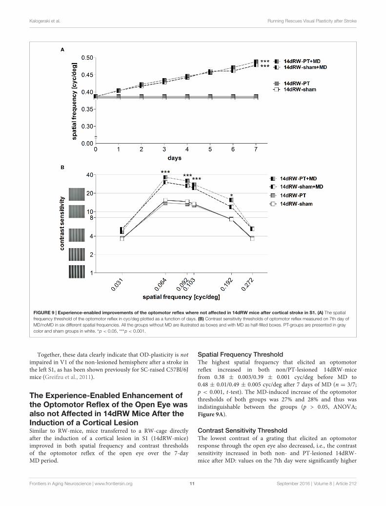
FIGURE 9 | Experience-enabled improvements of the optomotor reflex where not affected in 14dRW mice after cortical stroke in S1. (A) The spatialfrequency threshold of the optomotor reflex in cyc/deg plotted as a function of days. (B) Contrast sensitivity thresholds of optomotor reflex measured on 7th day ofMD/noMD in six different spatial frequencies. All the groups without MD are illustrated as boxes and with MD as half-filled boxes. PT-groups are presented in graycolor and sham groups in white. ∗p < 0.05, ∗∗∗p < 0.001.
Together, these data clearly indicate that OD-plasticity is notimpaired in V1 of the non-lesioned hemisphere after a stroke inthe left S1, as has been shown previously for SC-raised C57Bl/6Jmice (Greifzu et al., 2011).
The Experience-Enabled Enhancement ofthe Optomotor Reflex of the Open Eye wasalso not Affected in 14dRW Mice After theInduction of a Cortical LesionSimilar to RW-mice, mice transferred to a RW-cage directlyafter the induction of a cortical lesion in S1 (14dRW-mice)improved in both spatial frequency and contrast thresholdsof the optomotor reflex of the open eye over the 7-dayMD period.
Spatial Frequency ThresholdThe highest spatial frequency that elicited an optomotorreflex increased in both non/PT-lesioned 14dRW-micefrom 0.38 ± 0.003/0.39 ± 0.001 cyc/deg before MD to0.48 ± 0.01/0.49 ± 0.005 cyc/deg after 7 days of MD (n = 3/7;p < 0.001, t-test). The MD-induced increase of the optomotorthresholds of both groups was 27% and 28% and thus wasindistinguishable between the groups (p > 0.05, ANOVA;Figure 9A).
Contrast Sensitivity ThresholdThe lowest contrast of a grating that elicited an optomotorresponse through the open eye also decreased, i.e., the contrastsensitivity increased in both non- and PT-lesioned 14dRW-mice after MD: values on the 7th day were significantly higher
Frontiers in Aging Neuroscience | www.frontiersin.org 11 September 2016 | Volume 8 | Article 212
Kalogeraki et al. Running Rescues Visual Plasticity after Stroke
compared to noMD-mice at four out of the six spatial frequenciestested (p < 0.001 for spatial frequencies 0.064/0.092/0.103cyc/deg, and p < 0.01 for 0.192 cyc/deg; ANOVA, Figure 9B).
In detail, in 14dRW-sham/PT mice, contrast sensitivitythresholds of the open eye (at 0.031, 0.064, 0.092, 0.103,0.192 and 0.272 cyc/deg) improved to 5.2 ± 0.2 (19%contrast)/4.7 ± 0.1 (22%), 30.1 ± 1.8 (3%)/36.3 ± 2.2 (3%),26.4± 1.2 (4%)/31.8± 1.5 (3%), 24.3± 1.8 (4%)/27.8± 1.4 (4%),11.9 ± 0.9 (9%)/15.6 ± 0.7 (7%) and 5.2 ± 0.2 (19%)/5.3 ± 0.2(19%) on day 7 (p > 0.05, p < 0.001, p < 0.001, p < 0.001,p< 0.01, p > 0.05, compared to day 0, ANOVA). The experience-enabled increase in contrast sensitivity thresholds after MD wasindistinguishable between non- and PT-lesioned 14dRW-mice atany spatial frequency (p > 0.05, ANONA). Without MD, valuesof both groups remained stable over the 7-day measuring period(p > 0.05, for every frequency, ANOVA).
Amount of Running in 14dRW Mice Duringthe MD/noMD PeriodTo test: (i) whether a cortical lesion in S1 will affect the ability ofthe mice to run in a RW; and also (ii) if the amount of runninghas an effect on OD-plasticity after PT, the turns of the RW weremeasured daily in 14dRW-mice after the PT or sham treatment.Since there was no significant increase or decrease of runningamount over days after MD for both groups of 14dRW-mice(sham: p= 0.14; PT: p= 0.12, ANOVA), we calculated an averageamount of turns per day and animal. The 14dRW-sham mice(n = 6) ran on average 6.7 ± 1.2 km per day corresponding to16665 ± 3047 turns of the wheel, while the 14dRW-PT mice(n = 12) ran on average 4.9 ± 1.00 km per day correspondingto 12235± 2470 turns of the wheel. The running amount did notdiffer between the non- and PT-lesioned mice (p = 0.29, t-test),suggesting that the cortical lesion in S1 was not interfering withthe ability of mice to run on a RW. Furthermore, there wereno significant differences between the running amount of thenon and PT-lesioned mice after MD (sham + MD/PT + MD:4.9 ± 1.7/6.6 ± 1.2 km, n = 3/7; p = 0.427, t-test), and valueswithout or with MD were indistinguishable for both 14dRW-sham and 14dRW-PT mice (sham/sham + MD: p = 0.14, t-test;PT/PT + MD: p = 0.12, t-test), suggesting that the MD was notinterfering with running.
Finally, we tested whether the amount of running and theODIof individual mice were correlated, i.e., whether more runningmight cause stronger OD-shifts. To this end, the ODI of everymouse was plotted against the kilometers per day that eachmouse runs for all the animals that received an MD. Analysisof the data showed that there was no correlation between theindividual running amount and the ODI (p= 0.66, R2
= 0.00248,correlation test).
DISCUSSION
In the present study, we analyzed whether voluntary physicalexercise is beneficial for rescuing plasticity after a cortical stroke,in addition to being crucial for preserving visual plasticity intoadulthood. Our results provide clear evidence that this was
the case: raising mice in SCs equipped with a RW preventedimpairments of visual plasticity after a stroke in S1-in contrastto SC-mice (Greifzu et al., 2011; Pielecka-Fortuna et al., 2015b).In addition, we could demonstrate the therapeutic potential ofrunning after stroke: just 14 days of voluntary running afterstroke restored plasticity to SC-raised adult mice. Notably, visualplasticity was restored in both tested paradigms: compared toSC-mice without a RW, OD-plasticity was preserved/restored inV1 and the experience-enabled improvements of the optomotorreflex of the open eye were rescued in both long-term and short-term running adult mice.
Having access to a RW for all their life, our adult RW-PT-mice were protected against the plasticity impairments normallyoccurring in (even younger) SC-raised mice after a stroke in S1or secondary motor cortex (M2; Greifzu et al., 2011; Pielecka-Fortuna et al., 2015a). Notably, OD-plasticity was preserved intoadulthood, similar as in the non-lesioned control RW-animals.Thus, our RW-PT-mice reacted to the stroke like juvenileSC-mice or EE-mice (Greifzu et al., 2014). Physical exercise isknown to have beneficial effects on overall well-being, improvingcognition and delaying age-related memory decline in humans(Cotman et al., 2007; Hillman et al., 2008). Mice with access to aRW have increased cell proliferation, higher neuron survival andelevated levels of BDNF in the hippocampus (Kobilo et al., 2011;Mustroph et al., 2012). Many molecular factors that changedafter EE are also altered after physical exercise (Vivar et al.,2013); e.g., increased levels of BDNF (Berchtold et al., 2005), NGF(Neeper et al., 1996), and IGF (Carro et al., 2000). Interestingly,exposure of rats to either EE or treadmill exercise reduced thesusceptibility to cortical spreading depression (Santos-Monteiroet al., 2000; Lima et al., 2014), thereby possibly contributing toreduced plasticity-impairing effects of a cortical PT-lesion afterfree access to a RW.
Although many studies showed positive effects of physicalexercise on the brain, only recently the effect of runningon visual cortical activity and plasticity has been addressed.Locomotion increases firing rates in V1 (Niell and Stryker, 2010)and the lateral geniculate nucleus (Erisken et al., 2014), andenhancements of visual responses induced by locomotion aresufficient to promote recovery of visual function after long termMD (Kaneko and Stryker, 2014). Voluntary physical exercisealso preserved OD-plasticity into adulthood and restored thisplasticity in SC-raised adult mice (Kalogeraki et al., 2014).Additionally, the activity of a specific class of V1 interneuronsthat express vasoactive intestinal protein (VIP) is directlymodulated by locomotion (Fu et al., 2014). A neural circuitunderlying these effects was proposed, in which the specificrecruitment of VIP-cells by locomotion directly modulated V1-activity through a disinhibitory mechanism (Fu et al., 2014; Leeet al., 2014). It was also shown that providing adult rats withRWs for 3 weeks caused a decrease in GABA-release (Baroncelliet al., 2012). Extended running experience by raising mice in SCswith RWs may thus ‘‘chronically’’ reduce inhibitory drive ontopyramidal cells and thus promote cortical plasticity.
In addition to these plasticity-promoting effects of physicalexercise on a healthy brain, studies also pointed out the beneficialeffects of physical exercise after brain lesions. RW-exercise
Frontiers in Aging Neuroscience | www.frontiersin.org 12 September 2016 | Volume 8 | Article 212
Kalogeraki et al. Running Rescues Visual Plasticity after Stroke
increased the number of newborn hippocampal neurons afterPT-stroke inmice and improved spatiotemporal learning (Geibiget al., 2012). Mice with middle cerebral artery occlusion (MCAO)showed long-term functional and cognitive improvementsafter running (Gertz et al., 2006). Physical exercise was alsoneuroprotective: running 2–3 weeks before an MCAO-strokereduced cerebral infarct size and sensory-motor deficits inrodents (Wang et al., 2001; Endres et al., 2003).
One possibility how physical exercise may influenceOD-plasticity after stroke is through remodeling of theextracellular matrix by matrix metalloproteinase (MMP) activity.A recent study reported elevated levels of MMP9 after mildtreadmill exercise in rats (Nishijima et al., 2015), demonstratingthat running influences MMP activity. MMPs play an importantrole for plasticity in both the healthy and diseased brain (Huntley,2012). MMP-activity is a crucial component of OD-plasticityin the healthy juvenile and adult brain (Spolidoro et al., 2012;Pielecka-Fortuna et al., 2015b). After cerebral ischemia, theexpression of some MMPs is increased (Gasche et al., 1999; Heoet al., 1999; Montaner et al., 2001; Rosell et al., 2006). Inhibitionof MMPs with a broad-spectrum MMP-inhibitor immediatelybefore a stroke partially rescued experience-dependent barrelcortex plasticity in mice (Cybulska-Klosowicz et al., 2011).Recently, we revealed that an optimal level of MMP-activity iscrucial for adult visual cortex plasticity in the healthy and stroke-affected brain (Pielecka-Fortuna et al., 2015a): in healthy mice,inhibition of MMP-activity with a broad spectrum inhibitorduring the 7-day-MD-period resulted in lost OD-plasticity; incontrast, lowering stroke-induced elevated levels of MMPs withan inhibitor after stroke rescued OD-plasticity.
Stroke also affects the balance between excitation andinhibition in the affected neuronal network (Carmichael, 2006).High levels of the neurotransmitter glutamate after ischemicstroke lead to excitotoxicity and neuronal cell death (Laiet al., 2014). Additionally, a lesion in V1 caused reducedbasal GABAergic transmission measured away from the lesionborder (Imbrosci et al., 2015). It seems that changes in bothglutamatergic and GABAergic transmission after stroke can leadto negative consequences and thus may interfere with plasticity.EE-raising not only preserved a low GABAergic inhibitionand juvenile OD-plasticity into adulthood, it also protectedadult mice from stroke induced impairments of V1-plasticityafter an S1-lesion (Greifzu et al., 2014). Physical exerciseinfluencesmany neurotransmitter systems in the brain, includingglutamatergic (Farmer et al., 2004; Vasuta et al., 2007; Louet al., 2008) and GABAergic (Molteni et al., 2002). Altogether,these studies imply that a lower inhibitory tone after runningmay protect V1 from plasticity impairments after an S1-lesion.Likewise, short-term dark exposure reduced the inhibitory tonein V1 (He et al., 2006) and preserved OD-plasticity afteran S1-lesion (Stodieck et al., 2014). In summary, exposingmice to conditions which reduce the inhibitory tone in V1,protected them against stroke-induced impairments of visualplasticity. Interestingly, modification of excitatory circuits wasrecently shown to be also beneficial for preserving plasticity afterstroke: OD-plasticity in V1 was preserved after an S1-strokein mice lacking postsynaptic density protein-95 (Greifzu et al.,
2016b), a signaling scaffold protein present at mature excitatorysynapses necessary for synaptic maturation (Huang et al.,2015).
Furthermore, the basic parameters of spatial vision and theirexperience-enabled improvements were tested using optomotry(Prusky et al., 2004). After an S1-lesion, RW-mice showedsignificant enhancements in both the spatial frequency andcontrast sensitivity thresholds of the optomotor reflex of the openeye after MD. In contrast, an S1-lesion in adult SC-raised micewithout a RW abolished any threshold improvements (Greifzuet al., 2011). These improvements were rescued by either anti-inflammatory treatment or by waiting for 14 days between lesionand MD (Greifzu et al., 2011). In EE-raised mice, increases inoptomotor thresholds were also detected after stroke, but therescue was only partial (Greifzu et al., 2014). In contrast, inthe present study, both short-term and long-term running fullyrestored the optomotor threshold increases, and the final valueswere similar to those of non-lesioned SC- (Prusky et al., 2004) orRW-mice (Kalogeraki et al., 2014). Whether running has an anti-inflammatory effect remains to be determined.
Quantitative analyses showed no significant differences inlesion localization, length, depth and volume between lifelongand 14dRW-mice, only a smaller diameter in the 14dRW-mice.There was, however, no correlation between lesion volume andthe ODI after MD, in agreement with previous observations(Greifzu et al., 2011). Moreover, even a smaller and threetimes more distant lesion in M2, can affect OD-plasticity inV1 (Pielecka-Fortuna et al., 2015b). Thus, the smaller lesiondiameter in 14dRW-mice is most probably not causing thepreserved plasticity (lesion volume was similar to the long-termRW-group). In summary, the restoration of OD-plasticity in bothgroups of RW-mice is most likely due to their voluntary physicalexercise, and not caused by any differences in lesion location orvolume.
Summarizing, our data show that voluntary physical exercisewith RWs not only preserved visual plasticity into adulthood,but also restored OD-plasticity in adult mice after stroke.Additionally, physical exercise in stroke-lesioned mice restoredthe experience-enabled improvements of the spatial frequencyand contrast sensitivity thresholds of the optomotor reflex. Moreinterestingly, even short-term running for just 14 days after astroke was already effective in restoring both OD-plasticity andimprovements of the optomotor reflex in adult SC-raised mice.Interestingly, the magnitude of the induced OD-shifts and ofthe optomotor threshold improvements were indistinguishablebetween the long-term and short-term running groups. Thisdemonstrates that voluntary physical exercise can protect fromplasticity impairments caused by a cortical stroke, and is effectiveeven if the exercise starts only in the post-stroke period.Thus, the present data advertise voluntary physical exerciseas a promising candidate for post-stroke plasticity promotingtherapies.
AUTHOR CONTRIBUTIONS
Conception and design of the work (EK, JP-F, SL), acquisitionof data (EK, JP-F, JMH), analysis and interpretation of data
Frontiers in Aging Neuroscience | www.frontiersin.org 13 September 2016 | Volume 8 | Article 212
Kalogeraki et al. Running Rescues Visual Plasticity after Stroke
(EK, JP-F, JMH, SL), drafting and revising of the article(EK, JP-F, SL). All authors approved the final version of themanuscript.
FUNDING
This work was supported by the Federal Ministry of Educationand Research, Germany, Grants 01GQ0921 (to EK and JP-F)and 01GQ0810 (to SL), by an Alexander von HumboldtResearch Fellowship for Postdoctoral Researchers (to JP-F)
and by grants of the Deutsche Forschungsgemeinschaftthrough the Collaborative Research Center 889 ‘‘CellularMechanisms of Sensory Processing’’ to Siegrid Löwel(Project B5).
ACKNOWLEDGMENTS
We thankMatthias Schink for excellent animal care and technicalsupport.
REFERENCES
Baroncelli, L., Bonaccorsi, J., Milanese, M., Bonifacino, T., Giribaldi, F., Manno, I.,et al. (2012). Enriched experience and recovery from amblyopia in adultrats: impact of motor, social and sensory components. Neuropharmacol 62,2388–2397. doi: 10.1016/j.neuropharm.2012.02.010
Baroncelli, L., Sale, A., Viegi, A., Maya-Vetencourt, J. F., De Pasquale, R.,Baldini, S., et al. (2010). Experience-dependent reactivation of oculardominance plasticity in the adult visual cortex. Exp. Neurol. 226, 100–109.doi: 10.1016/j.expneurol.2010.08.009
Berchtold, N. C., Chinn, G., Chou, M., Kesslak, J. P., and Cotman, C. W. (2005).Exercise primes a molecular memory for brain-derived neurotrophic factorprotein induction in the rat hippocampus. Neuroscience 133, 853–861. doi: 10.1016/j.neuroscience.2005.03.026
Cang, J. H., Kalatsky, V. A., Löwel, S., and Stryker, M. P. (2005). Optical imaging ofthe intrinsic signal as ameasure of cortical plasticity in themouse.Vis. Neurosci.22, 685–691. doi: 10.1017/s0952523805225178
Carmichael, S. T. (2006). Cellular and molecular mechanisms of neural repairafter stroke: making waves. Ann. Neurol. 59, 735–742. doi: 10.1002/ana.20845
Carro, E., Nuñez, A., Busiguina, S., and Torres-Aleman, I. (2000). Circulatinginsulin-like growth factor I mediates effects of exercise on the brain. J. Neurosci.20, 2926–2933.
Cotman, C. W., Berchtold, N. C., and Christie, L. A. (2007). Exercise builds brainhealth: key roles of growth factor cascades and inflammation. Trends Neurosci.30, 464–472. doi: 10.1016/j.tins.2007.06.011
Cybulska-Klosowicz, A., Liguz-Lecznar, M., Nowicka, D., Ziemka-Nalecz, M.,Kossut, M., and Skangiel-Kramska, J. (2011). Matrix metalloproteinaseinhibition counteracts impairment of cortical experience-dependent plasticityafter photothrombotic stroke. Eur. J. Neurosci. 33, 2238–2246. doi: 10.1111/j.1460-9568.2011.07713.x
Dräger, U. C. (1978). Observations on monocular deprivation in mice.J. Neurophysiol. 41, 28–42.
Endres, M., Gertz, K., Lindauer, U., Katchanov, J., Schultze, J., Schröck, H., et al.(2003). Mechanisms of stroke protection by physical activity. Ann. Neurol. 54,582–590. doi: 10.1002/ana.10722
Erisken, S., Vaiceliunaite, A., Jurjut, O., Fiorini, M., Katzner, S., and Busse, L.(2014). Effects of locomotion extend throughout the mouse early visual system.Curr. Biol. 24, 2899–2907. doi: 10.1016/j.cub.2014.10.045
Espinosa, J. S., and Stryker, M. P. (2012). Development and plasticity ofthe primary visual cortex. Neuron 75, 230–249. doi: 10.1016/j.neuron.2012.06.009
Farmer, J., Zhao, X., van Praag, H., Wodtke, K., Gage, F. H., and Christie, B. R.(2004). Effects of voluntary exercise on synaptic plasticity and gene expressionin the dentate gyrus of adult male sprague-dawley rats in vivo. Neuroscience124, 71–79. doi: 10.1016/j.neuroscience.2003.09.029
Fu, Y., Tucciarone, J. M., Espinosa, J. S., Sheng, N., Darcy, D. P., Nicoll, R. A.,et al. (2014). A cortical circuit for gain control by behavioral state. Cell 156,1139–1152. doi: 10.1016/j.cell.2014.01.050
Gasche, Y., Fujimura, M., Morita-Fujimura, Y., Copin, J. C., Kawase, M.,Massengale, J., et al. (1999). Early appearance of activated matrixmetalloproteinase-9 after focal cerebral ischemia in mice: a possible rolein blood-brain barrier dysfunction. J. Cereb. Blood Flow Metab. 19, 1020–1028.doi: 10.1097/00004647-199909000-00010
Geibig, C. S., Keiner, S., and Redecker, C. (2012). Functional recruitment ofnewborn hippocampal neurons after experimental stroke. Neurobiol. Dis. 46,431–439. doi: 10.1016/j.nbd.2012.02.007
Gertz, K., Priller, J., Kronenberg, G., Fink, K. B., Winter, B., Schröck, H., et al.(2006). Physical activity improves long-term stroke outcome via endothelialnitric oxide synthase-dependent augmentation of neovascularization andcerebral blood flow. Circ. Res. 99, 1132–1140. doi: 10.1161/01.res.0000250175.14861.77
Gordon, J. A., and Stryker, M. P. (1996). Experience-dependent plasticity ofbinocular responses in the primary visual cortex of the mouse. J. Neurosci. 16,3274–3286.
Greifzu, F., Kalogeraki, E., and Löwel, S. (2016a). Environmental enrichmentpreserved lifelong ocular dominance plasticity, but did not improve visualabilities. Neurobiol. Aging 41, 130–137. doi: 10.1016/j.neurobiolaging.2016.02.014
Greifzu, F., Parthier, D., Goetze, B., Schlüter, O. M., and Löwel, S. (2016b). Oculardominance plasticity after stroke was preserved in PSD-95 knockout mice. PloSOne 11:e0149771. doi: 10.1371/journal.pone.0149771
Greifzu, F., Pielecka-Fortuna, J., Kalogeraki, E., Krempler, K., Favaro, P. D.,Schlüter, O. M., et al. (2014). Environmental enrichment extends oculardominance plasticity into adulthood and protects from stroke-inducedimpairments of plasticity. Proc. Natl. Acad. Sci. U S A 111, 1150–1155. doi: 10.1073/pnas.1313385111
Greifzu, F., Schmidt, S., Schmidt, K. F., Kreikemeier, K.,Witte, O.W., and Löwel, S.(2011). Global impairment and therapeutic restoration of visual plasticitymechanisms after a localized cortical stroke. Proc. Natl. Acad. Sci. U S A 108,15450–15455. doi: 10.1073/pnas.1016458108
He, H.-Y., Hodos, W., and Quinlan, E. M. (2006). Visual deprivation reactivatesrapid ocular dominance plasticity in adult visual cortex. J. Neurosci. 26,2951–2955. doi: 10.1523/JNEUROSCI.5554-05.2006
Heo, J. H., Lucero, J., Abumiya, T., Koziol, J. A., Copeland, B. R., anddel Zoppo, G. J. (1999). Matrix metalloproteinases increase very early duringexperimental focal cerebral ischemia. J. Cereb. Blood Flow Metabol. 19,624–633. doi: 10.1097/00004647-199906000-00005
Hillman, C. H., Erickson, K. I., and Kramer, A. F. (2008). Be smart, exercise yourheart: exercise effects on brain and cognition. Nat. Rev. Neurosci. 9, 58–65.doi: 10.1038/nrn2298
Huang, X., Stodieck, S. K., Goetze, B., Cui, L., Wong, M. H., Wenzel, C., et al.(2015). Progressive maturation of silent synapses governs the duration ofa critical period. Proc. Natl. Acad. Sci. U S A 112, E3131–E3140. doi: 10.1073/pnas.1506488112
Huntley, G. W. (2012). Synaptic circuit remodeling by matrix metalloproteinasesin health and disease. Nat. Rev. Neurosci. 13, 743–757. doi: 10.1038/nrn3320
Imbrosci, B., Wang, Y., Arckens, L., and Mittmann, T. (2015). Neuronalmechanisms underlying transhemispheric diaschisis following focal corticalinjuries. Brain Struct. Funct. 220, 1649–1664. doi: 10.1007/s00429-014-0750-8
Kalatsky, V. A., and Stryker, M. P. (2003). New paradigm for optical imaging:temporally encoded maps of intrinsic signal. Neuron 38, 529–545. doi: 10.1016/S0896-6273(03)00286-1
Kalogeraki, E., Greifzu, F., Haack, F., and Löwel, S. (2014). Voluntary physicalexercise promotes ocular dominance plasticity in adult mouse primaryvisual cortex. J. Neurosci. 34, 15476–15481. doi: 10.1523/JNEUROSCI.2678-14.2014
Frontiers in Aging Neuroscience | www.frontiersin.org 14 September 2016 | Volume 8 | Article 212
Kalogeraki et al. Running Rescues Visual Plasticity after Stroke
Kaneko, M., and Stryker, M. P. (2014). Sensory experience during locomotionpromotes recovery of function in adult visual cortex. Elife 3:e02798. doi: 10.7554/eLife.02798
Kobilo, T., Liu, Q.-R., Gandhi, K., Mughal, M., Shaham, Y., and van Praag, H.(2011). Running is the neurogenic and neurotrophic stimulus in environmentalenrichment. Learn. Mem. 18, 605–609. doi: 10.1101/lm.2283011
Lai, S., Panarese, A., Spalletti, C., Alia, C., Ghionzoli, A., Caleo, M., et al.(2014). Quantitative kinematic characterization of reaching impairments inmice after a stroke. Neurorehabil. Neural Repair 29, 382–392. doi: 10.1177/1545968314545174
Lee, A. M., Hoy, J. L., Bonci, A., Wilbrecht, L., Stryker, M. P., and Niell, C. M.(2014). Identification of a brainstem circuit regulating visual cortical statein parallel with locomotion. Neuron 83, 455–466. doi: 10.1016/j.neuron.2014.06.031
Lehmann, K., and Löwel, S. (2008). Age-dependent ocular dominance plasticity inadult mice. PloS One 3:e3120. doi: 10.1371/journal.pone.0003120
Lima, C. B., Saores Gde, S. F., Vitor, S. M., Andrade-da-Costa, B. L., Castellano, B.,and Guedes, R. C. A. (2014). Spreading depression features and Iba1immunoreactivity in the cerebral cortex of developing rats submitted totreadmill exercise after treatment with monosodium glutamate. Int. J. Dev.Neurosci. 33, 98–105. doi: 10.1016/j.ijdevneu.2013.12.008
Lou, S. J., Liu, J. Y., Chang, H., and Chen, P. J. (2008). Hippocampal neurogenesisand gene expression depend on exercise intensity in juvenile rats. Brain Res.1210, 48–55. doi: 10.1016/j.brainres.2008.02.080
Molteni, R., Ying, Z., and Gómez-Pinilla, F. (2002). Differential effects of acute andchronic exercise on plasticity-related genes in the rat hippocampus revealedby microarray. Eur. J. Neurosci. 16, 1107–1116. doi: 10.1046/j.1460-9568.2002.02158.x
Montaner, J., Alvarez-Sabin, J., Molina, C., Angles, A., Abilleira, S., Arenillas, J.,et al. (2001). Matrix metalloproteinase expression after human cardioembolicstroke: temporal profile and relation to neurological impairment. Stroke 32,1759–1766. doi: 10.1161/01.str.32.8.1759
Mustroph,M. L., Chen, S., Desai, S. C., Cay, E. B., DeYoung, E. K., and Rhodes, J. S.(2012). Aerobic exercise is the critical variable in an enriched environmentthat increases hippocampal neurogenesis and water maze learning in maleC57BL/6J mice. Neuroscience 219, 62–71. doi: 10.1016/j.neuroscience.2012.06.007
Neeper, S. A., Gómez-Pinilla, F., Choi, J., and Cotman, C. W. (1996). Physicalactivity increases mRNA for brain-derived neurotrophic factor and nervegrowth factor in rat brain. Brain Res. 726, 49–56. doi: 10.1016/0006-8993(96)00273-9
Niell, C. M., and Stryker, M. P. (2010). Modulation of visual responses bybehavioral state in mouse visual cortex. Neuron 65, 472–479. doi: 10.1016/j.neuron.2010.01.033
Nishijima, T., Kawakami, M., and Kita, I. (2015). A bout of treadmill exerciseincreases matrix metalloproteinase-9 activity in the rat hippocampus.Neurosci.Lett. 594, 144–149. doi: 10.1016/j.neulet.2015.03.063
Pielecka-Fortuna, J., Kalogeraki, E., Greifzu, F., and Löwel, S. (2015a). A smallmotor cortex lesion abolished ocular dominance plasticity in the adultmouse primary visual cortex and impaired experience-dependent visualimprovements. PloS One 10:e0137961. doi: 10.1371/journal.pone.0137961
Pielecka-Fortuna, J., Kalogeraki, E., Fortuna,M. G., and Löwel, S. (2015b). Optimallevel activity of matrix metalloproteinases is critical for adult visual plasticity inthe healthy and stroke-affected brain. Elife 4:e11290. doi: 10.7554/eLife.11290
Prusky, G. T., Alam, N. M., Beekman, S., and Douglas, R. M. (2004). Rapidquantification of adult and developing mouse spatial vision using a virtualoptomotor system. Invest. Ophthalmol. Vis. Sci. 45, 4611–4616. doi: 10.1167/iovs.04-0541
Rosell, A., Ortega-Aznar, A., Alvarez-Sabin, J., Fernandez-Cadenas, I., Ribó, M.,Molina, C. A., et al. (2006). Increased brain expression of matrixmetalloproteinase-9 after ischemic and hemorrhagic human stroke. Stroke 37,1399–1406. doi: 10.1161/01.str.0000223001.06264.af
Sale, A., Maya-Vetencourt, J. F., Medinin, P., Cenni, M. C., Baroncelli, L.,De Pasquale, R., et al. (2007). Environmental enrichment in adulthoodpromotes amblyopia recovery through a reduction of intracortical inhibition.Nat. Neurosci. 10, 679–681. doi: 10.1038/nn1899
Santos-Monteiro, J., Teodósio, N. R., and Guedes, R. C. A. (2000). Long-lastingeffects of early environmental stimulation on cortical spreading depression innormal and early malnourished adult rats. Nutr. Neurosci. 3, 29–40. doi: 10.1080/1028415x.2000.11747301
Scali, M., Baroncelli, L., Cenni, M. C., Sale, A., and Maffei, L. (2012). A richenvironmental experience reactivates visual cortex plasticity in aged rats. Exp.Gerontol. 47, 337–341. doi: 10.1016/j.exger.2012.01.007
Spolidoro, M., Putignano, E., Munafò, C., Maffei, L., and Pizzorusso, T.(2012). Inhibition of matrix metalloproteinases prevents the potentiation ofnondeprived-eye responses after monocular deprivation in juvenile rats. Cereb.Cortex 22, 725–734. doi: 10.1093/cercor/bhr158
Stodieck, S. K., Greifzu, F., Goetze, B., Schmidt, K.-F., and Löwel, S. (2014).Brief dark exposure restored ocular dominance plasticity in aging mice andafter a cortical stroke. Exp. Gerontol. 60, 1–11. doi: 10.1016/j.exger.2014.09.007
Vasuta, C., Caunt, C., James, R., Samadi, S., Schibuk, E., Kannangara, T.,et al. (2007). Effects of exercise on NMDA receptor subunit contributions tobidirectional synaptic plasticity in the mouse dentate gyrus. Hippocampus 17,1201–1208. doi: 10.1002/hipo.20349
Vivar, C., Potter, M. C., and van Praag, H. (2013). All about running: synapticplasticity, growth factors and adult hippocampal neurogenesis. Curr. Top.Behav. Neurosci. 15, 189–210. doi: 10.1007/7854_2012_220
Wang, R. Y., Yang, Y. R., and Yu, S. M. (2001). Protective effects of treadmilltraining on infarction in rats. Brain Res. 922, 140–143. doi: 10.1016/s0006-8993(01)03154-7
Watson, B. D., Dietrich, W. D., Busto, R., Wachtel, M. S., and Ginsberg, M. D.(1985). Induction of reproducible brain infarction by photochemically initiatedthrombosis. Ann. Neurol. 17, 497–504. doi: 10.1002/ana.410170513
Wiesel, T. N., and Hubel, D. H. (1963). Single-cell responses in striate cortex ofkittens deprived of vision in 1 eye. J. Neurophysiol. 26, 1003–1017.
Conflict of Interest Statement: The authors declare that the research wasconducted in the absence of any commercial or financial relationships that couldbe construed as a potential conflict of interest.
Copyright © 2016 Kalogeraki, Pielecka-Fortuna, Hüppe and Löwel. This is an open-access article distributed under the terms of the Creative Commons AttributionLicense (CC BY). The use, distribution and reproduction in other forums ispermitted, provided the original author(s) or licensor are credited and that theoriginal publication in this journal is cited, in accordance with accepted academicpractice. No use, distribution or reproduction is permitted which does not complywith these terms.
Frontiers in Aging Neuroscience | www.frontiersin.org 15 September 2016 | Volume 8 | Article 212