physics 1230: light and color · 2010-06-29 · what does accommodation of the eye have to do with...
TRANSCRIPT

1
Physics 1230: Light and Color
Lecture 13: The Eye, eyeglasses and other optical
instruments, start retina signal processing.
Reading: Chap. 9,10 Color perception.
Exam 4 cancelled: Exam extra credit
assignment will be due Wed. at 5PM
Extra credit to improve exam scores!
HW9: Due today, Monday, 5PM
FCQ at end of lecture.

Review: Parts of the human eye
Specialized optical instrument
and image analysis computer.

3
The Eye: Analogy
to the Camera
Lens and cornea
Iris (diaphram)
Ciliary muscle
(focus)
Retina (FILM??)
We will see that the
retina does FAR more
than film or CCD
4
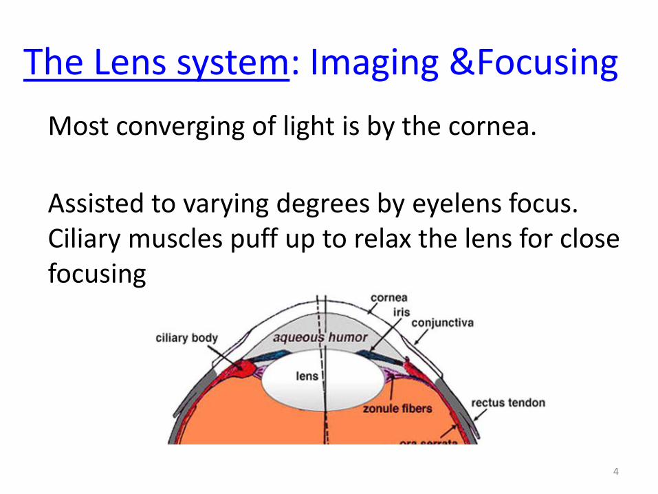
The Lens system: Imaging &Focusing
Most converging of light is by the cornea.
Assisted to varying degrees by eyelens focus.Ciliary muscles puff up to relax the lens for close focusing

What does accommodation of the eye have to do with
looking at me or your thumb? How does it work?(Lens represents combined cornea-eyelens system)
large f
smaller f
Focusing your eye on a nearby thumb requires shorter focal length (more bulgy) eyelens than focusing on the Prof far away, since rays must be bent more for image to fall on retina.
Thumb is out of focus
Prof is in focus
Thumb is in focus
Prof is out of focus

6
The Eye as a cool instrument: The eyelens
• Spherical aberration is mostly correctedCornea is not spherical surface (aspherical)Iris cuts out rays through the edge of the lensIndex of refraction is not uniform.
• Curvature of fieldretina is curved to correct for this
• Chromatic aberration:Bluest light is absorbed
We know that lenses suffer from various
aberrations. What happens in the eyelens?
Many of these tricks are now used in technology.

7
The Retina: Detecting the light and processing the images
Has 108 nerve endings to detect image
rods, for high sensitivity (night vision)
cones, for color and detail, 7 million
optic nerve = 106 transmission lines
fovea, region of best vision (cones)
The retina and optic nerves are recognized as actually
parts of the brain (like your olifactory bulb in the nose).
They start development IN the brain and migrate…
More nerves in your retina than some creatures
have in their entire brains. Processing Power.


Human versus cat retinas
Light comes in
from here
http://webvision.med.utah.edu/anatomy.html
Lots of specialization here for detection and
processing. More in the next couple of lectures…

10
Very common eye problems:
• Myopia, see close objects clearly, onlyfixed by a negative lens
• Hyperopia, see things far, onlyfixed by a positive lens
• Presbyopia, stiff lens, no accommodationBifocal glasses have near and far foci.
Issues in the lens focusing affect many
of us:
How do we fix these problems?

The remaining lectures:
11
• Ch. 5 (the eye),
• Ch. 6 (optical instruments),
• Ch. 7 (Retina and visual perception),
• Ch. 9 & 10 (color & color perception).
We
are
here

12
Optical instruments we’ll cover:
• Single lens instruments– Eyeglasses
– Magnifying glass
• Two lens– Telescope & binoculars
– Microscope
We
are
here

A near-sighted or MYOPIC eye produces an
image that is not far enough behind the lens,
so is blurry on the retina. Therefore, the eye
lens focal length is:
13
A) Too long for a focused image.
B) Too short for a focused image.
C) Actually, the iris is closed too much
D) None of these.

You have a lens with a short focal length
and you wish it was longer. You can make it
longer by using a second lens. The correct
choice for this case is:
14
A) A focusing lens of negative power
B) A diverging lens of positive power
C) A focusing lens of positive power
D) A diverging lens of negative power
Recall: 1 2
1 2
1 1 1
TOTAL
TOTAL
f f f
OR
D D D

15
Eyeglasses: Our most common optical instrument
For nearsighted people (can’t focus far away)
Eyeglasses are diverging (thinner in middle)
For farsighted people (can’t focus up close)
Eyeglasses are converging (thicker in middle)
Demo: eyeglasses
Normal vision: you can focus from 25 cm to infinity ()
18th century HUGE
improvement in quality of life

16
Eyeglass prescription is in diopters
Optometrists use diopters to measure the powerof a lens
Diopters [or D] = 1 / (focal length in meters)
Example: f = 50 cm or f = 0.5 m
D = 1/f = 2 diopters (units are 1/meters)
The example above would be:
(A) reading glasses (B) distance glasses

17
Astigmatism
Vertical and horizontal lines focus differently
This problem is fixed by a cylinder lens
Sharply focused
Out of focus
Focuses in one
direction, but not the
other!

18
Action of a cylinder lens
Focuses in one
direction, but not the
other!
If a cylinder lens is needed for your eyeglasses,
your cornea and eyelens is curved more in one
direction than in the other!

Play with your eyeglasses to see what they can do!
19

The Magnifying glass (again): New insight!
20
Ray optics lets us
determine the ray paths.
A model of the observer lets us predict
an image where rays converge.
Our first effort
to explain:

The Magnifying glass (again): Another view
21
Typical closest focus is 25 cm from the eye.
A magnifying glass is like READING GLASSES:
It lets you focus on closer things.
The eye perceives via focused images:
25 cm

The Magnifying glass (again): Another view
22Demo with thumb and eyepiece from telescope kits.
Best focused image alone.
The eye perceives via focused images:
25 cm
Focal
length
Magnifying glass
Bigger
image
25 cm
cmM
f
By similar triangles:
Or:

23
Magnification of a one-lens magnifier
Example:
5 cm focal length has a magnification of
cmin length focal
cm 25
Interesting thought:
2 mm focal length has 125 magnification
A) 5 B) 4 C) 25 D) 1/5 E) None of these

24
van Leeuwenhoek’s microscope
25 cm, reading distance,
approximately
Focal length, approximately

25
van Leeuwenhoek’s microscope
Years developed: 1660s
Tiny lens with 2 mm focal length
(a lens cannot be much bigger than the focal length)
Magnification = 125cm 0.2
cm 25
Problem: image was still small, and very dim.
Technology edge: Outstanding single lenses

26
Robert Hooke’s
microscope, also
circa 1660
Discovered: Blood cells,
Microbes, etc.
van Leeuwenhoek
No existing pictures
of Hooke…
A TWO LENS system.

27
Robert Hooke’s two-lens microscope
object
lens 1
Nosepiece
or
Objective
lens 2
eyepiece
lens 1 image
lens 2 image
A magnifying glass (the eyepiece) magnifies
the first image further.
The first lens, the
nosepiece, is used as
a projection lens.

28
Hooke’s discoveriesThe cell
Detailed structure of creatures.
Example: The flea (plague).

29
Modernbinocular
microscope is very much the same as
Hooke’s.
A beamsplitter, a half-
silvered mirror, sends
half the light to each
eyepiece.

Many optical instruments can be understood step by step, as we
did for Hooke’s microscope:
30
The first lens collects light and
produces an image.
The second lens produces a
new image of the first image.
The third lens produces a new
image of the second image…
And so on.

31
Galileo’s telescope (~1600)
Negative lens for eyepiece gives right-
side-up image.

32
Kepler’s telescope (~1600)
Positive lens for eyepiece gives upside down image.
It’s upside down, but brighter and is easier to see.
lens 1
Objective
lens 2
eyepiece
lens 1 image
lens 2 image

Your telescope kit makes a:
33
A)Galilean telescope
B)Keplerian telescope
C)Another type that we have not seen.

34
Galileo’s telescope
30 x magnification
Tiny lens means not
much light entered,
so image is dim.
Discoveries:
Sunspots
Craters on the Moon
Phases of Venus
Moons of Jupiter

Magnification of a telescope(refractor or reflector)
Magnification =
Example:
Objective focal length = 1 m = 100 cm
Eyepiece focal length = 1 cm
Magnification = 100
35
eyepiece oflength focal
mirroror lens objective oflength focal

Limits to magnification
A bigger fuzzy image is not useful (no additional information).
A high resolution image requires large lenses and mirrors.
36
8 inch reflector
telescope Hubble telescope, 96 inch
(2 meter) mirror
Smallest visible feature 1/(diameter of lens or mirror)

37
Yerkes observatory,
Largest refractor40 inch lens
1897
Larger lenses would
sag under their own
weight.
The lens was
achromatic (two kinds
of glass).

Yerkes Today
38
Still available for
observations in the
original observatory.
Lake Geneva Wisc.
COLD winters yield
great seeing.
Constant pressure
from developers for
the lakeshore.

39
Newton’s reflector telescope

40
Newton’s telescope
Advantage: no chromatic aberration

41
Palomar reflector - 5 meter mirror

42
South Africa Large Telescope (10 m)
This shows 7 of the 91 mirrors. Diameter is about 10 m.
Each mirror is a hexagon so that they pack closely.
Former CU student
Amanda Gulbis
uses this telescope.

43
Largest single mirrors are 8 m now
Prof. Roger Angel’s group. University of Arizona

44
Finished mirror(don’t sneeze)

45
KeckTelescope36 mirrors10 m dia.

46
Magellan Telescope (yr. 2018)24.5 m dia. segmented mirror

47
European Extremely Large Telescope (42 m dia.) would use many smaller mirrors

The remaining lectures:
48
• Ch. 5 (the eye),
• Ch. 6 (optical instruments),
• Ch. 7 (Retina and visual perception),
• Ch. 9 & 10 (color & color perception).
We
are
here

Stop here for the FCQs
49

The retina is where the image falls at the
back of your eyeball
• Inverted image falls on retinainstead of film.
– Can demonstrate this inversion
•Open left eye only.
•Press gently with left finger on eyeball just above tear duct.
•Observe dark spot in lower left corner of your field of view.
• Rods & cones packed into retina.
– Sensitive to light like camera film
• Optic nerve
– Nerve fibers connect rods & cones to brain. (transform light into electrical signals)
– Blind spot is where optic nerve leaves eyeball. Demo.
• How are rods and cones distributed in the retina? Fig. 5.12– The fovea is the small region near the
center of the retina
• Used for sharp, detailed viewing.
• Has the most cones (precise, color vision)
• Has no rods (used for low light, less precise viewing).
– Looking at someone means their image is on your fovea
• If their image is not on your fovea you see them "out of the corner of your eye."
• Eyeball moves to see a sharp image
– It scans to make all parts of an image eventually fall on your fovea
• Like TV image scanning

51
Retina
has 108 nerve endings to detect image
rods, for night vision
cones, for color and detail, 7 million
optic nerve = 106 transmission lines
fovea, region of best vision (cones)


Human and cat retinas
Light comes in
from here
http://webvision.med.utah.edu/anatomy.html

54
Rods and cones
• Rhodopsin, a photochemical, responds to lightIt is destroyed and reformed.Signal goes to a synapse, a gap between nerve cells
• There are 3 kinds of cones for 3 colors
red, green, blue

55
Retina details
Choroid, outside layer with blood supply
Photoreceptors: rods and cones
Plexiform layer, inside layer with nerves
Photopic vision, in bright light, cones are used
Scotopic, in low light, rods are usedmore rods per nerve combines signals

56

References
http://www.costaricacoffeeart.com/alternative_photograph
y_make_your_own_negative_film_or_plates.php
http://unblinkingeye.com/Articles/Emulsion/emulsion.html
http://en.wikipedia.org/wiki/Charge-coupled_device
http://www.camerapedia.org/wiki/CCD
57

58
Robert Hooke’s two-lens microscope
Magnification = M1 x M2
Example: Eyepiece M2 = 25
Nosepiece M1 = 40
Final magnification = 40 x 25 = 1000
M1, like for any projection lens, is Xi / Xo
M2, like for any magnifier, is 25 cm / focal length

59
Telescope drives
• Telescopes must rotate once every 24 hrs (approximately) to follow the stars, or the pictures will have streaks.
Northstar

Student telescopes
60
View of galaxyNGC1300

61
Catadioptric telescope
Also called Schmidt-Cassegrain Front glass lens corrects aberrationsWhy buy this? It’s shorter.

62
Hubble Space Telescope
image
Compare to student
telescope image.

63
Binoculars
Simply folded telescopes for each eye.

64
What does 7 x 50 binoculars mean?
• 7x is the magnification
• 50 is the diameter of the front lens (the objective lens) in millimeters
• 6 x 30 binoculars are easy to carry
• 7 x 50 binoculars are heavy, butthese give a brighter image
• 15 x 80 binoculars need a tripod

65
35 mm slide projector
Field lens is used to put the most light on the slide.
Field lensProjection
lensColor
slide
Mirror

66
Viewgraph projector
Curved mirror
Fresnel lens (condenser)
and viewgraph location
Projection
lens
Mirror

67
Eye problems
• Loss of accomodation: ability to focus from 10 inches to infinity
• Cataracts = cloudy eyelens, replacement lens does not accommodate
• Floaters = dead cells floating in vitreous humor (seen against a clear sky)
• Diseases of the eye components.
• Nerve damage
• Etc. …

68
A dash past The Iris
Higher light levels (say daytime): Closed down, f/8We sense colors!Fewer aberrationsMore depth of field
Low light levels (say at night!): Wide open, f/2 or f/3Get more of the available light!We lose our color vision.More aberrations from the larger lens openingLess depth of field
You can check depth of field:
Try it: Close one eye, hold up thumb, stuff behind
thumb is out of focus.