physiology of pain - semantic scholar · considered as an efferent neuro-effector system modulating...
TRANSCRIPT

"For personal study purposes only:" Extracted from Hemmings and Hopkins (Ed) "Foundations of Anesthesia", second edition, 2006 Elsevier Mosby
Physiology of Pain
Michael J Hudspith, Philip J Siddall, and Rajesh Munglani
TOPICS COVERED IN THIS CHAPTER Peripheral pain mechanisms Primary afferent nociceptors Visceral afferents Sympathetic nervous system Peripheral sensitisation/inflammation Role of nerve growth factor in peripheral sensitisation Silent nociceptors Peripheral nerve injury Clinical implications for peripheral analgesic action Dorsal horn mechanisms Termination sites of primary afferents Outputs from primary afferents Neurotransmitters and neuromodulators Excitatory nociceptor transmission Peptides Intracellular events Spinal mechanisms following nerve injury and inflammation Central sensitisation and synaptic plasticity Sensory and physiologic changes Sprouting Modulation at a spinal level The gate theory γ-aminobutyric acid and glycine Clinical implications for modulation of spinal sensitisation Ascending spinal tracts Target structures Clinical implications Supraspinal structures Thalamus Cortical structures Descending modulation Brain structures involved in descending inhibition Neurotransmitter mechanisms Clinical implications Clinical significance of inflammation and nerve injury Pre-emptive analgesia Chronic pain Peripheral nerve blockade in chronic pain Transitions from acute to chronic pain
A detailed understanding of sensory perception and the experience of pain is fundamental to the practice of anaesthesia. The discipline developed from a need to suppress or abolish the processing of noxious sensory input and the perception of pain during surgical procedures. Anaesthesia has flourished and evolved to encompass the management of pain itself in many settings, from acute postoperative pain to chronic pain states. In the past, pain was often regarded as a simple response by the brain to a noxious stimulus in the periphery; this nociceptive information was then transmitted along well-defined 'pain' pathways. The biologic processes involved in pain perception are, however, no longer viewed as a simple 'hard-wired' system with a pure 'stimulus-response' relationship. The International Association for the Study of Pain has defined pain as '...an unpleasant sensory and emotional experience associated with actual or potential tissue damage, or described in terms of such damage'. In consequence, the perception of pain and its threshold are the result of complex interactions between sensory, emotional, and behavioural factors. Inflammation and nerve injury can reduce pain thresholds and increase sensitivity to sensory stimuli. Conversely, 'battlefield analgesia', in which soldiers receive severe injuries with little immediate awareness of pain is a situation in which thresholds can increase. This chapter emphasizes the dynamic nature of sensory perception, with specific reference to pain and analgesia. Such changes of gain in pain perception can occur over a wide range of timescales: from milliseconds determined by a balance of ion-channel and receptor-operated channel function, through longer-term processes initiated by tropic factors over minutes to hours, leading ultimately to altered gene expression and altered neuronal phenotype and synaptic architecture within the CNS (i.e. synaptic plasticity).

What is the purpose or function of pain? A withdrawal reflex response to an acute noxious stimulus is an understandable and necessary reaction that has an obvious protective function even in the absence of conscious perception. More importantly, the experience of pain may lead to the avoidance of potentially harmful situations and possible injury. Immobility and withdrawal due to pain may serve to provide an environment in which healing and restoration of function can occur. The severe deformities developed by individuals with a rare congenital insensitivity to pain illustrate the useful protective function provided by the sensation of pain. However, chronic pain such as that following nerve injury, the pain associated with migraine, or where pain persists after healing of injury appears to serve no protective or restorative purpose; indeed, the pain itself becomes a disease process. Such chronic pain states are most difficult to treat. This appears to be related to the pathophysiologic processes that occur following inflammation and nerve injury, which are quite different from those seen following acute 'physiologic' pain. Current research efforts are directed toward understanding the pathophysiologic processes associated with chronic pain conditions, which are relevant to our understanding and management of pain in the clinical setting. Psychologic factors, which include emotional and behavioural responses, are fundamental components in the perception and expression of pain, and the person in pain should always be considered in the context of the interactions between biologic and psychosocial. processes. Attempts to manage pain that fail to take these interactions into account will inevitably lead to frustration and failure. Nevertheless, this chapter focuses on the neuro-biological (as opposed to the neuropsychological) processes in the peripheral nervous system, spinal cord, and higher centres, initiated by a variety of noxious insults that are key to appropriate pharmacologic pain management. A concise overview of the anatomy and physiology of sensory pathways will provide a background for more detailed discussion of central and peripheral adaptive processes pertinent to pain transduction. PERIPHERAL PAIN MECHANISMS Primary afferent nociceptors Stimuli that have the potential to cause damage (e.g. thermal, mechanical, or chemical stimuli) produce cutaneous pain by acting on primary afferent nociceptors; these are generally the initial structures involved in nociceptive processes. Nociceptors are widespread in skin, muscle, connective tissues, blood vessels, and viscera. Primary afferent nociceptors are pseudo-unipolar neurons with the cell body located in the dorsal root ganglion (DRG). The peripheral processes of these neurons ramify profusely and innervate a wide variety of tissues, where they lose their perineural sheath. Their central process projects to the spinal cord dorsal horn (Fig. 23.1). Whereas large-diameter myelinated afferents serving low-intensity mechanical stimulus transduction may develop specialized terminal structures (e.g. pacinian corpuscles), nociceptive afferents lack specialized terminal structures and are morphologically 'free' nerve endings. The peripheral axon terminal is not only a transducer of mechanical, thermal, or chemical energy into series of action potentials relayed to the spinal cord, it also releases peptides in response to injury (such as substance P, calcitonin gene-related peptide (CGRP), and neurokinin A) that contribute to peripheral inflammatory processes (Fig. 23.1).
There are two main categories of cutaneous receptor associated with noxious stimulation. The majority of nociceptors are C-fibre polymodal nociceptors and respond to different modes of stimuli, including noxious thermal (general above approximately 45oC), noxious mechanical, and noxious chemical stimuli. C fibres (<2 mm diameter) are unmyelinated with a conduction velocity of less than 2 m/s. However, not all unmyelinated fibres are nociceptors: some respond to heat in the non-noxious range, and some are activated by non-noxious mechanical stimuli. The other major group of nociceptors are thinly myelinated Ad fibres, which have a diameter of 2-5 mm and a conduction velocity of 6-30 m/s. The ratio of myelinated to unmyelinated fibres in cutaneous nerves is about 1:4. Most small-diameter primary afferents are mechanically sensitive although some are sensitive to thermal stimuli. Approximately 10% of cutaneous myelinated fibres and 90% of unmyelinated fibres are nociceptive. Brief cutaneous stimuli can result in separate and distinct sensations, which are sometimes referred to as 'fast' and 'slow' pains. Fast pain is thought to be caused by activation of faster-conducting cutaneous Ad fibres and is perceived as a short-lasting, pricking type of pain. Slow pain is believed to be caused by the activation of slower-conducting cutaneous C fibres, and is perceived as a dull, poorly localized, burning type of pain.
Figure 23.1 Neurogenic inflammation. Antidromic stimulation of nociceptive primary afferents results in the release of neuropeptides from primary afferent peripheral terminals; these neuropeptides bind to peripheral tachykinin receptors to produce vasodilatation, oedema, and hyperalgesia (triple response of Lewis). SP = substance P; CGRP = calcitonin gene-related peptide; NKA = neurokinin A.

Visceral afferents Information from nociceptors is transmitted by visceral afferents from visceral organs to the spinal cord and then in the spinothalamic tracts to the brain (Fig. 23.2). The cell bodies are located in the dorsal root ganglia, and fibres travel with sympathetic and parasympathetic axons. Stimuli that are usually painful when applied to the skin, such as thermal and mechanical stimuli, are not usually painful when applied to the viscera. For example, the brain or bowel can be cut without any sensation of pain. Visceral pain arising from hollow organs commonly results from distension or prolonged contraction of smooth muscle wall of the structure. In contrast to cutaneous pain, which is frequently sharp and well localized to the area of stimulation, visceral pain is diffuse, dull, and poorly localized. It is frequently associated with accentuated visceral autonomic reflexes, manifest as nausea and sweating. The poor localization of visceral pain may be a consequence of the low number of afferent fibres compared with the size of the surface that is innervated; these fibres converge on dorsal horn neurons over a
wide number of segments. Visceral afferents converge on second-order dorsal horn cells that also receive cutaneous spinal segmental input. This may give rise to the phenomenon of referred pain in dermatomal segments corresponding to their cutaneous innervation, and may result in allodynia and hyperalgesia in this skin area. Cutaneous referral may also be a consequence of considerable branching of peripheral visceral afferents, such that a single DRG cell may have axonal branches supplying both deep and superficial structures. Sympathetic nervous system Although visceral nociceptive afferents co-localize with sympathetic efferent nerves and are clearly involved in pelvic, abdominal, and thoracic visceral nociception described above, the sympathetic nervous system (SNS) is best considered as an efferent neuro-effector system modulating cardiovascular, bronchial, visceral, metabolic, and sudomotor function. The role of sympathetic neurons in pain and nociception is considerably more complex. It is well established that the SNS plays a critical role in global behavioural responses to noxious input such as confrontational defence or flight. Within the central nervous system hypothalamic and supra-hypothalamic noradrenergic neurons associated with the locus ceruleus (see below) mediate such responses, and their role in arousal, fear, and emotion will modulate the perception of nociceptive input to thalamic and cortical centres.
Much emphasis has been placed on the potential role of peripheral SNS dysfunction in the generation of diffuse burning pain and hyperalgesia following injury. Formerly classified as reflex sympathetic dystrophy and causalgia, depending on the absence or presence of macroscopic nerve injury, the current terminology is complex regional pain syndromes (CRPS) I and II, respectively. Simplistically, these pain syndromes have been ascribed to the development of sensitivity of peripheral nociceptive afferents to norepinephrine (noradrenaline) or other products of the efferent SNS (hence sympathetically maintained pain). Basic science studies have demonstrated a number of complex and incompletely
Figure 23.2 Visceral pain mechanisms: convergence of visceral and somatic nociceptive afferents on the same dorsal horn neuron. A small number of somatic nociceptive afferents may dually innervate both visceral and somatic structures, Reflex somatic motor activity results in muscle spasm and 'guarding'. Reflex sympathetic activity may result in altered visceral motility, sphincter spasm, and visceral ischemia, further exacerbating pain
Figure 23.3 Sympathetic pain mechanisms Injury may result in sympathetic activation of nociceptive afferents at multiple sites. (a) Altered tropic factor availability stimulates sympathetic neurons to form basket like outgrowths around dorsal root ganglion cells that may drive ganglion activity. (b) Expression of functional α-adrenoceptors at the site of nerve injury results in activation of nociceptive afferents via circulating catecholamines. (c) Peripheral nociceptor afferents may also express α-adrenoceptors and be activated by locally released or circulating catecholamines. Peripheral inflammation results in α-adrenergic-mediated release of prostaglandins from sympathetic terminals, with resultant sensitisation.

understood changes involving the SNS that may be responsible for the development of these features (Fig. 23.3). These include the expression of adrenoceptors on nociceptive afferents and the sprouting of sympathetic nerves into the DRG following nerve injury, which may provide in part an anatomic basis for sympathetically maintained pain syndromes. The clinical features of CRPS reflect neuropathic pain in the presence of autonomic dysfunction. Vasomotor and sudomotor changes, abnormalities of hair and nail growth, and osteoporosis are accompanied by sensory symptoms of spontaneous burning pain, hyperalgesia, and allodynia. There is frequently an associated disturbance of motor function, including weakness, dystonia, or tremor. Peripheral sensitisation / inflammation Thermal, mechanical, and chemical stimuli activate high-threshold nociceptors that signal information to the first relay in the spinal cord. Signal transduction mechanisms include the vanilloid receptor VRI (now TRPVI), a non-selective cation channel activated by both noxious heat, and capsaicin, the active constituent of chilli peppers; acid-sensing receptors (ASIC) respond to the low pH associated with ischemia and inflammation with increased Na+ conductance; similar but as yet uncharacterised receptors are proposed to transduce noxious mechanical stimuli.
Pain arising from direct activation or sensitisation of primary afferent neurons, especially C fibre polymodal nociceptors is a dynamic process. Nociceptor activation sets in train processes that modify responses to further stimuli; for example, a relatively benign noxious stimulus such as a scratch to the skin initiates peripheral inflammation that reduces the threshold for response of the nociceptor to subsequent sensory stimuli (Fig. 23.4). It is essential to appreciate that surgical or traumatic noxious stimuli are usually prolonged and associated with tissue damage of variable degrees. Clinical pain is therefore almost universally associated with peripheral sensitisation. Part of the inflammatory response is the release of intracellular contents from damaged cells and inflammatory cells such as macrophages, lymphocytes, and mast cells. Nociceptive stimulation also results in a neurogenic inflammatory response, with the release of compounds such as substance P, neurokinin A, and CGRP from the peripheral terminals of nociceptive afferent fibres. These peptides modify the excitability of sensory and sympathetic nerve fibres, induce vasodilatation and extravasation of plasma proteins, and promote the release of further chemical mediators by inflammatory cells (see Fig. 23. 1). These interactions result in a 'soup' of inflammatory mediators, including K+ and H+, serotonin, bradykinin, substance P, histamine, cytokines, nitric oxide, and products from the cyclo-oxygenase and lipoxygenase pathways of arachidonic acid metabolism (see Fig. 23.4). These chemicals then act to sensitise high-threshold nociceptors and produce the phenomenon of peripheral sensitisation. Following sensitisation, low-intensity mechanical stimuli that would not normally cause pain are now perceived as painful. There is also an increased responsiveness to thermal stimuli at the site of injury. This zone of 'primary hyperalgesia' surrounding the site of injury is a consequence of peripheral changes and is commonly observed following surgery and other forms of trauma. Peripheral sensitisation may include the SNS, and there is evidence that sympathetic nerve terminals may themselves release prostanoids and products of arachidonic acid metabolism after peripheral injury. This provides a potential link between the peripheral sympathetic efferent and the peripheral nociceptor in CRPS, where pain complaints may vary with sympathetic efferent activity. Role of nerve growth factor in peripheral sensitisation There is a central role for nerve growth factor (NGF) in the aetiology of inflammatory pain. Nerve growth factor belongs to the family of neurotropic peptides, including brain-derived neurotropic factor (BDNF), and neurotrophins 3, 4/5,
Figure 23.4 Peripheral sensitization. The gain of high threshold nociceptors can be modified in the periphery by a combination of chemical mediators. Tissue damage and inflammatory cell mediator release is supplemented by neuropeptide and catecholamine release from peripheral nociceptive afferent and sympathetic efferent terminals. (Adapted from Woolf CJ, Chong MS. Preemptive analgesia: treating postoperative pain by preventing the establishment of central sensitization. Anesth AnaIg. 1993; 77:362-79.)

and 6, which specify the phenotypic development of central and peripheral neurons. Neurotrophins interact with a low affinity p75 receptor, which may modulate the expression and function of specific high-affinity tyrosine kinase (Trk) receptors for each neurotrophin. The biologic effects of NGF are mediated via the TrkA receptor. The TrkA receptor is expressed on small unmyelinated nociceptive afferents that co-express the peptide CGRP and innervate a wide variety of peripheral tissues. Within these tissues there is constitutive expression of NGF at low levels by cell types such as fibroblasts, keratinocytes, immune cells, and Schwann cells. This constitutive production of NGF may determine nociceptor phenotype: the 'neurotropic hypothesis'. Inflammation via the cytokines IL-1β and TNF-α is associated with increased NGF expression and has been demonstrated in both animal models of inflammation and human disease, including arthritis and cystitis (Fig. 23.5).
The rapid onset of hyperalgesia following experimental subcutaneous administration of NGF strongly suggests a direct peripheral action mediating peripheral sensitisation. Tyrosine kinase A receptors are not restricted to nociceptive afferents but are expressed by both mast cells and post-ganglionic efferents. Nerve growth factor plays a central role in peripheral sensitisation mediated by both direct and indirect actions of inflammatory mediators on nociceptive afferents. Furthermore, growth factors may mediate up-regulation of various types of Na+ channel that are more likely to fire spontaneously (akin to pacemaker cells in the myocardium), but also are more sensitive to Na+ channel blockers such as lidocaine. The development of such Na+ channels may contribute to the features of spontaneous pain and extreme mechano-sensitivity seen in many pain states.
Axonal transport of NGF taken up by nerve terminals has tropic effects within the spinal cord dorsal horn, contributing to central sensitisation (see below).
Silent nociceptors Silent or 'sleeping' nociceptors are inactive under most circumstance but become active following inflammation and sensitisation by NGF. They have been identified in joint capsules and the walls of viscera. Following sensitisation they become responsive and discharge vigorously, even during ordinary movement or visceral distension within the physiological range; they also display changes in receptive fields. This class of nociceptor may contribute to the mechanical allodynia and hyperalgesia associated with peripheral inflammation in arthritis and visceral pain states, such as cystitis or inflammatory bowel disease. Peripheral nerve injury Peripheral nerve injury results in a number of biochemical, physiologic, and morphologic changes at the peripheral and spinal level that reflect altered afferent sensory input and which may themselves act as a generator of pain (Fig. 23.6). Nerve damage results in an inflammatory response around the site of injury, with increased production of compounds, including NGF and other tropic factors that normally modulate neuronal growth. Normal sensory processing and primary afferent phenotype are critically dependent on a balance of both retrograde and anterograde axonal transport of tropic factors typified by NGF. For example, following nerve injury there is a loss of the primary afferent peptide transmitters substance P and CGRP, and corresponding up-regulation of neuropeptide Y and galanin within the DRG and dorsal horn of the spinal cord. This complex pattern of neuropeptide changes reflects altered tropic factor availability, and the exogenous application of NGF after experimental nerve injury can partially reverse such phenotypic changes. Although altered neuronal phenotype within the DRG and dorsal horn may contribute to the development of pathologic pain states, certain changes, such as the up-regulation of neuropeptide Y, may also represent adaptive analgesic responses to injury. Peripheral nerve injury may initiate both excitotoxic and apoptotic death of neurons within the spinal cord dorsal horn. GABA-ergic interneurons are significantly depleted after peripheral nerve injury. GABA is the major inhibitory transmitter in the spinal cord, and a reduction in the population of GABA-ergic neurons in the spinal cord may contribute to
Inflammation produces a mixture of mediators that act to sensitise high-threshold nociceptors, thereby producing peripheral sensitisation
Figure 23.5 Role of nerve growth factor in peripheral sensitization. Nerve growth factor acts both directly and indirectly.

the hyperalgesia and allodynia seen in chronic pain. The vulnerability of the nervous system through loss of inhibitory control may be accentuated by the increased input from the periphery, as described above, as well as by increased activation of sympathetic nerves, which may 'fire up' the somatosensory system. Nerve injury involving loss of axonal integrity typically results in trophic factor-mediated sprouting of the peripheral end of the damaged fibres, which may result in neuroma formation. Neuromata express heterogeneous populations of spontaneously and repetitively active Na+ channels, which may contribute to spontaneous action potential generation after nerve injury. Furthermore, neuromata are typically mechano-sensitive and may be sensitive to norepinephrine and sympathetic nerve activity. Damage to the vasa nervorum causes a reduction in the blood supply to myelinated fibres, with resultant demyelination and the production of ectopic impulses.
Similar changes occur within the DRG. Partial ligation of a peripheral nerve results in spontaneous firing of these cells, at least in part as a consequence of altered synthesis and expression of Na+ channels, as described above. Peripheral nerve injury also induces the formation of abnormal basket-like terminations of sympathetic neurons around primary afferent cell bodies in the DRG that may contribute to SNS-mediated pain. Together, changes at the site of nerve injury and in the DRG may give rise to the perception of sharp, shooting, or burning pain in conditions such as diabetic neuropathy, postherpetic neuralgia, and peripheral nerve trauma. Contributing to this state of excitability is the feature of cross-excitation, in which DRG neuron ephaptic discharges may excite otherwise inactive neurons, further contributing to the hyperalgesic and allodynic state. Clinical implications for peripheral analgesic action NONSTEROIDAL ANTI-INFLAMMATORY DRUGS Nonsteroidal anti-inflammatory drugs (NSAIDs) are commonly used for 'peripheral' analgesia and have as one of their actions a reduction in the peripheral inflammatory response. Agents such as aspirin and other NSAIDs provide their anti-inflammatory action by blocking the cyclo-oxygenase pathway and it is now apparent that cyclo-oxygenase exists in several isoforms. Most notably COX-1 and COX-2 have been sequenced, but a COX-3 isoform that is a splice variant of COX-1 has recently been described. Whereas COX-1 is constitutively expressed in tissues, including the gastric mucosa, and plays a homeostatic 'housekeeping' role, COX-2 is predominantly induced by inflammation. This has presented an opportunity for the development of agents that have a selective anti-inflammatory effect without gastric side effects. The '-coxib' drugs do indeed demonstrate effective analgesic action with reduced gastrointestinal toxicity, although inhibition of constitutive COX-2 activity in renal and vascular endothelium may produce cardiovascular and renal side effects. Acetaminophen (paracetamol) has minimal effects on peripheral COX-1 or COX-2 in vitro, but may exert central analgesic actions through inhibition of COX-3. As well as the peripheral action of NSAIDs, there is increasing evidence that they exert their analgesic effects through central mechanisms involved in the development and maintenance of spinal cord sensitisation. This observation has led to the successful use of intrathecal and epidural NSAIDs in experimental animal pain models. OPIOIDS Although opioids traditionally have been considered centrally acting drugs, there is evidence for the action of opioids on peripheral nociceptor terminals after tissue damage (Fig. 23.7). Opioid receptors are synthesized in the cell body (DRG) and transported toward the central terminal in the dorsal horn and toward the periphery. Peripheral receptors become active within hours of local tissue damage. This occurs with unmasking of opioid receptors and the arrival of immuno-competent cells that possess opioid receptors and have the ability to synthesize opioid peptides. This finding has led to an interest in the peripheral administration of opioids for postoperative analgesia, both at the site of surgery and as supplements
Reduced inhibitory transmission in the spinal cord may contribute to the hyperalgesia and allodynia seen in chronic pain.
Figure 23.6 Possible responses to periphelp nerve injury. Transverse section of spinal cord and afferent input. 1) Peripheral nerve transection interrupts retrograde axonal transport of growth factors. Sprouting of proximal neuronal stumps produces neuromata expressing altered Na+ channel isoforms; similar changes occur in the DRG. 2) Sympathetic innervation and activation of DRG neurons (see Fig. 23.3). 3) Aβ fibers within laminae III and IV of the dorsal horn sprout and potentially form synaptic contact with nociceptive projection neurons of lamina II. Non-nociceptive (Aβ) afferent input may therefore activate nociceptive pathways, resulting in aliodynia. 4) Altered neuropeptide expression and loss of GABA-ergic inhibition within the dorsal horn may result in spontaneous activity of dorsal horn projection neurons.

to local anesthetics in nerve and plexus blocks. Systematic review of these techniques demonstrates that intra-articular administration of opioids following knee surgery or arthroscopy may be efficacious. Opioids with physicochemical properties that favour peripheral action are under development and may be useful for regional application.
LOCAL ANESTHETICS Systemic administration of local anesthetic agents such as lidocaine can result in a marked reduction in pain following peripheral nerve injury, and is a useful diagnostic tool in determining the aetiology of pain syndromes. Relatively low concentrations of local anesthetic can reduce ectopic activity in specific populations of Na+ channels in damaged nerves at concentrations below those required to produce conduction block at classic 'tetrodotoxin-sensitive' voltage-dependent Na+ channels. Although intravenous or subcutaneous lidocaine may be used as an analgesic adjunct in neuropathic malignant pain, oral congeners of local anesthetics such as mexiletine or flecainide and the anticonvulsant lamotrigine are more commonly used for long-term analgesia in neuropathic pain of non-malignant origin. SYMPATHETIC BLOCKADE Complex regional pain syndromes have been divided into those that are sympathetically maintained and those that are sympathetically independent. Pain problems that are sympathetically maintained may respond to sympathetic blockade by agents administered systemically, regionally, or around the sympathetic ganglion. Analgesia provided by sympathetic blocks may permit the mobilization and physiotherapy essential to the treatment of the condition. However, there is considerable dispute over the long-term efficacy of repeated sympathetic blocks, whether performed with local anesthetic, neurolytic solutions, or by radio-frequency thermocoagulation. DORSAL HORN MECHANISMS Termination sites of primary afferents The dorsal horns of the medulla and spinal cord are the major sites of termination of nearly all sensory afferents, irrespective of peripheral origin. Small myelinated and unmyelinated fibres tend to aggregate in the lateral aspect of the dorsal root and enter the dorsal horn laterally; larger fibres tend to travel medially. Whereas the principal route of entry for primary afferents is through the dorsal root, a significant number of primarily unmyelinated afferent neurons enter via the ventral root. In transverse section, the spinal cord grey matter is divided into 10 laminae according to Rexed's classification of their light microscopic morphology. The most superficial of these is lamina 1, and the dorsal horn extends to lamina VI. The ventral horn comprises laminae VII-IX, with lamina X being the region surrounding the central canal. The architecture of these laminae is of considerable significance when considering differing modalities of afferent sensory input to the spinal cord. Unmyelinated C fibre nociceptors terminate principally in lamina II (the substantia gelatinosa). Some unmyelinated fibres also ascend and descend several segments in Lissauer's tract before terminating on neurons that project to higher centres. Small myelinated Aδ nociceptors terminate principally in the superficial dorsal horn (lamina 1) and deeper in lamina V Nociceptors from joints terminate in lamina I as well as more deeply in laminae VI and VII. Large-fibre low-threshold mechanoreceptors, which transmit non-noxious information regarding fine touch, proprioception, and vibration, terminate mainly in lamina III and IV, or more rostrally in the dorsal column nuclei of the medulla oblongata. The terminations of primary afferent nociceptors transmit information to the first relay of neurons in the dorsal horn, sometimes known as second-order neurons. These are often divided into two main classes: 'nociceptive specific' or 'high threshold', and 'wide dynamic range' or 'convergent' neurons that have different response properties to afferent input and differential distributions in regions of the dorsal horn. Nociceptive-specific neurons are located predominantly within the superficial laminae of the dorsal horn and respond exclusively to noxious input from C and Aδ fibres. Neurons with
Figure 23.7 Opioid receptor synthesis in peripheral inflammation. Synthesis occurs within the dorsal root ganglia and receptors are transported to the periphery. Peripheral opioid receptors are activated by the release of endogenous opioid peptides from inflammatory cells at the site of injury. Local application of exogenous opoids therefore attenuates firing of sensitized nociceptors. (Adapted from Stein, 1995, Elsevier, Ltd.)
wide dynamic range represent the majority of nociceptive second-order neurons located in deeper laminae (IV-VI), and approximately 20% of nociceptive neurons in laminae 1-II of the dorsal horn. WDR neurons respond to both noxious and non-noxious inputs, including those from Aδ fibres, but normally do not signal pain in response to a tactile stimulus at a non-noxious level. However, if they become sensitised and hyper-responsive, they may discharge at a high rate following a tactile stimulus. If the activity of the neuron exceeds a threshold level following this stimulus, then the non-noxious tactile stimulus will be perceived as painful and give rise to the phenomenon of allodynia. Outputs from primary afferents There is a polysynaptic, intraspinal pathway that connects primary afferents to motor neurons. This is a basic pathway that underlies the withdrawal reflex and can occur even in the absence of pain perception, such as under anaesthesia or following spinal cord injury. It is heavily modified by local and descending inhibitory influences, and when descending controls are lost (as happens following complete spinal cord injury), the pathway can be activated by non-nociceptive afferents such that even innocuous stimuli will result in a flexor withdrawal response. An acute noxious stimulus also results in autonomic responses, such as a rise or fall in blood pressure and a change in respiration. Responses appear to be related to the structures involved: nociceptive stimuli from viscera frequently result in a fall in blood pressure, whereas cutaneous stimuli usually lead to an increase in blood pressure. These changes occur as a result of spinal and supraspinal activation of regions involved in autonomic regulation following nociceptor stimulation. The sensation and interpretation of pain requires activation of those brain regions associated with spatial discriminative and affective components of pain perception. This is clearly a potential (but not inevitable) consequence of activity of the primary afferent nociceptor, and involves integration of the polysynaptic output from the primary afferent through multiple ascending pathways. The exact location of specific supraspinal regions associated with pain perception is complex and incompletely understood, and will be discussed further below. Current research has focused on spinal mechanisms of pain transduction, and these will be reviewed. Neurotransmitters and neuromodulators The dorsal horn contains a host of peptide and amino acid neurotransmitters, neuromodulators, and their respective receptors. Neurotransmission within the dorsal horn encompasses: • excitatory transmitters released from the central terminals of primary afferent nociceptors; • excitatory transmission between neurons of the spinal cord; • inhibitory transmitters released by interneurons within the spinal cord; • inhibitory transmitters released from supraspinal sources. The concept of a single neuron releasing a single transmitter within the synaptic cleft clearly does not apply to the dorsal horn. Although exocytotic release of individual peptide or amino acid transmitters may occur, experimental data suggest that this rarely happens under physiologic conditions, and two or more compounds are commonly released at the same time. Differing ratios of co-transmitter release may occur, depending on the intensity of the stimulus. Neurotransmitters may be released in close proximity to pre- or postsynaptic receptors in the dorsal horn; however, it is clear that 'volume transmission' also occurs within the dorsal horn, where spatially distant receptors may be activated by transmitters outside a classic synapse. Excitatory nociceptor transmission EXCITATORY AMINO ACIDS Glutamate is the main CNS neurotransmitter and plays a major role in nociceptive transmission in the dorsal horn. Glutamate acts at α-amino-3-hydroxy-5-methyl-4-isoxazolepropionic acid (AMPA) receptors, N-methyl-D-aspartate (NMDA) receptors, kainate (KA), and metabotropic glutamate receptors. AMPA receptors are ligand-operated ion channels; the channel is not voltage dependent and permits the selective entry of Na+ under physiologic conditions. The result is a short-latency excitatory postsynaptic potential (EPSP), and AMPA receptors are responsible for 'fast' transmission of impulses in nociceptive and non-nociceptive pathways (Fig. 23.8). Such information may encode the onset, offset, and intensity of a noxious stimulus. AMPA receptors are not selectively localized to regions of the nervous system involved in nociception, and antagonists at the AMPA receptor may therefore have limited use as analgesics because of their widespread presence and function in the CNS. AMPA receptors may mediate responses in the 'physiologic' processing of sensory information. However, prolonged release of glutamate or concurrent activation of neurokinin receptors results in sustained activation of AMPA and/or neurokinin receptors. This appears to be crucial in the development of abnormal responses to further sensory stimuli by priming the NMDA receptor so that it reaches a state ready for activation.

The NMDA receptor complex is a multimeric channel permeable to Na+ and Ca++ that is both voltage and ligand gated. At a normal resting potential (-70 mV), Mg++ blocks the ionophore of the NMDA receptor, and binding of glutamate in the presence of its co-agonist glycine does not result in channel opening. Priming of the NMDA receptor occurs with depolarisation of the membrane to -30 mV, which enables Mg++ to leave the channel (Fig 23.8). This degree of depolarisation occurs when glutamate and peptides are co-released after intense afferent activation and act on AMPA and neurokinin receptors in the dorsal horn. Activation of the NMDA receptor causes large and prolonged depolarisation associated with Ca++ mobilization in neurons that are already partly depolarised. Activation of NMDA receptors at pre- and postsynaptic loci initiates processes that contribute to the medium- or long-term changes observed in chronic pain states, including central sensitisation, changes in peripheral receptive fields, induction of gene transcription, and long-term potentiation (LTP). This last refers to the changes in synaptic efficacy identified as a synaptic correlate of memory in the hippocampus and cerebral cortex, and may play a role in the development of a cellular 'memory' for pain or enhanced responsiveness to noxious inputs.
Metabotropic glutamate receptors comprise three groups (I-III) and at least eight subtypes, mGluRl - mGluR8. Group I are coupled to Gq proteins linked to phosphoinositide hydrolysis and protein kinase C activation, whereas groups II and III couple negatively to adenyl cyclase and cAMP signalling pathways through Gi / Go proteins. Their role in nociception is currently incompletely defined and they do not appear to be involved in acute 'physiologic' pain. However, there is now compelling evidence that spinal group I mGluRs play a modulatory role in nociceptive processing, central sensitisation, and pain behaviour. The role of group II and III mGIuR is less clear. Peptides Small-diameter nociceptive primary afferent fibres are characterized by a variety of peptide transmitters, including substance P, neurokinin A, and CGRP The release of substance P, which coexists in primary afferents with glutamate, occurs following cutaneous thermal, mechanical, or chemical noxious stimuli and is potentiated by peripheral inflammation. Although historically substance P was considered the major neurotransmitter involved spinal mechanisms of nociception, experimental data from animals lacking the substance P receptor (NK-1 receptor 'knock-out' mice) demonstrate that acute nociception persists in animals lacking substance P-mediated neurotransmission. Rather, substance P plays a modulatory role in nociception, modifying the gain in afferent transmission. Animal data demonstrate that substance P may play an important role in the transmission of prolonged or highly noxious stimuli. The actions of substance P may be potentiated by neurokinin A and CGRP within the dorsal horn, although the role of these peptides is less well understood. Disruption of
Figure 23.8 The pharmacology of spinal pain transcluction within the dorsal horn. Acute pain causes brief postsynaptic depolarization of dorsal horn neurons and activation of central pain pathways. More prolonged afferent input via Aδ and C fibers causes NMDA receptor activation. NMDA = N-methyl-D-aspartate; Glu = glutamate; NK = neurokinin 1; SP = substance P; AMPA = α-amino-3-hydroxy-5-methyl-4-isoxazoleproplonate; G = G, protein. (Adapted from Hudspith MJ. Glutamate: a role in normal brain function, anesthesia, analgesia and CNS injury. Br J Anaesth. 1997;78:731-747.)
Release of tachykinins is required to produce intense pain, making this system promising as a drug target.

the preprotachykinin A gene, which encodes for substance P and neurokinin A, significantly reduces the response to moderate-to-intense pain and abolishes neurogenic inflammation without affecting responses to mild pain. The release of these tachykinins from primary afferent nociceptors is therefore required to produce intense pain. Neurokinin A or substance P antagonists are promising targets for the development of new drugs to treat pain. Intracellular events NMDA receptor activation and Ca++ mobilization set in train a cascade of secondary events in the neuron that has been activated by prolonged and intense afferent input (Fig. 23.9). Subsequent changes in the neuron increase its responsiveness to further afferent input and lead to some or all of the phenomena described above.
Influx of Ca++ into the neuron activates a number of pathways involving second messengers, including inositol triphosphate (IP3), cGMP, eicosanoid, nitric oxide, and protein kinase C. The exact role of NO in nociceptive processing is unclear, and it does not appear to be important in acute nociception. However, production of NO is implicated in the induction and maintenance of chronic pain states. NO may act as a positive feedback mechanism, acting in conjunction with presynaptic NMDA receptors to up-regulate afferent input further, and thereby potentiate nociceptive input. Inhibition of NO synthesis results in a decrease in the behavioural correlates of pain in animal models of neuropathic pain.
Figure 23.9 Dorsal horn mechanisms involved in central sensitization and chronic pain. There are at least seven places at which changes causing central pain can occur: 1. Enhanced Ca++ entry into the postsynaptic neuron follows GLU release and NMDA receptor activation. Presynaptic NMDA receptors may mediate similar enhancement of Ca++ availability and potentiate glutamate release. 2. The Ca++ signal may be supplemented by Ca++ release from IP3-gated stores as a consequence of coactivation of mGlu receptors. 3. The rise in Ca++ concentration in the postsynaptic cell initiates a chain of events secondary to the activation of numerous Ca++ dependent enzymes. 4. Postsynaptic hyperexcitability follows phosphorylation of NMDA receptors, which alters their voltage gating characteristics. 5. Postsynaptic hyperexcitability may be augmented by release of retrograde transmitter(s) (NO and arachidonic acid?), causing the presynaptic nerve terminal to enhance its release of GLU and SP. Enhanced transmission occurs at the affected synapses. 6. Within the postsynaptic cell, protein kinase activation may lead to gene transcription, altered neuronal phenotype, and changes in synaptic morphology. 7. Within the postsynaptic ceil, protein kinase activation may lead to neuronal death, which may be immediate or delayed. KEY GLU = glutamate; NMDA = N-methyl-D-aspartate receptor; mGluR = metabotropic glutamate receptor; AMPA = α-amino-3-hydroxy-5-methyl-4-isoxazolepropionate receptor; PIP = phosphaticylinostol 4,5-bisphosphate; NO = nitric oxide; PLC = phospholipase C; IP3 = inostol 1,4,5-trisphosphate; IP3R = IP3 receptor; PLA2 = phospholipase A2; G = GTP binding protein; P = phosphorylation site; DAG = diacylglycerol; SP = substance P; NK1 = neurokinin I receptor. (Adapted from Hudspith MJ. Glutamate: a role in normal brain function, anesthesia, analgesia and CNS injury. Br J Anaesth. 1997;78:731 747.)

Immediate early gene induction and altered protein synthesis are key steps that follow Ca++ mobilization within the dorsal horn, and manifest as longer-term changes in neuronal excitability and ultimately in altered pain behaviour. Uncontrolled release of glutamate (as may occur after major nerve or CNS injury) may induce cell death within the CNS. This may be immediate (excitotoxicity); however, more significantly, neuronal and glial death may occur by programmed mechanisms involving protein synthesis (apoptosis) for periods long after the initial injury. Small GABA-ergic inhibitory interneurons appear to be particularly susceptible, with a net loss of inhibitory tone within the dorsal horn after nerve injury. SPINAL MECHANISMS FOLLOWING NERVE INJURY AND INFLAMMATION Central sensitisation and synaptic plasticity NMDA-mediated mechanisms underlying central sensitisation may occur at both pre- and postsynaptic sites in the dorsal horn. Presynaptic sensitisation, in conjunction with retrograde transmission by NO, may further enhance glutamate release from the primary afferent. At the postsynaptic site, NMDA receptor activation in second-order neurons results in phosphorylation of the NMDA receptor, such that the voltage gating of the receptor for subsequent stimuli is removed (see Fig. 23.9). Functionally this is manifest as increased gain of transmission for a given afferent input, and the earliest stages in this process are demonstrable electrophysiologically within the dorsal horn as 'wind-up'. Wind-up is a progressive increase in the frequency of firing of second-order spinal neurons elicited by a peripheral stimulus sufficient to excite C fibres at a frequency above 0.5 Hz. A key observation is that low doses of conventional analgesics such as µ- opioids administered prior to such stimulation prevent wind-up but do not reverse the established sensitised state. This has had a major impact on the current view of pain and has led to a surge of interest in approaches such as pre-emptive analgesia. The rationale behind pre-emptive analgesia is to prevent central sensitisation by blocking the acute pain stimulus, and thereby to inhibit the supposed progression to 'subacute' or chronic pain. Attempts to demonstrate pre-emptive analgesia in a clinical setting have met with only limited success, which in part reflects methodological difficulties. More significantly, postoperative and post-traumatic pain has a complex aetiology involving both inflammatory and neuropathic components, and will result in prolonged afferent input to the spinal cord. Although 'wind-up' and central sensitisation demonstrate that the response of cells in the dorsal horn can outlast the stimulus, they are relatively short-lived (minutes) phenomena, and ongoing afferent input hours or days after the initial injury probably has a key role in clinical pain mechanisms. Chronic pain necessitates pathophysiologic modulations of synaptic efficacy that persist for days or weeks, an example of which is long-term potentiation (LTP), a form of strengthening of the efficacy of synaptic transmission that occurs following activity across that synapse. LTP has been demonstrated to occur in the spinal cord, and shares many of the physiologic and biochemical features implicated above in the development of chronic pain. Indeed, we may now talk of 'memory traces' within the spinal cord. Such long-term functional changes may coexist with physical changes in synaptic architecture, exemplified by sprouting within the dorsal horn. Sensory and physiologic changes Changes that occur in the periphery following trauma lead to peripheral sensitisation and primary hyperalgesia. However, the altered processing of sensory input associated with trauma, inflammation, or nerve injury can only be partly explained by peripheral changes. Following injury, there is increased responsiveness to normally innocuous mechanical stimuli (allodynia) in a zone of 'secondary hyperalgesia' in uninjured tissue surrounding the site of injury. In contrast to the zone of primary hyperalgesia, there is no change in the threshold to thermal stimuli, and these changes are the behavioural manifestation of central sensitisation involving WDR neurons. WDR neurons have receptive fields with a central area that has a lower stimulus threshold than its periphery, and central sensitisation is associated with an expansion in receptive field size and a reduction in threshold, such that it responds to stimuli outside the region of cutaneous innervation that responds to nociceptive stimuli in the non-sensitised state. Furthermore, there is an increase in the magnitude and duration of the response to stimuli that are above threshold in strength. Consequently, stimuli that are non-noxious may activate neural pathways that normally signal nociceptive information, i.e. Aβ afferent input may result in WDR firing at a higher impulse frequency. These changes underlie the enhancement of postoperative pain by movement and coughing, and the perception of pain in dermatomes distant from the incision site; if unresolved, they will result in the development of chronic pain.

Sprouting Peripheral nerve injury induces morphologic changes in the superficial laminae of the dorsal horn (see Fig. 23.6). Axotomy or chemically induced C-fibre degeneration results in atrophy of laminae I-II nociceptor innervation. This has been shown to be associated with the sprouting of central terminals of myelinated Aβ afferents from lamina IV into lamina II. The resultant reorganization of the normal synaptic architecture, possibly as a result of altered NGF availability, creates the potential for functional contact between Aδ fibre terminals (which normally transmit non-noxious information) and second-order neurons of superficial laminae that normally receive nociceptive input. This mechanism, which develops (at least in animal models) over a period of weeks following nerve injury and persists beyond the period of peripheral nerve regeneration, would provide a mechanism for the pain and hypersensitivity to light touch (allodynia). However, the time course of dorsal horn sprouting does not follow that of the development of allodynia, which may manifest immediately after nerve injury. Furthermore, drugs that have anti-allodynic effects on animal behaviour may have little effect on Aδ evoked-evoked response in electrophysiological studies. Therefore, although sprouting may contribute to the persistence of allodynia under certain circumstances, it seems not to be critical to its initiation. MODULATION AT A SPINAL LEVEL The gate theory The transmission of nociceptive information is subject to modulation at all levels of the neuraxis, from the dorsal horn rostrally. Afferent impulses arriving in the dorsal horn initiate inhibitory mechanisms that limit the effect of subsequent impulses. Inhibition occurs through local inhibitory interneurons and descending pathways from the brain. A model of how this interaction occurs in relation to pain processing was proposed by Melzack and Wall in 1965, and has been termed the 'gate theory' (Fig. 23.10). Gate theory proposes that transmission or 'T' cells located in the dorsal horn project to the brain; that the output from these cells depends on information entering the dorsal horn in different types of primary afferent; and that such cells could be activated by noxious input from small-diameter primary afferents and by non-noxious information in large-diameter primary afferents. The output from transmission cells is regulated or modulated by inhibitory cells in the substantia gelatinosa, which also receive information from the primary afferents, but the effect on the inhibitory cell is dependent on whether it is non-noxious information in large-diameter afferents or noxious ,information in small-diameter afferents. Non-noxious input along large-diameter afferents primarily activates inhibitory cells, and therefore reduces output from transmission neurons. Noxious input along small-diameter afferents primarily inhibits the inhibitory cells, and therefore increases the output from transmission cells. Thus, the output from transmission cells to the brain is determined by the relative balance of activity in small- and large-diameter fibre afferents arriving at the dorsal horn. A further level of modulation in the gate theory is that descending pathways from the brain can also act to inhibit transmission of information by transmission cells. The gate theory has had a significant impact on concepts of pain and has helped to explain why pain may occur in some conditions and why some treatments (such as transcutaneous nerve stimulation and dorsal column stimulation) may be effective. However, it has been difficult experimentally to demonstrate some of the specific circuitry suggested in the original proposal. Although descending inhibitory controls have been demonstrated, most cells in the spinal cord respond to noxious and non-noxious stimuli and do not fit the proposed characteristics of transmission cells. Clinically, selective large-fibre loss often results in contradiction to that predicted by the gate theory. The theory also fails to explain why some people have pain after complete loss of afferent input, as occurs, for example, following complete spinal cord transection. Although it is an important and helpful advance in our understanding of pain, the gate theory does not completely resolve the specific mechanisms responsible for pain processing.
Figure 23.10 The gate theory of control. The activity of transmission cells is modulated by both excitatory and inhibitory links from the substantia gelatinosa and by descending inhibitory controls from the brainstem. The inhibitory link may involve both pre- and postsynaptic inhibition. All other connections are excitatory. (Adapted from Melzack R. Psychological aspects of pain. In: Cousins MJ, Bridenbaugh PO, eds. Neural blockade in clinical anesthesia and management of pain. Philadelphia 1998:781-91. Copyright Lippincott Williams and Wilkins.)

γ-aminobutyric acid and glycine Both GABA-ergic and glycinergic interneurons are involved in tonic inhibition of nociceptive input; down-regulation or loss of these neurons can result in features of neuropathic pain, such as allodynia. Although both GABAA and GABAB receptors have been implicated at both pre- and postsynaptic sites, GABAA receptor-mediated inhibition occurs through largely postsynaptic mechanisms. In contrast, GABAB mechanisms may be preferentially involved in presynaptic inhibition by suppressing excitatory amino acid release from primary afferent terminals. This finding may help to explain the disparity between laboratory findings, which demonstrate that GABAB receptor agonists such as baclofen have an antinociceptive action, and clinical experience, which has found that intrathecal baclofen is of limited use in the management of chronic pain. Particularly in neuropathic pain, where there is increased excitability of second-order neurons with no direct relationship to the amount of excitatory amino acids released by primary afferents, intrathecal administration of GABAA agonists may be more effective. Clinical implications for modulation of spinal sensitisation REDUCTION OF EXCITORY AMINO ACID RELEASE The initiation and, under some circumstances, maintenance of central sensitisation is dependent on NMDA receptor activation by endogenous glutamate. Riluzole (currently licensed for use in motor neuron disease) and the anticonvulsant lamotrigine attenuate glutamate release and have analgesic properties in experimental pain models. A number of clinical studies suggest that lamotrigine may be useful for the management of refractory neuropathic pain, such as trigerninal neuralgia and central post-stroke pain. NMDA ANTAGONISTS The NMDA receptor complex provides many potential targets for modulation of the initial stages of central sensitisation, including open channel blockers, competitive NMDA antagonists, and glycine and polyamine site allosteric modulators. The dissociative anesthetic ketamine, the antiparkinsonian drug memantine, the antitussive dextromethorphan, and the antiviral amantidine all bind to the open channel site and have analgesic efficacy in neuropathic pain. Recent evidence suggests that methadone also produces NMDA antagonist-mediated analgesia in addition to µ-opioid actions. Therapeutic manipulation of Mg++ concentration may influence NMDA receptor function, and Mg++ infusions reduce postoperative pain. Psychotornimetic side-effects limit the usage of ketamine and other experimental NMDA antagonists, even if administered epidurally or intrathecally. Concerns have also been raised about potential neurotoxicity, particularly with intrathecal administration of potent NMDA antagonists. There remains the potential for the development of NMDA receptor antagonists with a more acceptable side-effect profile, and several agents are being investigated, either for analgesia or for neuro-protection. NONSTEROIDAL ANTI-INFLAMMATORY AGENTS The production of arachidonic acid metabolites is part of the cascade that follows NMDA receptor activation. Although the peripheral effects of NSAIDs have been emphasized previously, there is considerable evidence for spinal cord targets for NSAID action. Spinally, NSAIDs inhibit cyclooxygenase-mediated processes involved in the maintenance of central sensitisation, and may also interact at the strychnine-insensitive glycine site of the NMDA receptor complex. Concerns regarding potential neurotoxicity preclude intrathecal or epidural administration of currently available NSAIDs. The report that COX-3 is abundantly expressed in human brain and spinal cord provides a putative central analgesic and antipyretic action of acetaminophen (paracetamol). MOLECULAR APPROACHES Traditional approaches to analgesia have focused on classic ligand-receptor blockade or enzyme inhibition as a means to reduce nociceptive input. The rapid progress in our understanding of the molecular and genetic mechanisms involved in nociception provides the potential for a new and powerful approach to pain management. Although currently in its infancy, the use of agents that modify tropic factor function, or antisense and siRNA strategies to limit gene expression, may make the selective targeting of factors involved in the transmission of nociceptive and neuropathic messages a potentially powerful analgesic tool. ASCENDING SPINAL TRACTS Target structures Fibres associated with the transmission of noxious information may ascend one or two segments from their point of origin before crossing in the dorsal commissure (see Fig. 23.5). Primary afferent nociceptors relay to projection neurons in
Gate theory proposes that pain transmission is modulated by inhibitory spinal mechanisms via a balance between modulatory afferents and descending pathways.

the dorsal horn, which ascend in the anterolateral funiculus to terminate in the thalamus. En route, collaterals of the projection neurons activate multiple higher centres, including the nucleus reticularis gigantocellularis. Neurons project from here to the thalamus, and also activate the nucleus raphe magnus and periaqueductal grey (PAG) of the midbrain. Descending fibres from the PAG project also to the nucleus raphe magnus and adjacent reticular formation. These neurons activate descending inhibitory neurons that are located in these regions and travel via the dorsolateral funiculus to terminate in the dorsal horn of the spinal cord. Descending projections also arise from a number of brainstem sites, including the locus ceruleus. Several other sites within what is often referred to as the limbic system receive projections from the spinal cord, such as the amygdaloid and septal nuclei. These projections to supraspinal sites are contained within the anterolateral funiculus in the contralateral quadrant of the spinal cord, comprising the spinothalamic, spinoreticular, spinomesencephalic, and spinolimbic tracts. The spinocervicothalamic tracts and the postsynaptic dorsal column pathway provide additional pathways for nociceptive input. SPINOTHALAMIC TRACT The spinothalamic tract is regarded as having a central role in pain perception and transmits information regarding pain, cold, warmth, and touch. The cells of origin of the spinothalamic tract are located predominantly within laminae I and IV-VI of the dorsal horn, with some in lamina X and the ventral horn. These cells project mainly to the contralateral thalamus, with some projecting ipsilaterally. The nuclei in the thalamus that receive these projections are located either laterally (ventral posterior lateral and ventral posterior inferior nuclei and medial posterior complex) or medially in the central lateral nucleus and other intralaminar nuclei. There appears to be a somatotopic organization within the spinothalamic tract, and spinothalamic projection neurons have restricted receptive fields. Fibres arising from more caudal segments tend to be located laterally, and those entering from more rostral segments tend to be located in the more medial and ventral part of the tract. They respond well to noxious mechanical and thermal stimuli, but many also respond to non-noxious mechanical stimuli. SPINORETICULAR TRACT The cells of origin of the spinoreticular tract are located in the deep layers of the dorsal horn and in laminae VII and VIII of the ventral horn. These cells send projections to several nuclei within the reticular formation of the brainstem, including the lateral reticular nucleus, nucleus gigantocellularis, nucleus paragigantocellularis lateralis in the medulla, the pontine nuclei oralis and caudalis, and the parabrachial region. Many spinoreticular neurons are activated preferentially by noxious input, but there is no clear somatotopic organization of the spinoreticular tracts. These projections terminate in close apposition to regions that are involved in blood pressure and motor control and the descending inhibition of pain. Therefore, it appears that this pathway is involved in the basic autonomic, motor, and endogenous analgesic responses to nociceptive input. Central processing of this information may contribute to the negative emotional arousal and behaviour associated with anxiety or threat. SPINOMESENCEPHALIC TRACT The cells of origin of the spinomesencephalic tract are located predominantly in laminae I and IV-VI of the dorsal horn, with some found in lamina X and the ventral horn. These cells project to several nuclei in the midbrain, including the PAG, cuneiform nucleus, red nucleus, superior colliculus, pretectal nuclei, and Edinger-Westphal nucleus. In contrast to the spinoreticular tract, the spinomesencephalic tract appears to be somatotopically organized, with projections from caudal body regions terminating in the caudal midbrain and projections from rostral. body regions terminating in more rostral regions of the midbrain. In contrast to the spinothalamic tract, cells in this tract have large and complex receptive fields. The sites of termination of this tract suggest that some of its components are involved in a range of more organized and integrated motor, autonomic, and antinociceptive responses to noxious input, such as orienting, quiescence, defence, and confrontation. SPINOLIMBIC PATHWAY Ascending projections from the brainstern relay information from the spinoreticular tract to the medial thalamus, hypothalamus, and other structures in the limbic system. As well as this multi-synaptic pathway, there are direct projections from the spinal cord to the hypothalamus, nucleus accumbens, septal nuclei, and amygdala. Projections to the hypothalamus arise chiefly from cells in the deep dorsal horn and lateral spinal nucleus, as well as from cells in laminae I, VII, and X. Projections to the amygdala arise from cells in the deep dorsal horn and lamina X. These projections may be responsible for the motivational or affective responses associated with pain perception. SPINOCERVICOTHALAMIC PATHWAY The spinocervicothalamic pathway comprises neurons that have cells of origin in lamina IV of the spinal dorsal horn and that ascend in the dorsal part of the lateral funiculus. The fibres terminate in the lateral cervical nucleus at the level of CI and C2 before crossing and ascending with the medial lemniscus to terminate in the contralateral ventral posterior lateral nucleus and medial part of the posterior complex of the thalamus. Most cells in this tract respond to light touch and do not appear to have a primary role in nociception. However, some do respond to nociceptive stimuli, and therefore it may serve as a potential pathway for the transmission of nociceptive information.
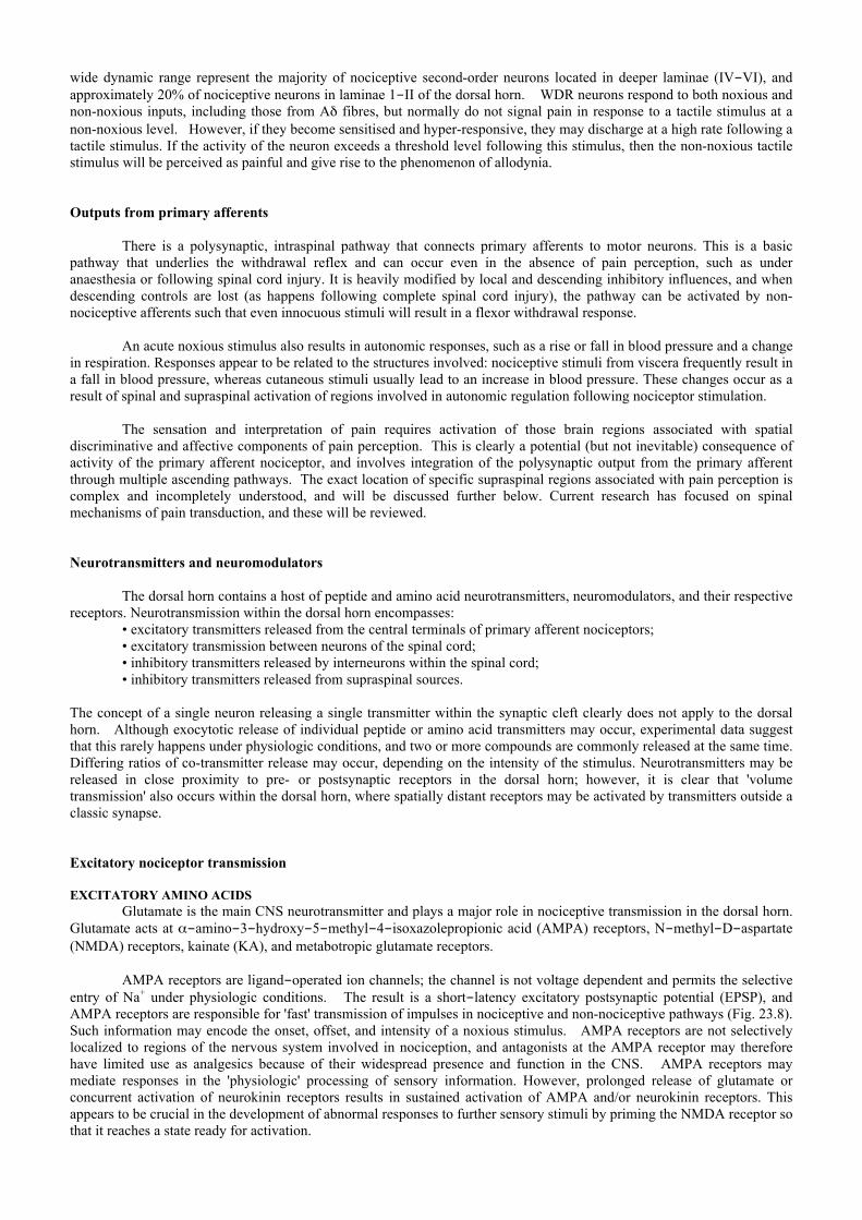
POSTSYNAPTIC DORSAL COLUMN PATHWAY The dorsal columns and nuclei (gracile and cuneate nuclei) are generally considered to be the pathway for information regarding the non-noxious sensations of fine touch, proprioception, and vibration. Stimulation of the dorsal columns is normally reported to produce a sensation of vibration rather than pain, although there are reports that mechanical stimulation of the medial aspect of the nucleus gracilis may result in pain. However, recent evidence points to a role of the dorsal column pathway in visceral nociception, and cells in the gracile nucleus respond to noxious stimulation of the viscera. The pathway that supplies this information is referred to as the postsynaptic dorsal column pathway, and has cells of origin in lamina III of the dorsal horn as well as just lateral to lamina X. Further evidence for a role for the dorsal column nuclei in pain perception comes from the demonstration that a phenotypic change occurs in neurons projecting through the dorsal column following nerve injury. Damage to large myelinated. fibres results in the de novo synthesis of substance P within these neurons and may be part of the mechanism responsible for the hypersensitivity to light touch that occurs following peripheral nerve injury. Clinical implications Many surgical and percutaneous procedures have been employed to disrupt specific tracts in the spinal cord. These include cordotomy, extralemniscal myelotomy, and commissural myelotomy. The distribution of fibres associated with pain transmission within the anterolateral quadrant would suggest that section of these tracts using an anterolateral cordotomy should be a useful procedure in abolishing or relieving pain. This concept is based on the cartesian or 'private line' model of pain perception, and results are variable and often transient. Sometimes excellent relief can be obtained in the short term, but long-term results are usually disappointing and complications include the return of pain, motor weakness, and loss of bladder and bowel function. Consequently, these procedures are usually limited to the treatment of cancer pain. Electrical stimulation of the fibres of the dorsal columns (dorsal column stimulation, DCS) may activate the proposed gating mechanisms of the dorsal horn and inhibit the nociceptive input associated with neuropathic pain states. The mechanism involves enhanced release of endogenous GABA and attenuation of the release of excitatory amino acids. A direct inhibitory effect on the postsynaptic dorsal column pathway may also contribute to analgesia, although visceral pain is rarely considered an indication for DCS. SUPRASPINAL STRUCTURES Thalamus Axons within the spinothalamic pathway are divided into two main groups, depending on their terminations. One group travels in the anterolateral funiculus and, together with projections from the medulla, pons, and midbrain, terminates more laterally in the ventroposterior nuclei and posterior complex of the thalamus. These axons are believed to be involved in the sensory discriminative component of pain (Fig. 23.11). Another group terminates more medially in the intralaminar nuclei, including the centrolateral, ventroposterolateral, and submedian nuclei, which project to the somatosensory cortex. The centromedian nuclei project more diffusely, including projections to the limbic system, and are believed to be involved in the affective-motivational aspects of pain. The medial thalamus also receives projections from the
spinoreticular and spinomesencephalic tracts. Projections from the spinocervical and postsynaptic dorsal column pathways terminate in the ventral posterior lateral nucleus and posterior complex. Animal studies indicate that there is a spinothalamic projection with terminations in the ventroposterior thalamic nucleus. Neurons in this region have restricted receptive fields and respond to noxious stimuli. These findings suggest that nuclei in this region have a role in the discriminative aspects of pain processing. However, it is interesting that stimulation of the ventrocaudal nucleus (analogous to the ventroposterior nucleus in animals, and supposedly part of the 'pain' pathway) in awake humans rarely results in pain, except in those who have central deafferentation pain. In contrast to cells in the lateral thalamus,
Figure 23.11 Rostral projections of nociceptive progression. Ascending projections terminate in the thalamic nuclear complex. The descending fibers inhibit the transmission of nociceptive information between primary afferents and projection neurons in the dorsal horn. In addition to direct neural connections, endorphins are released into the cerebrospinal fluid and blood, where they can exert an inhibitory effects at multiple centers. (With permission from Siddall PJ, Cousins MJ. introduction to pain mechanisms. In: Cousins MJ, Bridenbaugh PO, eds. Neural blockade in clinical anesthesia and management of pain. Philadelphia 1998: 675 713. Copyright (D Lippincott Williams and Wilkins.)

cells in the medial thalamus have large, often bilateral, receptive fields, suggesting a minor role in discriminative aspects of pain perception. It has been reported that a nucleus within the medial thalamus is specific for pain and temperature sensation. Positron emission tomography (PET) studies have identified a number of subcortical structures that are presumed to be involved in nociceptive transmission and pain perception. These include the thalamus, putamen, caudate nucleus, hypothalamus, amygdala, PAG, hippocampus, and cerebellum. Although previous physiologic and anatomic experiments have suggested that some of these structures are responsible for pain transmission, the role of others is unclear. The activation of these subcortical structures may vary with differing pain complaints: studies indicate that whereas an acute experimental painful stimulus results in increased activity in the thalamus, chronic pain caused by cancer and chronic neuropathic pain are associated with a decrease in activity in the thalamus. Cortical structures The effect of cortical stimulation and lesions on pain perception is confusing and intriguing. Patients who have had a complete hernispherectomy can have almost normal pain sensation. In the awake human, stimulation of the primary somatosensory cortex typically evokes non-painful sensations. Neurosurgical lesions of cortical regions produce varying effects, depending on the region ablated. Lesions of the frontal lobe and cingulate cortex result in a condition in which pain perception remains. However, the suffering component of pain appears to be reduced, the person only reports pain when queried, and spontaneous requests for analgesia are reduced. These effects contrast with those seen following lesions of the medial thalamus and hypothalamus, in which there is pain relief but no demonstrable analgesia. Both PET and functional magnetic resonance imaging (fMRI) have been helpful in elucidating the cortical regions involved in pain processing, and although there is some inconsistency in the results (perhaps because of the differing stimuli used in various studies) it is clear that no specific 'pain centre' or homunculus can be identified where nociceptive signals ultimately reach conscious perception. Painful stimuli result in activation of somato-sensory, motor, premotor, parietal, frontal, occipital, insular, and anterior cingulate regions of the cortex, in concert with activation of subcortical structures mentioned above. Although it is by no means clear, it has been suggested on the basis of PET findings that the parietal regions of the cortex are responsible for evaluation of the temporal and spatial features of pain, and the frontal cortex, including the anterior cingulate, is responsible for the emotional response to pain. Current neurophysiological constructs of consciousness propose massively parallel processing of sensory input by neural networks involving thalamocortical and thalamolimbic loops. The multiple destinations of nociceptive projections from the spinal cord to thalamic, limbic, and cortical targets indicate that pain 'emerges' from the coordinated processing of this information in areas involved in sensation, emotion, and is therefore an inherently complex affective emotional as well as spatial discriminative facets. Descending modulation There are powerful inhibitory influences arising from the brain that descend in the spinal cord to modulate spinal reflexes. Interest from those studying pain strengthened with the demonstration in the late 1960s that electrical stimulation of the PAG of the midbrain enabled abdominal surgery to be performed on animals with little evidence of discomfort. It is now known that there are powerful inhibitory (as well as facilitatory) influences on nociceptive transmission acting at many levels of the neuraxis. The PAG receives projections from a number of brain regions, including the amygdala, frontal and insular cortex, and hypothalamus, and acts in concert with the rostral ventromedial medulla (RVM) to provide a descending pain modulatory system. In addition to direct neural connections, endorphins synthesized in the pituitary are released into the cerebrospinal fluid and where they can exert an inhibitory effect at several centres, including the PAG. Descending inhibition may be activated by external factors such as stress (stress-induced analgesia) and noxious input (diffuse noxious inhibitory controls), or can be induced by peripheral or central nervous stimulation. Although the role of descending systems has been emphasized, there is also evidence for ascending modulation of 'higher' structures. For example, stimulation in the PAG can produce inhibition of the responses of neurons in the medial thalamus. Although it is possible that this inhibition may occur through the activation of descending pathways, it indicates that there are interactions at many levels of the nervous system. Brain structures involved in descending inhibition Descending inhibitory influences arise from a number of supraspinal structures, including the hypothalamus, PAG, locus cereleus, and RVM nuclei, including nucleus raphe magnus and nucleus paragigantocellularis lateralis. Electrical stimulation and microinjection of excitatory amino acids into these regions inhibits nociceptive responses. Descending inhibition does not require 'turning on'. Even though descending inhibition is activated by external stimuli, it is also

tonically active and maintains a resting level of inhibitory function. This is demonstrated by reversible spinal cord block, which results in an increased responsiveness of spinal cord neurons. The midbrain PAG appears to have a major role in descending inhibition. Stimulation in this region results in antinociception; although the PAG does not project directly to the spinal cord, descending fibres from the PAG relay in several structures that are also implicated in descending inhibition. These include the nucleus raphe magnus of the RVM and the paragigantocellular nucleus situated lateral to it. The PAG also appears to be highly organized. It has been demonstrated that analgesia obtained from the lateral PAG is non-opioid in nature, whereas opioid analgesia is obtained from stimulation of the medial PAG.
As with the PAG, electrical or chemical stimulation of the midline nucleus raphe magnus or ventrolateral (paragigantocellular nucleus) medulla results in antinociception. In the RVM, two types of neuron, on-cells and off-cells, have been identified as pain modulatory. On-cells exhibit a sudden increase in firing, whereas off-cells cease firing immediately prior to initiation of a nociceptive response. Fibres descend directly from these regions to the dorsal horn of the spinal cord and may exert a bi-directional control over nociception. Tracts from these structures descend mainly in the dorsolateral funiculus of the spinal cord and terminate in laminae 1, II, and V. Stimulation of the locus ceruleus also results in inhibition of nociceptive-evoked dorsal horn activity. This nucleus receives inputs from a number of brain structures and provides descending inputs to the spinal dorsal horn. These descending fibres travel ipsilaterally in the ventrolateral funiculus. Terminations of descending pathways interact with several different neural elements in the dorsal horn. These include projection neurons that transmit information from the spinal cord to the brain, local interneurons within the spinal cord, and primary afferent terminals. Inhibition may occur pre-synaptically by modulation of transmitter release, or post-synaptically by either excitation of local inhibitory interneurons or direct inhibition of second-order projection neurons. The bulk of evidence suggests that descending inhibition is exerted via postsynaptic mechanisms. Neurotransmitter mechanisms OPIOIDS Opioid receptors modulate nociceptive input at many sites in the CNS, although functionally these can be divided into supraspinal and spinal sites of analgesia. Supraspinally, there is a high density of opioid receptors in the PAG, nucleus raphe magnus, and locus ceruleus. The profound analgesia produced by electrical stimulation of these regions is reversed by the opioid antagonist naloxone, which confirms the importance of endogenous opioid agonists in nociceptive behaviour. However, microinjection of opioid agonists into these regions results in complex alterations in pain behaviour, reflecting both inhibition and disinhibition (the inhibition of an inhibitory interneuron) of pathways involved in nociception. Subregional differences, dose dependence, and action at specific opioid receptor subtypes further increase the complexity of supraspinal analgesia. For example, high doses of morphine in the clinical setting have occasionally been reported to result in hyperalgesia rather than analgesia. Opioids act at a spinal level in the dorsal horn by activating presynaptic opioid receptors, which inhibit glutamate and neurokinin release from primary afferent terminals, and postsynaptic receptors, which inhibit second-order neuron depolarisation. Particularly within the spinal cord dorsal horn, opioid receptor density and distribution are dynamic: inflammation may enhance and nerve injury markedly reduce opioid-mediated spinal analgesia. Changes in presynaptic opioid receptor number and function, at least in part, underlie these effects. Opioid analgesic mechanisms are themselves modulated by the release of cholecystokinin (CCK) acting on CCK1 and CCK2 receptors in the dorsal horn and at supraspinal sites such as the RVM. Microinjection of CCK into the RVM of experimental animals produces behavioural signs of neuropathic pain, and pathologic up-regulation of this system has been proposed as a mechanism contributing to opioid-resistant chronic pain states in humans. SEROTONIN Serotonin has long been suggested to be a major neurotransmitter in descending controls. Serotonin is contained in a high proportion of nucleus raphe magnus cells, and in terminals of descending fibres in the dorsal horn. Electrical stimulation of the nucleus raphe magnus increases the release of serotonin in the spinal cord. Agents that block serotonin synthesis attenuate stimulation-produced analgesia, and the application of some serotonin agonists in the spinal cord inhibits cells responsive to nociceptive stimuli. Although these findings appear highly suggestive of a role for serotonin in descending inhibition, several other findings indicate that it is not conclusive. Antinociception produced by stimulation of descending pathways cannot always
The midbrain periaqueductal grey matter is important in descending inhibition of spinal nociceptive input.

be blocked by serotonin antagonists. Under certain circumstances intrathecal serotonin can facilitate behavioural nociceptive responses, and serotonin agonists can have an excitatory effect on cells in the superficial dorsal horn. These conflicting results may result from motor effects associated with some behavioural responses, differing effects associated with different serotonin receptor subtypes, and observation of effects on inhibitory neurons. NOREPINEPHRINE Norepinephrine is also an important neurotransmitter for descending inhibitory controls, and most central noradrenergic neurons originate within the locus ceruleus (LC). Descending projections from the LC give rise to the majority of noradrenergic fibres in the spinal cord. The noradrenergic stress response hypothesis proposes that stimuli that threaten the physical or psychological integrity of the individual generate a central noradrenergic response mediated by the LC: the associated hypervigilance and defensive response is associated with enhanced antinociception. Direct stimulation of noradrenergic cell groups in the brainstem, notably the locus ceruleus, produces antinociception, and norepinephrine-containing terminals are found in the superficial laminae of the spinal dorsal horn. Iontophoretic application of norepinephrine inhibits the activation of dorsal horn neurons by noxious stimuli, and intrathecal norepinephrine results in inhibition of nociceptive responses. Conversely α2-adrenoceptor antagonists and compounds that reduce the amount of norepinephrine in the spinal cord increase nociceptive responses. OTHER NEUROTRANSMITTERS Other transmitters that appear to be important in descending inhibition are acetylcholine, GABA, thyrotropin-releasing hormone, and somatostatin. Clinical implications ANTIDEPRESSANTS Tricyclic antidepressants, typified by amitriptyline, produce spinally and supraspinally mediated analgesia in chronic pain states by mechanisms distinct from those that enhance mood. Tricyclic antidepressants have actions that include the inhibition of uptake of both norepinephrine and serotonin, in conjunction with muscarinic anticholinergic effects. The selective serotonin-reuptake inhibitors (SSRI) have weaker analgesic actions than traditional tricyclic antidepressants, suggesting that noradrenergic mechanisms are a more significant mode of tricyclic analgesia than the potentiation of serotonergic mechanisms. However, amitriptyline and similar tricyclic antidepressants are moderately potent Na+ channel blockers and also bind to NMDA receptors with an affinity similar to that of ketamine. The analgesic action of tricyclic antidepressants is therefore likely to involve multiple mechanisms, and they remain a mainstay of pharmacological pain management. OPIOIDS Opioids remain the primary analgesics for the management of acute, severe nociceptive pain. Respiratory depression and sedation are supraspinally mediated and limit maximally tolerated systemic doses. Selective targeting of opioid receptors in the dorsal horn via epidural or intrathecal administration is therefore a logical means to achieve analgesia and reduce supraspinally mediated side effects. Opioid tolerance results in a requirement for increasing doses to achieve a given degree of analgesia for a constant nociceptive stimulus. The mechanisms include opioid-mediated enhancement of cAMP levels, protein phosphorylation, and subsequent up-regulation of NMDA receptor mechanisms within the dorsal horn and supraspinal sites. In addition, the accumulation of the morphine metabolite morphine-3-glucuronide, which may antagonize the analgesic action normally produced by opioid receptor activation, may play a supplementary role. Tolerance is rarely of clinical significance in acute pain management, and although frequently described in the context of chronic pain, it is not an inevitable consequence of long-term opioid administration. Neuropathic pain states with pre-existing NMDA receptor activation may predispose to the development of opioid tolerance or insensitivity. Animal studies indicate that the administration of an NMDA antagonist can prevent both the development of tolerance to morphine and the withdrawal syndrome in morphine-dependent rats. Therefore, co-administration of NMDA antagonists such as dextromethorphan or ketamine with opioids may attenuate the development of opioid tolerance and potentiate opioid analgesic mechanisms. The observation that methadone enantiomers are analgesic as a result of NMDA receptor antagonism in addition to µ-opioid receptor activation provides a logical basis for its long-term administration. NITROUS OXIDE Nitrous oxide produces analgesia via complex mechanisms involving supraspinal opioid-mediated activation of descending noradrenergic pathways and direct activation of spinal α2- adrenoceptors. In addition, recent evidence suggests that nitrous inhibits NMDA receptor function. CLONIDINE α2-adrenergic agonists clonidine and dexmedetomidine exert potent analgesic actions at a spinal level when administered by epidural or intrathecal routes. The activation of pre- and postsynaptic α2-adrenoceptors inhibits primary afferent transmitter release and second-order neuronal depolarisation. Clonidine potentiates spinal opioid analgesia, and

combination therapy may be efficacious in neuropathic pain. Epidural and intrathecal clonidine have been demonstrated to provide analgesia in CRPS states that are opioid insensitive. STIMULATION-PRODUCED ANALGESIA The demonstration that powerful inhibition of nociceptive responses can be obtained by activation of descending pathways led to a resurgence of interest in techniques such as acupuncture and transcutaneous electrical nerve stimulation. It also led to the development and investigation of new techniques, such as dorsal column stimulation and deep brain stimulation. Although not widely used, deep brain stimulation involves the insertion of fine electrodes into brain regions where inhibition can be obtained. Initial studies focused on the PAG and periventricular grey matter, but other sites, such as the thalamus, are also used and provide significant analgesia in selected patients. CLINICAL SIGNIFICANCE OF INFLAMMATION AND NERVE INJURY Peripheral and central sensitisation resulting from inflammation or nerve injury is a common consequence of surgical or traumatic injury. In most individuals acute postoperative pain is amenable to 'conventional' therapy with combinations of opioids, NSAIDs, and local anesthetics, and is self-limiting. The resolution of the injury is generally associated with resolution of pain, indicating that, at least at the behavioural level, the manifestations of central sensitisation are reversible. Experimental evidence indicating that phenotypic changes outlast behavioural changes in pain models suggests that adaptive analgesic mechanisms usually compensate within the spinal cord. Pre-emptive analgesia There is evidence that severe postoperative pain may be a significant predictor of long-term pain, and that steps that reduce or abolish noxious input to the spinal cord during a painful event such as surgery may reduce or minimize spinal cord changes and thereby lead to a reduction of postoperative, and possibly chronic, pain. However, the duration or degree of noxious input that is required before these long-term changes occur remains unclear. Furthermore, it is uncertain how much these long-term changes depend on the afferent barrage during surgery, and how much they depend on continuing inputs from the wound after surgery. At both stages there will be sustained noxious input and, therefore, the potential to produce central sensitisation; however, it would be expected that interventions that pre-empt central sensitisation and seek to prevent it, rather than attempts to treat it after it has occurred, should be more successful. The pre-emptive effects of local anesthetics, opioids, and NSAIDs (alone or in combination) administered locally, epidurally, intrathecally, or systemically at a variety of time points before, during, or after surgery have been studied. A number of trials have shown that pre-emptive analgesia results in reduced pain, decreased analgesic requirements, improved morbidity, and decreased hospital stay. However, these are balanced by a similar number that have failed to show benefit. The variability in agent, timing, and method of administration, as well as differences in the type of surgery and anesthetic procedure, have made it difficult to compare many studies of pre-emptive analgesia. Furthermore, many studies have been flawed methodologically. As examples, nitrous oxide and volatile agents may influence central sensitisation, and the increased anesthetic requirement in patients not receiving pre- or intraoperative analgesia may influence outcome. Similarly, basic science studies suggest that NSAIDs are equally effective at attenuating sensitisation, whether administered pre- or post-stimulus: postoperative administration of NSAID may mask or minimize opioid pre-emptive effects. Therefore, despite the logical appeal of pre-emptive analgesia and its ready clinical application, further trials are necessary before a definitive statement can be made regarding its benefits and advantages. Much of the focus of pre-emptive analgesia has been on reducing acute pain in the early postoperative period. Pre-emptive analgesia may also be important in reducing chronic pain, but this has not been systematically evaluated. The finding that preoperative epidural blockade of patients undergoing lower limb amputation resulted in a lower incidence of phantom limb pain at 6 and 12 months after surgery than that seen in a control group having intraoperative block alone generated much interest. Although there are inadequacies in the study design and not all subsequent studies have confirmed their findings, it demonstrates that pre-emptive analgesia may have the potential to prevent the development of chronic pain states. Further studies are required to address this important question. Chronic pain ONGOING NOCICEPTIVE INPUT Perhaps the simplest explanation of chronic pain is that of pure ongoing peripheral nociceptive input. This is typified by chronic inflammatory degenerative conditions (e.g. rheumatoid arthritis or degenerative osteoarthritis). Ongoing activation of peripheral nociceptors with or without the presence of inflammatory mediators results in ongoing peripheral sensitisation. However, this is commonly supplemented by central sensitisation. Clinical manifestations include ongoing peripheral mechano-thermal hyperalgesia, and allodynia. Peripheral nociceptor sensitisation may respond to NSAIDs, whereas opioids will attenuate primary afferent transmitter release in the dorsal horn. Conditions of pure peripheral

nociceptor activation should respond to conventional analgesics. Opioid resistance suggests the involvement of central sensitisation mechanisms. PERIPHERAL MAINTENANCE OF CENTRAL SENSITIZATION Central sensitisation, once established, can clearly be maintained by ongoing peripheral activation of nociceptive afferents where a peripheral inflammatory or nociceptive stimulus persists. Following nerve injury, spontaneously discharging neuromata and spontaneously active DRG cells provide an alternative mechanism for the maintenance of central sensitisation. Sympathetic efferent activity may play a similar role in CRPS pain states, where sprouting of sympathetic neurons results in activation of primary afferent cell bodies within the DRG. Furthermore, sensitised dorsal horn neurons with enlarged receptive fields can be activated by non-noxious input; an effect that may be potentiated by sprouting of Aβ fibres. Non-noxious mechanical and proprioceptive afferent input may thereby maintain central sensitisation after resolution of the initial injury. These mechanisms underlie zygapophysial facet joint-generated spinal pain, and possibly myofascial pain. Specific blockade of the neuronal mechanisms that maintain sensitisation (e.g. local anesthetic blockade of neuroma, facet joint, or efferent SNS innervation) may markedly attenuate spontaneous pain, allodynia, and hyperalgesia and provide insight into the aetiology of the pain complaint. ONGOING CENTRAL SENSITIZATION Pain perception requires activity only in cortical and associated supraspinal regions many synapses distant from the peripheral nociceptor. Therefore, ongoing afferent input into the spinal cord is not a prerequisite for ongoing pain. If the normal spinal inhibitory mechanisms are overwhelmed by the magnitude of the initial stimulus (e.g. massive loss of inhibitory interneurons), and there is a failure in adaptive analgesic mechanisms that might oppose otherwise uncontrolled excitation in pain pathways, then a self-maintaining state of central sensitisation may be established: this may involve abnormal up-regulation of endogenous CCK and dynorphin pathways at both spinal and supraspinal sites. The determinants leading to the 'centralization' of pain are imprecisely understood, but the severity and chronicity of noxious stimulus in individuals with genetically determined 'vulnerable neurochemistry' are proposed as key factors. The concept of a balance between excitation and inhibition is of prime importance in the pathophysiology of pain; for example, cerebrovascular accident, head injury, and spinal cord injury are frequently associated with 'central' pain syndromes (e.g. 'thalamic pain') that reflect aberrant excitatory activity in central neurons involved in pain transduction, resulting in spontaneous pain. Furthermore, a spontaneous loss of spinal and/or supraspinal inhibitory tone and consequent disinhibition of projection neurons should result in spontaneous pain, hyperalgesia, and allodynia. Fibromyalgia and similar diffuse pain syndromes that are not clearly linked to an initial nociceptive event could, at least in part, be explained by such mechanisms. Centrally mediated and maintained pain will be 'referred' to dermatomes representative of pathologically activated central neurons. Peripheral neural blockade of afferent input from these dermatomes will not abolish spontaneous pain sensation, although hyperalgesia and allodynia may be relieved and the quality of pain altered. Such 'central' pain states rarely respond to conventional analgesic strategies. They are associated with down-regulation of opioid receptor number and function, and pain is likely to be independent of primary afferent excitatory amino acid transmitter release, which is a primary target of opioid analgesia. Potentiation of spinal and supraspinal noradrenergic and GABA ergic systems, together with suppression of ectopic Na+ channel activation, may provide significant analgesia, necessitating combination therapy with tricyclic antidepressants, anticonvulsants, and Na+ channel antagonists. Peripheral nerve blockade in chronic pain Peripheral nerve blockade is often used as a diagnostic procedure to predict outcome for nerve section or neuro-ablative procedures. However, isolated uncontrolled nerve blocks have little diagnostic or predictive value in the assessment of sciatic pain caused by lumbosacral disease. The recent evidence for physiologic and morphologic changes within the spinal cord following sustained peripheral input suggests that there are pitfalls in diagnostic neural blockade. First, chronic pain caused by peripheral nerve damage may no longer be dependent on peripheral input, and there is no evidence that treatments aimed at the periphery modify established central sensitisation. For example, a longstanding intercostal neuralgia will not be expected to respond to intercostal neurectomy; indeed, the pain may increase as the processes of central sensitisation may be enhanced. Second, diagnostic nerve blocks seek to identify a 'pain' source and can isolate the person from the disease process thought to be responsible for the pain. Using diagnostic nerve blocks in this way can mean that there is little recognition of the complex psychologic issues that can underlie the chronic pain presentation. It may even mean that a psychologic 'diagnosis' is made on the basis of the person's response to a diagnostic procedure. Therefore, diagnostic procedures must be performed in the context of a multidisciplinary assessment, with an understanding of the complex biopsychosocial components of chronic pain and placebo response to interventions.
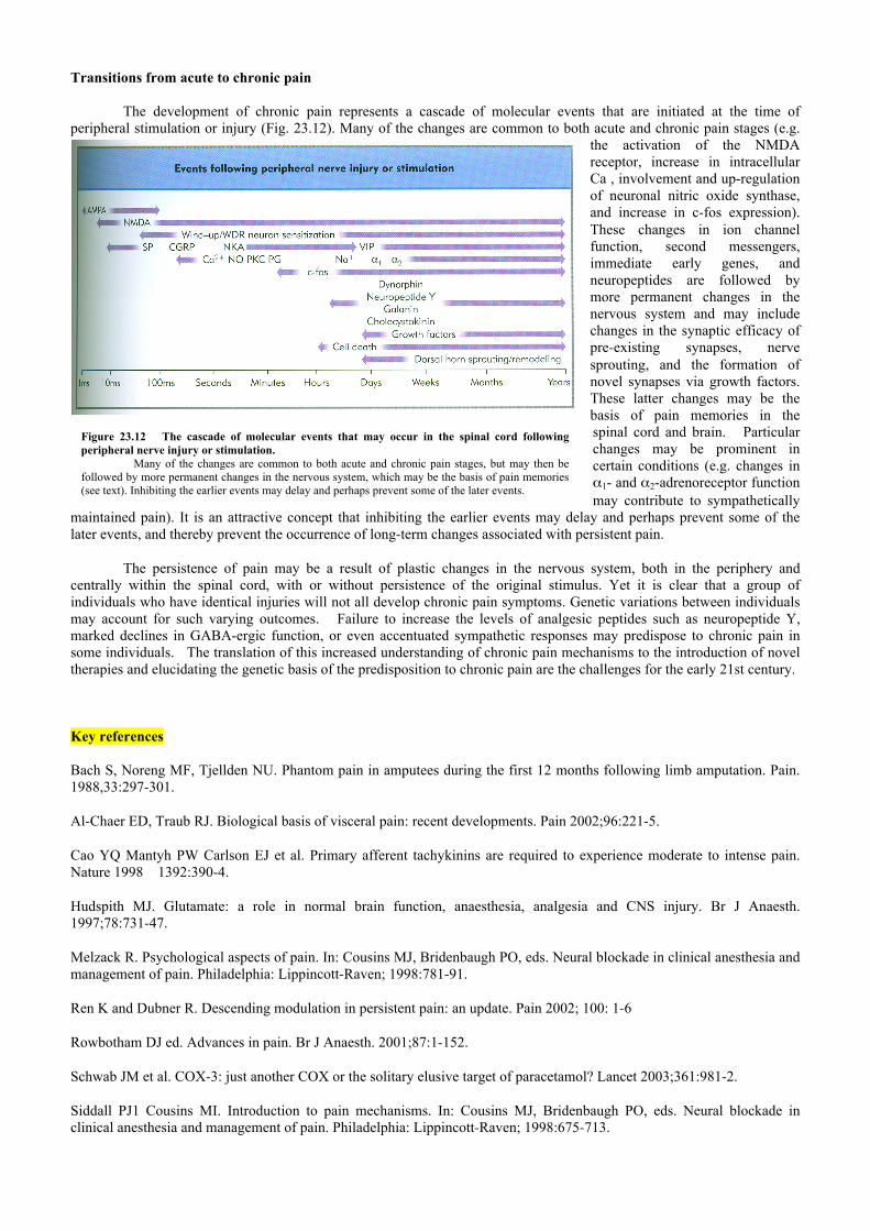
Transitions from acute to chronic pain The development of chronic pain represents a cascade of molecular events that are initiated at the time of peripheral stimulation or injury (Fig. 23.12). Many of the changes are common to both acute and chronic pain stages (e.g.
the activation of the NMDA receptor, increase in intracellular Ca , involvement and up-regulation of neuronal nitric oxide synthase, and increase in c-fos expression). These changes in ion channel function, second messengers, immediate early genes, and neuropeptides are followed by more permanent changes in the nervous system and may include changes in the synaptic efficacy of pre-existing synapses, nerve sprouting, and the formation of novel synapses via growth factors. These latter changes may be the basis of pain memories in the spinal cord and brain. Particular changes may be prominent in certain conditions (e.g. changes in α1- and α2-adrenoreceptor function may contribute to sympathetically
maintained pain). It is an attractive concept that inhibiting the earlier events may delay and perhaps prevent some of the later events, and thereby prevent the occurrence of long-term changes associated with persistent pain. The persistence of pain may be a result of plastic changes in the nervous system, both in the periphery and centrally within the spinal cord, with or without persistence of the original stimulus. Yet it is clear that a group of individuals who have identical injuries will not all develop chronic pain symptoms. Genetic variations between individuals may account for such varying outcomes. Failure to increase the levels of analgesic peptides such as neuropeptide Y, marked declines in GABA-ergic function, or even accentuated sympathetic responses may predispose to chronic pain in some individuals. The translation of this increased understanding of chronic pain mechanisms to the introduction of novel therapies and elucidating the genetic basis of the predisposition to chronic pain are the challenges for the early 21st century. Key references Bach S, Noreng MF, Tjellden NU. Phantom pain in amputees during the first 12 months following limb amputation. Pain. 1988,33:297-301. Al-Chaer ED, Traub RJ. Biological basis of visceral pain: recent developments. Pain 2002;96:221-5. Cao YQ Mantyh PW Carlson EJ et al. Primary afferent tachykinins are required to experience moderate to intense pain. Nature 1998 1392:390-4. Hudspith MJ. Glutamate: a role in normal brain function, anaesthesia, analgesia and CNS injury. Br J Anaesth. 1997;78:731-47. Melzack R. Psychological aspects of pain. In: Cousins MJ, Bridenbaugh PO, eds. Neural blockade in clinical anesthesia and management of pain. Philadelphia: Lippincott-Raven; 1998:781-91. Ren K and Dubner R. Descending modulation in persistent pain: an update. Pain 2002; 100: 1-6 Rowbotham DJ ed. Advances in pain. Br J Anaesth. 2001;87:1-152. Schwab JM et al. COX-3: just another COX or the solitary elusive target of paracetamol? Lancet 2003;361:981-2. Siddall PJ1 Cousins MI. Introduction to pain mechanisms. In: Cousins MJ, Bridenbaugh PO, eds. Neural blockade in clinical anesthesia and management of pain. Philadelphia: Lippincott-Raven; 1998:675-713.
Figure 23.12 The cascade of molecular events that may occur in the spinal cord following peripheral nerve injury or stimulation. Many of the changes are common to both acute and chronic pain stages, but may then be followed by more permanent changes in the nervous system, which may be the basis of pain memories (see text). Inhibiting the earlier events may delay and perhaps prevent some of the later events.

Vanderah TW Ossipov MH et al. Mechanisms of opioid-induced pain and antinociceptive tolerance: descending facilitation and spinal dynorphin. Pain 2001;92:5-9. Woolf CJ, Salter MW Neuronal plasticity: increasing the gain in pain. Science 2000;288:1765-8. Woolf CJ, Shortland P, Coggeshall RE. Peripheral nerve injury triggers central sprouting of myelinated afferents. Nature 1992;355:75-8. Further reading Brodal P. The central nervous system: structure and function, 2nd edn. Oxford: Oxford University Press; 1998. Dickenson A, Besson JM. The pharmacology of pain. Handbook of experimental pharmacology. Berlin: Springer Verlag; 1997. Hansson PT, Fields HL et al. (eds) Neuropathic pain: pathophysiology and treatment. Progress in pain research and management, Vol 21. Seattle: IASP Press; 2001. Munglani R. Advances in chronic pain therapy with special reference to low back pain. In: Kaufman L, Ginsburg R, eds. Anaesthetic review, Vol. 14. Edinburgh: Churchill Livingstone; 1998:153-74. Munglani R, Hunt SP, Jones GJ. Spinal cord and chronic pain. In: Kaufman L, Ginsburg R, eds. Anaesthetic review, Vol. 12. Edinburgh: Churchill Livingstone; 1996:53-76. Willis WD Jr, Coggeshall RE. Sensory mechanisms of the spinal cord, 2nd edn. London: Plenum Press,- 1991.