pico - the early studies - surgical devices and advanced ... the... · pico - the early studies. 2...
TRANSCRIPT

PICO - The early studies

2
Contents
1. Forward by Professor Donald Hudson, Dr Kevin Adams 3 and Dr Adriaan van Huyssteen
2. The evolution of Negative Pressure Wound Therapy 4
3. What is PICO™? 5
4. What does research on PICO tell us? 7
1. Transmission of negative pressure levels at the wound bed 8
2. Tissue contraction 9
3. Establishing a characteristic pattern of peri-wound blood flow 10
Traumaticwound
1. Use of PICO following suture of a dog bite wound 12
Skingraft
2. Use of PICO following post burn skin graft to the knee 13
Post-surgicalwounds
3. Use of PICO on a post-surgical wound following breast reconstruction: 1 14
4. Use of PICO on a post-surgical wound following breast reconstruction: 2 15
5. Use of PICO following incisional hernia repair 16
6. Use of PICO following post hip implant surgical wound: 1 17
7. Use of PICO following hip implant: 2 18
8. Use of PICO following hip implant: 3 19
9. Use of PICO following hip implant: 4 20
10. Use of PICO following knee implant: 1 21
11. Use of PICO following knee implant: 2 22
12. Use of PICO after revision of knee replacement 23

1. Forward by Professor Donald Hudson and Dr Kevin Adams at Academy of Plastic Surgery, Claremont, South Africa and Dr Adriaan van Huyssteen at Panorama Mediclinic, Panorama, South Africa“We have been closely involved with Negative Pressure Wound Therapy (NPWT) for many years, have tested and indeed helped to develop a number of systems for use on our patients in Cape Town”.
“Combining the clinical effectiveness of NPWT with the known benefits of advanced wound care dressings always seemed to be an ideal solution for some patients, especially those at high risk in the critical days following surgery. Recently we were very pleased to have been able to examine the performance of just such a prototype device on a number of patients in our clinics”.
“This booklet describes 12 recent case histories using PICO™, a single use NPWT system provided by Smith & Nephew, beginning with some background on NPWT, before describing PICO and moving on to the clinical cases. We hope that you will agree that PICO opens up some very interesting possibilities in treating many kinds of small to medium sized wounds in both hospital and outpatient settings”.
3
Contents
1. Forward by Professor Donald Hudson, Dr Kevin Adams 3 and Dr Adriaan van Huyssteen
2. The evolution of Negative Pressure Wound Therapy 4
3. What is PICO™? 5
4. What does research on PICO tell us? 7
1. Transmission of negative pressure levels at the wound bed 8
2. Tissue contraction 9
3. Establishing a characteristic pattern of peri-wound blood flow 10
Traumaticwound
1. Use of PICO following suture of a dog bite wound 12
Skingraft
2. Use of PICO following post burn skin graft to the knee 13
Post-surgicalwounds
3. Use of PICO on a post-surgical wound following breast reconstruction: 1 14
4. Use of PICO on a post-surgical wound following breast reconstruction: 2 15
5. Use of PICO following incisional hernia repair 16
6. Use of PICO following post hip implant surgical wound: 1 17
7. Use of PICO following hip implant: 2 18
8. Use of PICO following hip implant: 3 19
9. Use of PICO following hip implant: 4 20
10. Use of PICO following knee implant: 1 21
11. Use of PICO following knee implant: 2 22
12. Use of PICO after revision of knee replacement 23

4
2. The evolution of Negative Pressure Wound Therapy Overthepast15years,NegativePressureWoundTherapy,(NPWT),hasprovidedclinicianswithapowerfulnewresourcetomanagecomplexwounds.HereisabroadsummaryoftheknowneffectsofNPWT:
Much research is being done in many of these fields and every year we get a better overall understanding of the mechanisms of action, role and future potential of NPWT (see Malmsjö and Borgquist 2010, Henderson et al., 2010 for some easy to understand reviews).
For some of the most challenging wounds, NPWT is becoming a first line therapy - but what about wounds which are still important but of a less serious nature? Are the costs of therapy or the restrictions on the daily activities of patients justified for less severe wounds? PICO™ is a new simplified NPWT system that combines the associated benefits of NPWT with the simplicity of an advanced wound care dressing for small to medium size wounds with low to moderate levels of exudate.
Promotion of a closed moist environment. (Morykwas et al., 1997) Reduction of tissue oedema. (Kamolz et al., 2004) Contraction of the wound edges. (Malmsjö et al., 2009) Mechanical stimulation of the wound bed. (Saxena et al., 2004)
Alteration of blood flow at the wound edges. (Wackenfors et al., 2004) Stimulation of angiogenesis. (Greene et al., 2006) Formation of granulation tissue. (Armstrong and Lavery 2005) Physical splinting of grafts. (Llanos et al., 2006); and incisional
wounds (Gomoll et al., 2006)
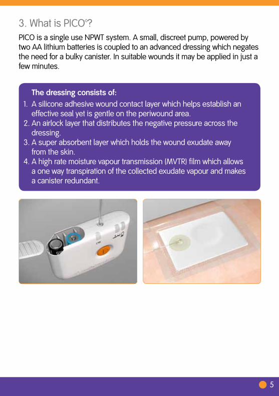
3. What is PICO™? PICO is a single use NPWT system. A small, discreet pump, powered by
two AA lithium batteries is coupled to an advanced dressing which negates the need for a bulky canister. In suitable wounds it may be applied in just a few minutes.
5
Thedressingconsistsof:1. A silicone adhesive wound contact layer which helps establish an
effective seal yet is gentle on the periwound area. 2. An airlock layer that distributes the negative pressure across the
dressing. 3. A super absorbent layer which holds the wound exudate away from the skin. 4. A high rate moisture vapour transmission (MVTR) film which allows
a one way transpiration of the collected exudate vapour and makes a canister redundant.
6
PICO™ is supplied in a pack that can be taken off the shelf when required. It contains a single use pump, which lasts for 7 days and two individually packed dressings and fixation strips (allowing for the wound to be inspected during those 7 days).
The pump is operated through a single orange button. It works with normal lithium AA batteries. The batteries maybe recycled whilst the pump should be disposed of as non-clinical waste when treatment is finished (see www.mypico.co for more information).
Three lights let you know how PICO is working – a green light which flashes constantly to tell you it is working properly and two amber alarm lights which flash if there is an issue. One indicates an air leak and a second shows that the battery charge is low. The batteries may be replaced within the 7 day life of the pump.
The pressure is nominally set at -80mmHg. Research shows that physiological effects are near maximal at this level (Borgquist et al., 2010a; Borgquist et al., 2010b):
A single push of the orange button starts the therapy. If the button is pushed again, the therapy will pause and then will automatically restart after an hour if the button is not pressed again before this time.
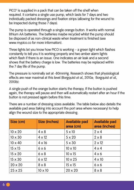
There are a number of dressing sizes available. The table below also details the available pad area (taking into account the port area where necessary) to help align the wound size to the appropriate dressing.
Size(cm) Size(inches) Availablepadarea(cm)
Availablepadarea(inches)
10 x 20 4 x 8 5 x 10 2 x 410 x 30 4 x 12 5 x 20 2 x 810 x 40 4 x 16 5 x 30 2 x 1215 x 15 6 x 6 10 x 10 4 x 415 x 20 6 x 8 10 x 15 4 x 615 x 30 6 x 12 10 x 25 4 x 1020 x 20 8 x 8 15 x 15 6 x 625 x 25 10 x 10 20 x 20 8 x 8
7
4. What does research on PICO™ tell us? At the University of Lund in Sweden, scientists examined whether PICO delivers
NPWT in the same manner as traditional devices such as RENASYS™ or VACTM. In making such an assessment they tested 3 factors which have been established as diagnostic for NPWT.
The transmission of negative pressure to the base of the wound (Malmsjo et al., 2009a)
Tissue contraction (Malmsjo et al., 2009a); Establishing a characteristic pattern of peri-wound blood flow (Morykwas et al.,
1997; Borgquist et al., 2010b):
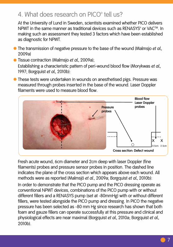
These tests were undertaken in wounds on anesthetised pigs. Pressure was measured through probes inserted in the base of the wound. Laser Doppler filaments were used to measure blood flow.
Fresh acute wound, 6cm diameter and 2cm deep with laser Doppler (fine filaments) probes and pressure sensor probes in position. The dashed line indicates the plane of the cross section which appears above each wound. All methods were as reported (Malmsjö et al., 2009a; Borgquist et al., 2010b):
In order to demonstrate that the PICO pump and the PICO dressing operate as conventional NPWT devices, combinations of the PICO pump with or without different fillers and a RENASYS pump (set at -80mmHg) with or without different fillers, were tested alongside the PICO pump and dressing. In PICO the negative pressure has been selected as -80 mm Hg since research has shown that both foam and gauze fillers can operate successfully at this pressure and clinical and physiological effects are near maximal (Borgquist et al., 2010a; Borgquist et al., 2010b).
Pressureprobes
x
BloodflowLaserDopplerprobes
x0.5cm 2.5cm
Crosssection:Defectwound

8
1. Transmission of negative pressure levels at the wound bed
In each combination the pressure levels achieved at the wound bed were virtually identical to the operating set point of the PICO™ pump. This shows that the PICO pump and dressing (orange bar) will operate to deliver specified negative pressure to the wound bed with or without foam and gauze fillers (Graph A).
85
80
75
70
65
60
Wou
nd b
ed p
ress
ure
(-m
mH
g)
PICO pump set point -80mmHg
RENASY
SPIC
O
Graph A: Pre-clinical evidence: Pressure transmission
Foam GauzePICODressing
Gauze Foam
PICO system (Pump and dressing with no filler)
RENASY
SPIC
ORE
NASYS
PICO
RENASY
SPIC
ORE
NASYS
PICO
FILLERS
INTERFACES
PUMPS
PICODressing
PICODressing

9
2. Tissue contraction
Tissue contraction recorded for the PICO™ pump and dressing is highlighted by the orange bar. In the defect wound, tissue contraction was observed for all pump and dressing combinations including the PICO pump and dressing combination. Slightly greater contraction (90%) was seen with negative pressure applied to foam fillers than with gauze filler (92%) or the PICO pump and dressing (92%). This verifies that PICO delivers tissue contraction comparable with conventional NPWT devices operating on defect wounds (Graph B).
Wou
nd s
urfa
ce a
rea
(% c
hang
e)
RENASY
SPIC
O
Graph B: Pre-clinical evidence: Tissue contraction
Foam Gauze
Gauze Foam
PICO system (Pump and dressing with no filler)
RENASY
SPIC
ORE
NASYS
PICO
RENASY
SPIC
ORE
NASYS
PICO
100
95
90
85
FILLERS
INTERFACES
PUMPS
PICODressing
PICODressing
PICODressing
10
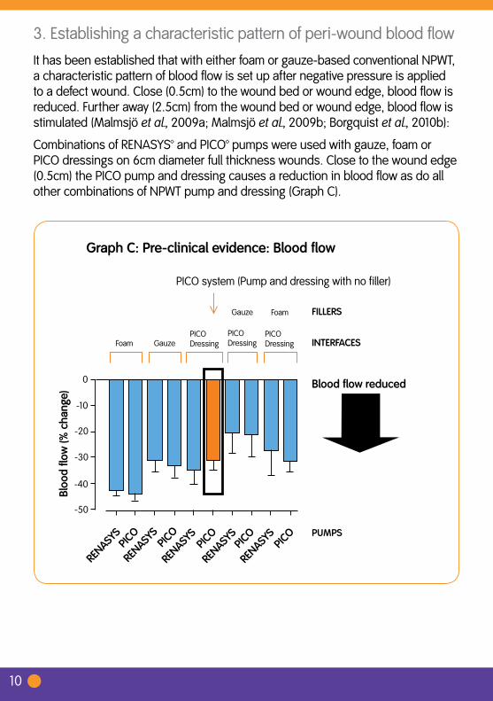
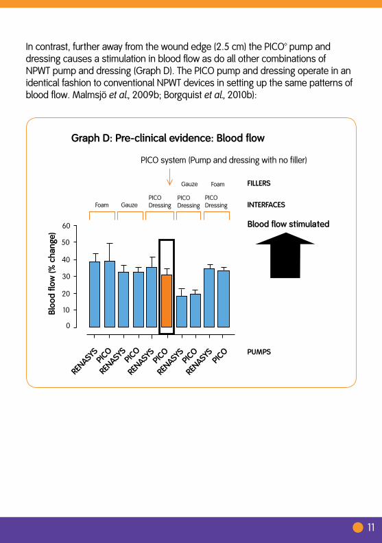
3. Establishing a characteristic pattern of peri-wound blood flow It has been established that with either foam or gauze-based conventional NPWT, a characteristic pattern of blood flow is set up after negative pressure is applied to a defect wound. Close (0.5cm) to the wound bed or wound edge, blood flow is reduced. Further away (2.5cm) from the wound bed or wound edge, blood flow is stimulated (Malmsjö et al., 2009a; Malmsjö et al., 2009b; Borgquist et al., 2010b):
Combinations of RENASYS™ and PICO™ pumps were used with gauze, foam or PICO dressings on 6cm diameter full thickness wounds. Close to the wound edge (0.5cm) the PICO pump and dressing causes a reduction in blood flow as do all other combinations of NPWT pump and dressing (Graph C).
RENASY
SPIC
O
Graph C: Pre-clinical evidence: Blood flow
Foam Gauze
Gauze Foam
PICO system (Pump and dressing with no filler)
RENASY
SPIC
ORE
NASYS
PICO
RENASY
SPIC
ORE
NASYS
PICO
-20
-40
-50
Bloo
d flo
w (%
cha
nge)
-30
-10
0 Blood flow reduced
FILLERS
INTERFACES
PUMPS
PICODressing
PICODressing
PICODressing
11
In contrast, further away from the wound edge (2.5 cm) the PICO™ pump and dressing causes a stimulation in blood flow as do all other combinations of NPWT pump and dressing (Graph D). The PICO pump and dressing operate in an identical fashion to conventional NPWT devices in setting up the same patterns of blood flow. Malmsjö et al., 2009b; Borgquist et al., 2010b):
RENASY
SPIC
O
Graph D: Pre-clinical evidence: Blood flow
Foam Gauze
Gauze Foam
PICO system (Pump and dressing with no filler)
RENASY
SPIC
ORE
NASYS
PICO
RENASY
SPIC
ORE
NASYS
PICO
50
40
30
20
10
0
Bloo
d flo
w (%
cha
nge)
60 Blood flow stimulated
FILLERS
INTERFACES
PUMPS
PICODressing
PICODressing
PICODressing
12
Thefollowing12casesillustratehowtreatmentwithPICO™hasworkedonavarietyofwoundsincludingsurgicalincisions.
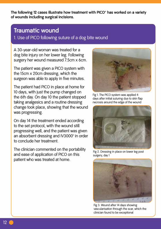
Fig 1. The PICO system was applied 4 days after initial suturing due to skin flap necrosis around the edge of the wound
A 30-year-old woman was treated for a dog bite injury on her lower leg. Following surgery her wound measured 7.5cm x 6cm.
The patient was given a PICO system with the 15cm x 20cm dressing, which the surgeon was able to apply in five minutes.
The patient had PICO in place at home for 10 days, with just the pump changed on the 6th day. On day 10 the patient stopped taking analgesics and a routine dressing change took place, showing that the wound was progressing.
On day 14 the treatment ended according to the set protocol, with the wound still progressing well, and the patient was given an absorbent dressing and IV3000™ in order to conclude her treatment.
The clinician commented on the portability and ease of application of PICO on this patient who was treated at home.
Fig 2. Dressing in place on lower leg post surgery, day 1
Fig 3. Wound after 14 days showing vascularisation through the scar, which the clinician found to be exceptional
Traumaticwound1. Use of PICO following suture of a dog bite wound
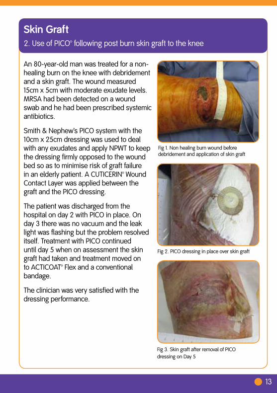
Fig 1. Non healing burn wound before debridement and application of skin graft
An 80-year-old man was treated for a non- healing burn on the knee with debridement and a skin graft. The wound measured 15cm x 5cm with moderate exudate levels. MRSA had been detected on a wound swab and he had been prescribed systemic antibiotics.
Smith & Nephew’s PICO system with the 10cm x 25cm dressing was used to deal with any exudates and apply NPWT to keep the dressing firmly opposed to the wound bed so as to minimise risk of graft failure in an elderly patient. A CUTICERIN™ Wound Contact Layer was applied between the graft and the PICO dressing.
The patient was discharged from the hospital on day 2 with PICO in place. On day 3 there was no vacuum and the leak light was flashing but the problem resolved itself. Treatment with PICO continued until day 5 when on assessment the skin graft had taken and treatment moved on to ACTICOAT™ Flex and a conventional bandage.
The clinician was very satisfied with the dressing performance.
Fig 2. PICO dressing in place over skin graft
Fig 3. Skin graft after removal of PICO dressing on Day 5
SkinGraft2. Use of PICO™ following post burn skin graft to the knee
13
14
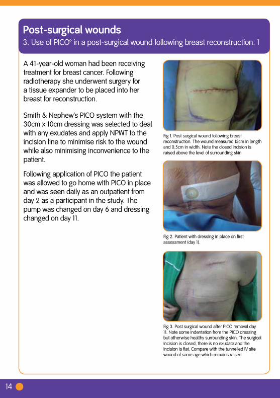
Fig 1. Post surgical wound following breast reconstruction. The wound measured 15cm in length and 0.5cm in width. Note the closed incision is raised above the level of surrounding skin
A 41-year-old woman had been receiving treatment for breast cancer. Following radiotherapy she underwent surgery for a tissue expander to be placed into her breast for reconstruction.
Smith & Nephew’s PICO system with the 30cm x 10cm dressing was selected to deal with any exudates and apply NPWT to the incision line to minimise risk to the wound while also minimising inconvenience to the patient.
Following application of PICO the patient was allowed to go home with PICO in place and was seen daily as an outpatient from day 2 as a participant in the study. The pump was changed on day 6 and dressing changed on day 11.
Fig 2. Patient with dressing in place on first assessment (day 1).
Fig 3. Post surgical wound after PICO removal day 11. Note some indentation from the PICO dressing but otherwise healthy surrounding skin. The surgical incision is closed, there is no exudate and the incision is flat. Compare with the tunnelled IV site wound of same age which remains raised
Post-surgicalwounds3. Use of PICO™ in a post-surgical wound following breast reconstruction: 1

15
Fig 1. Wound immediately following breast reconstruction surgery
A 27-year-old woman had a surgical wound measuring 14.1cm x 0.1cm following a skin sparing mastectomy and immediate prosthetic breast reconstruction. The PICO system was applied.
On the first day after application, the pump and dressing were changed. The wound was already seen to be progressing to closure at this point. No further dressing changes were needed until day 6, while on day 5, the patient was discharged from hospital.
With a third PICO system applied on day 6, treatment continued until day 11, when the wound was seen to be closed and treatment with PICO was ended.
Overall the surgeon was very satisfied with the performance of the dressing.
Fig 2. PICO dressing in place on day 7
Fig 3. Wound on day 14, 3 days after closure
Post-surgicalwounds4. Use of PICO™ on a post- surgical wound following breast reconstruction: 2

16
Fig 1. Post surgical wound on 52-year-old male before application of PICO. The clinicians noted a wide undermining of skin and tight closure of the abdominal wall.
A 52-year-old man was treated after suffering from diverticular disease and an incisional hernia. He had had a colectomy and a surgical wound in order to repair his hernia. The wound measured 26cm x 5cm x 0.1cm and was closed by suture.
After suturing the wound, the patient was given a PICO system with the 10cm x 30cm dressing.
Treatment continued for 6 days, when at the routine first dressing change, the wound was seen to be closed with no exudates or infection present. As the wound was now closed, treatment with PICO was discontinued at this point.
Overall the clinicians were satisfied and found PICO to perform better than they might have expected advanced wound dressings to in this instance.
Fig 2. PICO dressing over incisional hernia after first application
Fig 3. Wound closed after removal of PICO dressing on day 6
Post-surgicalwounds5. Use of PICO™ following incisional hernia repair

17
Fig 1. Hip wound of 53 year old man immediately after surgery
A 53-year-old man suffering from osteoarthritis was treated following a post hip implant surgical wound. His wound, closed by suture and Steri-StripsTM, measured 17.5cm x 0.5cm.
He was given a PICO system with the dressing measuring 10cm x 30cm.
A routine dressing change was performed on day 3. At this point his wound was progressing to closure, with no infection and light exudate levels, although the surrounding skin was inflamed.
The patient remained comfortable, although on day 5 some bruising was noted around the lower aspect of the dressing, which remained for a few days.
At the routine dressing change on day 10, the wound was found to be closed.
Overall the clinician was very satisfied with the treatment. Fig 2 a and 2 b. Wound on day 3, before
and after new dressing
Fig 3. Post -surgical hip wound closed on day 10
Post-surgicalwounds6. Use of PICO™ following post hip implant surgical wound: 1
18
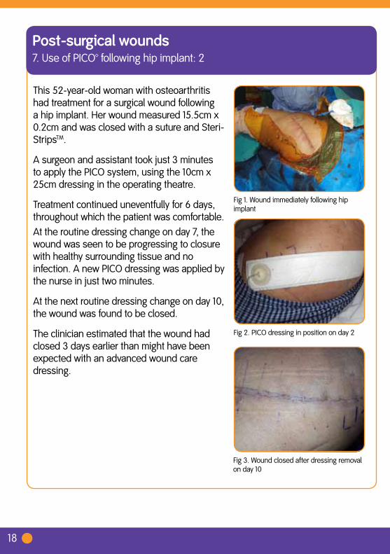
Fig 1. Wound immediately following hip implant
This 52-year-old woman with osteoarthritis had treatment for a surgical wound following a hip implant. Her wound measured 15.5cm x 0.2cm and was closed with a suture and Steri-StripsTM.
A surgeon and assistant took just 3 minutes to apply the PICO system, using the 10cm x 25cm dressing in the operating theatre.
Treatment continued uneventfully for 6 days, throughout which the patient was comfortable.At the routine dressing change on day 7, the wound was seen to be progressing to closure with healthy surrounding tissue and no infection. A new PICO dressing was applied by the nurse in just two minutes.
At the next routine dressing change on day 10, the wound was found to be closed.
The clinician estimated that the wound had closed 3 days earlier than might have been expected with an advanced wound care dressing.
Fig 2. PICO dressing in position on day 2
Post-surgicalwounds7. Use of PICO™ following hip implant: 2
Fig 3. Wound closed after dressing removal on day 10
19
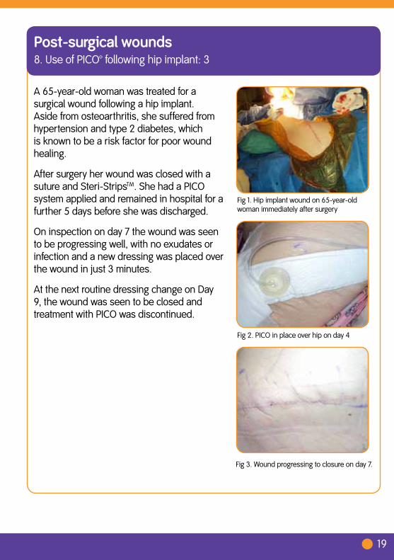
Fig 1. Hip implant wound on 65-year-old woman immediately after surgery
A 65-year-old woman was treated for a surgical wound following a hip implant. Aside from osteoarthritis, she suffered from hypertension and type 2 diabetes, which is known to be a risk factor for poor wound healing.
After surgery her wound was closed with a suture and Steri-StripsTM. She had a PICO system applied and remained in hospital for a further 5 days before she was discharged.
On inspection on day 7 the wound was seen to be progressing well, with no exudates or infection and a new dressing was placed over the wound in just 3 minutes.
At the next routine dressing change on Day 9, the wound was seen to be closed and treatment with PICO was discontinued.
Fig 2. PICO in place over hip on day 4
Post-surgicalwounds8. Use of PICO™ following hip implant: 3
Fig 3. Wound progressing to closure on day 7.
20
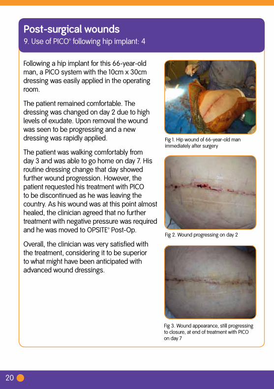
Fig 1. Hip wound of 66-year-old man immediately after surgery
Following a hip implant for this 66-year-old man, a PICO system with the 10cm x 30cm dressing was easily applied in the operating room.
The patient remained comfortable. The dressing was changed on day 2 due to high levels of exudate. Upon removal the wound was seen to be progressing and a new dressing was rapidly applied.
The patient was walking comfortably from day 3 and was able to go home on day 7. His routine dressing change that day showed further wound progression. However, the patient requested his treatment with PICO to be discontinued as he was leaving the country. As his wound was at this point almost healed, the clinician agreed that no further treatment with negative pressure was required and he was moved to OPSITE™ Post-Op.
Overall, the clinician was very satisfied with the treatment, considering it to be superior to what might have been anticipated with advanced wound dressings.
Fig 2. Wound progressing on day 2
Post-surgicalwounds9. Use of PICO™ following hip implant: 4
Fig 3. Wound appearance, still progressing to closure, at end of treatment with PICO on day 7
21
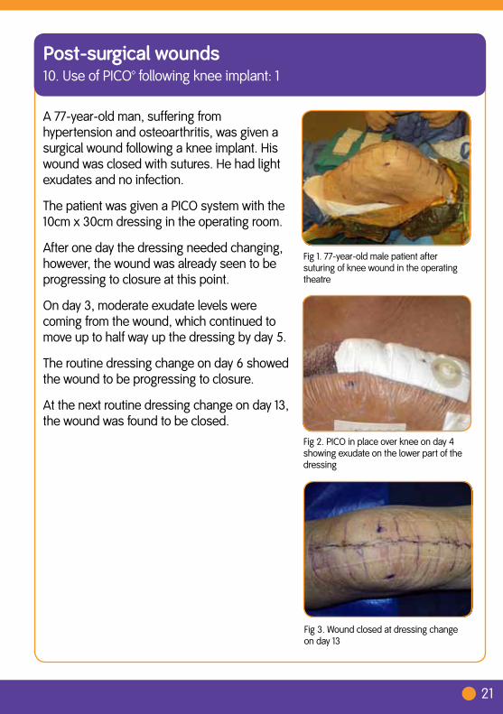
Fig 1. 77-year-old male patient after suturing of knee wound in the operating theatre
A 77-year-old man, suffering from hypertension and osteoarthritis, was given a surgical wound following a knee implant. His wound was closed with sutures. He had light exudates and no infection.
The patient was given a PICO system with the 10cm x 30cm dressing in the operating room.
After one day the dressing needed changing, however, the wound was already seen to be progressing to closure at this point.
On day 3, moderate exudate levels were coming from the wound, which continued to move up to half way up the dressing by day 5.
The routine dressing change on day 6 showed the wound to be progressing to closure.
At the next routine dressing change on day 13, the wound was found to be closed.
Fig 2. PICO in place over knee on day 4 showing exudate on the lower part of the dressing
Post-surgicalwounds10. Use of PICO™ following knee implant: 1
Fig 3. Wound closed at dressing change on day 13

22
Fig 1. Knee immediately post surgery
A 48-year-old woman was treated with PICO after knee implant surgery.
Her dressing was easy to apply and treatment continued uneventfully until her routine dressing change on day 6, where her wound was seen to be progressing well.
On day 10 the vacuum light started to flash shortly after the pump was changed, however at this dressing change and on inspection the wound was found to be closed and no further treatment was needed.
The clinician commented that PICO had a superior healing speed to that expected of advanced wound dressings in this case.
Fig 2. Knee wound at dressing change on day 6
Post-surgicalwounds11. Use of PICO™ following knee implant: 2
Fig 3.Knee wound at dressing change on day 10
23
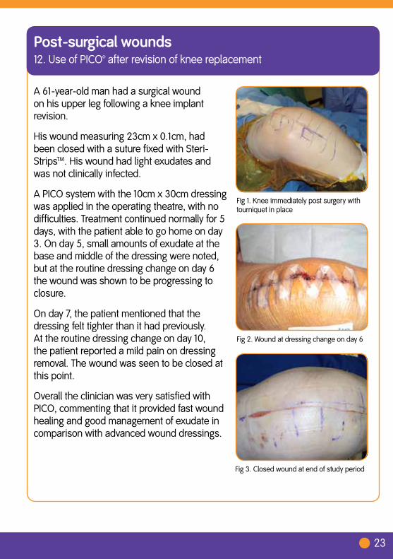
Fig 1. Knee immediately post surgery with tourniquet in place
A 61-year-old man had a surgical wound on his upper leg following a knee implant revision.
His wound measuring 23cm x 0.1cm, had been closed with a suture fixed with Steri-StripsTM. His wound had light exudates and was not clinically infected.
A PICO system with the 10cm x 30cm dressing was applied in the operating theatre, with no difficulties. Treatment continued normally for 5 days, with the patient able to go home on day 3. On day 5, small amounts of exudate at the base and middle of the dressing were noted, but at the routine dressing change on day 6 the wound was shown to be progressing to closure.
On day 7, the patient mentioned that the dressing felt tighter than it had previously. At the routine dressing change on day 10, the patient reported a mild pain on dressing removal. The wound was seen to be closed at this point.
Overall the clinician was very satisfied with PICO, commenting that it provided fast wound healing and good management of exudate in comparison with advanced wound dressings.
Fig 2. Wound at dressing change on day 6
Post-surgicalwounds12. Use of PICO™ after revision of knee replacement
Fig 3. Closed wound at end of study period

WoundManagement www.smith-nephew.com/woundSmith & NephewMedical Ltd ™Trademark of Smith & Nephew101 Hessle Road TMAll trademarks of acknowledgedHull HU3 2BN © Smith & Nephew March 2011 27177T +44 (0)1482 225181F +44 (0)1482 328326
ReferencesArmstrong DG, Lavery LA; Diabetic Foot Study Consortium. 2005 Negative pressure wound therapy after partial diabetic foot amputation: a multicentre, randomised controlled trial. Lancet. 366(9498):1704-10.Borgquist O, Gustafsson L, Ingemansson R, Malmsjö M. Micro- and macromechanical effects on the wound bed of negative pressure wound therapy using gauze and foam. Ann Plast Surg. 2010a;64(6):789-93.Borgquist O, Ingemansson R, Malmsjö M. Wound edge microvascular blood flow during negative-pressure wound therapy: examining the effects of pressures from -10 to -175 mmHg. Plast Reconstr Surg. 2010b; 125(2):502-9.Gomoll AH, Lin A, Harris MB. Incisional vacuum-assisted closure therapy. J Orthop Trauma. 2006; 20(10):705-9.Greene AK, Puder M, Roy R, Arsenault D, Kwei S, Moses MA, Orgill DP. Microdeformational wound therapy: effects on angiogenesis and matrix metalloproteinases in chronic wounds of 3 debilitated patients. Ann Plast Surg. 2006; 56(4):418-22.Henderson V, Timmons J, Hurd, T, Deroo K, Maloney S, Sabo S. NPWT in everyday practice Made Easy. Wounds International 2010; 1(5):Kamolz LP, Andel H, Haslik W, Winter W, Meissl G, Frey M. Use of subatmospheric pressure therapy to prevent burn wound progression in human: first experiences. Burns. 2004; 30(3):253-8.Llanos S, Danilla S, Barraza C, Armijo E, Piñeros JL, Quintas M, Searle S, Calderon W (2006) Effectiveness of negative pressure closure in the integration of split thickness skin grafts: a randomized, double-masked, controlled trial. Ann Surg. Nov;244(5):700-5Malmsjö M, Borgquist O. NPWT settings and dressing choices made easy. Wounds International 2010; 1(3):Malmsjö M, Ingemansson R, Martin R, Huddleston E. Negative Pressure Wound Therapy using gauze or open-cell polyurethane foam: similar early effects on pressure transduction and tissue contraction in an experimental porcine wound model. Wound Repair Regen. 2009a; 17:200-5. Malmsjö M, Ingemansson R, Martin R, Huddleston E. Wound edge microvascular blood flow: effects of negative pressure wound therapy using gauze or polyurethane foam. Ann Plast Surg. 2009b; 63(6):676-81 Morykwas MJ, Argenta LC, Shelton-Brown EI, McGuirt W. Vacuum-assisted closure: a new method for wound control and treatment: animal studies and basic foundation. Ann Plast Surg. 1997; 38(6):553-62.Saxena V, Hwang CW, Huang S, Eichbaum Q, Ingber D, Orgill DP. Vacuum-assisted closure: microdeformations of wounds and cell proliferation. Plast Reconstr Surg. 2004; 114(5):1086-96; discussion 1097-8.Wackenfors A, Sjögren J, Gustafsson R, Algotsson L, Ingemansson R, Malmsjö M. Effects of vacuum-assisted closure therapy on inguinal wound edge microvascular blood flow. Wound Repair Regen. 2004; 12(6):600-6. A prospective, open, non-comparative, multi-centre study to evaluate the functionality and dressing performance of a new negative pressure enhanced dressing (NPED) in acute wounds.
http://gettag.mobi