piezo1 induced apoptosis of type ii pneumocytes during ards
TRANSCRIPT

RESEARCH Open Access
Piezo1 induced apoptosis of type IIpneumocytes during ARDSGuo-Peng Liang1†, Jing Xu1†, Li-li Cao2, Yi-Hua Zeng3, Bai-Xu Chen1, Jing Yang1, Zhong-Wei Zhang1*† andYan Kang1*†
Abstract
Objective: The mechanisms of lung injury in acute respiratory distress syndrome (ARDS) are not wellunderstood.Piezo1 was recently identified as a mechanotransduction protein. The present study found theexpression of Piezo1 in type II pneumocytes and investigated its role in mediating ARDS-related lung injury.
Methods: Sprague-Dawley rats were used to establish an ARDS model, the expression of Piezo1,lung injuries,apoptosis as well as calcium influx were assessed.
Results: Piezo1 was expressed in type II pneumocytes as shown by immunofluorescence staining andexpression was increased in the ARDS model. Knockdown of Piezo1 reduced apoptosis which was related tothe elevation of Bcl-2.Calcium influx played a vital role in Piezo1-induced apoptosis.
Conclusion: Piezo1 was expressed in type II pneumocytes. Mechanical stretch of alveoli during ARDS inducedactivation of the Piezo1 channel,which resulted in calcium influx. The increased intracellular Ca2+ induced theapoptosis of type II pneumocytes, which may be related to the Bcl-2 pathway.
Keywords: Acute respiratory distress syndrome, Lung injury;Piezo1;apoptosis;Ca2+
BackgroundAcute respiratory distress syndrome (ARDS) is a com-mon and life-threatening respiratory disorder of inflam-matory lung injury responsible for 10% of all ICUadmissions and a has a mortality rate of nearly 40% [1].It is a type of acute diffuse, inflammatory lung injury,leading to increased pulmonary vascular permeability,increased lung weight, and loss of aerated lung tissue[2]. Recent evidence suggested that the main pathogen-esis of ARDS included cytokine imbalance oxidativestress, and apoptosis of alveolar epithelial cells [3].How-ever, the mechanisms of ARDS-related lung injury havenot been clarified well.Piezo1 was first detected in murine neuroblastoma
cells and was recently identified as a component ofmechanically-activated cation channels [4]. which were
inherently mechanosensitive without relying on alter-nate cellular components for sensing mechanical stim-uli [5]. Application of the force on cells caused calciumto rapidly enter cells through Piezo1 channels [6–8],which played an important role in sensing mechanicalstretch. Piezo1 expressed in platelets, this contributedto Ca2+ entry and thrombus formation under arterialshear [9]. Piezo1 was found to be expressed in endothe-lial cells of developing blood vessels, and knockout ofPiezo1 was embryonically lethal with defects in vascularremodeling [10]. In addition,Piezo1 expressed in arter-ial smooth muscle cells was able to sense the fluidshear stress exerted by blood flow and affect arterialstructure(diameter and wall thickness) by releasingATP and regulating of NO formation to control vascu-lar tone and blood pressure [11–13] . Piezo1 also sensesextension of the bladder urothelium [14], intraluminalpressure changes and urine flow sensing [15]. Thesestudies showed that Piezo1 is capable of detecting vari-ous kinds of mechanical stress and is involved in bio-logical processes and maintenance of normal structuresof human organs.
© The Author(s). 2019 Open Access This article is distributed under the terms of the Creative Commons Attribution 4.0International License (http://creativecommons.org/licenses/by/4.0/), which permits unrestricted use, distribution, andreproduction in any medium, provided you give appropriate credit to the original author(s) and the source, provide a link tothe Creative Commons license, and indicate if changes were made. The Creative Commons Public Domain Dedication waiver(http://creativecommons.org/publicdomain/zero/1.0/) applies to the data made available in this article, unless otherwise stated.
* Correspondence: [email protected]; [email protected]†Guo-Peng Liang and Jing Xu contributed equally to this work and shouldbe considered co-first authors.†Yan Kang and Zhong-Wei Zhang contributed equally to this work.1Department of Critical Care Medicine, West China School of Medicine andWest China Hospital, Sichuan University, Chengdu 610041, ChinaFull list of author information is available at the end of the article
Liang et al. Respiratory Research (2019) 20:118 https://doi.org/10.1186/s12931-019-1083-1

During respiration, alveoli and airways are stretchedrepetitively, and are constantly under mechanical stressgenerated by air and the surrounding tissues. Respiratorydisorders tend to enhance mechanical stretch by a seriesof pathophysiological mechanisms, that lead to lung in-jury. Mechanical ventilation is another important factorthat may increase lung injury due to tidal hyperinflation[16, 17], and cyclic recruitment/derecruitment [18].Given that Piezo1 is a mechanosensitive ion channelwith the ability to sense mechanical stretch, we assumedthat Piezo1 was also expressed in the lung and may be apotential mechanism of lung injury. The mRNA expres-sion profile indicated that Piezo1 may be expressed inthe lung [4]. However, to the best of our knowledge, nostudies have identified the physiological functions ofPiezo1 in lung injury.Therefore, the present study was designed to investi-
gate the expression and functions of Piezo1 in the lungand study the potential mechanisms of Piezo1 in ARDS-related lung injury. We attempted to find a new targetfor prevention and treatment of ARDS.
Materials and methodsAnimals and experimental designThe protocol was approved by the Institutional AnimalCare and Treatment Committee of Sichuan University(Chengdu, P.R. China). Health adult Sprague-Dawley (SD)rats weighing 250 ± 10 g(Experimental Animal Center ofAcademy of Military Medical Sciences of the People’s Lib-eration Army, China) were housed in cages with free ac-cess to water and food and divided randomly into fourgroups: 1) control group, 2)ARDS group, 3)ARDS+ hightidal volume(HV)group and 4)ARDS+ low tidal volu-me(LV) group,(five rats per group). Control rats wereinjected with 0.1mL/kg body weight physiological salinewhile those in experimental groups were given 0.1mL/kgbody weight oleic acid by intravenous injection and therats were spun around 10 times to establish ARDS modelwhich was then confirmed by assessing partial pressure ofoxygen in the arterial blood after 3 h. ARDS+HV andARDS+LV rats were ventilated with a tidal volume of 10mL/kg and 6mL/kg respectively,with positive end-expiratory pressure (PEEP) 3 cmH2O, and respiratory fre-quency of 25/min for 4 h. Next, rats in all groups wereanaesthetized, and lung sections were collected.
Assessment of lung injuryBlood(50ul) was collected from aorta abdominalis andpartial pressure of oxygen and carbon dioxide(PaO2,andPaCO2,respectively)was measured by blood gas analyzer(Abbott i-STAT). Oxygenation index was calculated asPaO2/FiO2, and lung morphology was observed byhematoxylin and eosin(HE)staining of left lung tissue. Thewet/dry ratio (W/D ratio) was calculated to reflect the
severity of pulmonary edema. Wet weights of right lowerlungs were measured upon collection of lung tissue, andthen lungs were incubated in an oven (60 °C for 72 h) tomeasure dry weight. The levels of serum TNF-α and IL-1βwere measured by ELISA assay(MD6692,MD6693).
Cell culture and transfectionHuman lung epithelial cell line A549 was seeded on 10cm dishes in DMEM, and cultured at 37°C and 5% CO2.Cells were transiently transfected with either Piezo1siRNA or control siRNA using Lipofectamine 2000(Invi-trogen, Carlsbad, CA, USA;11,668,019),the sequence ofsiRNA is provided in Additional file 1: Figure S2-A.
RNA isolation, real-time q-PCR, and western blotsAfter 24-48 h post transfection, total RNA was extractedusing Trizol(Invitrigen)following the manufacturers’ in-structions. Complementary DNAs (cDNAs) were synthe-sized using the HiScript® II One Step RT-PCR Kit (VazymeBiotech, Nanjing, China)and amplified using specificprimers for Piezo1.qPCR was performed with a LightCycler480 system(Roche, Basel, Switzerland). GAPDH was usedas a reference gene for relative quantification of Piezo1 geneexpression. The primer sequences are as follows:Piezo1-F 5′-GCTGTACCTGCCTGATTTCTTCC-3′.Piezo1-R 5′-CATGCGCTGCCACTCCTCT-3′;GAPDH-F 5′-CACATGGCCTCCAAGGAGTAA-3′;
and.GAPDH-R 5′-TGAGGGTCTCTCTCTTCCTCTTGT-
3′.Western blots were performed as previously described
[19]. Antibodies included Piezo1 (Santa Cruz Biotechnol-ogy, Dallas, TX, USA; sc-164,319),dilution ratio 1:1000),Bcl-2(Abgent, San Diego, CA, USA, dilution ratio 1:1000),cas-pase3 + cleaved caspase3(Cell Signaling Technology, Dan-vers, MA, USA; 9664 t, dilution ratio 1:2000) and GAPDH(Abcam, Cambridge, UK; Ab8245; dilution ratio 1:1000).
Isolation of lung type II pneumocytesType II pneumocytes were isolated from SD rats by a modi-fied method as previously described [20]. Briefly, the can-nula was inserted into the pulmonary artery through theright ventricular and the right lung was lavaged with normalsaline supplemented with heparin for 2–3min, then theheart and lungs were quickly severed together with the tra-chea and taken out from the body. Lavage was performedthrough trachea cannula using normal saline(5mL× 8), then2ml PBS containing 0.1% collagenase and 0.125% trypsinwas infused into the right lung and the lung was incubatedthe lung in 5% CO2 at 37°C for 25min. The lung was cutinto small pieces and FBS was used to inactivate trypsin,then the lung was transferred into 20mL PBS containing 1mL DNAse I, and incubated in a 37°C water bath for 5min.Lung single suspension were filtered through 60,150 and
Liang et al. Respiratory Research (2019) 20:118 Page 2 of 10
280 mesh filters in turn and the supernatant was discardedafter centrifugation. The pellet was resuspended in DMEMmedia and seeded in 10 cm dish for 1 h. Then, the unad-hered cells were collected and transferred into another dish.Finally, the cells were identified by immunofluorescencestaining of pulmonary surfactant protein C(SP-C),themarker of type II pneumocytes.
Immunofluorescence and immunohistochemistryExpression of Piezo1 was first examined by immunohisto-chemical staining in lung tissues and then examined by im-munofluorescence staining. For immunohistochemistry,paraffin-embedded and dewaxed lung sections were ex-posed to citrate buffer for antigen unmasking, then blockedfor 30min using BSA. Sections were incubated overnight at4 °C with Piezo1 and SP-C primary antibodies, then incu-bated with horseradish peroxidase (HRP) for 30min thenext day. For immunofluorescence, lung sections were pre-pared in the same manner but incubated with secondaryantibodies at 37°C for 30min the next day. Then, sectionswere stained with DAPI (Beyotime,Shanghai,China;C1002)and the images were captured using a fluorescence micro-scope(Leica, Wetzlar, Germany;DM500) .
Apoptosis assayFor primary cells derived from SD rats, single cell sus-pensions were prepared as mentioned in the “Isolationof lung type II pneumocytes cells” section. For cell lines,A549 cells were digested by trypsin without EDTA. Thecell concentration was adjusted to 3 × 10 [5]/mL andstained with a 100ul binding solution containing 5ulannexin V-fluorescein isothiocyanate and 5ul PI solutionin a 100ul binding solution. Apoptosis was assayed usinga Novo Cyte flow cytometry (Acea Biosciences, SanDiego, CA, USA) .
Mechanical stretch and calcium imagingBioFlex culture plates were coated with type I collagen in4 °C for 16-24 h. A549 (5 × 10 [5])cells were then seeded onthe plates for 24 h, and fresh serum-free medium was usedbefore applying mechanical stretch. FX-4000 (FlexercellCorporation, Burlington, NC, USA) was used to producemechanical stretch on cells with a square wave.The mech-anical force intensity was set up as 15%,frequency 0.5HZ,and lasted for 4 h. The control group was subjected to thesame procedure but without stretch stimulation. After theexperiment, cells were collected and washed twice withPBS and resuspended in 1mL PBS, and then 500ul of10 μM calcium probes (Fluo-3/AM, Beyotime; S1056) wasadded into cell suspensions and incubated in the dark at37°C for 30min. Confocal imaging (TCS SP8,Leica,German)was performed to detect Ca2 + .Fluo-3 AM fluorescencesignals were measured at 488 nm excitation wavelength and528 nm emission wavelength. We chose eight visual fields
and calculated the fluorescence intensity and cell countswith Image Pro Plus 6.0.The calcium signal was calculatedas total fluorescence intensity/ cell counts.
Statistical analysisThe data were analyzed using SPSS 19.0 software andexpressed as mean ± standard error. The results in differ-ent groups were compared by one-way ANOVA andpost hoc Tukey was used for multiple comparisons. P <0.05 was considered statistically significant.
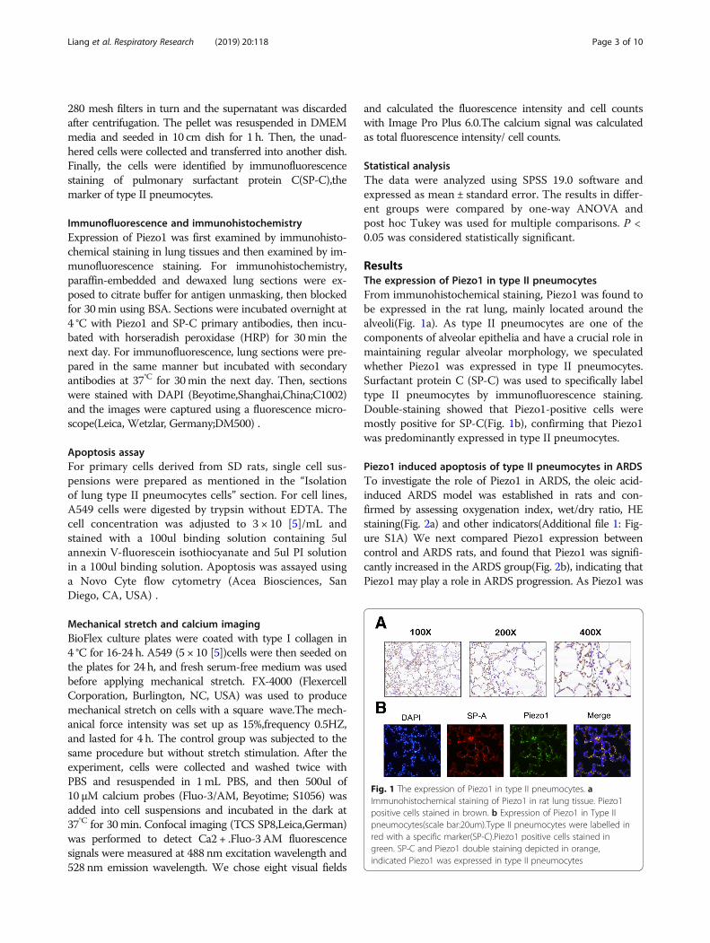
ResultsThe expression of Piezo1 in type II pneumocytesFrom immunohistochemical staining, Piezo1 was found tobe expressed in the rat lung, mainly located around thealveoli(Fig. 1a). As type II pneumocytes are one of thecomponents of alveolar epithelia and have a crucial role inmaintaining regular alveolar morphology, we speculatedwhether Piezo1 was expressed in type II pneumocytes.Surfactant protein C (SP-C) was used to specifically labeltype II pneumocytes by immunofluorescence staining.Double-staining showed that Piezo1-positive cells weremostly positive for SP-C(Fig. 1b), confirming that Piezo1was predominantly expressed in type II pneumocytes.
Piezo1 induced apoptosis of type II pneumocytes in ARDSTo investigate the role of Piezo1 in ARDS, the oleic acid-induced ARDS model was established in rats and con-firmed by assessing oxygenation index, wet/dry ratio, HEstaining(Fig. 2a) and other indicators(Additional file 1: Fig-ure S1A) We next compared Piezo1 expression betweencontrol and ARDS rats, and found that Piezo1 was signifi-cantly increased in the ARDS group(Fig. 2b), indicating thatPiezo1 may play a role in ARDS progression. As Piezo1 was
Fig. 1 The expression of Piezo1 in type II pneumocytes. aImmunohistochemical staining of Piezo1 in rat lung tissue. Piezo1positive cells stained in brown. b Expression of Piezo1 in Type IIpneumocytes(scale bar:20um).Type II pneumocytes were labelled inred with a specific marker(SP-C).Piezo1 positive cells stained ingreen. SP-C and Piezo1 double staining depicted in orange,indicated Piezo1 was expressed in type II pneumocytes
Liang et al. Respiratory Research (2019) 20:118 Page 3 of 10

Fig. 2 (See legend on next page.)
Liang et al. Respiratory Research (2019) 20:118 Page 4 of 10
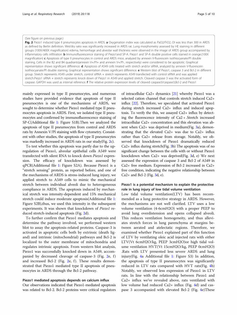
mainly expressed in type II pneumocytes, and numerousstudies have provided evidence that apoptosis of type IIpneumocytes is one of the mechanisms of ARDS, wesought to determine whether Piezo1 mediated type II pneu-mocytes apoptosis in ARDS. First, we isolated type II pneu-mocytes and confirmed by immunofluorescence staining ofSP-C(Additional file 1: Figure S1B).Then we analyzed theapoptosis of type II pneumocytes from control and ARDSrats by Annexin V/PI staining with flow cytometry. Consist-ent with other studies, the apoptosis of type II pneumocyteswas markedly increased in ARDS rats in our study(Fig. 2c).To test whether this apoptosis was partly due to the up-
regulation of Piezo1, alveolar epithelial cells A549 weretransfected with silent RNA to knock down Piezo1 expres-sion. The efficacy of knockdown was assessed byqPCR(Additional file 1: Figure S2A). Because Piezo1 is a“stretch sensing” protein, as reported before, and one ofthe mechanisms of ARDS is stress-induced lung injury, weapplied stretch to A549 cells to mimic the mechanicalstretch between individual alveoli due to heterogeneouscompliance in ARDS. The apoptosis induced by mechan-ical stretch was intensity-dependent and 15% mechanicalstretch could induce moderate apoptosis(Additional file 1:Figure S2B),thus, we used this intensity in the subsequentexperiments. It was shown that knockdown of Piezo1 re-duced stretch-induced apoptosis (Fig. 2d).To further confirm that Piezo1 mediates apoptosis and
determine the pathway involved, we performed westernblot to assay the apoptosis-related proteins. Caspase-3 isactivated in apoptotic cells both by extrinsic (death lig-and) and intrinsic (mitochondrial) pathways and Bcl-2 islocalized to the outer membrane of mitochondria andregulates intrinsic apoptosis. From western blot analysis,Piezo1 was successfully knocked down in A549, accom-panied by decreased cleavage of caspase-3 (Fig. 2e, f )and increased Bcl-2 (Fig. 2e, f ). These results demon-strated that Piezo1 mediated type II apoptosis of pneu-mocytes in ARDS through the Bcl-2 pathway.
Piezo1 mediated apoptosis depends on calcium influxOur observations indicated that Piezo1-mediated apoptosiswas related to Bcl-2. Bcl-2 proteins were critical regulators
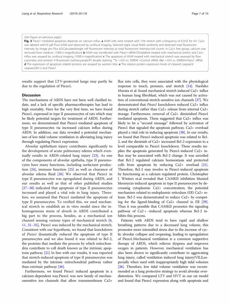
of intracellular Ca2+ dynamics [21] whereby Piezo1 was aselected cation channel that controls stretch induced Ca2+influx [22]. Therefore, we speculated that activated Piezo1during stretch increased Ca2+ influx and induced apop-tosis. To verify the this, we assayed Ca2+ influx by detect-ing the fluorescence intensity of Ca2 + .Stretch increasedintracellular Ca2+ concentration and this elevation was ab-sent when Ca2+ was deprived in medium(Fig. 3a), demon-strating that the elevated Ca2+ was due to Ca2+ influxrather than Ca2+ release from storage. Notably, we ob-served that knockdown of Piezo1 dramatically reducedCa2+ influx during stretch(Fig. 3b) The apoptosis was of nosignificant change between the cells with or without Piezo1knockdown when Ca2+ was deprived(Fig. 3d, e) We nextassessed the expression of caspase 3 and Bcl-2 of A549 inCa2+ free medium. Expression of Bcl-2 increased in Ca2+free condition, indicating the negative relationship betweenCa2+ and Bcl-2 (Fig. 3d, e).
Piezo1 is a potential mechanism to explain the protectiverole in lung injury of low tidal volume ventilationLow tidal volume ventilation(LTV) has been recom-mended as a lung protective strategy in ARDS. However,the mechanisms are not well clarified. LTV uses a lowvolume ventilation (4-6cmH2O) with a proper PEEP toavoid lung overdistension and opens collapsed alveoli.This reduces ventilation homogeneity, and thus allevi-ates stretch forces in lung parenchyma at margins be-tween aerated and atelectatic regions. Therefore, weexamined whether Piezo1 explained part of this functionof LTV by ventilating oleic acid injected rats with eitherLTV(Vt 6cmH2O/kg, PEEP 3cmH2O)or high tidal vol-ume ventilation HVT(Vt 15cmH2O/kg, PEEP 0cmH2O).Rats with LTV presented less severe ARDS and lunginjury(Fig. 4a Additional file 1: Figure S3) In addition,the apoptosis of type II pneumocytes was significantlyreduced in LTV rats compared with HVT rats(Fig. 4b)Notably, we observed less expression of Piezo1 in LTVrats. In line with the relationship between Piezo1 andCa2+ influx we’ve revealed above, rats ventilated withlow volume had reduced Ca2+ influx (Fig. 4d) and cas-pase 3 accompanied with elevated Bcl-2 (Fig. 4e)These
(See figure on previous page.)Fig. 2 Piezo1 induced type II pneumocytes apoptosis in ARDS. a Oxygenation index was calculated as PaO2/FiO2, OI was less than 300 in ARDSas defined by Berlin definition. Wet/dry ratio was significantly increased in ARDS rat. Lung morphometry assessed by HE staining in differentgroups (100X/400X magnification) edema, hemorrhage and alveolar wall thickness were observed in the image of ARDS group accompanied byinflammatory cells infiltration .b Immunofluorescence staining of Piezo1and SP-A. Piezo1 and SP-A double positive cells stained in orange.(100Xmagnification) c Apoptosis of type II pneumocytes in control and ARDS mice, analyzed by annexin V-fluorescein isothiocyanate/PI doublestaining. Cells in the B2 and B4 quadrants(annexin V+/PI+ and annexin V+/PI-, respectively) were considered to be apoptotic. Graphicalrepresentation shows significant difference. d. Apoptosis of A549 cells treated with stretch and/or siRNA, analyzed by annexin V-fluoresceinisothiocyanate/PI double staining. Graphical representation shows significant difference. e Western blot of Piezo1, caspase 3 and Bcl-2 in differentgroup. Stretch represents A549 under stretch, control siRNA + stretch represents A549 transfected with control siRNA and was appliedstretch.Piezo1 siRNA + stretch represents knock down of Piezo1 in A549 and applied stretch. Cleaved caspase 3 was the activated form ofcaspase. GAPDH was used as internal reference. f The relative protein expression levels of cleaved caspase3/caspase3,Bcl-2 and Piezo1
Liang et al. Respiratory Research (2019) 20:118 Page 5 of 10

Fig. 3 (See legend on next page.)
Liang et al. Respiratory Research (2019) 20:118 Page 6 of 10
results support that LTV-protected lungs may partly bedue to the regulation of Piezo1.
DiscussionThe mechanisms of ARDS have not been well clarified to-date, and a lack of specific pharmacotherapies has lead tohigh mortality. Here for the very first time, we have foundPiezo1, expressed in type II pneumocytes of rats which maybe likely potential targets for treatment of ARDS. Further-more, we demonstrated that Piezo1-mediated apoptosis oftype II pneumocytes via increased calcium influx duringARDS. In addition, our data revealed a potential mechan-ism of low tidal volume ventilation in alleviating lung injurythrough regulating Piezo1 expression.Alveolar epithelium injury contributes significantly to
the development of acute pulmonary edema which even-tually results in ARDS-related lung injury [23]. As oneof the components of alveolar epithelia, type II pneumo-cytes have many functions, including surfactant produc-tion [24], immune function [25] as well as clearance ofalveolar edema fluid [26] We observed that Piezo1 intype II pneumocytes was upregulated during ARDS, andour results as well as that of other published studies[27–30] indicated that apoptosis of type II pneumocytesincreased and played a vital role in lung injury. There-fore, we assumed that Piezo1 mediated the apoptosis oftype II pneumocytes. To verified this, we used mechan-ical stretch to establish an in vitro model since the in-homogeneous stress of alveoli in ARDS contributed abig part to the process, besides, as a mechanical ionchannel sensing various types of mechanical stretch [6,11, 31–35], Piezo1 was induced by the mechanical stretch.Consistent with our hypothesis, we found that knockdownof Piezo1 dramatically reduced the apoptosis of type IIpneumocytes and we also found it was related to Bcl-2,the proteins that mediate the process by which mitochon-dria contribute to cell death known as the intrinsic apop-tosis pathway [21] In line with our results, it was reportedthat stretch-induced apoptosis of type II pneumocytes wasmediated by the intrinsic mitochondrial pathway ratherthan extrinsic pathway [36].Furthermore, we found Piezo1 induced apoptosis in a
calcium-dependent way.Piezo1 was new family of mechan-osensitive ion channels that allow transmembrane Ca2+
flux into cells, they were associated with the physiologicalresponse to touch, pressure, and stretch [14]. NaohikoMurata et al. found mechanical stretch induced Ca2+ influxin human lung fibroblast, which was not caused by activa-tion of conventional stretch-sensitive ion channels [37]. Wedemonstrated that Piezo1 knockdown reduced Ca2+ influxduring stretch rather than Ca2+ release from internal Ca2+storage. Furthermore, removal of Ca2+ diminished Piezo1mediated apoptosis. These suggested that Ca2+ influx waslikely to be a “second message” followed by activation ofPiezo1 that signaled the apoptosis pathway. Ca2+ overloadplayed a vital role in inducing apoptosis [38]. In our results,we found that Piezo1-induced apoptosis was related to Bcl-2, and the diminish of Ca2+ increased Bcl-2 expression to alevel comparable to Piezo1 knockdown. These results im-plies the apoptosis generated by Piezo1-induced Ca2+ in-flux may be associated with Bcl-2 change. It was unveiledthat Bcl-2 regulated calcium homeostasis and protectedcells from apoptosis by reducing Ca2+ overload [21].Therefore, Bcl-2 may involve in Piezo1-induced apoptosisby functioning as a calcium regulated protein. ChristopherJ. Winters et.al revealed that CAMKII inhibition bluntedbleomycin-induced apoptosis in type II pneumocytes by de-creasing cytoplasmic Ca2+ concentration, the potentialmechanism related to endoplasmic reticulum (ER) Ca2+ re-lease. Bcl-2 was demonstrated to reduce Ca2+ by compet-ing for the ligand-binding of Ca2+ channel in ER [39].Thus it was possible that CAMKII promotes the signalingpathway of Ca2 + −induced apoptosis whereas Bcl-2 in-hibits this process.Patients with ARDS tend to have rapid and shallow
breathing patterns due to a decreased lung volume, thispromotes more intensified stress due to the increase of cyc-lic alveolar collapse and reopening, leading to upregulationof Piezo1.Mechanical ventilation is a common supportivetherapy of ARDS, which relieves dyspnea and improvesoxygen in patients. However, mechanical ventilation hasalso been shown to significantly contribute to aggravatinglung injury, called ventilation induced lung injury(VILI),es-pecially when used with inappropriately high tidal volumes[26]. Therefore, low tidal volume ventilation was recom-mended as a lung protective strategy to avoid alveolar over-distention. We compared LTV and HVT in our rat modeland found that Piezo1 expression along with apoptosis and
(See figure on previous page.)Fig. 3 Piezo1 mediated apoptosis depends on calcium influx. a A549 cells were treated with 15% stretch with a frequency of 0.5HZ for 4 h. Ca2+was labeled with10 μM Fluo-3/AM and observed by confocal imaging. Selected eight visual fields randomly and detected total fluorescentintensity by Image pro Plus 6.0.Calculatedsingle cell fluorescent intensity as total fluorescent intensity/cell counts. In Ca2+ free group, calcium wasremoved from medium. (1000 ×magnification) b A549 was transfected with Piezo1-siRNA72hrsbefore treated with mechanical stretch and Ca2+influx was assayed by confocal imaging. (1000 ×magnification) c The apoptosis of A549 treated with mechanical stretch was assessed by flowcytometry and annexin V-fluorescein isothiocyanate/PI double staining. **p < 0.01.vs. DMEM +Control siRNA; ##p < 0.01.vs. DMEM+Piezo1 siRNA.d The expression of apoptosis related proteins are assayed by western blot. e The relative protein expression levels of cleaved caspase3/caspase3,Bcl-2 and Piezo1
Liang et al. Respiratory Research (2019) 20:118 Page 7 of 10

Ca2+ influx was significantly reduced when low tidal vol-ume was used. This supports that Piezo1 may be a potentialmechanism to explain the protective role of LTV.
ConclusionsThe results of our studies demonstrated that Piezo1played an pivotal role in lung injury. Activation of Piezo1by mechanical stretch induced the apoptosis of type IIpneumocytes via Ca2+ influx. Inhibition of Piezo1 orCa2+ influx may become a potential target for preventiveor therapeutic interventions aiming at relieving lung injuryin ARDS. The Piezo1 mediated apoptosis was related toBcl-2, but further research is required to support this.
Additional file
Additional file 1: Figure S1. The expression of Piezo1 on rat type IIpneumocytes. A)The level of IL-1β and TNF-a increased in ARDS rats,tested by ELISA assay. B)Identification of Type II pneumocytes isolatedfrom lung. Nuclei stained with DAPI, Positive staining depicted in blue.SP-C containing cells stained in green. SP-C is a specific marker for type IIpneumocytes(1000 ×magnification). Figure S2. Piezo1 induced type IIpneumocytes apoptosis in ARDS.A)The efficacy of knockdown of piezo1was evaluated by Q-PCR and siRNA-1 was used in the following experi-ments. B) The apoptosis of A549 cells strength of mechanical stretch,assayed by annexin V-fluorescein isothiocyanate/PI double staining. Fig-ure S3. Piezo1 was a potential mechanism to explain the protective rolein lung injury of LTV. The level of IL-1βand TNF-a in different groups,tested by ELISA assay. (DOCX 605 kb)
AbbreviationsARDS: acute respiratory distress syndrome; HE: hematoxylin and eosin; IL-1β: interleukins-1β; MV: mechanical ventilation; PEEP: positive end-expiratorypressure; SD: Sprague-Dawley; SP-A: pulmonary surfactant protein A; TNF-α: tumor necrosis factor-a; W/D: wet-to-dry ratio
AcknowledgementsNot applicable.
Authors’ contributionsG-PL and JX conceived, designed and performed the experiments, draftedthe manuscript; L-LC assisted in parts of the experiments;L-LC and Y-HZhelped analyze the statistics; YK,B-XC, JY helped in the draft of the article; KYand Z-ZW designed the experiments and revised the article. All authors readand approved the final manuscript.
Fig. 4 Piezo1 was a potential mechanism to explain the protectiverole in lung injury of LTV. a Wet/dry ratio decreased andoxygenation index (calculated as PaO2/FiO2) in different group.Lung morphometry assessed by HE staining (100X/400Xmagnification) b The apoptosis of type II pneumocytes assayed byflow cytometry. c Immunofluorescence staining of Piezo1 and SP-A.Piezo1 and SP-A double positive cells stained in orange.(100 ×magnification) d Ca2+ influx detected by confocal microscope andthe quantitation of immunofluorescence presented as a bar graph.(1000 ×magnification) e The expression of caspase 3,Bcl-2 andPiezo1 in rats assessed by western blot (cleaved caspase 3 was anactivated form of caspase 3) (NS, not significant. *, p < 0.05.**, p <0.001.LV low tidal volume, HV high tidal volume.) f The relativeprotein expression levels of cleaved caspase3/caspase3,Bcl-2and Piezo1
Liang et al. Respiratory Research (2019) 20:118 Page 8 of 10
FundingThis study was supported by National Natural Science Foundation ofChina (31301116); Project of Sichuan Provincial Education Department(11ZA297);Project of Sichuan Science and Technology(2019YFS0235).
Availability of data and materialsThe datasets used and/or analyzed during the current study are availablefrom the corresponding author on reasonable request.
Ethics approval and consent to participateThe protocol was approved by the Institutional Animal Care and TreatmentCommittee of Sichuan University (Chengdu, P.R. China).
Consent for publicationNot applicable.
Competing interests“Author Li-li Cao” is currently receiving a grant (31301116) from NationalNatural Science Foundation of China and “Author Z. W Zhang” is currentlyreceiving a grant(11ZA297) from Education Department of Sichuan Province(CN). "Author GuoPeng Liang "is currently receiving a grant(2019YFS0235)from Project of Sichuan Science and Technology.The remaining authors havedisclosed that they do not have anyconflicts of interest.
Publisher’s NoteSpringer Nature remains neutral with regard to jurisdictional claims inpublished maps and institutional affiliations.
Author details1Department of Critical Care Medicine, West China School of Medicine andWest China Hospital, Sichuan University, Chengdu 610041, China. 2ChengduMedical College, Chengdu 610041, Sichuan, China. 3Department ofRespiratory Critical Care Medicine, West China School of Medicine and WestChina Hospital, Sichuan University, Chengdu 610041, China.
Received: 8 March 2019 Accepted: 24 May 2019
References1. Bellani G, Laffey JG, Pham T, et al. Epidemiology, patterns of care, and
mortality for patients with acute respiratory distress syndrome in intensivecare units in 50 countries. JAMA. 2016;315(8):788–800.
2. Force ADT, Ranieri VM, Rubenfeld GD, et al. Acute respiratory distresssyndrome: the Berlin definition. JAMA. 307(23):2526–33.
3. Sawa T. The molecular mechanism of acute lung injury caused byPseudomonas aeruginosa: from bacterial pathogenesis to host response. JIntensive Care. 2014;2(1):10.
4. Coste B, Mathur J, Schmidt M, et al. Piezo1 and Piezo2 are essentialcomponents of distinct mechanically activated cation channels. Science. Oct01 2010;330(6000):55–60.
5. Syeda R, Florendo MN, Cox CD, et al. Piezo1 channels are inherentlymechanosensitive. Cell Rep. 2016;17(7):1739–46.
6. Cahalan SM, Lukacs V, Ranade SS, Chien S, Bandell M, Patapoutian A. Piezo1links mechanical forces to red blood cell volume. Elife. May 22 2015;4.
7. Li J, Hou B, Tumova S, et al. Piezo1 integration of vascular architecture withphysiological force. Nature. Nov 13 2014;515(7526):279–82.
8. Faucherre A, Kissa K, Nargeot J, Mangoni ME, Jopling C. Piezo1 plays a rolein erythrocyte volume homeostasis. Haematologica. Jan 2014;99(1):70–5.
9. Ilkan Z, Wright JR, Goodall AH, Gibbins JM, Jones CI, Mahaut-Smith MP.Evidence for shear-mediated Ca2+ entry through mechanosensitive cationchannels in human platelets and a megakaryocytic cell line. J Biol Chem.Apr 17 2017.
10. Ranade SS, Qiu Z, Woo SH, et al. Piezo1, a mechanically activated ionchannel, is required for vascular development in mice. Proc Natl Acad Sci US A. 2014;111(28):10347–52.
11. Retailleau K, Duprat F, Arhatte M, et al. Piezo1 in smooth muscle cells isinvolved in Hypertension-dependent arterial remodeling. Cell Rep. Nov 102015;13(6):1161–71.
12. Wang S, Chennupati R, Kaur H, Iring A, Wettschureck N, Offermanns S.Endothelial cation channel PIEZO1 controls blood pressure by mediatingflow-induced ATP release. J Clin Invest. Dec 01 2016;126(12):4527–36.
13. Hypertension ASJ. Mechanosensation by PIEZO1 in blood pressure control.Nat Rev Nephrol. 2017;13(1):3.
14. Miyamoto T, Mochizuki T, Nakagomi H, et al. Functional role forPiezo1 in stretch-evoked ca(2)(+) influx and ATP release in urothelialcell cultures. J Biol Chem. 2014;289(23):16565–75.
15. Bagriantsev SN, Gracheva EO, Gallagher PG. Piezo proteins: regulatorsof Mechanosensation and other cellular processes. J Biol Chem. Nov14 2014;289(46):31673–81.
16. Chiumello D, Carlesso E, Cadringher P, et al. Lung stress and strainduring mechanical ventilation for acute respiratory distress syndrome.Am J Respir Crit Care Med. Aug 15 2008;178(4):346–55.
17. Oba Y, Salzman GA. Ventilation with lower tidal volumes as comparedwith traditional tidal volumes for acute lung injury. N Engl J Med.2000;343(11):813 author reply 813-814.
18. Caironi P, Cressoni M, Chiumello D, et al. Lung opening and closingduring ventilation of acute respiratory distress syndrome. Am J RespirCrit Care Med. Mar 15 2010;181(6):578–86.
19. Wang YY, Zhang H, Ma T, et al. Piezo1 mediates neuron oxygen-glucose deprivation/reoxygenation injury via ca/calpain signaling.Biochem Biophys Res Commun. 2019;513(1):147–53.
20. Belinsky SA, Lechner JF, Johnson NF. An improved method for theisolation of type II and Clara cells from mice. In Vitro Cell Dev BiolAnim. May 1995;31(5):361–6.
21. Hardwick JM, Soane L. Multiple functions of BCL-2 family proteins.Cold Spring Harb Perspect Biol. Feb 1 2013;5(2).
22. Cinar E, Zhou S, DeCourcey J, Wang Y, Waugh RE, Wan J. Piezo1regulates mechanotransductive release of ATP from human RBCs. ProcNatl Acad Sci U S A. Sep 22 2015;112(38):11783–8.
23. Matthay MA, Zemans RL. The acute respiratory distress syndrome:pathogenesis and treatment. Annu Rev Pathol. 2011;6:147–63.
24. Wright JR, Hawgood S. Pulmonary surfactant metabolism. Clin ChestMed. 1989;10(1):83–93.
25. Harbeck RJ, Gegen NW, Struhar D, Mason R. Class II molecules on ratalveolar type II epithelial cells. Cell Immunol. 1988;111(1):139–47.
26. Gill SE, Yamashita CM, Veldhuizen RA. Lung remodeling associated withrecovery from acute lung injury. Cell Tissue Res. 2017;367(3):495–509.
27. Steinberg H, Greenwald RA, Sciubba J, Das DK. The effect of oxygen-derived free radicals on pulmonary endothelial cell function in theisolated perfused rat lung. Exp Lung Res. 1982;3(2):163–73.
28. Li X, Shu R, Filippatos G, Uhal BD. Apoptosis in lung injury andremodeling. J Appl Physiol (1985). 2004;97(4):1535–42.
29. Galani V, Tatsaki E, Bai M, et al. The role of apoptosis in thepathophysiology of acute respiratory distress syndrome (ARDS): anup-to-date cell-specific review. Pathol Res Pract. Mar 15 2010;206(3):145–50.
30. Song Y, Mao B, Qian G. The role of apoptosis and Fas/FasL in lungtissue in patients with acute respiratory distress syndrome. ZhonghuaJie He He Hu Xi Za Zhi. Oct 1999;22(10):610–2.
31. Martins JR, Penton D, Peyronnet R, et al. Piezo1-dependent regulationof urinary osmolarity. Pflugers Arch. Jul 2016;468(7):1197–206.
32. Wright JR, Amisten S, Goodall AH, Mahaut-Smith MP. Transcriptomicanalysis of the ion channelome of human platelets andmegakaryocytic cell lines. Thromb Haemost.Aug 01 2016;116(2):272–84.
33. Peyronnet R, Martins JR, Duprat F, et al. Piezo1-dependent stretch-activatedchannels are inhibited by Polycystin-2 in renal tubular epithelial cells. EMBORep. Dec 2013;14(12):1143–8.
34. Michishita M, Yano K, Tomita KI, Matsuzaki O, Kasahara KI. Piezo1 expressionincreases in rat bladder after partial bladder outlet obstruction. Life Sci. Dec01 2016;166:1–7.
35. Miyamoto T, Nakagomi H, Kira S, et al. Piezo1, a novel Mechanosensor inthe bladder Urothelium. Neurourol Urodynam. 2012;31(6):1015–7.
36. Kuhn H, Nieuwenhuijsen H, Karthe B, Wirtz H. Stretch-induced apoptosis inrat alveolar epithelial cells is mediated by the intrinsic mitochondrialpathway. Experimental lung research. 2017;43(1):49–56.
37. Murata N, Ito S, Furuya K, et al. Ca2+ influx and ATP release mediated bymechanical stretch in human lung fibroblasts. Biochem Bioph Res Co. 2014;453(1):101–5.
Liang et al. Respiratory Research (2019) 20:118 Page 9 of 10
38. Rong YP, Aromolaran AS, Bultynck G, et al. Targeting Bcl-2-IP3 receptorinteraction to reverse Bcl-2's inhibition of apoptotic calcium signals. MolCell. Jul 25 2008;31(2):255–65.
39. Ivanova H, Wagner LE, Tanimura A, et al. Bcl-2 and IP compete for theligand-binding domain of IPRs modulating ca signaling output. Cellular andmolecular life sciences : CMLS. 2019.
Liang et al. Respiratory Research (2019) 20:118 Page 10 of 10