pigment epithelium-derived factor in the monkey retinal
TRANSCRIPT
Pigment epithelium-derived factor in the monkey retinal pigment
epithelium and interphotoreceptor matrix: apical secretion and distribution
S. Patricia Becerraa,*, Robert N. Farissb, Yan Q. Wua, Luis M. Montuengac,d,Paul Wonga,e, Bruce A. Pfeffera,f
aLaboratory of Retinal Cell and Molecular Biology, NEI, NIH, Building 6, room 308, 6 Center Drive, MSC 2740, Bethesda, MD, 20892-0706, USAbLaboratory of Mechanisms of Ocular Diseases, NEI, NIH, Bethesda, MD, USA
cDCS, NCI, NIH, Bethesda, MD, USAdDepartment of Histology and Pathology, University of Navarra, Pamplona, Navarra, Spain
eDepartment of Biological Sciences, University of Alberta, Edmonton, CanadafBausch and Lomb, Rochester, NY, USA
Received 16 April 2003; accepted in revised form 29 October 2003
Abstract
Pigment epithelium-derived factor (PEDF) is an extracellular protein derived from the retinal pigment epithelium (RPE), a tissue formed
by polarized cells that release growth and trophic factors in a directional fashion. We have investigated the distribution and directional
release of PEDF protein by the monkey RPE. We established primary cultures of monkey RPE cells that expressed the PEDF gene, and that
synthesized and secreted the PEDF protein. Northern analysis of RPE cultures and monkey ocular tissues showed that PEDF transcripts were
highly expressed in RPE as compared with several other monkey ocular tissues, being even more abundant in cultured cells than they were in
the native RPE. The differentiated RPE cells in culture secreted protein that shared the immunological, biochemical and biological
characteristics of PEDF. The overall PEDF levels in the RPE conditioned media reached 6·5 mg ml2 after 8 days in culture (i.e. 1·1 pg of
PEDF per RPE cell). RPE cells were cultivated on permeable supports as monolayers forming a barrier between apical and basal
compartments. Apical and basal culture media were sampled at three or four-day intervals for 18 cycles, and the PEDF content was
quantified. Most of the PEDF protein was significantly higher in the apical than in the basal medium (.4 times) at the initial recovery
intervals, to be detected only in the apical medium at the latter intervals. In the native monkey eye, the concentration of soluble PEDF in the
interphotoreceptor matrix (144 nM) was 7-fold and 25-fold greater than in vitreous and aqueous, respectively. PEDF was abundant in the
interphotoreceptor matrix surrounding rod and cone outer segments, and was detectable at lower levels in the RPE as visualized by confocal
microscopy. We concluded that PEDF synthesized by the RPE is secreted preferentially from the apical surface and is distributed apically to
the RPE bordering the outer segments of photoreceptors. PEDF can be a useful marker for RPE polarization and differentiation. The
polarization of RPE may be an important mechanism to control PEDF secretion and our results offer interesting possibilities on regulation of
PEDF.
q 2004 Elsevier Ltd. All rights reserved.
Keywords: retinal pigment epithelium; pigment epithelium-derived factor; secretion; polarization; interphotoreceptor matrix; monkey eye; apical; basal
1. Introduction
The retinal pigment epithelium (RPE), situated between
the neurosensory retina and the vascularized choroid,
maintains the photoreceptors and the choriocapillaris in
the normal eye, and is involved in the pathogenesis of retinal
degeneration and choroidal neovascularization in age-
related macular degeneration. It performs functions essen-
tial for photoreceptor survival and secretes factors important
for homeostasis of the outer retina. Formed by cells that
polarize, the RPE can secrete components in a directional
fashion. For example, hyaluronan, matrix metalloprotei-
nase-2 and tissue inhibitor of metalloproteinase-1 syn-
thesized by the RPE are secreted preferentially from the
apical surface (retina facing) toward the photoreceptors
0014-4835/$ - see front matter q 2004 Elsevier Ltd. All rights reserved.
DOI:10.1016/j.exer.2003.10.013
Experimental Eye Research 78 (2004) 223–234
www.elsevier.com/locate/yexer
* Corresponding author. Dr S. Patricia Becerra, Laboratory of Retinal
Cell and Molecular Biology, NEI, NIH, Building 6, room 308, 6 Center
Drive MSC 2740 Bethesda, MD, USA.
E-mail address: [email protected] (S. Patricia Becerra).
(Padgett et al., 1997; deS Senanayake et al., 2001), while
tissue inhibitor of metalloproteinase-3, fibroblast growth
factor 5, interleukin-6 and interleukin-8 are secreted from
the basolateral surface of these cells facing the choroid
(Fariss et al., 1997; Dunn et al., 1998; Holtkamp et al.,
1998). Its polarization is under constant regulation by
signaling molecules coming from the choroid and the retina
or extracellular stimuli. For example, vasoactive intestinal
peptide, a 28-amino acid neuropeptide and differentiation
promotor during development of the functional RPE,
stimulates macromolecule secretion at the apical surface
and fluid transport from the apical to the basal surface of the
RPE sheet (Koh, 2000); oxygen or extracellular matrix
components from RPE modulate the polarity of secretion of
angiogenic factors, such as vascular endothelial growth
factor, from the RPE (Mousa et al., 1999; Blaauwgeers et al.,
1999). Thus the polarity of the RPE plays a role in
maintaining a balance of secreted factors essential for the
functional RPE.
One component secreted by the RPE is pigment
epithelium-derived factor (PEDF), a protein with neuro-
trophic and antiangiogenic activities expressed mainly in
compartments of the eye. PEDF acts in neuronal survival
and differentiation on photoreceptor and neuronal cells of
the retina and central nervous system (Becerra, 1997;
Cayouette et al., 1999; Houenou et al., 1999; Cao et al.,
2001; Crawford et al., 2001). It is also a major inhibitor of
neovascularization and is responsible for excluding vessels
from invading the retina, vitreous and cornea (Dawson et al.,
1999; Stellmach et al., 2001; Bouck, 2002). These
biological activities are of great importance for the
development and maintenance of normal physiological
functions in the eye. Biochemical and immunochemical
studies have identified PEDF as a soluble extracellular
protein localized in washes of the interphotoreceptor matrix
(IPM), vitreous and aqueous of several mammalian species
(Tombran-Tink et al., 1995; Wu et al., 1995; Ortego et al.,
1996; Alberdi et al., 1998; Karakousis et al., 2001). PEDF,
discovered as a product secreted by cultured RPE cells from
fetal human eyes (Tombran Tink, 1991), can be released
from a variety of cells, including stably transfected
eukaryotic cells with full length PEDF cDNA under the
control of recombinant transcriptional promoters (Stratikos
et al., 1996; Perez-Mediavilla et al., 1998). By sequence
homology, PEDF is a member of the serpin superfamily
(Steele et al., 1993), formed by proteins with a common
overall protein conformation with a variety of biological
functions, most of which are extracellular serine protease
inhibitors. Like serpins, PEDF is a globular and compact
protein of ,50 kDa that is highly resistant to proteolytic
cleavage except for an exposed loop towards its carboxyl-
end; however, it does not behave as an inhibitor of proteases
(Becerra et al., 1995). It has binding affinity for components
of extracellular matrixes, such as glycosaminoglycans
(Alberdi et al., 1998) and collagens (Meyer et al., 2002),
which represents its molecular association with the IPM,
and other extracellular matrixes. More interestingly, PEDF
has binding affinity for receptors on the surface of cells from
retina and the CNS known to respond to its stimuli, such as
retinoblastoma cells, cerebellar granule cell neurons,
photoreceptors, motor neurons (Alberdi et al., 1999;
Aymerich et al., 2001; Bilak et al., 2002). While the
deposition of PEDF in extracellular ocular compartments
coincides with their avascular nature, the discrete distri-
bution of PEDF binding sites in the bovine retina to the
inner segments of photoreceptors (Aymerich et al., 2001) is
consistent with PEDF’s in vivo effects on survival of
photoreceptors (Cayouette et al., 1999; Cao et al., 2001).
Given the neurotrophic and antiangiogenic activities of
PEDF and its potential exploitation as a therapeutic agent
for retinal diseases triggered by photoreceptor degener-
ations and abnormal neovascularization, it is of interest to
determine its levels and distribution in the RPE/retina for
physiological studies and development of therapeutics.
Using the monkey eye as a non-human primate model, we
have (1) compared the levels of PEDF gene expression
among ocular tissues; (2) determined PEDF protein levels in
extracellular compartments of the eye; (3) characterized in
detail the secreted monkey PEDF protein; and (4)
investigated a directional delivery of PEDF from RPE in
culture and in situ. We discuss the implications of a polar
secretion and distribution of PEDF by the RPE on the retina.
2. Materials and methods
2.1. Tissue samples
Eyes were from juvenile Rhesus monkeys (aged 3–5
years) using procedures in strict compliance with the NIH
Guidelines for Care and Use of Laboratory Animals. Eyes
were processed for immunolabeling or ocular tissues
dissected while chilled on ice. Aqueous and vitreous
extracts were obtained as described by Wu and Becerra
(1996). IPM lavages were obtained after dissection of the
anterior portion, removal of vitreous and retina (Wu et al.,
1995), or alternatively, by cannulation (Pfeffer et al., 1983),
a method known to assure an IPM lavage free of
intracellular contamination maintaining the topological
integrity of the IPM compartment (Adler, 1989).
2.2. RPE cell culture
Confluent first passage Rhesus monkey RPE was utilized
to generate conditioned media samples and for extraction of
total RNA. The methodology used for establishing primate
RPE cultures was as described in detail by Pfeffer (1990). In
summary, cells were maintained as stable monolayers in 12-
well clusters for at least two weeks and then experiments
were performed after cultured cells had achieved biochemi-
cal and physiological markers of the differentiated tissue
in vivo. These markers include de novo melanization,
S.P. Becerra et al. / Experimental Eye Research 78 (2004) 223–234224

formation of domes (emblematic of vectorial fluid transport),
morphological apical–basal polarization, and expression of
cellular retinaldehyde-binding protein. Confluent wells
contained 6 £ 105 cells each, as calculated from hemocyt-
ometer counting of parallel cultures. For production of
conditioned media, RPE cells were maintained in protein-
free defined medium (Pfeffer, 1990) modified as follows: (1)
serum, bovine retinal extract, insulin and transferrin were
omitted, (2) Albumax was replaced by Chemically Defined
Lipid Concentrate (0·1% (v/v); Gibco-BRL, Gaithersburg,
MD, USA); and (3) 0·05% (w/v) each of dimethyl beta-
cyclodextrin and hydroxypropyl beta-cyclodextrin (Cyclo-
dextrin Technologies Development, High Springs, FL, USA)
were added The monolayer was washed twice with protein-
free medium, and then 0·75 ml were added per well. After the
third and sixth days, glucose, fructose, essential amino acids,
and vitamins were supplemented to the cultures, along with
water to correct for volume loss by evaporation. Throughout
the eight-day period, the pH of the medium remained
constant and cell morphology was unchanged. Medium was
collected after eight days in culture, immediately frozen in
dry ice and stored at 2808C.
2.3. Culture of RPE Cells on permeable supports
To investigate the polarization release of PEDF, RPE
monolayers were established in complete medium (Pfeffer,
1990) on 9 mm diameter cell culture inserts containing
0·4 mm pore size polyethylene terephthalate membranes
(Cyclopore, Falcon, Lincoln Park, NJ, USA). Cultures were
monitored visually under an inverted microscope to assess
cell proliferation and morphology at confluence. Upon
seeding in the inserts, the RPE cells displayed low levels of
pigmentation. A stable, confluent monolayer was attained
by 12 days and the culture medium was replaced in both
chambers with fresh medium. After that, the harvesting of
medium from apical (0·5 ml) and basal (1·2 ml) compart-
ments, and its replenishment were repeated in cycles of
alternating 4 and 3 days. At 19 days following seeding, de
novo pigmentation was noticeable in the cell and was
retained during the remaining 15 cycles of medium
exchanges. Conditioned media were stored at 2808C.
Similar preparations have been shown, with the use of a
Millicell-Electrical Resistance System (Millipore, Bedford,
MA, USA), to develop a transepithelial resistance in the
range of 30–50 ohm-cm2.
2.4. Northern blotting
The method used was as described in Wong et al., 2000.
Briefly, individual ocular tissues were dissected from
twenty juvenile monkey eyes and their RNA extracted for
northern blot analysis. Blots were probed with a 32P-
radiolabeled human PEDF cDNA fragment. Total RNA
from each tissue was pre-stained with ethidium bromide
prior to electrophoresis and gels were photographed after
electrophoresis. The relative levels of PEDF mRNA for
each tissue were calculated as the ratio of densitometric
intensity of the PEDF divided by the combined 18S and 28S
RNA intensities (indicated as ‘ratio’).
2.5. Western blotting and quantitation of PEDF protein
The methods used have been described before by Wu
et al. (1995). Briefly, western transfers were immunoreacted
with Ab-rPEDF with an enzymatic colorimetric method for
detection. Alternatively, a monoclonal antibody to PEDF
(MAB1059; Chemicon) was used, as described before
(Meyer et al., 2002). Quantitation was performed by
densitometric scanning of immunoreactive stains and
relative to those of known amounts of bovine or human
PEDF protein as standards. Quantitation of total protein was
performed using a BioRad Protease Assay kit (BioRad).
2.6. PEDF protein purification
To optimize a purification scheme for PEDF from
monkey RPE conditioned media and vitreous extracts we
modified previous methods for the purification of bovine
PEDF (Wu et al., 1995; Wu and Becerra, 1996). These
methods involved ammonium sulfate fractionation, cation-
exchange column chromatography and size-exclusion
column chromatography. In contrast with the previous
methods, monkey PEDF did not partition into one fraction
by ammonium sulfate saturation, not even with 90% of the
salts (unpublished observations). Therefore, for recovery
purposes, concentration by ammonium sulfate precipitation
was avoided for PEDF from monkey sources. The protein
was purified to near homogeneity by two purification steps.
First, proteins were fractionated by S-Sepharose column
chromatography as described (Wu et al., 1995). PEDF-
containing fractions were then pooled and subjected to size-
exclusion ultrafiltration using Centricon-100 to exclude
large molecules and the filtrate applied to centricon-30 to
exclude small molecules and concentrate the protein. The
final sample yielded a highly pure protein at 1 mg PEDF per
ml of monkey vitreous, and 2·6 mg PEDF per ml media with
a 40% recovery. We have observed that the recovery and
purity of the final PEDF sample decreases with increasing
concentration of serum in the medium (Wu and Becerra,
personal observations). In a similar fashion, PEDF was
purified from IPM lavages and aqueous extracts. However,
the purity was lower than for vitreous and the RPE
conditioned media, e.g., 0·5 mg of PEDF in 115 mg of
total protein from IPM lavages from three eyes. The final
samples were stored at 2808C.
2.7. Controlled proteolysis
To examine the overall protein conformation of the
purified protein we performed the typical assay for serpin
proteins, controlled proteolysis, as described (Becerra et al.,
S.P. Becerra et al. / Experimental Eye Research 78 (2004) 223–234 225
1995). Briefly, purified monkey PEDF protein was mixed
with limited concentrations of chymotrypsin in a 15 ml
reaction containing 20 mM Tris HCl, pH 7·5, 150 mM NaCl,
1 mM EDTA, and incubated at 258C for the indicated
periods of time. The reactions were stopped by addition of
SDS-PAGE sample buffer and freezing in dry ice. After
heating at 1008C for 3–5 min, the reaction mixtures were
subjected to SDS-PAGE to compare migration patterns of
the products. A parallel set of samples was subjected to N-
terminal sequence determination of peptides using an
Applied Biosynthesis model 477 sequencer (Foster City,
CA, USA) following the manufacturer’s protocols.
2.8. Biological assay for PEDF
Neurite outgrowth from retinoblastoma cells was as
described by Becerra et al. (1993). Briefly, human Y-79
cells exponentially growing in serum-containing medium
were washed twice with phosphate buffered saline and
plated (1·25 £ 105 cells per ml) in serum-free minimal
essential medium supplemented with a mix of insulin,
transferrin and selenium. Effectors were then added to the
cultures. After 7 days at 378C in CO2, cells were attached to
poly-D-lysine coated plates with fresh serum-free medium.
The differentiation state of the cultures was monitored at
various intervals after attachment by light microscopy.
2.9. Immunolabeling
For immunofluorescence, following enucleation, monkey
eyes were dissected and fixed by immersion for 4 hr in 4%
formaldehyde in PBS pH 7·3. Tissue was stored in 0·5%
formaldehyde in PBS at 48C. Retina/RPE/choroid samples
were excised and washed in PBS and then embedded in 7%
agarose in PBS according to Hale and Matsumoto (1993). A
Leica vibrating microtome was used to cut 100 mm thick
slices of tissue for immunofluorescence. Sections were
blocked briefly in 5% normal goat serum in PBS and then
immunolabeled with a monoclonal antibody to PEDF
(Chemicon) diluted 1:100 in ICC buffer (PBS with 0·5%
BSA, 0·2% Tween-20, pH 7·3) and incubated overnight at
48C. Biotinylated peanut agglutinin (PNA) diluted 1:200
(Vector Labs) and streptavidin Cy5 diluted 1:200 (Jackson
ImmunoResearch) were used to label cone matrix sheaths.
Alexa568 phalloidin (Molecular Probes) was diluted 1:80
and used to label filamentous actin. Sections were washed
repeatedly and labeled in the dark for 4 hr with the nuclear
dye DAPI (40,6-diamidino-2-phenyl-indole, dihydrochlor-
ide) (Molecular Probes) (1 mg ml21) and secondary antibody
-goat anti-mouse Cy3 (Jackson ImmunoResearch) or goat
anti-mouse Alexa488 (Molecular Probes). Following
repeated washing, sections were mounted in Gel Mount
(Bio-Meda) and protected with a coverslip. Primary antibody
was omitted from sections used as negative controls Speci-
mens were analyzed on a Leica SP2 laser scanning confocal
microscope equipped with Nomarski optics. Samples were
scanned in sequential scan mode to reduce bleed-through
artifacts. PEDF-immunolabeled and negative control sec-
tions were imaged under identical scanning conditions. Files
were imported into Photoshop and converted to psd format
for layout purposes.
For immunohistochemistry the fixed monkey retina
sections were immunoreacted with a polyclonal antiserum
to human recombinant PEDF Ab-rPEDF (Wu et al., 1995)
and detection was by the peroxidase-mediated diaminoben-
zidine (DAB) precipitation method was as described before
(Montuenga et al., 1997). Negative controls were immunor-
eacted without primary antibody or with preabsorbed Ab-
rPEDF. Ab-rPEDF was preabsorbed by incubating the
antiserum with PEDF protein covalently attached to resin
3M-Emphazed (6 mg rhuPEDF ml21 resin, and prepared as
described by Alberdi et al. (1999) for 16 hr at 48C. The resin
mixture was transferred to a disposable column and
unbound antisera proteins were eluted by gravity flow and
collected as preabsorbed Ab-rPEDF. Immunolabeling was
visualized by light microscopy.
3. Results
3.1. Distribution of PEDF gene expression and PEDF
protein in monkey eyes
To identify the major potential sites of PEDF protein
synthesis we analyzed the distribution of PEDF transcripts
in the monkey eye. We established primate RPE cultures
and utilized confluent first passage Rhesus monkey RPE.
Northern blot analyses of individual ocular monkey tissues
and cultured RPE cells detected PEDF transcripts of 1·5 kb,
in agreement with the size for the human PEDF mRNA
(Steele et al., 1993; Pignolo et al., 1993), detected in all
tissues tested at different levels (Fig. 1). Expression was the
highest in trabecular meshwork and RPE, followed by
sclera, ciliary body, orbital muscle and retina, and the
lowest in iris, limbus and cornea, based on densitometry.
The levels of PEDF mRNA in the cultured RPE cells were
.3-fold higher than in the RPE/choroid tissue, and both
.5-fold higher than in retina.
Because PEDF is an extracellular protein we compared
the protein obtained from the culturing medium of RPE
cells and ocular fluids IPM, vitreous and aqueous.
Western analysis of all the samples with monoclonal or
polyclonal antibodies to PEDF revealed immunoreactivity
migrating as a protein of 50 kDa (Fig. 2), which was
blocked when challenged with recombinant human PEDF
protein (data not shown) indicating specific immunor-
eactivity for PEDF in monkey RPE conditioned medium,
IPM, vitreous and aqueous. PEDF-immunoreactive bands
migrating with slightly different speeds for vitreous and
aqueous in Fig. 2(B) lanes 3 and 4 resemble two species
of PEDF with differences in glycopeptides containing N-
acetylneuraminic acid (NeuAc) and N-acetylhexosamine
S.P. Becerra et al. / Experimental Eye Research 78 (2004) 223–234226

(HexNAc), as reported by Duh et al. (2002). However, an
apparent single band was observed when a gradient
polyacrylamide gel was used (see Fig. 2(C), lane 2). A
summary and analysis of the purification of monkey
PEDF from RPE conditioned media and vitreous are
shown in Table 1 and Fig. 2(A). PEDF-immunoreactivity
of 50-kDa was detected in monkey IPM lavages only after
separation of PEDF from other IPM proteins by cation-
exchange column chromatography (Fig. 2(B); lanes 1 and
2), suggesting that IPM proteins comigrating with PEDF
might have obstructed the PEDF-antibody interaction in
westerns of the total IPM lavage. Similarly, PEDF was
detected in western blots of monkey retina and RPE tissue
extracts only after cation-exchange chromatography (Fig.
2(C)). PEDF was estimated in the IPM after purification
by cation-exchange column chromatography and in the
aqueous humor. The concentrations and amounts of PEDF
in monkey IPM, vitreous and aqueous were calculated and
are summarized in Table 2.
3.2. Characterization of monkey PEDF protein
We then characterized the purified monkey PEDF protein
in detail. The size of the purified protein was determined by
SDS-PAGE using several gels of a variety of polyacryl-
amide concentrations and under reducing conditions. The
calculated size was 50 700 ^ 1000-Mr and is in agreement
with the size for the native and recombinant bovine and
human PEDF proteins (Wu et al., 1995, and Wu and
Becerra, 1996; Stratikos et al., 1996; Perez-Mediavilla et al.,
1998). The monkey PEDF behaved as a monomeric protein
by size-exclusion ultrafiltration.
Controlled proteolysis of the purified protein with
chymotrypsin rendered limited proteolytic products of
,46-kDa (Fig. 3(A) and (B)). However, trypsin treatments
did not produce a discrete limited product (data not shown).
Edman degradation of the limited chymotryptic reactions
Fig. 1. Distribution of PEDF gene expression in monkey eye tissues.
Northern blot of RNA from monkey ocular tissues shows detection of
hybridization of a 32P-radiolabeled human PEDF cDNA fragment (right
panel). Arrow indicates the migration position of the PEDF transcript.
Migration of size RNA markers is indicated at the top. Left and middle
panels show ethidium bromide staining of the 28S and 18S RNA. The
relative levels of PEDF mRNA per the combined 18S and 28S RNA
intensities are indicated at the bottom as ‘ratio’. TM corresponds to
trabecular meshwork.
Fig. 2. Monkey PEDF in extracellular ocular compartments. (A) Secretion of PEDF from cultured RPE. Media from monkey RPE cells after 8 days in culture
was collected and PEDF protein was quantified and purified. SDS/12·5% PAGE followed with Coomassie Blue staining and immunoblotting with antiserum
Ab-rPEDF are shown, as indicated to the bottom. Lanes 1 and 3 correspond to proteins from medium, and lanes 2 and 4 to the purified sample. (B) Western
blots of ocular extracts showing immunoreactivity of antiserum Ab-rPEDF of a 50-kDa protein in the IPM, vitreous and aqueous of juvenile monkey eyes.
SDS/10–20% PAGE was for lanes 1 and 2; SDS/12·5% PAGE was for lanes 3 and 4. Lane 1 corresponds to IPM lavage concentrated 5-fold (20 ml); lane 2 to
IPM eluate of S-Sepharose column concentrated 10-fold (20 ml); lane 3 to vitreous concentrated 4-fold (5 ml); lane 4 to aqueous concentrated 5-fold (15 ml).
Arrow indicates the migration position of a 50-kDa PEDF protein. (C) Western blot of soluble tissue extracts before and after being subjected to S-sepharose
column chromatography showing immunoreactivity of monoclonal antibody to PEDF. SDS/10–20% PAGE of samples applied to lanes as indicated to the
bottom of the panel, and rhuPEDF corresponds to purified recombinant human PEDF. PEDF in monkey IPM lavage, RPE and retina soluble extracts was
detected only after S-sepharose column chromatography. Migration positions of SDS-PAGE molecular weight standards to the left of each panel.
S.P. Becerra et al. / Experimental Eye Research 78 (2004) 223–234 227

revealed only one peptide sequence, TF?LDYHLNQP-
FIFVLRDT, which mapped to an internal region starting at
Thr-383 of the human PEDF sequence. This polypeptide
fragment corresponded to a 4 kDa chymotryptic product
from the carboxy-end of PEDF. Because sequencing of the
intact protein did not yield an amino-terminal sequence,
the 46-kDa polypeptide was assumed to correspond to the
chymotryptic product from the amino-end of the PEDF.
These observations imply a single chymotryptic cleavage
site available in the monkey PEDF between serpin positions
P1–P10(382–383, human PEDF numbering), typical of
human and bovine PEDF that have P1 ¼ leucine amino acid
residue (Becerra, 1997) (Fig. 3(B)).
Neurite-outgrowth assays corroborated that the monkey
protein from RPE and vitreous has the typical neurotrophic
activity on retinoblastoma cells (Fig. 3(C)–(E)) described
for PEDF from human and bovine sources (Becerra et al.,
1993; Wu et al., 1995).
Together these results demonstrate that the purified
monkey protein (1) shared size and the overall protein
folded conformation typical of PEDF, and serpins in
general; (2) had identical amino acid sequence downstream
from the P1 position to the human, bovine and mouse
PEDF; (3) immunoreacted with specific antibodies to
human PEDF; and (4) had the biological activity of
PEDF. Thus the monkey RPE-secreted PEDF protein has
similar biochemical, immunological, structural and func-
tional characteristics to the naturally occurring PEDF of the
monkey eye and those described previously for bovine and
human PEDF proteins.
3.3. Steady-state polarity of PEDF protein in cultured
RPE cells
A vectorial release of PEDF by cultured RPE was
investigated. We cultivated monkey RPE cells to confluence
on permeable supports to allow the epithelial cells to form a
barrier between apical and basal medium compartments and
to promote RPE polarization. Medium from both compart-
ments was removed at intervals of 3–4 days and replaced
with fresh medium. The conditioned medium was subjected
to western blotting with antibodies to PEDF. As shown in
Fig. 4, PEDF was detected in the apical media with levels of
immunoreactivity remaining constant throughout a period
of 63 days. In contrast, the levels of PEDF that accumulated
in the basal media were lower than in the apical media and
decreased during the first 42 days, after which it was
virtually undetectable. During the initial phase of our study,
the concentration of PEDF was to be estimated about 14 and
1·5 mg ml21 in the apical and basal medium, respectively.
Based on the intensity of the PEDF immunoreactivity in our
western blots (Wu et al., 1995) the apical:basal ratio of
PEDF was determined. Considering the volumes in each
chamber, the apical:basal ratio started at 3·9:1 (i.e. 80% of
the total PEDF was detected in the apical compartment) and
it then increased to a greater ratio as PEDF levels were
detected only in the apical compartment during the latter
course of the study. To demonstrate that this pattern of
secretion polarity was specific to PEDF, the distribution of
another RPE secreted protein, TRPM-2/clusterin, was
followed in the same cultures, and showed a different
pattern, being preferentially distributed in the basal media
compartment with an apical:basal ratio of 1:2 at the end of
the 63-day sampling period, (Wong et al., 2000). These
observations imply that the distribution of these individual
proteins was maintained at ratios specific for each secreted
macromolecule, and our results were not due to a passive
process of general secretion into the medium by the RPE
cells. Thus, the steady-state polarity of PEDF from RPE
cells in culture indicates higher accumulations of PEDF to
the apical side and suggests preferential secretion of PEDF
from the apical membrane of RPE cells.
3.4. Distribution of PEDF in the RPE-retina in situ
We used confocal microscopy to examine the distri-
bution of PEDF in the RPE/retina region of monkey eyes.
Immunofluorescence of monkey retina with monoclonal
antiserum to a carboxy-terminal end fragment of human
PEDF, anti-PEDF, showed a strong and specific immunor-
eactivity in the IPM (Fig. 5). Less intense immunolabeling
was also observed in the RPE and in retinal ganglion cells.
At higher magnification, the distribution of PEDF within the
IPM can be seen in greater detail (Fig. 6(A) and (B)). PEDF
is most abundant surrounding rod and cone outer segments
and in the supra-cone space (arrow). Very low levels of
PEDF immunolabeling were observed in the extracellular
Table 1
Purification of monkey PEDF
Total protein
(mg)
PEDF protein
(mg)
Yield
(mg)
Recovery
(%)
RPE-CMa 44 650 260 40
Vitreousa 4·5 65 25 38
a Purification through S-Sepharose column chromatography followed by
exclusion ultrafiltration using 30–100 kDa limits. Starting material was
100 ml for RPE conditioned media for 8 days (RPE-CM), and 65 ml for
vitreous (54 eyes).
Table 2
PEDF protein in the monkey eye
PEDF protein
(mg per eye)
PEDF protein
(mg/ml)
PEDF
protein (nM)
PEDF/Total
protein (%)
IPMa 0·45 7·2 144 0·14
Vitreousa 1·2 1 20 1·4
Aqueousa 0·06 0·3 5·7 nd
a The estimated volume of IPM, vitreous and aqueous were 0·063, 1·2 and
0·2 ml, respectively. Starting material was three eyes for IPM, 54 eyes for
vitreous and aqueous.
S.P. Becerra et al. / Experimental Eye Research 78 (2004) 223–234228

space surrounding the rod and cone inner segments, an area
that has been shown previously to contain PEDF receptors
(Aymerich et al., 2001). Low to moderate levels of PEDF
were also observed in the basal region of the RPE. The
lectin PNA, which specifically labels the cone matrix sheath
(Fariss et al., 1990), was used to delineate this extracellular
domain surrounding cone photoreceptors. PEDF immuno-
labeling was not detected in the cone matrix sheath but was
abundant in the narrow region between the cone outer
segment and the cone matrix sheath, as well as the supra-
cone space. Alexa 568 phalloidin was used in double-
labeling studies with antibodies to PEDF to evaluate the
intracellular and extracellular distribution of PEDF (Fig.
7(A) and (B)). Given that Phalloidin has binding affinity for
filamentous actin, it can be used to demonstrate the
dimensions of the RPE actin cytoskeleton (Vaughan and
Fisher, 1987). At high magnification the apical and basal
surfaces of the RPE were visible (Fig. 7(B)). The thin line of
phalloidin labeling along the basal surface of the RPE
showed the basal boundary of these cells. PEDF immuno-
labeling in this area was clearly intracellular. Very low but
detectable PEDF immunolabeling was also observed in
Bruch’s membrane. The intensity of the fluorescence for
PEDF in the RPE decreased sharply at the interface with the
apical surface of the RPE. This might be due to melanin
present in that area of RPE cells, which can quench
fluorescence (personal observations, R. Fariss). Immuno-
histochemistry of the monkey RPE confirmed
specific labeling of PEDF distributed through the RPE
cells, although still somehow difficult to distinguish
under the area of melanin accumulation (compare
Fig. 7(C) and (D)).
Fig. 3. Characterization of monkey PEDF. (A) Controlled proteolysis of monkey PEDF. Purified protein was incubated with chymotrypsin at 258C in a 15 ml
reaction containing 20 mM Tris HCl, pH 7·5, 150 mM NaCl, and 1 mM EDTA, as indicated. The reactions were stopped by addition of SDS-PAGE sample
buffer and the products analyzed by SDS/10–20% PAGE and Coomassie Blue staining. Arrows correspond to migration positions of PEDF substrate and
product, and numbers to the left to migration positions of SDS-PAGE molecular weight standards. (B) Scheme of partial amino acid sequence of monkey PEDF
and alignment with human, bovine and mouse PEDF sequences. Chymotryptic cleavage between P1 and P10 located towards the carboxy-end of the PEDF
molecule, illustrated by an arrow, yields limited products of 46-kDa and 4-kDa. Sequence comparison and alignment using a BLAST program (Altschul et al.,
1997) of the 4-kDa product downstream from position P1 of monkey PEDF and GenBanke data bases revealed that the highest identity was with human,
bovine and mouse PEDF sequences. This region constitutes the signature sequence of serpins, further confirming that the monkey protein is the serpin PEDF.
(C) and (D) Biological activity of monkey PEDF. Representative fields of human retinoblastoma Y-79 cells treated with monkey PEDF at 9-day post
attachment showing typical morphological differentiation induced by PEDF. (C) 100 ng ml21 monkey PEDF purified from vitreous plus 125 mg ml21 BSA;
(D) 125 mg ml21 BSA.
S.P. Becerra et al. / Experimental Eye Research 78 (2004) 223–234 229

4. Discussion
The present study indicates that in the monkey eye the
RPE preferentially releases the mature PEDF protein from
its apical side, and suggests that the RPE targets PEDF
towards the photoreceptors of the neural retina. This
conclusion is supported by several facts: (1) PEDF
accumulates preferentially in the apical media of polarized
RPE cell cultures; (2) IPM lavages have high concentrations
of soluble PEDF; and (3) strong and specific immunor-
eactivity for PEDF is detected to the apical side of the RPE
in the IPM area surrounding the photoreceptor rods and
cones in the retina. Although the steady-state polarity of
PEDF from the cultured RPE cells indicates that secretion of
PEDF from the apical side of RPE cells is preferred over the
basal side, we cannot discard the possibilities that (1) the
basal side could have proteases resulting in degradation of
the PEDF released into the basal compartment; and (2) upon
basally-directed secretion, PEDF could bind to components
of extracellular matrix normally found in basement
membranes (such as collagens, glycosaminoglycans) pre-
venting diffusion of the secreted PEDF into the medium.
Nonetheless, the distribution of PEDF in the RPE and IPM
in vivo is in agreement with a preferred apicolateral
secretion by the RPE. It is clear that further information is
required to confirm intracellular targeting of the precursor
PEDF to the apical membrane of polarized RPE cells.
However, the present results demonstrate that the polarized
RPE releases mature PEDF protein toward the adjacent high
affinity binding sites in photoreceptors that are available to
interact with the secreted PEDF ligand (Aymerich et al.,
2001), and suggest a role of PEDF in a paracrine relation
between the RPE and the photoreceptors. These obser-
vations are consistent with the known trophic function of
Fig. 4. Directional release of PEDF by polarized RPE cells. Polarized monkey RPE cell cultures were cultivated using permeable supports in serum-containing
media. Media were collected from the apical and basal compartments and replaced with fresh media in cycles of alternating 4 and 3 days after a stable,
confluent monolayer was attained. Collected apical and basal media from each cycle were analyzed for PEDF content. (A) Immunoblot analysis with antiserum
Ab-rPEDF. The numbers at the top of each lane correspond to days post-attaining confluence. PEDF in each medium was quantified by densitometric analysis
of the immunoreactivity and the apical:basal ratio of PEDF secretion was determined from these values for each time point. The apical:basal ratio started with
3·8:1 and increased with time to .28:1. (B) Total protein analysis of the media by SDS-PAGE and Coomassie Blue staining. Replicate apical and basal
samples in panel A at 42 days post-attaining confluence are shown (as indicated). Migration positions of SDS-PAGE molecular weight standards are to the left.
Fig. 5. Confocal immunofluorescence localization of PEDF in monkey retina. PEDF immunolabeling, located in the layers containing photoreceptors outer
segments (OS) and inner segments (IS), is red. Nuclei in the neural retina and RPE, labeled with the DNA-binding fluorescent dye DAPI, are in blue. PEDF
protein is abundant in the IPM. Left panel, minus monoclonal antibody to PEDF; right panel, plus monoclonal antibody to PEDF.
S.P. Becerra et al. / Experimental Eye Research 78 (2004) 223–234230
the RPE in the maintenance of the photoreceptors and point
to a role for PEDF in normal eye functioning. In addition,
PEDF may be employed as a marker for RPE polarization in
culture.
Several lines of evidence point to the importance of
PEDF for the retina and its exploitation as a therapeutic
agent for retinal diseases triggered by photoreceptor
degenerations and abnormal neovascularization. To our
knowledge this is the first presentation of data providing
comprehensive information on levels and concentrations of
PEDF in ocular compartments of the primate eye. The levels
of PEDF in monkey vitreous are within the range of those
reported previously in humans, e.g. 23·7 ^ 0·7 nM (Spran-
ger et al., 2001), 1·7 ^ 0·22 mg ml21 (Ogata et al., 2002),
with the exception of one, about 16 mg ml21 (Holekamp
et al., 2002). It is expected that the determinations of PEDF
concentrations in the monkey IPM and aqueous are also
within the range of those in their human counterparts. In the
monkey eye, the concentrations of soluble PEDF in IPM are
7- and 25-fold higher than in vitreous and aqueous,
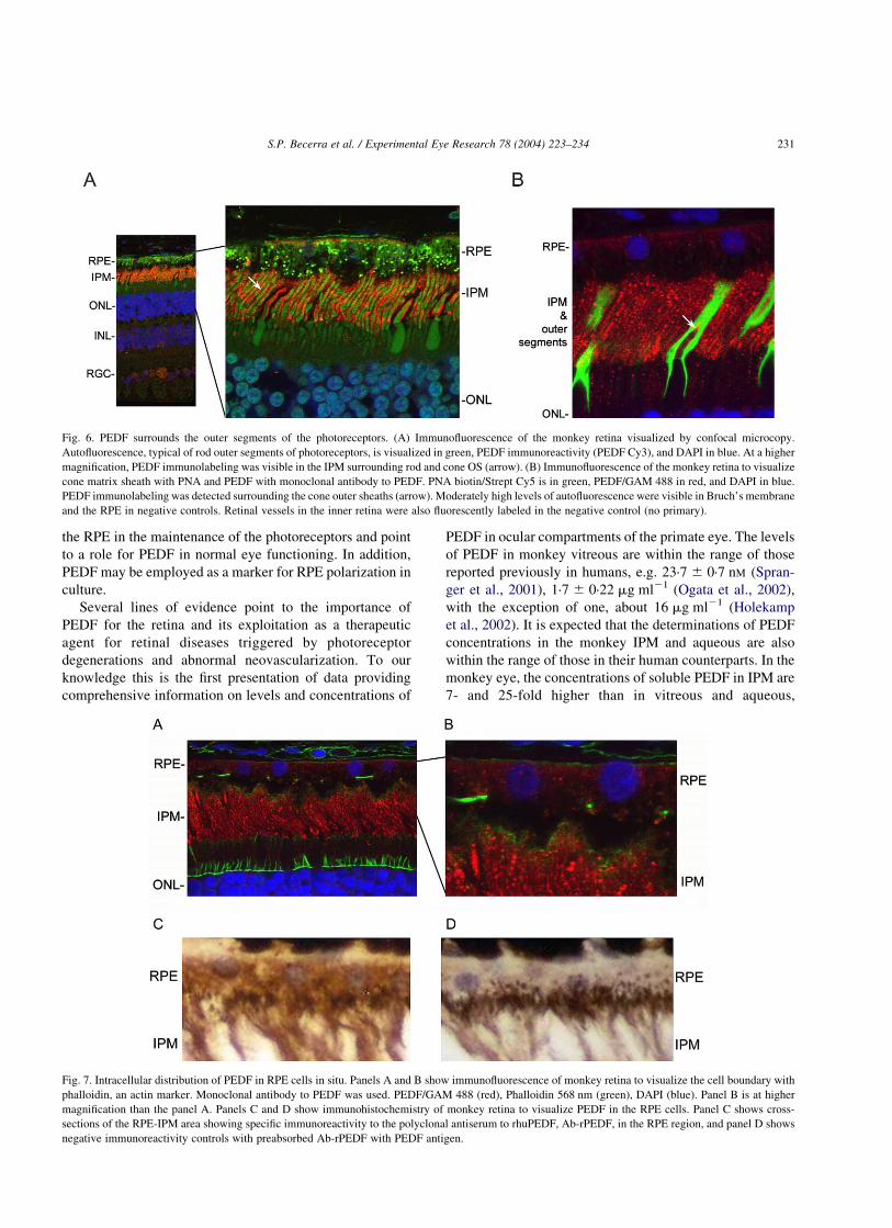
Fig. 6. PEDF surrounds the outer segments of the photoreceptors. (A) Immunofluorescence of the monkey retina visualized by confocal microcopy.
Autofluorescence, typical of rod outer segments of photoreceptors, is visualized in green, PEDF immunoreactivity (PEDF Cy3), and DAPI in blue. At a higher
magnification, PEDF immunolabeling was visible in the IPM surrounding rod and cone OS (arrow). (B) Immunofluorescence of the monkey retina to visualize
cone matrix sheath with PNA and PEDF with monoclonal antibody to PEDF. PNA biotin/Strept Cy5 is in green, PEDF/GAM 488 in red, and DAPI in blue.
PEDF immunolabeling was detected surrounding the cone outer sheaths (arrow). Moderately high levels of autofluorescence were visible in Bruch’s membrane
and the RPE in negative controls. Retinal vessels in the inner retina were also fluorescently labeled in the negative control (no primary).
Fig. 7. Intracellular distribution of PEDF in RPE cells in situ. Panels A and B show immunofluorescence of monkey retina to visualize the cell boundary with
phalloidin, an actin marker. Monoclonal antibody to PEDF was used. PEDF/GAM 488 (red), Phalloidin 568 nm (green), DAPI (blue). Panel B is at higher
magnification than the panel A. Panels C and D show immunohistochemistry of monkey retina to visualize PEDF in the RPE cells. Panel C shows cross-
sections of the RPE-IPM area showing specific immunoreactivity to the polyclonal antiserum to rhuPEDF, Ab-rPEDF, in the RPE region, and panel D shows
negative immunoreactivity controls with preabsorbed Ab-rPEDF with PEDF antigen.
S.P. Becerra et al. / Experimental Eye Research 78 (2004) 223–234 231

respectively, similar to the concentrations previously
reported in the bovine eye (Wu et al., 1995; Wu and
Becerra, 1996). These values represent the basal levels of
this interesting neurotrophic and antiangiogenic factor in the
primate eye. Changes in PEDF levels in the eye with
angiogenic retina or retinal degenerations may contribute to
the development or progression of these disorders. Evidence
for an association between decreased PEDF and induced or
diseased-related angiogenesis (Spranger et al., 2001; Gao
et al., 2002; Ogata et al., 2002; Holekamp et al., 2002)
makes the present information on physiological PEDF
levels more valuable for diagnostic and therapeutic studies.
Our immunolabeling studies of monkey retina demon-
strate the localization of PEDF in the IPM, and at low to
moderate levels in the RPE cytoplasm and retinal ganglion
cells. These results were obtained with a monoclonal anti-
PEDF antibody that recognized a single band on westerns of
monkey retinal and RPE extracts. Using the same
monoclonal anti-PEDF antibody, Karakousis et al. (2001)
reported PEDF-immunolabeling in the IPM of adult human
retina. However, they were unable to detect PEDF-
immunolabeling in the RPE due to high levels of lipofuscin
autofluorescence in their samples. Using a polyclonal
antibody to PEDF, they reported widespread distribution
of this protein in photoreceptor inner and outer segments,
rod nuclei, rod and cone synapse, a subset of cells in the
inner nuclear layer and ganglion cell layer. However, this
polyclonal antibody was shown by these authors to label
multiple bands migrating slower and faster than PEDF on
westerns of human retinal extracts. PEDF localization in the
IPM has been reported previously in weanling rats (Becerra
et al., 1999). Thus, the distribution of PEDF in the monkey
IPM is in agreement with those previously reported for other
species, and the present study provides new information on
the distribution of PEDF in the RPE.
We have established a polarized RPE cell culture that
expresses, synthesizes and produces high levels of PEDF.
Expressed in comparative quantitative terms, 2·6 mg of
purified monkey PEDF protein can be obtained from one
liter of RPE conditioned media, vitreous from 2167 eyes or
IPM lavage from 15 600 eyes. The average concentration of
PEDF protein in the culture medium of RPE cells attached
to plastic after 8 days in culture reached 6·5 mg ml21, a high
value obtained from primary cultures. Considering the
number of cells cultured we estimate that the media has
1·1 pg of PEDF per RPE cell. Note that in polarized cells,
the steady-state levels at the apical side were maintained
during the course of the study of 63 days. Thus, very
stringent conditions for culturing these cells are optimal for
high expression of the PEDF gene and secretion of the
protein.
The data reported here also provide biochemical,
structural, immunological and biological evidence that the
protein purified from monkey sources is PEDF. These
observations suggest that this protein is the monkey
homologue of the previously described human, bovine and
mouse PEDF that contains the structural determinants for
biological function. Because both humans and monkeys are
within the same taxonomic order, it is expected that monkey
and human PEDF share a high degree of sequence
similarity. In this regard, the monkey PEDF shares with
the human the lack of a tryptic site in the serpin-exposed
loop, which is present in the bovine PEDF. The tryptic
recognition site in the bovine PEDF is an arginine in
position P2, which in the human PEDF is occupied by a
histidine and is likely to be conserved in the monkey, thus
losing the tryptic site. However, it will be necessary to
isolate and sequence cDNA clones for monkey PEDF to
provide evidence on the degree of identity between the full-
length monkey and human PEDF, as well as among other
species.
Our data offer interesting possibilities on regulation of
PEDF. The polarization of RPE may be an important
mechanism to control PEDF secretion, as is the rate of
PEDF gene expression and PEDF protein synthesis. The
data obtained so far on the action of PEDF on the retina
may have not considered the polar secretion of PEDF by
RPE. Regulation of RPE polarization by extracellular
stimulus or signaling molecules from the choroid or neural
retina, may affect the levels of PEDF secreted by the RPE.
The fact that extracellular molecules or oxygen can switch
the polarization of RPE from apical to basal and vice versa
(Mousa et al., 1999; Blaauwgeers et al., 1999; Koh, 2000)
suggests the possibility of the existence of a stimulus that
could specifically target the change of polarity for PEDF
secretion to the choroid (basal side) or increase the
secretion to the IPM (apical side), thus increasing PEDF
intrinsic activities for the choroid or neural retina. Another
possibility for regulation might be at the secretion level. In
this regard the apicolateral secretion of PEDF and specific
distribution within the IPM entail the possibility of co-
release and targeting in association with the glycosami-
noglycans also synthesized by the RPE. For example, the
non-sulfated glycosaminoglycan hyaluronan, with binding
affinity for PEDF (Becerra et al., 1998), is also secreted in
a polar fashion to the apical side of human fetal RPE cells
in culture and is distributed to the IPM (deS Senanayake,
2001), suggesting an association of these two molecules
intracellularly and secretion in concert. Thus, the loss or
increase of glycosaminoglycan expression could affect the
amount of PEDF secretion. In the IPM, the glycosami-
noglycans may serve as a depository matrix for PEDF
located in proximity to the PEDF-receptor in photoreceptor
cells, and selective glycosaminoglycans can be positive
modulators of the interactions between PEDF and its
receptors (Alberdi et al., 2003) by increasing its activity.
Finally, the structural determinants for secretion, identified
in a recent mutagenic study at a region of the serpin
exposed loop of human PEDF (Shao et al., 2003), would
recognize the secretory components polarized to the apical
membrane of the RPE cells. The existence of natural PEDF
variants with mutations or deletions of this region would
S.P. Becerra et al. / Experimental Eye Research 78 (2004) 223–234232
have a decrease in secretion rate, resulting in a decrease in
antiangiogenic and neurotrophic activity in the IPM. These
possibilities are discussed under the consideration that
some of them may have important implications if PEDF, or
molecules derived from it, are to be used in the future as
therapeutic agents.
In conclusion, the observation that RPE cells secrete
PEDF in a polarized fashion towards the neural retina may
represent a mechanism to prevent damage to the adjacent
fragile retinal tissue. This is consistent with the known
trophic function of the RPE in the maintenance of the
photoreceptors and points to a role for PEDF in normal
eye functioning. However, loss of polarity of PEDF
production may play a role in the pathogenesis of retinal
degenerations or/and choroidal or retinal
neovascularization.
Acknowledgements
We thank Dr Steve Bernstein for help in the dissection of
the ocular tissues from monkey eyes, Dr Vicente Notario for
help in the biological assay, Ms Patricia Spinella for
performing amino acid sequencing, and Drs Barbara
Wiggert and Juan Amaral for interesting discussions and
proof reading the manuscript.
References
Adler, A., 1989. Selective presence of acid hydrolases in the interphotor-
eceptor matrix. Exp. Eye Res. 49, 1067–1077.
Alberdi, E., Hyde, C.C., Becerra, S.P., 1998. Pigment epithelium-derived
factor (PEDF) binds to glycosaminoglycans: analysis of the binding
site. Biochemistry 37, 10643–10652.
Alberdi, E., Aymerich, M.S., Becerra, S.P., 1999. Binding of pigment
epithelium-derived factor (PEDF) to retinoblastoma cells and cerebellar
granule neurons. J. Biol. Chem. 274, 31605–31612.
Alberdi, E.M., Weldon, J.E., Becerra, S.P., 2003. Glycosaminoglycans in
human retinoblastoma cells: heparan sulfate, a modulator of the
pigment epithelium-derived factor-receptor interactions. Biochemistry
4, 1.
Altschul, S.F., Madden, T.L., Schaffer, A.A., Zhang, J., Zhang, Z., Miller,
W., Lipman, D.J., 1997. Gapped BLAST and PSI-BLAST: a new
generation of protein database search programs. Nucleic Acids Res. 25,
3389–3402.Review.
Aymerich, M.S., Alberdi, E.M., Martinez, A., Becerra, S.P., 2001.
Evidence for pigment epithelium-derived factor receptors in the neural
retina. Invest. Ophthalmol. Vis. Sci. 42, 3287–3293.
Becerra, S.P., Alberdi, E., Martinez, A., Montuenga, L.M., Cayouette, M.,
Gravel, C., 1999. Pigment epithelium-derived factor (PEDF) in the
retina: Protective effect against photoreceptor cell degeneration. In:
Hollyfield, J.G., et al. (Eds.), In Retinal Degenerative Diseases and
Experimental Therapy, Kluwer Academic/Plenum, New York, pp.
519–526.
Becerra, S.P., Hollyfield, J.G., Iza-Azcarate, I., Perez-Mediavilla, L.A.,
1998. Pigment epithelium-derived factor (PEDF) has binding affinity
for hyaluronan. Invest. Ophthalmol. Vis. Sci. 40, 42.
Becerra, S.P., Palmer, I., Kumar, A., Steele, F., Shiloach, J., Notario, V.,
Chader, G.J., 1993. Overexpression of pigment epithelium-derived
factor in Escherichia coli. A functionally active neurotrophic factor.
J. Biol. Chem. 268, 23148–23156.
Becerra, S.P., Sagasti, A., Spinella, P., Notario, V., 1995. Pigment
epithelium-derived factor behaves like a noninhibitory serpin. Neuro-
trophic activity does not require the serpin reactive loop. J. Biol. Chem.
270, 25992–25999.
Becerra, S.P., 1997. Structure–function studies on PEDF. A noninhibitory
serpin with neurotrophic activity. Adv. Exp. Med. Biol. 425, 223–237.
Bilak, M.M., Becerra, S.P., Vincent, A.M., Moss, B.H., Aymerich, M.S.,
Kuncl, R.W., 2002. Identification of the neuroprotective molecular
region of pigment epithelium-derived factor and its binding sites on
motor neurons. J. Neurosci. 22, 9378–9386.
Blaauwgeers, H.G., Holtkamp, G.M., Rutten, H., Witmer, A.N., Koolwijk,
P., Partanen, T.A., Alitalo, K., Kroon, M.E., Kijlstra, A., van Hinsbergh,
V.W., Schlingemann, R.O., 1999. Polarized vascular endothelial
growth factor secretion by human retinal pigment epithelium and
localization of vascular endothelial growth factor receptors on the inner
choriocapillaris. Evidence for a trophic paracrine relation. Am. J. Pathol.
155, 421–428.
Bouck, N., 2002. PEDF: anti-angiogenic guardian of ocular function.
Trends Mol. Med., 330–334. Review.
Cao, W., Tombran-Tink, J., Elias, R., Sezate, S., Mrazek, D., McGinnis,
J.F., 2001. In vivo protection of photoreceptors from light damage by
pigment epithelium-derived factor. Invest. Ophthalmol. Vis. Sci. 42,
1646–1652.
Cayouette, M., Smith, S.B., Becerra, S.P., Gravel, C., 1999. Pigment
epithelium-derived factor delays the death of photoreceptors in mouse
models of inherited retinal degenerations. Neurobiol. Dis. 6,
523–532.
Crawford, S.E., Stellmach, V., Ranalli, M., Huang, X., Huang, L., Volpert,
O., De Vries, G.H., Abramson, L.P., Bouck, N., 2001. Pigment
epithelium-derived factor (PEDF) in neuroblastoma: a multifunctional
mediator of Schwann cell antitumor activity. J. Cell. Sci. 114,
4421–4428.
Dawson, D.W., Volpert, O.V., Gillis, P., Crawford, S.E., Xu, H., Benedict,
W., Bouck, N.P., 1999. Pigment epithelium-derived factor: a potent
inhibitor of angiogenesis. Science 285, 245–248.
deS Senanayake, P., Calabro, A., Nishiyama, K., Hu, J.G., Bok, D.,
Hollyfield, J.G., 2001. Glycosaminoglycan synthesis and secretion by
the retinal pigment epithelium: polarized delivery of hyaluronan from
the apical surface. J. Cell. Sci. 114, 199–205.
Duh, E.J., Yang, H.S., Suzuma, I., Miyagi, M., Youngman, E., Mori,
K., Katai, M., Yan, L., Suzuma, K., West, K., Davarya, S., Tong,
P., Gehlbach, P., Pearlman, J., Crabb, J.W., Aiello, L.P.,
Campochiaro, P.A., Zack, D.J., 2002. Pigment epithelium-derived
factor suppresses ischemia-induced retinal neovascularization and
VEGF-induced migration and growth. Invest. Ophthalmol. Vis. Sci.
43, 821–829.
Dunn, K.C., Marmorstein, A.D., Bonilha, V.L., Rodriguez-Boulan, E.,
Giordano, F., Hjelmeland, L.M., 1998. Use of the ARPE-19 cell line as
a model of RPE polarity: basolateral secretion of FGF5. Invest.
Ophthalmol. Vis. Sci. 39, 2744–2749.
Fariss, R.N., Anderson, D.H., Fisher, S.K., 1990. Comparison of
photoreceptor-specific matrix domains in the cat and monkey retinas.
Exp. Eye Res. 51, 473–485.
Fariss, R.N., Apte, S.S., Olsen, B.R., Iwata, K., Milam, A.H., 1997. Tissue
inhibitor of metalloproteinases-3 is a component of Bruch’s membrane
of the eye. Am. J. Pathol. 150, 323–328.
Gao, G., Li, Y., Fant, J., Crosson, C.E., Becerra, S.P., Ma, J.X., 2002.
Difference in ischemic regulation of vascular endothelial growth factor
and pigment epithelium-derived factor in brown norway and sprague
dawley rats contributing to different susceptibilities to retinal
neovascularization. Diabetes 51, 1218–1225.
Holekamp, N.M., Bouck, N., Volpert, O., 2002. Pigment epithelium-
derived factor is deficient in the vitreous of patients with choroidal
neovascularization due to age-related macular degeneration. Am.
J. Ophthalmol. 134, 220–227.
S.P. Becerra et al. / Experimental Eye Research 78 (2004) 223–234 233

Holtkamp, G.M., Van Rossem, M., de Vos, A.F., Willekens, B., Peek, R.,
Kijlstra, A., 1998. Polarized secretion of IL-6 and IL-8 by human retinal
pigment epithelial cells. Clin. Exp. Immunol. 112, 34–43.
Houenou, L.J., D’Costa, A.P., Li, L., Turgeon, V.L., Enyadike, C., Alberdi,
E., Becerra, S.P., 1999. Pigment epithelium-derived factor promotes the
survival and differentiation of developing spinal motor neurons.
J. Comp. Neurol. 412, 506–514.
Karakousis, P.C., John, S.K., Behling, K.C., Surace, E.M., Smith, J.E.,
Hendrickson, A., Tang, W.X., Bennett, J., Milam, A.H., 2001.
Localization of pigment epithelium derived factor (PEDF) in develop-
ing and adult human ocular tissues. Mol. Vis. 7, 154–163.
Koh, S.M., 2000. VIP enhances the differentiation of retinal pigment
epithelium in culture: from cAMP and pp60(c-src) to melanogenesis
and development of fluid transport capacity. Prog. Retin. Eye Res. 19,
669–688.
Hale, I.L., Matsumoto, B., 1993. Resolution of subcellular detail in thick
tissue sections: immunohistochemical preparation and fluorescence
confocal microscopy. Meth. Cell. Biol., 289–324. Review.
Montuenga, L.M., Martinez, A., Miller, M.J., Unsworth, E.J., Cuttitta, F.,
1997. Expression of adrenomedullin and its receptor during embry-
ogenesis suggests autocrine or paracrine modes of action. Endocrin-
ology 138, 440–451.
Meyer, C., Notari, L., Becerra, S.P., 2002. Mapping the type I collagen-
binding site on pigment epithelium-derived factor. Implications for its
antiangiogenic activity. J. Biol. Chem. 277, 45400–454007.
Mousa, S.A., Lorelli, W., Campochiaro, P.A., 1999. Role of hypoxia and
extracellular matrix-integrin binding in the modulation of angiogenic
growth factors secretion by retinal pigmented epithelial cells. J. Cell.
Biochem. 74, 135–143.
Ogata, N., Nishikawa, M., Nishimura, T., Mitsuma, Y., Matsumura, M.,
2002. Unbalanced vitreous levels of pigment epithelium-derived factor
and vascular endothelial growth factor in diabetic retinopathy. Am.
J. Ophthalmol. 134, 348–353.
Ortego, J., Escribano, J., Becerra, S.P., Coca-Prados, M., 1996. Gene
expression of the neurotrophic pigment epithelium-derived factor in the
human ciliary epithelium. Synthesis and secretion into the aqueous
humor. Invest. Ophthalmol. Vis. Sci. 37, 2759–2767.
Padgett, L.C., Lui, G.M., Werb, Z., LaVail, M.M., 1997. Matrix
metalloproteinase-2 and tissue inhibitor of metalloproteinase-1 in the
retinal pigment epithelium and interphotoreceptor matrix: vectorial
secretion and regulation. Exp. Eye Res. 64, 927–938.
Perez-Mediavilla, L.A., Chew, C., Campochiaro, P.A., Nickells, R.W.,
Notario, V., Zack, D.J., Becerra, S.P., 1998. Sequence and expression
analysis of bovine pigment epithelium-derived factor. Biochim.
Biophys. Acta 1398, 203–214.
Pfeffer, B.A., 1990. Improved methodology for cell culture of human and
monkey retinal pigment epithelium. In: Osborne, N.N., Chader, G.J.
(Eds.), Progress in Retinal Research, vol. 10. Pergamon Press, Oxford,
UK, pp. 251–291.
Pfeffer, B., Wiggert, B., Lee, L., Zonnenberg, B., Newsome, D., Chader, G.,
1983. The presence of a soluble interphotoreceptor matrix retinol-
binding protein (IRBP) in the retinal interphotoreceptor space. J. Cell.
Physiol. 117, 333–341.
Pignolo, R.J., Cristofalo, V.J., Rotenberg, M.O., 1993. Senescent WI-38
cells fail to express EPC-1, a gene induced in young cells upon entry
into the G0 state. J. Biol. Chem. 268, 8949–8957.
Shao, H., Schvartz, I., Shatiel, S., 2003. Secretion of pigment epithelium-
derived factor: mutagenic study. Eur. J. Biochem. 270, 822–831.
Spranger, J., Osterhoff, M., Reimann, M., Mohlig, M., Ristow, M., Francis,
M.K., Cristofalo, V., Hammes, H.P., Smith, G., Boulton, M., Pfeiffer,
A.F., 2001. Loss of the antiangiogenic pigment epithelium-derived
factor in patients with angiogenic eye disease. Diabetes 50, 2641–2645.
Steele, F.R., Chader, G.J., Johnson, L.V., Tombran-Tink, J., 1993. Pigment
epithelium-derived factor: neurotrophic activity and identification as a
member of the serine protease inhibitor gene family. Proc. Nat. Acad.
Sci. USA 90, 1526–1530.
Stellmach, V.V., Crawfordm, S.E., Shou, W., Bouck, N., 2001. Prevention
of ischemia-induced retinopathy by the natural ocular antiangiogenic
agent pigment epithelium-derieved factor. Proc. Nat. Acad. Sci. USA
98, 2593–2597.
Stratikos, E., Alberdi, E., Gettins, P.G., Becerra, S.P., 1996. Recombinant
human pigment epithelium-derived factor (PEDF): characterization of
PEDF overexpressed and secreted by eukaryotic cells. Protein Sci. 5,
2575–2582.
Tombran Tink, J., Chader, G.G., Johnson, L.V., 1991. PEDF: a pigment
epithelium-derived factor with potent neuronal differentiative activity.
Exp. Eye Res. 53, 411–414.
Tombran-Tink, J., Shivaram, S.M., Chader, G.J., Johnson, L.V., Bok, D.,
1995. Expression, secretion, and age-related downregulation of pigment
epithelium-derived factor, a serpin with neurotrophic activity.
J. Neurosci. 15, 4992–5003.
Vaughan, D.K., Fisher, S.K., 1987. The distribution of F-actin in cells
isolated from vertebrate retinas. Exp. Eye Res. 44, 393–406.
Wu, Y.Q., Notario, V., Chader, G.J., Becerra, S.P., 1995. Identification of
pigment epithelium-derived factor in the interphotoreceptor matrix of
bovine eyes. Protein Exp. Purif. 6, 447–456.
Wu, Y.Q., Becerra, S.P., 1996. Proteolytic activity directed toward
pigment epithelium-derived factor in vitreous of bovine eyes.
Implications of proteolytic processing. Invest. Ophthalmol. Vis. Sci.
37, 1984–1993.
Wong, P., Pfeffer, B.A., Bernstein, S.L., Chambers, M.L., Chader, G.J.,
Zakeri, Z.F., Wu, Y.Q., Wilson, M.R., Becerra, S.P., 2000. Clusterin
protein diversity in the primate eye. Mol. Vis. 6, 184–191.
S.P. Becerra et al. / Experimental Eye Research 78 (2004) 223–234234