plant basal resistance: genetics, biochemistry and impacts ...€¦ · plant basal resistance:...
TRANSCRIPT

Plant basal resistance: genetics, biochemistry and impacts on plant-biotic interactions
Shakoor Ahmad
Plant basal resistance: genetics, biochemistry and impacts on plant-biotic interactions
Shakoor Ahmad

Most of the research described in this thesis was conducted at the Department of Biological Chemistry and Crop Protection, Rothamsted Research, Harpenden, Herts, AL5 2JQ, United Kingdom and was funded by the UK Biotechnology and Biological Sciences Research Council (BBSRC) Institute Career Path Fellowship (grant no. BB/E023959/1) to J.T.
Printed by: Proefschriftmaken.nl || Printyourthesis.comLayout: Shakoor AhmadISBN: 978-90-8891-423-2
Plant basal resistance: genetics, biochemistry and impacts on plant-biotic interactions
Basale ziekteresistentie: genetica, biochemie en de effecten op plant-biotische interacties
(met een samenvatting in het Nederlands)
Proefschrift
ter verkrijging van de graad van doctor aan de Universiteit Utrecht op gezag van de rector magnificus, prof. dr. G. J. van der Zwaan, ingevolge het besluit van het college voor promoties
in het openbaar te verdedigen op maandag 18 juni 2012 des middags te 12.45 uur
door
Shakoor Ahmad
geboren op 14 april 1976te Bannu, Pakistan

Promotor: Prof. dr. Ir. C.M.J. Pieterse
Co-promotoren: Dr. J.Ton
Dr. R.Gordon-Weeks


Contents
Chapter 1 General introduction and thesis outline 9
Chapter 2 Genetic dissection of basal defence responsiveness in acces sions of Arabidopsis thaliana 31
Chapter 3 Benzoxaziniod metabolites regulates innate immunity against aphids and fungi in maize 59
Chapter 4 Benzoxaziniods in root exudates of maize attract Pseudomonas putida to the rhizosphere 85
Chapter 5 General discussion 107
References 123
Summary 139
Samenvatting 141
Acknowledgments 145
Curriculum vitae 147
List of publications 149


10
CHAPTER 1
General Introduction
Adapted from:Ahmad S, Gordon-Weeks R, Pickett J, Ton J.
Natural variation in priming of basal resistance: from evolutionary origin to agricultural exploitation. Molecular Plant Pathology 11(6), 817-827 (2010).

11
General introduction
THE PLANT IMMUNE SYSTEM
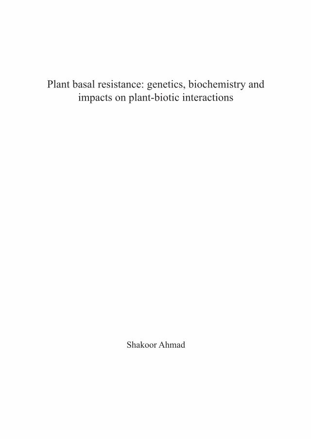
Plants are constantly interacting with potentially hostile organisms, such as viruses, bacteria, fungi, oomycetes, nematodes, and insects. Over the course of millions of years of co-evolution, these plant-microbe interactions have shaped the plant immune system. This regulatory system is complex and involves multiple inducible defence mechanisms, which are active at different stages of colonization by the plant attacker (Figure 1; Ton et al., 2009). To establish a successful parasitic interaction, pathogens and herbivores need to penetrate the host tissue. Plant viruses mostly depend on vectors to penetrate plant tissues, but many fungi, oomycetes and aphids can penetrate cell walls directly, whereas pathogenic bacteria often depend on natural openings, such as stomata or wound sites. At this stage of infection, a rapid closure of the stomata can form a first pre-invasive defence barrier (Melotto et al., 2008). After successful entry of the plant tissue, plant attackers often face an early-acting, post-invasive defence barrier that is marked by localized defence responses, such as the accumulation of reactive oxygen species, defence gene induction and deposition of callose-rich papillae (Eulgem et al., 1999; Flors et al., 2005; Torres et al., 2006). Upon further colonization, plants undergo a large-scale transcriptional and metabolic reprogramming that coincides with the biosynthesis of defence regulatory hormones, such as salicylic acid (SA) or jasmonic acid (JA), and complementary long-distance signals to regulate a broad spectrum of local and systemic defence mechanisms (Heil and Ton, 2008).
The default mode of the plant immune system: non-host resistance The most common type of disease resistance in plants is non-host resistance, which is effective against a very wide range of attackers and provides the most effective form of plant defence (Mysore and Ryu, 2004). Non-host resistance can result from the fact that the host plant simply does not provide the right environment for the attacking pathogen. However, in addition to this passive form of non-host resistance, research over the past decade has uncovered that non-host resistance also relies on active inducible defence mechanisms (Lipka et al., 2005). This non-host defence response is typically activated by conserved microbial features, such as flagellin, chitin, glycoproteins or lipopolysaccharides, which are referred to as “pathogen-associated molecular patterns” (PAMPs, synonymously called MAMPs for “microbe-associated molecular patterns”). Defence responses to herbivores can be triggered by herbivore-associated molecular patterns (HAMPs; Mithoefer and Boland, 2008), but are more commonly triggered by the perception of endogenous plant elicitors that are released upon tissue damage, which are called damage-associated molecular patterns (DAMPs; Heil, 2009; Heil et al., 2012). Much about the perception machinery of HAMPs remains unknown (Mithoefer and Boland, 2008). PAMPS and DAMPS are thought to be recognised by plasma membrane-localised pattern-recognition receptors

12
Chapter 1
Figure 1: Induced plant defence is a multi-layered phenomenon involving a multitude of defence mechanisms that are activated at different stages of the plant-microbe interaction. Upon first contact with a microbial pathogen, plants can express pre-invasive defence mechanisms. A well-known example is the rapid closure of stomata upon recognition of pathogen-associated molecular patterns (PAMPs). When the invading pathogen is capable of penetrating into the host tissue, it faces a second layer of inducible plant defences. This relatively early post-invasive defence is marked by accumulation of reactive oxygen species (ROS), often directly followed by deposition of callose-rich papillae. If the attacking pathogen is able to suppress and/or evade this early post-invasive defence barrier, it will encounter a third layer of inducible defences. This relatively late post-invasive defence is associated with the activation a wide range of defence mechanisms that are under control by de novo produced signalling hormones, such as salicylic acid. Late post-invasive defence is also associated with the generation of vascular long-distance signals that can prime systemic plant parts against upcoming pathogen attack. Red cells indicate defence-expressing cells and orange cells indicate those that are being successfully parasitized. The figure is adopted from Ton et al. (2009).
(PRRs; Gomez-Gomez and Boller, 2000; Scheer and Ryan, 2002; Huffaker et al., 2006; Miya et al., 2007). Although immune response triggered by these defence elicitors are commonly referred to as PAMP-triggered immunity (PTI), the term ‘pattern-triggered immunity’ would be more appropriate as it more collectively reflects responses to PAMPs, MAMPs, DAMPs and HAMPs. The PTI response is associated with a wide range of quickly activated defence mechanisms, such as localized callose deposition, reactive oxygen species accumulation and single cell death responses (Schwessinger and Zipfel, 2008).

13
General introduction
Co-evolution between virulent pathogens and plant immunity “zigzags” between basal resistance and effector-triggered immunityAs mentioned above, PTI is a nonspecific defence response and is able to stop the majority of hostile microbes. Virulent pathogens, however, have evolved the ability to suppress PTI through the use of pathogen effectors (Jones and Dangl, 2006). This effector-triggered susceptibility (ETS) reduces the efficiency of the plant immune response to basal resistance, which is insufficient to provide effective protection against disease. To counteract ETS, selected plant varieties have evolved resistance (R) proteins, which can detect pathogen effectors directly, or can guard the targets of pathogen effectors, thereby indirectly recognizing the activity of effectors (McDowell and Woffenden, 2003). Activation of R proteins often gives rise to a hypersensitive response (HR) that can block virulent pathogens at relatively early stages of infection. This so called effector-triggered immunity (ETI) is extremely effective against biotrophic pathogens and has, therefore, been studied extensively over the past decades. However, a major limitation of ETI is that it only protects against specific races of biotrophic pathogens (Lukasik and Takken, 2009), whereas it can be ineffective or even disease-promoting in response to necrotrophic pathogens (Kliebenstein and Rowe, 2008). Moreover, avirulent biotrophs are under constant selective pressure to break ETI, which limits the durability of this defence strategy. Pathogens can break ETI by evolving alternative effectors that suppress ETI, or that are no longer recognized by R proteins (Abramovitch et al., 2006; Fu et al., 2007; Cui et al., 2009; Houterman et al., 2009). Consequently, ETI is reverted to basal resistance, thereby imposing further selection pressure on the host plant to evolve improved R proteins that are capable of recognising the newly evolved effectors. The resulting arms race between plants and their (a)virulent pathogens manifests as an on-going oscillation in the effectiveness of plant defence and is referred to as the “zigzag” model (Jones and Dangl, 2006).
PRIMING OF DEFENCE: AN ALTERNATIVE DEFENCE STRATEGY TO COPE WITH BIOTIC STRESS
Although ETI can be extremely effective against biotrophic pathogens, plants can also counteract pathogens through a sensitization of their basal immune system. This priming of defence causes a faster and stronger induction of defensive mechanisms upon subsequent attack (Conrath et al., 2006; Frost et al., 2008). Priming of defence, also known as sensitization of defence, is a physiological state that enables plants to respond to a low level of environmental stress in a more efficient manner (Conrath, 2011). Similar to PTI, priming of defence is effective against a broad spectrum of plant attackers, suggesting that primed resistance is at least partially based on an augmented expression of PTI mechanisms. However, some forms of defence priming have also been shown to reduce lesion formation

14
Chapter 1
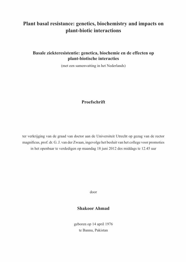
by avirulent pathogens (Ross, 1961; Hoffland et al., 1996), suggesting that priming can boost both PTI and ETI mechanisms. Since basal resistance has been defined as the sum of resistance by PTI and ETI, minus the susceptibility by ETS (Jones and Dangl, 2006), priming of defence can best be defined as an augmented capacity to express basal resistance mechanisms (Figure 2A). If the augmented basal defence response precedes the delivery of pathogen effectors, priming can provide full immunity against otherwise virulent pathogens (Figures 2B). Indeed, this has been reported for some forms of chemically-induced priming (Zimmerli et al., 2000; Conrath et al., 2006). In most cases, however, primed defence expression slows down the colonisation by virulent pathogens to a larger extent than basal resistance (Conrath et al., 2006). Most priming-inducing stimuli can trigger defence mechanisms directly if applied in higher doses. For instance, relatively high soil-drench concentrations of beta-aminobutyric acid (BABA) trigger PR-1 gene induction directly in Arabidopsis, whereas lower concentrations of BABA merely prime the induction of PR-1 (Van Hulten et al., 2006). Furthermore, transient induction of direct defence can give rise to longer-lasting priming of defence (Bruce et al., 2007; Heil and Ton, 2008). Hence, many induced resistance phenomena are based on a combination of direct defence and priming and their relative contribution depends on the dose of the resistance inducing stimulus and the time point after induction.
Biologically induced priming of defencePriming of defence can be induced by various biological agents and is often expressed in plant parts distal from the initial site of stimulation. For example, localised attack by pathogenic microbes can elicit a broad-spectrum systemic acquired resistance (SAR) response that is associated with priming of defence responses (Kohler et al., 2002; Conrath et al., 2006; Jung et al., 2009; Conrath, 2011). SAR is triggered by localised pathogen attack and develops in uninfected distal parts of the plant as against a broad spectrum of pathogens (Durrant and Dong, 2004). During this process, leaves/tissue under pathogen attack produce a systemic signal which is transported to uninfected distal plant parts, where it primes the tissues for SA-dependent defences (Jung et al., 2009). The 1st systemic study of SAR in Nicotiana benthamiana demonstrated that the phenomenon lasts for up to 20 days after primary infection (Ross, 1961). Studies in the following decades have mostly focused on the onset of SAR, which requires accumulation of plant stress hormone, SA and an intact NPR1 protein (Durrant and Dong, 2004). More recent studies have revealed that SAR establishment requires additional signals, which precede systemic accumulation of SA, such as jasmonates (Truman et al., 2007) and indole-derived metabolites (Truman et al., 2010). The exact nature of the mobile SAR signal, however, remains debatable, even within the same Arabidopsis-based pathosystem (Attaran et al., 2009). Apart from MeSA (Vlot et al., 2008), glycerolipids (Chaturvedi et al., 2008), azelaic acid (Jung et al., 2009), and glycerol-3-phosphate (Chanda

15
General introduction
Figure 2: Priming of basal resistance provides protection against virulent pathogens. (A) Basal resistance against virulent pathogens results from a residual level of host defence after defence suppression by disease-promoting pathogen effectors (blue arrows). Priming of basal resistance leads to a faster and stronger induction of basal defence mechanisms, providing enhanced resistance against the invading pathogen. In most cases, priming of basal resistance cannot prevent the delivery of pathogen effectors entirely, and thereby only slows down the introgression of the pathogen (‘moderately primed defence response’). However, if the primed defence response precedes the delivery of pathogen effectors, this defence strategy can prevent pathogen infection and provide full protection against otherwise virulent pathogens (‘strongly primed defence response’). Red plant cells indicate the expression of basal defence mechanisms. (B) The ‘zigzag’ model describes basal resistance as the sum of pathogen-associated molecular pattern (PAMP)-triggered immunity (PTI) and weak effector-triggered immunity (ETI) minus effector-triggered susceptibility (ETS) (Jones and Dangl, 2006). Apart from newly evolved R proteins that recognize effectors or their activities, ETS can be counteracted by the priming of defence, causing faster and stronger induction of basal defence mechanisms after pathogen attack. A moderately primed defence response merely augments the PTI/ETI response, but would still allow ETS to take place (shown in orange), whereas a strongly primed defence reaction can prevent ETS entirely (shown in red).
et al., 2011) have been reported to act as mobile signals. As a possible explanation for this controversy, Liu et al. (2011) recently suggested that SAR is mediated by an interaction between two mobile signals: MeSA and a complex formed between the lipid transfer protein DIR1 and glycerolipid and/or lipid derivatives. Hence, the signalling pathways controlling systemic defence priming during SAR are mediated by complex signalling networks.
16
Chapter 1
Selective non-pathogenic root-colonizing microbes can induce systemic defence priming as well (Van Wees et al., 2008). The resulting disease resistance is commonly referred to as induced systemic resistance (ISR). For instance, non-pathogenic rhizobacteria can prime Arabidopsis against a wide range of plant pathogens, like bacteria, oomycetes, fungi, viruses and even herbivores (Pieterse et al., 1996; Ton et al., 2002; Van Wees et al., 2008). A more recent example by (Verhagen et al., 2010) showed that beneficial bacteria such as Pseudomonas fluorescens CHA0 and Pseudomonas aeruginosa 7NSK2 mediate ISR in grapevine through potentiating oxidative burst and phytoalexin production (i.e. resveratrol and viniferin) after attack by Botrytis cinerea. Arbuscular micorhizal fungi (AMF) can also induce defence priming in plants (Pozo and Azcón-Aguilar, 2007; Pozo et al., 2009). The interaction between Arabidopsis and Pseudomonas fluorescens WCS417r has served as a biological model system to study the signalling transduction pathways controlling ISR. Research on this model system revealed P. fluorescens WCS417r–mediated ISR in Arabidopsis requires responsiveness to jasmonate and ethylene (ET) and is dependent on NPR1 (Pieterse et al., 1998). A transcriptome analysis for P. fluorescens WCS417r-inducible genes led to the identification of the ISR responsive transcription factor gene MYB72 (Verhagen et al., 2004). Subsequent analysis of mutant lines in MYB72 revealed that this transcription factor gene plays a critical role for the early signalling events leading to elicitation of the systemic signal (Van der Ent et al., 2008). The same authors reported that P. fluorescens WCS417r–mediated priming in Arabidopsis thaliana against Hyaloperonospora arabidopsidis, an oomycete pathogen that is unaffected by JA- and ET-dependent defences (Thomma et al., 1998; Ton et al., 2002), is based on priming of callose deposition, which requires intact abscisic acid (ABA) signalling (Van der Ent et al., 2009). Apart from defence priming against pathogens, systemic defence priming can also be effective against herbivore attack. When plants are subjected to damage by herbivorous insects, they emit a complex blend of airborne chemical signals, known as volatile organic compounds (VOCs). VOCs serve primarily to attract natural enemies of the herbivore (Turlings and Ton, 2006), but they have also been shown to induce defence priming in systemic tissues and even neighbouring plants (Engelberth et al., 2004; Heil and Silva Bueno, 2007; Ton et al., 2007; Heil and Ton, 2008). Three green leaf volatiles, (Z)-3-hexenal, (Z)-3-hexen-1-ol, and (Z)-3-hexenyl acetate, in particular have been linked to elicitation of defence priming by herbivore-induced VOCs (Engelberth et al., 2004). VOC-induced priming augments JA-inducible defences (Frost et al., 2008). Interestingly, however, only a sub-set of JA-dependent genes is responsive to priming by VOCs in maize (Ton et al., 2007). The latter observation suggests that priming-inducing VOCs target specific components in the JA response pathway.

17
General introduction
Chemically induced defence primingInduced resistance by pathogens, rhizobacteria and herbivores can be mimicked by selective chemical agents (Oostendorp et al., 2001). Table I lists an overview of publications reporting defence priming in response to exogenous application of chemical compounds. In addition to the pathogen-, herbivore-, or damage associated patterns triggering the above-mentioned biological priming responses, defence priming can also be mimicked by endogenous plant signalling metabolites, such as JA, SA, and functional analogues thereof (Kauss et al., 1994; Mur et al., 1996; Kohler et al., 2002). Treatment with thiamine (Vitamin B1) has been reported to prime Arabidopsis, causing augmented accumulation of hydrogen peroxide (H2O2), callose-rich papillae, and PR-1 transcript following pathogen infection (Ahn et al., 2007). Recently, cytokinins have emerged as another plant-endogenous priming signal. These plant hormones control specification of cells, maintenance of meristematic cells, shoot formation and development of plant vasculature, but recent evidence suggests that these compounds also regulate defence in plants (Choi et al., 2011). Interestingly, the defensive targets of cytokinins seem to depend on the plant species under investigation. Whereas cytokinins prime for JA-controlled defences in poplar (Dervinis et al., 2010), they prime for SA-dependent gene expression in Arabidopsis (Choi et al., 2011), and they were recently reported to control pathogen-induced biosynthesis of JA- and SA-independent phytoalexins in tobacco (Großkinsky et al., 2011). Finally, azelaic acid has been shown to act as a long-distance priming signal during the onset of SAR. Upon exogenous application, this dicarboxylic acid primes Arabidopsis for SA-dependent defences and confers systemic resistance against P. syringae (Jung et al., 2009). There are also xenobiotic chemicals that can trigger defence priming in plants. For instance, the chemical Probenazole (PBZ; 3-allyloxy-1,2-benzisothiazole-1,1-dioxide), which is the active ingredient in Oryzemate, has been used widely in Asia to induce resistance in rice against Magnaporthe grisea. Its mode of action relies on enhanced biosynthesis of SA (Yoshioka et al., 2001; Iwai et al., 2007), which by itself serves as an endogenous priming signal. PBZ is metabolised by plants into saccharin (1,2-benzisothiazole-1,1-dioxide), a compound that is best known for its application as an artificial sweetener. However, saccharin can also induce resistance in plants against various diseases (Oostendorp et al., 2001; Boyle and Walters, 2006; Srivastava et al., 2011). In barley, saccharin induces resistance to powdery mildew fungus, which is associated with priming of cinnamyl alcohol dehydrogenase activity (Walters et al., 2008). Another xenobiotic chemical capable of inducing resistance through defence priming is BABA. Application of this non-protein amino acid protects against an exceptionally broad spectrum of plant diseases (Jakab et al., 2001), including crop diseases that are difficult to control by conventional strategies of disease management, such as late blight disease (Liljeroth et al., 2010). BABA is active at relatively low concentrations and acts in an enantiomer-specific manner (Cohen, 2002). These findings suggest that BABA mimics

18
Chapter 1
an endogenous plant signalling metabolite, or that it activates a plant regulatory protein controlling multiple immune responses simultaneously. Indeed, research on BABA-induced defence priming in Arabidopsis revealed that BABA not only mimics SAR-related priming of SA-dependent defences, but it also primes for pathogen-induced deposition of callose-containing papillae (Zimmerli et al., 2000; Ton et al., 2005). This priming of cell wall defence functions independently of SA and JA, but requires intact biosynthesis and perception of the plant hormone ABA (Ton and Mauch-Mani, 2004; Van der Ent et al., 2009)
Table I. Chemicals that trigger priming of defence in plants after exogenous application.
Chemical Stimulus Primed defence response Plant Species Reference Benzothiadiazole
(BTH)PAL gene induction Arabidopsis (Kohler et al., 2002)
Probenazole SA-inducible genes Rice (Iwai et al., 2007)
Saccharin Cinnamyl alcohol dehydro-genase activity
Barley (Boyle and Walters, 2006)
Beta amino butyric acid (BABA)
SA-inducible genes and callose deposition
Arabidopsis (Zimmerli et al., 2000; Ton and Mauch-Mani, 2004)
Thiamine (Vitamin B1)
ROS accumulation, cal-lose deposition, and SA-
induced expression
Arabidopsis (Ahn et al., 2007)
Cytokinins SA-inducible genes Arabidopsis (Choi et al., 2011)
JA-inducible genes Poplar (Dervinis et al., 2010)
Scopoletin and Capsidiol Tobacco (Großkinsky et al., 2011)
Azelaic acid SA-inducible genes Arabidopsis (Jung et al., 2009)
Quercetin ROS accumulation, callose deposition, and PR1 gene
induction
Arabidopsis (Jia et al., 2010)
Molecular mechanisms of primingIn contrast to research on innate plant defences that are directly responsive to pathogens and herbivores, the majority of research on priming of defence has remained limited to a description of the phenomenon after treatment with resistance-inducing agents, along with an assessment of its effectiveness in terms of disease resistance (Conrath et al., 2006; Frost et al., 2008). Only a few research groups have begun to address the mechanistic basis of defence priming (Conrath, 2011). Consequently, there are still many open questions about defence priming in plants, particularly with respect to the signalling mechanisms controlling the onset and long-term maintenance of the phenomenon.

19
General introduction
enhanced accumulation of signalling proteins. One common hypothesis postulates that priming is based on an increased accumulation of inactive defence signalling proteins, thereby providing enhanced defence signalling capacity (Conrath et al., 2006). Subsequent exposure to environmental stress would then lead to a faster and stronger defence signalling cascade, ultimately resulting in the augmented defence response. Indeed, SAR-related priming is associated with an increased accumulation of two inactive MAP protein kinases, MPK3 and MPK6, which show enhanced kinase activity upon secondary stress application (Beckers et al., 2009). Simultaneously, a genome-wide profiling of transcription factor (TF) genes was performed, which demonstrated that induction of ISR-related priming is associated with augmented expression of JA-regulatory TF genes (Van der Ent et al., 2009). The same study showed that BABA-induced priming was associated with enhanced expression of WRKY transcription factor genes, which encode transcription factors that regulate SA-induced defence gene transcription (Van Verk et al., 2011). Although enhanced accumulation of defence-related signalling TFs can contribute to a faster and stronger transcriptional activation of defence genes after pathogen attack, these signalling proteins typically have a limited half-life. Hence, their enhanced accumulation after application of a single priming stimulus does not provide a satisfactory explanation for the long-lasting nature of priming phenomena. epigenetic mechanisms. A recent study showed that SAR-related priming in Arabidopsis is associated with post-translational changes of histone H3 and H4 tails at gene promoters of defence-regulatory transcription factor genes (Jaskiewicz et al., 2011). Although these chromatin modifications were monitored relatively shortly after SAR induction, such epigenetic regulatory mechanism provides an attractive explanation for the long-lasting nature of the priming phenomenon. Indeed, recent evidence demonstrated that priming is an epigenetic phenomenon. Three independent research groups demonstrated that the primed defence state in Arabidopsis can be transmitted to following generations from iso-genic plant lines (Luna et al., 2012; Rasmann et al., 2012; Slaughter et al., 2012). Moreover, expression of trans-generational priming of SA-dependent defence genes was associated with chromatin remodelling at the corresponding gene promoters (Luna et al., 2012), whereas priming of JA-dependent defence requires intact biogenesis of small interfering RNAs (Rasmann et al., 2012). glycolylation of secondary metabolites. It is also conceivable that secondary metabolites contribute to long-lasting priming of defence. The chemical defence capacity of plants can be enhanced by an increased accumulation of inactive defence metabolite conjugates, such as glucosinolates and plant hormone-glucosides. Consequently, pathogen- or wounding-induced activity of hydrolytic glucosidase enzymes would lead to a faster and greater release of active aglycone metabolites. signalling cross-talk. Priming can also result from a shift in the cross-talk balance

20
Chapter 1
between defence signalling pathways. For instance, suppression of the SA-dependent pathway by mycorrhizal fungi results in a potentiation of JA-dependent defences (Pozo and Azcón-Aguilar, 2007), while trans-generational priming of the SA response in Arabidopsis coincides with a repression of JA response (Luna et al., 2012) Interestingly augmented levels of JA have been associated with primed callose deposition in grapevine against Plasmopara (Hamiduzzaman et al., 2005). Similarly priming of papillae formation was observed in the roots of mycorrhiza-infected tomatoes with Phytophthora (Cordier et al., 1998). These observations point to a mechanism by which suppression of the SA-response results in a beneficial side effect: systemic priming of JA-dependent defences and callose deposition. reduced scavenging capacity of reactive oxygen species. Several recent studies have pointed to an important role of reactive oxygen species (ROS) in priming of defence. Thiamine (vitamin B1) induces resistance against Pseudomonas syringae pv. tomato DC3000 (PstDC3000), which is associated with hydrogen peroxide (H2O2)-dependent priming of defence genes and callose deposition (Ahn et al., 2007). Vitamin B2 (riboflavin) induces a phenotypically similar resistance response that is associated with priming of ROS production, callose deposition and SA-inducible genes (Zhang et al., 2009). The plant secondary metabolite quercetin has also been demonstrated to induce SA- and NPR1-dependent resistance against PstDC3000, which is associated with augmented deposition of ROS, callose, PR1 and PAL gene transcripts (Jia et al., 2010). A recent study by Mukherjee et al. (2010) provided a plausible mechanism for ROS-dependent regulation of priming. The authors performed a phenotypic analysis of different alleles of the ascorbic acid deficient mutant vtc1 and demonstrated that the enhanced disease resistance of this mutant is based on priming of pathogen-induced accumulation of ROS, SA and NPR1 gene transcripts (Mukherjee et al., 2010). The authors suggested that the reduced ROS scavenging capacity of vtc1 causes constitutive priming of pathogen-induced H2O2, thereby causing augmented SA accumulation and enhanced defence induction.
Costs & benefits of primingThe full development of an inducible defence response requires energy and, therefore, involves costs on growth and reproduction. Apart from allocation costs, costs can also arise from toxicity of the defence to the plant’s own metabolism, or when the defence response affects the plant’s interaction with beneficial organisms (Heil, 2002). It is commonly accepted that plants only express inducible defences if the benefits (i.e. protection against the attackers) outweigh the associated costs (Heil, 2002; Walters and Boyle, 2005). Van Hulten et al. (2006) conducted a laboratory study to compare the costs and benefits of defence priming versus direct induction of defence in Arabidopsis. By using low doses of BABA to induce priming and high doses of either BABA or BTH to induce defence expression directly, it was found that priming is associated with relatively minor costs on plant growth and seed

21
General introduction
set. Moreover, the protective benefits of priming outweighed its costs under conditions of high disease pressure. It was thus concluded that priming is a cost-efficient defence strategy in disease-imposing environments. Interestingly, the outcome of this laboratory study was subsequently tested under agronomical field conditions by Walters et al. (2008), who subjected saccharin-primed barley to varying degrees of disease by the hemi-biotrophic fungus Rhynchosporium secalis and monitored fitness levels by plant growth and grain yield. As predicted, primed plants displayed significantly higher fitness than un-primed plants, thereby extending our laboratory demonstration that priming is a beneficial defence strategy in hostile environment.
PLANT DEFENCE STRATEGIES AND THEIR ADAPTIVE VALUES IN HOSTILE ENVIRONMENTS
Naturally occurring plant species can often be sub-divided into genetically distinct geographic varieties. Although these so-called ecotypes are similar enough to be considered as one species, they differ genetically in some traits due to variant selection pressures from their environments of origin. In this context, plant defence strategies can have adaptive values that vary according to the environmental conditions (Figure 3). The concept that priming of defence provides benefits in hostile environments suggests that plants in these environments are under pressure to evolve a constitutively enhanced responsiveness of basal defence mechanisms. Since priming protects against a wide variety of diseases and pests (Conrath et al., 2006), this selection pressure would be most pronounced under pressure by a wide range of different pathogens and herbivores (Figure 3; strategy A). There are, however, alternative defence strategies that could provide similar or even greater benefits, depending on the nature of the environment. For instance, PAMPs from plant-beneficial microbes have been demonstrated to trigger defensive responses (Van Loon et al., 2008), suggesting that plants with primed defence responsiveness to PAMPs may risk compromising their interaction with plant-beneficial micro-organisms. Indeed, various studies have reported negative impacts of SA-dependent resistance on rhizobial and mycorrhizal symbioses with legumes (Stacey et al., 2006; Jin et al., 2009; Faessel et al., 2010). Hence, there could be a counteracting selection against constitutive priming to maintain associations with mycorrhiza or N-fixing bacteria. Therefore, an increased ability to attract and interact with micro-organisms that are capable of suppressing pathogens directly through nutrient competition or antibiosis (Handelsman and Stabb, 1996; Weller et al., 2002), could be an alternative defence strategy in hostile environments (Figure 3; strategy B). In support of this, Rudrappa et al. (2008) demonstrated that Arabidopsis can attract disease-suppressing rhizobacteria through exudation of L-malic acid, which is further boosted by aboveground infection by P. syringae pv. tomato. Secondly, priming

22
Chapter 1
rarely provides complete protection against one pathogen or pathogen race, whereas ETI typically does. Hence, ETI would be more efficient in environments with disease pressure from one predominant pathogen species (Figure 3; strategy C). Thirdly, although priming is less costly than direct induction of defence, it is still associated with minor costs under
Figure 3: Model of plant defence strategies and their adaptive values under different biotic stress conditions. Plants in environments with relatively high disease pressure from a wide array of different attackers benefit from constitutive priming of basal resistance mechanisms, which provide broad-spectrum protection against pests and diseases (strategy A). However, this defence strategy may affect the plant’s ability to associate with plant-beneficial microbes, such as mycorrhizae, N-fixing bacteria or plant-growth promoting rhizobacteria. In this situation, plants would benefit more from an increased ability to attract and associate with plant-beneficial microbes with disease-suppressing traits (strategy B). Plants in environments with a constant pressure from one dominant biotrophic pathogen benefit from effector-triggered immunity (ETI; strategy C). ETI can be broken and give rise to an ongoing “zigzag” evolution, as described by Jones and Dangl (2006). Inducible defence priming upon perception of stress-indicating signals provides a cost-efficient adaptation to environments with variable degrees of disease pressure (strategy D). Because priming of defence and induction of defence are both associated with costs on plant growth and reproduction, a relatively un-responsive immune system would be beneficial in environments with relatively low disease pressure (strategy E).
23
General introduction
conditions of low disease pressure (Van Hulten et al., 2006). Consequently, plants exposed to variable levels of disease pressure would benefit from an inducible priming response (Figure 3; strategy D). As variable degrees of disease pressure are the reality in many natural plant environments, priming mainly manifests as an inducible resistance response. Finally, the selection for any of the above defence strategies is likely to be influenced by the plant’s abiotic environment. For example, the plant hormone ABA not only controls tolerance to abiotic stress, but also plays a multifaceted role in the fine-tuning of resistance to diseases and pests (Ton et al., 2009). Most plants are capable of expressing combinations of different defence strategies. The importance of each of these strategies depends on the environment. For instance, many plant-beneficial micro-organisms have the ability to protect plants through a combination of direct disease suppression and induction of defence priming in the host plant (ISR; Van Wees et al., 2008; Zamioudis and Pieterse, 2011). Colonisation by these microbes causes a constitutive level of systemic priming that is phenotypically similar to genetically acquired priming, thus combining the advantages of two defence strategies: direct disease suppression by plant-beneficial microbes and constitutive defence priming. Furthermore, the expression of one defence strategy can give rise to induction of another. For example, localised expression of PTI results in the development of SAR (Mishina and Zeier, 2007), which is largely based on priming of defence (Jung et al., 2009; Kohler et al., 2002).
Natural selection for constitutively primed basal resistance? Although the above examples justify the general conclusion that natural variation in responsiveness of basal resistance mechanisms is prevalent, this does not necessarily prove that hostile environments select for constitutively primed immune systems. In order to demonstrate that constitutive defence priming has evolved from inducible priming under constant levels of disease pressure, more evidence is required from both the molecular level and the plant community level (Shindo et al., 2007). For instance, patterns of single nucleotide polymorphisms in alleles contributing to natural variation in basal resistance could provide indications of past selective pressures. If the degree of nucleotide diversity deviates from the estimated diversity under neutral selection, this could be interpreted as evidence for environmental selection pressures. Typically, reduced levels of nucleotide polymorphisms indicate selective sweeps, during which newly evolved gene variants outcompete others (Nielsen, 2005). On the other hand, enhanced levels of nucleotide polymorphisms suggest balancing selection, which maintains ancient genetic variation (Mitchell-Olds and Schmitt, 2006). Although these methods provide useful indications for past selective forces on genes, fitness assays under different disease pressures would still be necessary to establish what gene variants provide which adaptive phenotypes. As was outlined by Holub (2007), a major challenge for the future is to apply currently available

24
Chapter 1
genetic resources for Arabidopsis (i.e. fully genotyped recombinant mapping populations or association mapping populations) to field experimentation. For instance, Arabidopsis mapping populations in which defence traits segregate could be grown under different field conditions with varying degrees of disease pressure by one or multiple pathogens and/or herbivores. Subsequent fitness evaluation may reveal defence-regulating QTLs that provide selective benefits under specified environmental conditions. With more and more Arabidopsis accessions being genome-sequenced, another promising approach arises from genome-wide association mapping approaches, which are based on associations between phenotypes and DNA sequence variants within individuals or isogenic populations (Nordborg and Weigel, 2008; Atwell et al., 2010). Particularly if defence phenotypes can be related to ecological stress parameters from the accessions’ geographical origins, this technique has the potential to assign measurable ecological significance to defence regulatory alleles.
SECONDARY DEFENCE METABOLITES: BIOSYNTHETIC ORIGINS AND CHEMICAL CLASSIFICATION
Irrespective of the type of resistance response expressed in plants, secondary metabolites are ubiquitous “tools” in plant defence. Figure 4 provides a generic overview of the biosynthesis pathways involved in the production of defence secondary compounds in plants. Unlike primary metabolites, which play a role in the process of photosynthesis, respiration, solute transport, nutrient assimilation and differentiation, secondary metabolites have no recognised role in plant processes that are essential for growth. The distribution of secondary metabolites across the plant kingdom is diverse and varies between plant species and taxa. Based on chemical structure, plant secondary metabolites can be divided into four major groups: terpenes, phenolics, nitrogen- and sulphur-containing compounds and oxyilipins. Figure 4 shows a generic overview of the different biochemical pathways controlling these plant compounds.
Terpenes This class of metabolites is immensely diverse and includes more than 30,000 lipophilic compounds (Kennedy and Wightman, 2011). Their structure includes one or more 5-carbon isoprene (C5H8) units, which are synthesized in plants by both the mevalonate and dexy-d-xylulose pathways (Rohmer, 1999). Classifications of terpenoids are based on the number of isoprene units they contain. Hemiterpenes incorporate 1 isoprene unit, monoterpenes incorporate 2 units, sesquiterpenes incorporate 3 units, diterpenes incorporate 4 units, sesterpenes comprise 5 units, triterpenes include 6 units, and tetraterpenes incorporate 8 units. Terpenes exhibit a broad range of ecological roles in the plant kingdom. Their roles include antimicrobial properties, attraction of pollinator, parasitoic or predator insects, and

25
General introduction
PhenolicsApproximately 10,000 plant secondary metabolites are known to contain phenolic ring structures (Kennedy and Wightman, 2011). These are derivatives of the pentose phosphate pathway, the shikimate pathway, and the phenylpropanoid pathway. Structurally, natural phenolic compounds from plants have at least 1 aromatic hydrocarbon ring with one or more hydroxyl groups attached. Phenolics range from simple low molecular weight compounds like phenylpropanoids, coumarins, and benzoic acid derivatives, to more complex structures like flavanoids, stilbenes, and tannins. Phenolics play diverse roles in plant defence, such as responding to bacteria/fungal attack, providing scent/colour/flavour to attract beneficial insects/deter herbivores, and acting as semiochemicals during interactions with plant-beneficial microbes (Treutter, 2006).
Sulphur and Nitrogen containing secondary metabolites This is a large group of secondary metabolites containing more than 15,000 molecules. They include alkaloids, cyanogenic glucosides, and non-protein amino acids. S- and N-containing
Figure 4: A simplified scheme of the major biosynthetic pathways controlling plant secondary metabolites with representative examples and structures from each class. Secondary metabolites are derived from different pathways: the shikimate pathway, the malonic acid pathway, mavalonic acid pathway, the MEP/DOXP (2-C-methyl-D-erythritol 4-phosphate/1-deoxy-D-xylulose 5-phosphate) pathway, and the oxylipin pathway. Based on structural characteristics, the resulting secondary metabolites can be divided into four main classes: nitrogen (N) - and sulphur (S) containing compounds, phenolic compounds, terpenes, and oxylipins.
activities as allelopathic chemicals (De Almeida et al., 2010; Martino et al., 2010).

26
Chapter 1
metabolites are biosynthesised from common amino acids. For example, N-containing alkaloids are usually synthesized from aspartic acid, lysine, tyrosine and tryptophan (Facchini, 2001) These compounds play important roles in plant-pest interactions in different plant families , such as Brassicaceae, Alliaceae and Asteraceae (Burow et al., 2008). Two well-known examples of S- and N-containing defence metabolites are glucosinolates in Brassicaceae and the Alliins in Alliaceae (Burow et al., 2008). S- and N-containing secondary metabolites offer an array of defence compounds that activate direct and/or indirect defences against a broad range of harmful microbes/insects.
OxylipinsOxylipins encompass a large family of oxygenated metabolites that are derived from fatty acids. Oxylipins are best known for their role in plant defence signalling pathways (Blée, 2002), and are produced by oxidation of fatty acids, mainly linolenic acid and linoleic acid, followed by secondary modification (Vicente et al., 2011). The oxylipin biosynthesis pathway converts linoleic acid or linolenic acid into hydroperoxide substrates, such as 9-HPOD (hydroperoxy-octadecadienoic acids), 9-HPOT (hydroperoxy-octadecatrienoic acids), 13-HPOT and 13-HPOD. These compounds are subsequently utilized by different pathway branches that are under control by HPL-hydroperoxide lyase, AOS-allene oxide synthase, DES-divinyl synthase, and P0X-peroxygenase, respectively (Figure 4). The AOS pathway generates the plant defence hormone JA, which is essential for activation of direct and indirect defences against necrotrophic pathogens and insects (Pozo et al., 2005). In addition to jasmonates, the oxylpin pathway produces antimicrobial leaf aldehydes or divinyl ethers and herbivore-induced volatiles, such as green leaf volatiles (Liavonchanka and Feussner, 2006).
SECONDARY DEFENCE METABOLITES: FUNCTIONAL CLASSIFICATION
The function of secondary metabolites in plant defence ranges from direct to indirect. Metabolites can act directly as anti-proliferative agents for pathogenic microorganisms (González-Lamothe et al., 2009). Secondary metabolites can also act as feeding deterrents against herbivores, during which the metabolites offer a bitter taste, or are directly toxic to the herbivore (Michael, 2003). On the other hand, secondary metabolites can contribute to plant defence indirectly, by stimulating the interaction with disease-suppressing organisms. This form of defence includes certain tritrophic interactions, where herbivore-infested plants emit volatile metabolites that attract natural enemies of the attacking herbivore (Turlings and Ton, 2006). Volatile metabolites also function to attract pollinating insects, which by themselves can have an herbivory-suppressing effect (Tautz and Rostas, 2008). On the basis of their activity, secondary defence metabolites can roughly be

27
General introduction
divided into three general classes: phytoanticipins and phytoalexins, which contribute to direct defence, and semiochemicals, which function in indirect defence. Phytoanticipins are constitutively produced and are commonly stored in the inactive glycosulated form, whereas phytoalexins are inducible defence metabolites that are synthesised de novo upon pathogen and/or insect attack (VanEtten et al., 1994). As is demonstrated in chapters III and IV of this thesis, some secondary metabolites fulfil multiple tasks in plant defence.
Phytoalexins Phytoalexins are low molecular weight secondary metabolites with antimicrobial properties and are synthesized in plants in response to environmental stress. Phytoalexins are widely distributed among crop species, have broad-spectrum antimicrobial effects, and are commonly used as biochemical markers for expression of plant defence (Ahuja et al., 2012). Camalexin (3-thiazol-2-yl-indole) is the major phytoalexins in Arabidopsis and is derived from tryptophan. Depending on attacking pathogen, different signalling pathways are involved in the activation of camalexin biosynthesis (Heck et al., 2003; Denby et al., 2005; Rowe et al., 2010). Camalexin biosynthesis itself is regulated via a MAPK signalling cascade (Ren et al., 2008; Xu et al., 2008). Mao et al., (2011) demonstrated that induction of camalexin is controlled by a MPK3- and MPK6-dependent signalling cascade via phosphorylation of the WRKY33 transcription factor, which in turn binds to the promoter of the camalexin biosynthesis gene PAD3. Camalexin is effective against broad range of biotrophic and necrotrophic fungi and oomycetes (Glazebrook et al., 1997; Van Baarlen et al., 2007; Sanchez-Vallet et al., 2010; Schlaeppi et al., 2010), but is not effective against hemi-biotrophic P. syringae bacteria and generalist insects, such as Myzus persicae and Spodoptera littoralis (Ahuja et al., 2012). While only two phytoalexins are known to be induced by pathogens in Arabidopsis (camalexin and rapalexin A; Pedras and Adio, 2008)), the range of phytoalexins found in crops is typically more diverse. Phytoalexins have been studied in Brassicaceae, Fabaceae, Solanaceae, Vitaceae and Poaceae. The most recent are kauralexins and zealexins from Zea mays (Huffaker et al., 2011; Schmelz et al., 2011).
Phytoanticipins In contrast to phytoalexins, which are induced in response to environmental stress, phytoanticipins are present in pre-existing quantities. These low-molecular weight compounds are present either in their active aglycone form, or they are converted into an inactive form by glucosyltransferase activity. Phytoanticipons encompass a diverse group of secondary metabolises and can be annotated to several structural groups, such as terpenoids (e.g. sclareol, episclareol), N- and S-containing metabolites (e.g. benzoxzenone, glucosinolate) and aromatics (e.g. sakuranetin). Some compounds can be classified as both phytoalexins and phytoanticipins, such as the flavanone sakuranetin. This compound is
28
Chapter 1
constitutively produced in blackcurrant leaves, but is pathogen-inducible in rice leaves after infection (Kodama et al., 1988). Glucosinolates are N- and S- containing indolic phytoanticipins that are exclusively found in Brassicaceae. Upon tissue damage, glucosinolates are hydrolysed by endogenous β-thioglucoside glucohydrolases, also known as myrosinases, which results in the accumulation of toxic metabolites, such as isothiocyanates, thiocyanates and nitriles (Halkier and Gershenzon, 2006). Glucosinolates can be directly toxic, but their activity is mostly based on deterrence of plant attackers, including mammals, birds, insects, mollusks, nematodes, bacteria and fungi (Halkier and Gershenzon, 2006). The most abundant class of phytoanticipins in Poaceae are benzoxazinones (BXs). These phenolic compounds have broad-spectrum defence activity against insects, nematodes, bacteria and insects (Niemeyer, 1988, 2009). Their biosynthesis originates from indole and is mostly under developmental control, which leads to accumulation of less inactive BX-glucosides in the vacuole (Frey et al., 2009). BX-glucosides are hydrolysed by β-glucosidases upon tissue disruption, which leads to the release of biocidal aglycone BXs (Nikus and Jonsson, 1999).
SemiochemicalsSemiochemicals are naturally produced low molecular weight compounds used as signals in communication between organisms. Based on their effects, they can be divided into: pheromones (chemical cues used for intra-species communication), kairomones (chemcials used for host identification and location), allomones (defence secretions which only serve the producing organism itself) and allelochemicals (signalling chemicals used for communication between individuals of different species). Semiochemicals can be volatile or non-volatile. Volatile semiochemicals can act over long distances, while non-volatile semiochemicals more likely act over shorter ranges (Romeis and Zebitz, 1997). Plant-derived semiochemicals can originate from wide range of biosynthetic pathways, but predominantly come from the lipoxygenase and isoprenoid pathways. Well know examples of above-ground semiochemicals are monoterpenes, such as (E)-ocimene, sesquiterpenes, such as germacrene D, (E)-β-farnesene, and the aromatic compounds methyl salicylate. The emission of volatile semio-chemicals signals is not restricted to above-ground plant parts, the sesquiterpene (E)-β-caryophyllene was found to be released from maize root upon feeding by the Western Corn Rootworm, which can attract entomapathogenic nematodes (Rasmann et al., 2005).

29
General introduction
OUTLINE OF THIS THESIS
Chapters 2 and 3 address different aspects of plant basal resistance, whereas Chapter 4 describes the role of a defence-related metabolite during plant-rhizobacteria interactions. In Chapter 2, six different Arabidopsis ecotypes were tested for their responsiveness of relatively early post-invasive defence (marked by PAMP-induced callose deposition) and relatively late post-invasive defence (marked by SA-induced PR-1 induction). This analysis revealed considerable natural variation in the responsiveness of these post-invasive defence layers. Surprisingly, there was an inverse relationship between early and late-acting defence responses amongst these accessions: those that were primed to activate PAMP-induced callose were relatively un-responsive in their activation of the SA-inducible PR-1 gene, and vice versa. To explore the genetic basis of this natural variation, we analysed 164 recombinant inbred lines from a cross between accession Bur-0 and Col-0 and identified QTLs influencing both early and late defences. One QTL controlling SA responsiveness was found to contribute to basal resistance against P. syringae pv. tomato.
Chapter 3 describes the role of maize BXs during expression of basal resistance against aphids and fungi, using mutants in the first biosynthetic step of BX biosynthesis. Mutants in the BX1 gene were more susceptible to cereal aphids and northern blight fungus. Treatment with the fungal/insect-derived PAMP chitosan stimulated the conversion of 2,4-dihydroxy-7-methoxy-2H-1,4-benzoxazin-3(4H)-one-glucoside (DIMBOA-glc) into N-O-methylated 2-hydroxy-4,7-dimethoxy-1,4-benzoxazin-3-one-glucoside (HDMBOA-glc) and DIMBOA, which was particularly pronounced in apoplastic fractions. Furthermore, bx1 mutants were strongly reduced in chitosan-induced callose, and infiltration with DIMBOA, but not HDMBOA-glc, elicited callose deposition.
In chapter 4, the role of BXs in maize-rhizobacteria interactions is described. Chromatographic analysis revealed that DIMBOA is the dominant BX compound in root exudates of maize. Growth analysis of the rhizobacterial strain Pseudomonas putida KT2440, a competitive colonizer of the maize rhizosphere with plant-beneficial traits, revealed that P. putida KT2440 is relatively tolerant to DIMBOA and accelerates DIMBOA breakdown. Transcriptome analysis of P. putida KT2440 after exposure to DIMBOA revealed increased transcription of genes controlling benzoate catabolism and chemotaxis. Bacterial chemotaxis assays confirmed motility of P. putida KT2440 cells towards DIMBOA. Moreover, BX-deficient bx1 mutants of maize allowed less bacterial colonization than roots of wild type plants when cultivated in soil that had been supplemented with P. putida KT2440 bacteria. This difference was also apparent in a competitive (non-sterilised) soil environment, demonstrating that DIMBOA acts as a belowground plant semio-chemical, which recruits plant-beneficial rhizobacteria from the soil.
Finally, in Chapter 5, the results of my PhD work are discussed in a context of the

30
Chapter 1
latest insights. This Chapter also presents some additional results, which have not been presented in the experimental Chapters 2 to 4.

31

32
CHAPTER 2
Genetic dissection of basal defence responsiveness in accessions of Arabidopsis thaliana
Shakoor Ahmad¶1, 2, Marieke Van Hulten¶2, Janet Martin1, Corné M.J. Pieterse2, 3, Saskia C.M. Van Wees2 and Jurriaan Ton1
1Rothamsted Research, Centre of Sustainable Pest and Disease Management, West Common, AL5 2JQ, Harpenden, Herts, United Kingdom.
2 Plant-Microbe Interactions, Institute of Environmental Biology, Faculty of Science, Utrecht University, P.O. Box 800.56, 3508 TB Utrecht, The Netherlands.
3 Centre for BioSystems Genomics, P.O. Box 98, 6700 AB Wageningen, The Netherlands.¶ These authors contributed equally
Plant, Cell & Environment (2011), 34 (7): 1191-1206.

33
Genetic dissection of basal defence responsiveness
ABSTRACT
Basal resistance involves a multitude of pathogen- and herbivore-inducible defence mechanisms, ranging from localized callose deposition to systemic defence gene induction by salicylic acid (SA) and jasmonic acid (JA). In this study, we have explored and dissected genetic variation in the responsiveness of basal defence mechanisms within a selection of Arabidopsis accessions. Responsiveness of JA-induced PDF1.2 gene expression was associated with enhanced basal resistance against the necrotrophic fungus Plectosphaerella cucumerina and the herbivore Spodoptera littoralis. Conversely, accessions showing augmented PR-1 induction upon SA treatment were more resistant to the hemi-biotrophic pathogen Pseudomonas syringae, and constitutively expressed defence-related transcription factor (TF) genes. Unexpectedly, accessions with primed responsiveness to SA deposited comparatively little callose after treatment with microbe-associated molecular patterns. A quantitative trait locus (QTL) analysis identified two loci regulating flagellin-induced callose and one locus regulating SA-induced PR-1 expression. The latter QTL was found to contribute to basal resistance against P. syringae. None of the defence regulatory QTLs influenced plant growth, suggesting that the constitutive defence priming conferred by these loci is not associated with major costs on plant growth. Our study demonstrates that natural variation in basal resistance can be exploited to identify genetic loci that prime the plant’s basal defence arsenal.

34
Chapter 2
INTRODUCTION
The plant immune system governs a wide range of defence mechanisms that are activated after recognition of pathogen-associated molecular patterns (PAMPs). This PAMP-triggered immunity (PTI) protects the plant against the majority of potentially harmful micro-organisms (Jones and Dangl, 2006). However, a small minority of virulent pathogens have evolved ways to suppress PTI by using effectors that interfere with PTI signalling components (Nomura et al., 2005), rendering the host plant susceptible. To counteract this effector-triggered susceptibility (ETS), plants have co-evolved the ability to recognize and respond to these pathogen effectors (Jones and Dangl, 2006). This immune response is dependent on specific resistance (R) proteins that can recognize the presence or activity of effectors, resulting in effector-triggered immunity (ETI). Pathogens that are resisted by ETI can break this immune response by evolving alternative effectors that suppress ETI, or that are no longer recognized by the host’s R proteins (Abramovitch et al., 2006; Fu et al., 2007; Cui et al., 2009; Houterman et al., 2009). In this situation, ETI is reverted to basal resistance, which is too weak to protect against disease, thereby putting the susceptible host plant under selective pressure to evolve alternative R proteins. The resulting arms race between plants and their (a)virulent pathogens manifests as an ongoing oscillation in the effectiveness of plant defence and is referred to as the zigzag model (Jones and Dangl, 2006). PTI, ETI and basal resistance involve multiple defensive mechanisms that are activated at different stages of infection. Induced defence can already be active before the host tissue is colonized. Rapid closure of stomata can form a first pre-invasive defence barrier against bacterial pathogens (Melotto et al., 2006; Melotto et al., 2008). After successful entry of the host tissue, plant attackers often encounter early-acting post-invasive defence barriers, such as accumulation of reactive oxygen species, followed by depositions of callose-rich papillae (Eulgem et al., 1999; Ton et al., 2009; Luna et al., 2011). Upon further colonization, plants undergo a large-scale transcriptional reprogramming that coincides with the generation of long-distance defence signals and de novo biosynthesis of the regulatory plant hormones salicylic acid (SA) and jasmonic acid (Heil and Ton, 2008). This relatively late-acting post-invasive defence involves expression of wide range of local and systemic defence mechanisms. Hence, induced defence is a multilayered phenomenon that includes a wide range of resistance mechanisms, which are regulated by a complex cellular signalling network (Pieterse et al., 2009). Arabidopsis thaliana displays substantial natural variation in basal resistance against a variety of pathogens, such as Pseudomonas syringae pv. tomato DC3000 (Kover and Schaal, 2002; Perchepied et al., 2006; Van Poecke et al., 2007), Erysiphe pathogens (Adam et al., 1999), Fusarium graminearum (Chen et al., 2006), Plectosphaerella cucumerina (Llorente et al., 2005), Botrytis cinerea (Denby et al., 2004) and Alternaria brassicicola (Kagan and

35
Genetic dissection of basal defence responsiveness
Hammerschmidt, 2002). Quantitative trait locus (QTL) mapping of this natural variation have identified novel regulatory loci. Llorente et al. (2005) revealed that genetic variation in basal resistance to P. cucumerina is largely determined by the ERECTA gene, which encodes for a LRR receptor like kinase protein. QTL analysis of natural variation in basal resistance against Pseudomonas syringae pv. tomato DC3000 (Pst DC3000) has identified various QTLs that mapped to genomic regions containing putative R and/or PRR genes (Kover et al., 2005; Perchepied et al., 2006). This suggests that natural variation in basal resistance against Pst DC3000 is based on differences in the perception of the pathogen. However, downstream signal transduction components can contribute to natural variation in basal resistance as well. For instance, variation in basal resistance against necrotrophic fungi has been reported to originate from accumulation levels of the phytoalexin camalexin (Kagan and Hammerschmidt, 2002; Denby et al., 2004), which are due to variations in signalling, rather than synthesis per se (Denby et al., 2004). Furthermore, Koornneef et al. (2008) reported natural variation between Arabidopsis accessions in the level of cross-talk between SA and JA signalling, suggesting that differences in signalling downstream of plant hormones can contribute to natural variation in basal resistance. The relative weakness of basal resistance imposes selective pressure on plants to evolve alternative defensive strategies (Ahmad et al., 2010). Apart from ETI, plants have evolved the ability to enhance their basal defence capacity after perception of selected environmental signals. This so-called priming of defence results in a faster and/or stronger expression of basal resistance upon subsequent attack by pathogenic microbes or herbivorous insects (Conrath et al., 2006). Priming is typically induced by signals that indicate upcoming stress, such as localised attack by pathogens (Van Wees et al., 1999; Jung et al., 2009), or wounding-induced volatiles that are released by neighbouring, insect-infested plants (Engelberth et al., 2004; Ton et al., 2007). However, there are also examples where interactions with plant beneficial microorganisms trigger defence priming, such as non-pathogenic rhizobacteria (Van Wees et al., 1999; Verhagen et al., 2004; Pozo et al., 2008) or mycorrhizal fungi (Pozo et al., 2009). Finally, most biologically induced priming phenomena can be mimicked by applications of chemicals, such as low doses of SA (Mur et al., 1996), methyl jasmonate (MeJA; Kauss et al., 1994) and β-aminobutyric acid (BABA; Jakab et al., 2001). The primed defence state is associated with enhanced expression of defence regulatory protein kinases that remain inactive until a subsequent stress stimulus is perceived, (Conrath et al., 2006; Beckers et al., 2009). Furthermore, we recently demonstrated that induction of rhizobacteria- and BABA-induced priming coincides with enhanced expression of defence-regulatory transcription factor (TF) genes (Van der Ent et al., 2009). Accumulation of these signalling proteins can contribute to an augmented induction of defence-related genes after pathogen attack. Previously, we demonstrated that priming of defence is associated with minor

36
Chapter 2
fitness costs when compared to expression of induced defence (Van Hulten et al., 2006). In addition, we found that the costs of priming are outweighed by the benefits of protection under conditions of disease pressure (Van Hulten et al., 2006). Together, these findings suggest that defence priming entails a beneficial defence strategy in hostile environments. Accordingly, it can be predicted that selected plant accessions have adapted to hostile environments by acquiring a constitutively primed immune system (Ahmad et al., 2010). This hypothesis prompted us to investigate whether natural variation in basal resistance of Arabidopsis is associated with variation in responsiveness of basal defence mechanisms. To this end, we selected six Arabidopsis accessions that had previously been reported to differ in basal resistance against Pst DC3000 (Supplementary information Table S1) and tested them for basal resistance against different attackers and responsiveness to exogenously applied JA, SA, and PAMPs. We show that natural variation in basal resistance against pathogens and herbivores is associated with variation in the sensitivity of basal defence responses. Further genetic dissection of this variation identified two QTLs controlling PAMP-induced callose and one QTL regulating SA-induced defence gene induction and basal resistance against Pst DC3000.
MATERIALS AND METHODS
Cultivation of plants, pathogens, and herbivoresArabidopsis accessions Col-0, Can-0, No-0, Bur-0, Sf-2 and Ws-2 (Supplementary information Table S1) from the Nottingham Arabidopsis Stock Centre (UK) were grown in sand for 2 weeks and subsequently transferred to 60-mL pots containing a compost soil/sand mixture, as described previously by Pieterse et al. (1998). Plants were cultivated in a growth chamber with an 8-h day (24°C) and 16-h (20°C) night cycle at 60-70% relative humidity (RH). Pseudomonas syringae pv. tomato DC3000 (Pst DC3000; Whalen et al., 1991) and luxCDABE-tagged P. syringae pv. tomato DC3000 (Pst DC3000-lux; Huckelhoven, 2007) were cultured as described by Van Wees et al. (1999) and P. cucumerina was cultured as described by Ton and Mauch-Mani (2004). Spodoptera littoralis eggs were provided by Dr. Ken Wilson (Lancester University, UK) and reared on artificial diet as described (Shorey and Hale, 1965).
Pseudomonas syringae pv. tomato DC3000 bioassaysFive-week-old plants were inoculated by dipping the leaves in a bacterial suspension containing 108 colony-forming units (CFU).mL-1 in 10 mM MgSO4 and 0.01% (v/v) Silwet L-77 (Van Meeuwen Chemicals BV, Weesp, the Netherlands), or by pressure infiltration of a bacterial suspension containing 5 × 10⁵ colony-forming units.mL-1 in 10 mM MgSO4. After inoculation, plants were maintained at 100% RH. At 4 days after dip-inoculation, the percentage of diseased leaves per plant was determined (n=35). Leaves were scored as

37
Genetic dissection of basal defence responsiveness
diseased when showing water-soaked lesions surrounded by chlorosis. Bacterial proliferation over a 3 day time interval was determined as described by Ton et al. (2005). Colonisation by bioluminescent Pst DC3000-lux was quantified at 3 days after dip inoculation, using a liquid nitrogen cooled CCD detector (Princeton Instruments, Trenton, NJ, USA) at maximum sensitivity. Digital photographs of inoculated leaves were taken under bright light (exposure time 0.1 s) and in darkness (exposure time 300 s), using WinView/32 software at fixed black and white contrast settings. Bacterial titres in each plant were expressed as the number of bioluminescent pixels in their leaves, standardised to the total number of leaf pixels from bright light pictures, using Photoshop CS3 software as described previously (Luna et al., 2011).
Plectosphaerella cucumerina bioassaysFive-week-old plants were inoculated by applying 6-μl droplets containing 5 x 10⁵ spores mL-1 onto 6 - 8 fully expanded leaves and maintained at 100% RH. Seven days after inoculation, each leaf was examined for disease severity. Disease rating was expressed as intensity of disease symptoms: I, no symptoms; II, moderate necrosis at inoculation site; III, full necrosis size of inoculation droplet, and IV, spreading lesion. Leaves were stained with lactophenol trypan blue and examined microscopically as described previously (Ton and Mauch-Mani, 2004).
Spodoptera littoralis bioassaysTwo independent experiments were performed using 3.5- and 5-week-old plants (n = 45), divided over three 250 mL-pots per accession. Third-instar S. littoralis larvae of equal size were selected, starved for 3 h, weighted and divided between the 6 different accessions (4 caterpillars per pot; 12 caterpillars per genotype). After 18 h of infestation, caterpillars were re-collected, weighted and plant material was collected for photographic assessment of leaf damage. Caterpillar regurgitant was collected by anesthetising caterpillars with CO2 and gently centrifuging at 800-1000 rpm for 5 minute in 50-mL tubes containing fitted sieves to separate the regurgitant from caterpillars.
Statistical analysis of bioassaysStudent’s t-tests, χ2 tests, ANOVA, and multiple regression analysis were performed using IBM SPSS statistics 19 software (IBM, SPSS, Middlesex, UK).
RNA blot analysis of hormone-induced gene expression Plant hormone treatments were performed by dipping the rosettes of 5 to 6-week-old plants in a solution containing 0.01 % (v/v) Silwet L-77 and SA (sodium salicylate), JA, or MeJA at the indicated concentrations. Plants were placed at 100% RH and leaves from 3 – 5 rosettes

38
Chapter 2
were collected at 6 h (for SA) and 4 h (for JA or MeJA) after treatment. RNA extraction, RNA blotting, and labelling of specific probes for PR-1 and PDF1.2 were performed as previously described by Ton et al. (2002). Equal loading was verified by ethidium bromide staining of the gels.
Gene expression assays by reverse transcription-quantitative PCR (RT-qPCR)Basal TF gene expression profiles in accessions were based on 3 biologically replicate samples, each consisting of 3 to 5 rosettes from 5-week-old plants. TF gene expression profiling of water- and BABA-treated Col-0 plants were based on 3 similar biologically replicate samples, collected at 2 days after soil-drench treatment of 4-week-old plants with water or 80 μM BABA. Analysis of PDF1.2 and VSP2 gene induction was based on 3 biologically replicate samples, each consisting of 6 leaves of similar age from 3 different plants of 5 weeks old, which were collected at the indicated time-points after spraying 0.01 % (v/v) Silwet L-77 solution with 0, 200, or 500 μM JA, or after mechanical wounding by forceps (1 wounding site per leaf), with or without 5 μL caterpillar regurgitant pipetted onto the wounded leaf areas. Gene expression analysis of RILs was performed by cultivating 15 – 18 plants of each RIL (maximally 20 RILs per screen) along with both parental accessions. Leaves of 4-week-old plants were sprayed with water, 200 uM JA, or 0.5 mM SA, each supplemented with 0.01% (v/v) Silwet L-77. At 4 h and 7 h after treatment, 3 biologically replicate samples, each consisting of 6 leaves from 3 different plants, were collected for analysis of PDF1.2 and PR-1 gene expression, respectively. RNA extraction, cDNA synthesis and qPCR reactions were performed as described by Van der Ent et al. (2009). Primers were similar as described previously (Czechowski et al., 2004; Czechowski et al., 2005), or designed using Primer3 software (http://frodo.wi.mit.edu/primer3/input.htm) with a Tm between 60.5 and 62, and a product size <175 bp. Two technical replicates of each sample were subjected to the qPCR reaction. Two technical replicates of each sample were subjected to qPCR reaction. PCR efficiency (E) of primer pairs was estimated from data obtained from multiple amplification plots using the equation (1+E) = 10slope (Ramakers et al., 2003) and were confirmed to consistently provide (1+E) values close to 2 (ranging from 1.92 to 2.0). Transcript levels were calculated relative to the reference genes At1G13320 or GAPDH (Czechowski et al., 2005), using the 2ΔΔCt method, as described (Livak and Schmittgen, 2001; Schmittgen and Livak, 2008), or the 2ΔCt method, where ΔCt = Ct (reference gene) - Ct (gene of interest).
Callose assaysVapour phase sterilised seeds were cultivated in sterile 12-wells plates, containing filter-sterilised MS medium (without vitamins), supplemented with 0.5 % sucrose and 0.5% MES hydrate (pH = 5.7 - 5.8). Seedlings were cultivated under at 16 h / 8 h day/night cycle at 20 ºC 150 µM.m-2 s-1 light intensity. At day 7, medium was replaced by fresh MS medium and

39
Genetic dissection of basal defence responsiveness
one day later seedlings were treated with 0.01 mM flg22 or 0.01% chitosan. Cotyledons (8 to 15 from different plants) were collected at 24 h after PAMP treatment, stained with alinine blue, and quantified for callose intensity as described by (Luna et al., 2011).
Cluster analysis of TF gene expression profilesTF gene profiles were analysed using TIGR Multi-experiment Viewer (tmev) software (Saeed et al., 2003). Analyses were based on the log-transformed values of the fold inductions of each gene, relative to the mean expression value of three independent, un-induced Col-0 samples. Differences in TF gene expression between accessions were tested for statistical significance using a Student’s t-tests, or a non-parametric Wilcoxon Mann-Whitney test when values did not follow normal distributions.
QTL mapping analysisQTL mapping was performed with the Bur-0 x Col-0 core population from INRA Versailles Genomic Resource Centre (Bouchabke et al., 2008). This mapping population was genotyped with 87 molecular markers at an average genetic marker distance of 4.4 cM (~1.4 Mb) and has a global allelic equilibrium of 51.3 % of Col-0 and 48.7 % Bur-0 (http://dbsgap.versailles.inra.fr/vnat/Documentation/20/DOC.html). GenStat software (12th edition) was used for analysis of genetic linkage. Gene expression values (2ΔCt), callose intensities, and rosette diameters for each RIL were standardised to corresponding average values from the parental accessions in each screen and up-loaded as phenotypic data. After calculation of the genetic predictors, an initial genome scan produced candidate QTL positions by simple interval mapping, which were used as cofactors in a subsequent genome scan by composite interval mapping. A logaritham of odds (LOD) score of 2.94 was used as threshold of significance, corresponding to a genome-wide significance of P = 0.05 for normally distributed data.
Comparison of Col-0 and Bur-0 genomic sequencesGenomic polymorphisms between Col-0 and Bur-0 were based on the fully sequenced genome of accession Bur-0 (Ossowski et al., 2008) and visualized using polymorph (http://polymorph.weigelworld.org/).
RESULTS
Natural variation in JA-induced PDF1.2 expression is associated with basal resistance against the nectrophic fungus Plectosphaerella cucumerina and the generalist herbivore Spodoptera littoralisSix Arabidopsis accessions were selected on the basis of previously reported natural variation in basal resistance (Supplementary information Table S1). To test the response of
40
Chapter 2
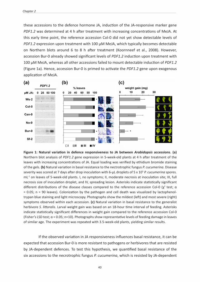
these accessions to the defence hormone JA, induction of the JA-responsive marker gene PDF1.2 was determined at 4 h after treatment with increasing concentrations of MeJA. At this early time point, the reference accession Col-0 did not yet show detectable levels of PDF1.2 expression upon treatment with 100 μM MeJA, which typically becomes detectable on Northern blots around 6 to 8 h after treatment (Koornneef et al., 2008). However, accession Bur-0 already showed significant levels of PDF1.2 induction upon treatment with 100 μM MeJA, whereas all other accessions failed to mount detectable induction of PDF1.2 (Figure 1a). Hence, accession Bur-0 is primed to activate the PDF1.2 gene upon exogenous application of MeJA.
Figure 1: Natural variation in defence responsiveness to JA between Arabidopsis accessions. (a) Northern blot analysis of PDF1.2 gene expression in 5-week-old plants at 4 h after treatment of the leaves with increasing concentrations of JA. Equal loading was verified by ethidium bromide staining of the gels. (b) Natural variation in basal resistance to the nectrotrophic fungus P. cucumerina. Disease severity was scored at 7 days after drop inoculation with 6-μL droplets of 5 x 10⁵ P. cucumerina spores.mL-1 on leaves of 5-week-old plants. I, no symptoms; II, moderate necrosis at inoculation site; III, full necrosis size of inoculation droplet, and IV, spreading lesion. Asterisks indicate statistically significant different distributions of the disease classes compared to the reference accession Col-0 (χ2 test; α = 0.05; n = 90 leaves). Colonization by the pathogen and cell death was visualized by lactophenol-trypan blue staining and light microscopy. Photographs show the mildest (left) and most severe (right) symptoms observed within each accession. (c) Natural variation in basal resistance to the generalist herbivore S. littoralis. Larval weight gain was based on an 18-hour time interval of feeding. Asterisks indicate statistically significant differences in weight gain compared to the reference accession Col-0 (Fisher’s LSD test; α = 0.05; n=10). Photographs show representative levels of feeding damage in leaves of similar age. The experiment was repeated with 3.5-week-old plants, yielding similar results.
If the observed variation in JA responsiveness influences basal resistance, it can be expected that accession Bur-0 is more resistant to pathogens or herbivores that are resisted by JA-dependent defences. To test this hypothesis, we quantified basal resistance of the six accessions to the necrotrophic fungus P. cucumerina, which is resisted by JA-dependent
41
Genetic dissection of basal defence responsiveness
defence mechanisms (Berrocal-Lobo et al., 2002). At 6 days after inoculation of 5-week-old plants, the accessions showed variation in symptom severity. Amongst all accessions, Ws-2 was most severely affected by the pathogen, whereas accession Bur-0 and, to a lesser extent, accession Sf-2 were more resistant than Col-0 (Figure 1b). Microscopic analysis of lactophenol trypan-blue stained leaves confirmed that the differences in disease symptoms are consistent with differences in tissue damage and colonization by the fungus (Figure 1b). To examine levels of basal resistance against JA-resisted herbivores, we quantified larval weight gain and leaf damage on 5-week-old plants upon 18 h of infestation by Spodoptera littoralis, a generalist herbivore that is resisted by JA-controlled defences (Mewis et al., 2005; Bodenhausen, 2007; Bodenhausen and Reymond, 2007). As is shown in Figure 1c, larval growth on accession Bur-0 was the lowest of all combinations and differed statistically from the larval growth values on Col-0 plants (Figure 1c). Furthermore, levels of leaf damage appeared relatively severe on accessions Ws-2 and Col-0, whereas Can-0, No-0 and Sf-2 showed intermediate levels of damage. Consistent with the larval weight gain values, accession Bur-0 showed the lowest degree of damage by the caterpillars (Figure 1c). The caterpillar experiment yielded identical results when repeated with 3.5-week-old plants (data not shown), indicating that the variation in herbivore resistance is un-related to plant age. Hence, the enhanced responsiveness of accession Bur-0 to JA is associated with increased levels of basal resistance against attackers that are controlled by JA-dependent defence mechanisms.
Accession Bur-0 is primed to activate ERF1/ORA59-dependent PDF1.2 but is repressed in MYC2-dependent induction of VSP2Induction of PDF1.2 gene expression is regulated by the transcription factors (TFs) ORA59 and ERF1, which integrate JA- and ET-dependent defence signals (Lorenzo et al., 2003; Pre et al., 2008). Conversely, induction of JA-dependent VSP2 is regulated by MYC2, a TF that integrates JA- and abscisic acid (ABA)-dependent signals (Lorenzo et al., 2004). Both branches of the JA response pathway act antagonistically on each other (Lorenzo et al., 2004; Lorenzo and Solano, 2005). To investigate whether the primed PDF1.2 gene responsiveness of Bur-0 is caused by a shift in negative cross-talk between ERF1/ORA59- and MYC2-dependent signalling branches, we monitored expression of PDF1.2 and VSP2 in Col-0 and Bur-0 at different time points after application of JA. Again, accession Bur-0 showed strongly augmented levels of JA-induced PDF1.2 expression in comparison to Col-0 (Figure 2a). In contrast, expression of VSP2 remained consistently lower in Bur-0 at all-time points after JA application. A similar transcription profile was observed after leaf wounding and combined treatment of wounding + S. littoralis regurgitant (Figure 2b). The antagonistic induction profiles of PDF1.2 and VSP2 indicate that accession Bur-0 is primed to activate the ERF1/ORA59-dependent branch of the JA response, but is repressed in the MYC2-dependent

42
Chapter 2
JA response. Nevertheless, we did not detect consistent differences in expression levels of ORA59, ERF1, or MYC2 between Bur-0 and Col-0 (Supplementary information Figure S1a), nor did we find differences in genomic coding sequences of these TF genes (Supplementary information Figure S1b). Hence, the differentially regulated JA response in accession Bur-0 is caused by modulating factors acting up- or downstream of ORA59, ERF1, or MYC2.
Figure 2: Arabidopsis accession Bur-0 is primed to activate PDF1.2 gene expression, but is repressed in VSP2 gene expression. (a) RT-qPCR analysis of PDF1.2 and VSP2 expression in 5-week-old Col-0 and Bur-0 plants at different time points after application of increasing concentrations of JA to the leaves. Data presented are average fold-change values (n=3; ± SEM) relative to the mean expression level in control-treated Col-0 at 2 hpt. (b) RT-qPCR analysis of PDF1.2 and VSP2 gene expression in 5-week-old Col-0 and Bur-0 plants at different time points after leaf wounding, or combined treatment of wounding and S. littoralis regurgitant. Data presented are average fold-change values (n=3; ± SEM) relative to the mean expression level in control-treated Col-0 at 2 hpt.
Natural variation in SA-induced PR-1 expression is associated with basal resistance against the hemi-biotrophipc pathogen Pst DC3000 To assess natural variation in responsiveness to the plant defence hormone SA, the six accessions were examined for levels of PR-1 gene induction at 6 h after treatment of the leaves with increasing concentrations of SA. In the first experiment, accessions Bur-0, Can-0 and Sf-2 showed enhanced levels of PR-1 gene induction by SA, whereas accessions Col-0, No-0 and Ws-2 exhibited relatively moderate levels of PR-1 induction (Figure 3a). In an independent second experiment, similar results were obtained for all accessions except No-0, which showed constitutive PR-1 gene expression in the control group. The latter finding is likely caused by the occasional development of spontaneous lesions in No-0 under our greenhouse conditions (data not shown). Thus, despite the variable behaviour of accession
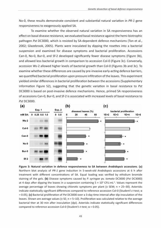
43
Genetic dissection of basal defence responsiveness
No-0, these results demonstrate consistent and substantial natural variation in PR-1 gene responsiveness to exogenously applied SA. To examine whether the observed natural variation in SA responsiveness has an effect on basal disease resistance, we evaluated basal resistance against the hemi-biotrophic pathogen Pst DC3000, which is resisted by SA-dependent defence mechanisms (Ton et al., 2002; Glazebrook, 2005). Plants were inoculated by dipping the rosettes into a bacterial suspension and examined for disease symptoms and bacterial proliferation. Accessions Can-0, No-0, Bur-0, and Sf-2 developed significantly fewer disease symptoms (Figure 3b), and allowed less bacterial growth in comparison to accession Col-0 (Figure 3c). Conversely, accession Ws-2 allowed higher levels of bacterial growth than Col-0 (Figures 3b and 3c). To examine whether these differences are caused by pre-invasive early-acting defence barriers, we quantified bacterial proliferation upon pressure infiltration of the leaves. This experiment yielded similar differences in bacterial proliferation between the accessions (Supplementary information Figure S2), suggesting that the genetic variation in basal resistance to Pst DC3000 is based on post-invasive defence mechanisms. Hence, primed SA responsiveness of accessions Can-0, Bur-0, and Sf-2 is associated with increased levels of basal resistance to Pst DC3000.
Figure 3: Natural variation in defence responsiveness to SA between Arabidopsis accessions. (a) Northern blot analysis of PR-1 gene induction in 5-week-old Arabidopsis accessions at 6 h after treatment with different concentrations of SA. Equal loading was verified by ethidium bromide staining of the gels. (b) Disease symptoms caused by P. syringae pv. tomato DC3000 (Pst DC3000) at 4 days after dipping the leaves in a suspension containing 5 × 10⁵ CFU.mL-1. Values represent the average percentage of leaves showing chlorotic symptoms per plant (± SEM; n = 25–30). Asterisks indicate statistically significant differences compared to reference accession Col-0 (Student’s t-test; α = 0.05). (c) Bacterial proliferation of Pst DC3000 over a 3-day time interval after dip-inoculation of the leaves. Shown are average values (± SE; n = 5–10). Proliferation was calculated relative to the average bacterial titer at 30 min after inoculation (dpi). Asterisks indicate statistically significant differences compared to reference accession Col-0 (Student’s t-test; α = 0.05).

44
Chapter 2
Accessions with primed PR-1 gene responsiveness express enhanced levels of priming-related TF genesPreviously, we demonstrated that priming of SA-dependent defence upon treatment with the chemical priming agent BABA is marked by enhanced expression of 28 defence-regulatory TF genes (Van der Ent et al., 2009). Because Can-0, Bur-0, and Sf-2 are primed to respond to SA in comparison to Col-0 (Figure 3a), we used RT-qPCR to examine whether this phenotype is similarly marked by enhanced expression of TF genes. Out of the 28 TF genes tested, 21, 23 and 24 genes were significantly enhanced in Can-0, Sf-2 and Bur-0, respectively (p<0.05; Figure 4 and supporting information Figure S3). In contrast, accession Ws-2, which exhibited similar or reduced SA responsiveness compared to Col-0 (Figure 3a), displayed a mostly unaltered or decreased expression of these marker genes (Figure 4). To examine whether the basal transcription patterns of the 28 TF genes in accessions Bur-0, Can-0 and Sf2 are similar to those of chemically primed Col-0 plants, we used multivariate cluster analysis. As expected, the transcription profiles of Bur-0, Can-0 and Sf-1 resembled the BABA-induced transcription profile of Col-0 (Supplementary information Figure S3), demonstrating that the constitutively primed SA response in these accessions is marked by elevated expression of the same set of TF genes that are induced upon chemical defence priming by BABA.
Figure 4: Accessions with primed responsiveness to salicylic acid (SA) show enhanced transcription of priming-related transcription factor (TF) genes. Transcriptional levels of 28 priming-related TF genes, previously identified as markers for beta-aminobutyric acid (BABA)-induced priming of SA-dependent defence (Van der Ent et al., 2009). Data presented are log-transformed fold-change values (±SEM) relative to the mean expression level in accession Col-0. Asterisks indicate statistically enhanced levels of expression compared to Col-0 (Mann–Whitney–Wilcoxon test or Student’s t-test; α = 0.05).

45
Genetic dissection of basal defence responsiveness
Natural variation in responsiveness of callose deposition to PAMPsTo examine genetic variation in sensitivity of locally expressed defence responses that are not directly under control by JA or SA, we quantified callose intensities upon treatment with the fungal PAMP elicitor chitosan and the bacterial PAMP elicitor flg22. Seedlings of the six accessions were cultivated in a hydroponic growth medium and examined for levels of callose deposition at 24 h after mock or PAMP treatment, using a standardised quantification method as described previously (Luna et al., 2011). Although all accessions showed a statistically significant increase in callose deposition after chitosan treatment, there was substantial variation in the intensity of this defence response (Figure 5). On average, Col-0 and Ws-2 deposited 5-fold more callose than accessions Bur-0, No-0, Sf-2 and Can-0. Similar patterns were observed upon treatment with flg22 (Figure 5), with the exception of accession Ws-2, which lacked flg22-induced callose due to a dysfunctional FLS2 receptor (Gomez-Gomez and Boller, 2000). Interestingly, when comparing natural variation in PAMP-induced callose to natural variation in SA-induced PR-1 expression (Figure 3), an inverse relationship can be noted: accessions with a modest callose response are primed to respond to SA, while accessions with a primed callose responsive are relatively un-responsive to SA.
Figure 5: Natural variation in PAMP-induced callose deposition. Eight-days-old seedlings were treated with either 0.1% chitosan or 1µM flg22. After 24 h, cotyledons were stained for callose with analine blue. Callose intensities were digitally quantified from photographs by UV epifluorescence microscopy. Values represent averages of the relative number of callose-corresponding pixels (n = 15; ± SEM).
Identification of a locus regulating responsiveness of SA-induced PR-1 expression To dissect the genetic variation in defence gene responsiveness, we performed QTL mapping analysis, using a fully genotyped core population of 164 RILs from a cross between

46
Chapter 2
accessions Col-0 and Bur-0 (Simon et al., 2008). For each gene expression screen, 15 to 20 RILs were cultivated along with the parental accessions, and examined for PDF1.2 or PR-1 gene expression at 4 h after treatment with 200 µm JA, or 7 h after treatment with 0.5 mM SA, respectively. At both time-points, leaves from mock treated plants were collected to assess basal levels of PDF1.2 and PR-1 expression. Although accession Bur-0 showed consistently higher levels of JA-induced PDF1.2 expression than Col-0, this difference appeared to be too variable between screens to allow for reliable QTL analysis of the pooled data set (data not shown). On the other hand, inter-experiment variation in PR-1 gene expression remained marginal. PR-1 expression values in each RIL screen were standardized to the averaged value from the parental accessions (Col-0 and Bur-0). Composite interval mapping of these standardized values revealed statistically significant linkage between SA-induced PR-1 expression and a locus at chromosome IV (LOD score = 5.64; Figure 6 and Table 1). RILs carrying the Bur-0 alleles at this locus showed higher levels of SA-induced PR-1 expression than RILs carrying the Col-0 alleles at this locus. This direction indicates either a suppressive effect from the Col-0 parent, or a stimulatory locus from the Bur-0 parent (Table 1). No genetic linkage was found with levels of basal PR-1 gene expression (Figure 6a). Hence, the locus at chromosome IV influences responsiveness of the PR-1 gene to SA, but has no influence on basal levels of PR-1 expression. Interestingly, the locus maps closely to a cluster of TIR-NB-LRR genes (At4g16860–At4g16990), of which 10 show nonsynonymous polymorphisms between Col-0 and Bur-0 (Supplementary information Table S2).The locus also maps closely to the highly polymorphic ACD6 gene (At4g14430; Supplementary information Table S2), which was recently identified as a source of natural allelic variation causing vegetative growth reduction and SA-dependent disease resistance (Todesco et al., 2010).
Table 1 Chromosome locations and significance of QTLs influencing the responsiveness of SA-induced PR1 expression, PAMP-induced callose, and plant growth.
TRAIT CHROM. CM POSITION (BP)
LOD SCORE
DIRECTION1
FLG22-INDUCED CALLOSE I 0.0 592,939 3.01 BUR+COL-
FLG22-INDUCED CALLOSE III 34.2 10,995,664 16.22 BUR-COL+
GROWTH III 63.5 22,146,585 3.19 BUR+COL-
GROWTH IV 0.0 641,363 3.76 BUR+COL-
SA-INDUCED PR1 EXPRESSION IV 30.9 8,929,959 5.64 BUR+COL-
1 Bur+Col-: a positive effect from the Bur-0 alleles or a negative effect from the Col-0 alleles; Bur-Col+: a negative effect from the Bur-0 alleles or a positive effect from the Col-0 alleles.

47
Genetic dissection of basal defence responsiveness
Figure 6: LOD scores and chromosome positions of QTLs influencing PR-1 gene expression, callose deposition, and plant growth. The QTL mapping analysis was based on 164 RILs from a cross between accessions Col-0 and Bur-0. (a) QTLs controlling basal and SA-induced PR-1 expression. (b) QTLs controlling basal and flg22-induced callose. (c) QTLs controlling plant growth estimated from rosette sizes of 4 week-old-plants. The RPP4/SCN1/RPP5 cluster of R genes is indicated at the top. Dotted black lines represent threshold levels of statistically significant LOD scores (2.94).
Identification of QTLs regulating responsiveness of flg22-induced callose To explore the genetic basis of natural variation in responsiveness of PAMP-induced callose, we screened the RIL population for levels of basal and flg22-induced callose. For each experiment, callose intensities were standardised to the averaged values from the parental

48
Chapter 2
accessions. Composite interval mapping of the pooled data did not revealed genetic linkage with basal callose levels in mock-treated seedlings. On the other hand, strong linkage was found between flg22-induced callose and a locus at chromosome III (LOD score = 16.22; Figure 6). An additional weaker influence was detected at a locus on the top of chromosome I (LOD score = 3.01; Figure 6b). The direction of both QTLs suggests opposite effects (Table 1): the major-effect QTL at chromosome III mediates a repressive effect from the Bur-0 parent or a stimulatory effect from the Col-0 parent, whereas the weaker QTL at chromosome I exerts a stimulatory effect from Bur-0 or a repressive effect from Col-0.
QTLs regulating defence responsiveness are not associated with reduced growthInduction of defence priming is associated with minor costs on plant growth (Van Hulten et al., 2006; Walters et al., 2008). To examine whether the constitutive defence priming in accession Bur-0 is associated with direct costs on plant growth, we scored rosette sizes of the RILs over a growth period of 4 weeks. Two relatively weak QTLs were identified at the bottom and top of chromosomes III and IV (LOD scores 3.19 and 3.76, respectively), which did not correspond to the QTLs controlling flg22-induced callose or SA-induced PR-1 expression (Figure 6; Table 1). This indicates that the priming-inducing loci from accession Bur-0 are not associated with major costs on plant fitness.
Priming by the SA response locus on chromosome IV boosts basal resistance against Pst DC3000The major callose QTL at chromosome III is based on either a repressive effect by the Bur-0 alleles, or a stimulatory effect from the Col-0 alleles (Table 1). Because accession Bur-0 is considerably more resistant against Pst DC3000 than accession Col-0 (Figure 3), it is unlikely that this locus contributes to basal resistance against this pathogen. In contrast, the direction of the SA response locus at chromosome IV is consistent with a positive effect on basal resistance against Pst DC3000. To confirm this, we compared levels of basal resistance between five RILs carrying the Bur-0 alleles of the SA response locus, five RILs carrying the Col-0 alleles of this locus, and the two parental accessions. Basal resistance assays were performed using a bioluminescent luxCDABE-tagged P. syringae pv. tomato DC3000 strain (Pst DC3000-lux), which enable in planta quantification of bacterial colonization (Fan et al., 2008). At 3 days after dipping the leaves in a bacterial suspension, Pst DC3000-lux colonisation in leaves of the Bur-0 lines was consistently lower than in leaves of the Col-0 lines (Figure 7a). Furthermore, bacterial growth values in all genotypes were inversely related to levels of SA-induced PR-1 transcription (Figures 7b and 7c). Multiple regression analysis of the bacterial growth values confirmed a statistically significant effect of the SA response locus (P < 0.001), whereas variation between lines with similar genotypes for this locus had no significant influence on bacterial growth (P = 0.258). These results indicate
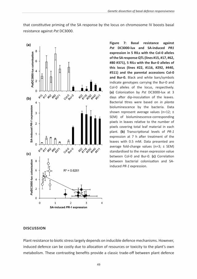
49
Genetic dissection of basal defence responsiveness
that constitutive priming of the SA response by the locus on chromosome IV boosts basal resistance against Pst DC3000.
Figure 7: Basal resistance against Pst DC3000-lux and SA-induced PR1 expression in 5 RILs with the Col-0 alleles of the SA response QTL (lines #15, #17, #62, #80 #371), 5 RILs with the Bur-0 alleles of this locus (lines #22, #116, #292, #440, #511) and the parental accessions Col-0 and Bur-0. Black and white bars/symbols indicate genotypes carrying the Bur-0 and Col-0 alleles of the locus, respectively. (a) Colonization by Pst DC3000-lux at 3 days after dip-inoculation of the leaves. Bacterial titres were based on in planta bioluminescence by the bacteria. Data shown represent average values (n=12; ± SEM) of bioluminescence-corresponding pixels in leaves relative to the number of pixels covering total leaf material in each plant. (b) Transcriptional levels of PR-1 expression at 7 h after treatment of the leaves with 0.5 mM. Data presented are average fold-change values (n=3; ± SEM) standardised to the mean expression value between Col-0 and Bur-0. (c) Correlation between bacterial colonisation and SA-induced PR-1 expression.
DISCUSSION
Plant resistance to biotic stress largely depends on inducible defence mechanisms. However, induced defence can be costly due to allocation of resources or toxicity to the plant’s own metabolism. These contrasting benefits provide a classic trade-off between plant defence

50
Chapter 2
and development (Heil, 2002; Heil and Baldwin, 2002). Priming of induced defence is associated with relatively minor costs, which are outweighed by the benefits of increased resistance under conditions of disease pressure (Van Hulten et al., 2006; Walters et al., 2008). Furthermore, priming provides resistance against a broad spectrum of pathogens and herbivores (Conrath et al., 2006; Frost et al., 2008). It is, therefore, plausible that selected plant varieties have evolved constitutively primed immune systems to adapt to environments that impose constant pressure from a wide range of pathogens (Ahmad et al., 2010). In support of this, our present study revealed genetic variation in the sensitivity of basal defence reactions amongst naturally occurring Arabidopsis accessions. Genetic dissection of this variation identified QTLs that regulates the responsiveness of these basal defences (Figure 6; Table 1). Interestingly, none of these QTLs had a detectable influence on plant growth (Figure 6), which supports our notion that constitutively primed defence is not associated with major fitness costs. Priming of defence can be based on enhanced expression of signalling proteins that remain inactive until an environmental stress signal is perceived by the plant (Conrath et al., 2006; Beckers et al., 2009). In agreement with this, we previously found that BABA-induced priming of SA-dependent defence is marked by enhanced expression of a set of defence-related TF genes (Van der Ent et al., 2009). In this study, we demonstrated that accessions Bur-0, Can-0 and Sf-2 express enhanced responsiveness to SA, are more resistant to PstDC3000, and show elevated expression of BABA-inducible TF genes (Figure 4). This suggests that the primed defence state of Bur-0, Can-0, and Sf-2 is based on similar signalling mechanisms as BABA-induced defence priming. In addition, these results illustrate that the set of BABA-inducible TF genes can serve as a marker to identify plants with primed immune systems. Accessions with primed responsiveness to SA expressed relatively high basal resistance against PstDC3000 (Figure 3). Van Leeuwen et al. (2007) reported similar natural variation between Arabidopsis accessions in the transcriptional response to exogenously applied SA. In this study, we used QTL mapping analysis to dissect this natural variation and identified a locus on chromosome IV that contributes to basal resistance against PstDC3000 (Figures 6 and 7). Interestingly, this locus maps to a cluster of 9 TIR-NB-LRR genes at chromosome IV (Figure 6; Supplementary information Table S2), which includes the Leptosphaeria maculans R gene RLM3 (Staal et al., 2008), the Hyaloperonospora arabidopsidis R genes RPP4 and RPP5 (Parker et al., 1997; Van Der Biezen et al., 2002), and the suppressor of npr1-1, constitutive 1 (SNC1) gene. Mutations in SNC1 have been reported to constitutively activate SA-dependent disease resistance (Li et al., 2001; Zhang et al., 2003). It is, therefore, tempting to speculate that the non-synonymous polymorphisms in between Col-0 and Bur-0 in this gene cluster (Supplementary information Table S2) are responsible for the differences in SA-induced PR-1 gene expression and basal resistance
51
Genetic dissection of basal defence responsiveness
against Pst DC3000 between these accessions. Furthermore, natural allelic variation in the ACD6 gene (At4g14420) has recently been reported to cause constitutive expression of SA-dependent defence and reduced vegetative growth (Todesco et al., 2010). Not only does ACD6 map closely to the SA response QTL, accessions Col-0 and Bur-0 also show 10 non-synonymous polymorphisms in the ACD6, of which four confer biochemically dissimilar amino acids (Supplementary information Table S2). However, unlike accession Est-1, Bur-0 is not reduced in vegetative growth (Figure 6) and does not express SA-dependent defence constitutively (Figure 3). It remains, nonetheless, possible that the Bur-0 alleles of ACD6 constitute a less extreme gene variant that merely primes for SA-dependent defences. Jones & Dangl (2006) defined basal resistance as residual resistance that is activated by virulent pathogens after defence suppression by disease-promoting pathogen effectors (i.e. basal resistance = PTI - ETS + weakened ETI). A recent study by Zhang et al. (2010) suggested that basal resistance against PstDC3000 is mostly determined by weakened ETI, while PTI has relatively little contribution. In agreement with this, we mapped a regulatory locus for SA responsiveness and basal resistance against PstDC3000 to a cluster of ETI-associated TIR-NB-LRR genes. Furthermore, two previous studies on natural variation in basal resistance against PstDC3000 identified QTLs at other genomic regions enriched in R and/or PRR genes (Kover et al., 2005; Perchepied et al., 2006). Together, these results suggest that natural variation in basal resistance against Pst DC3000 originates from the ETI component of basal resistance. Interestingly, accessions with relatively high basal resistance to Pst DC3000 deposited comparatively low levels of PAMP-induced callose (Figures 3 & 5). Moreover, our QTL analysis identified a major callose-promoting locus from the more susceptible Col-0 parent (Figure 6; Table 1), suggesting that virulent Pst DC3000 is not significantly resisted by PAMP-induced callose. Indeed, other studies have demonstrated that Pst DC3000 is extremely efficient in suppressing callose deposition through type III effectors (Zhang et al., 2007; Guo et al., 2009; Xiang et al., 2010). In contrast, dissection of natural variation in resistance against non-host Pseudomonas syringae pv. phaseolicola identified a major influence from the flagellin receptor FLS2 (Forsyth et al., 2010), suggesting that PTI has a more prominent role in non-host resistance against P. syringae pathogens. It thus seems that natural variation in resistance against virulent P. syringae strains stems from ETI-related defence mechanisms, whereas natural variation in resistance against non-host P. syringae strains is based on PTI-related defence mechanisms. Accession Bur-0 is primed to activate both SA- and JA-dependent defences (Figures 1 – 3), even though both pathways are mutually antagonistic in Arabidopsis (Koornneef and Pieterse, 2008). This phenotype demonstrates that enhanced responsiveness of SA- and JA-dependent defences is not affected by the negative cross-talk between both pathways. In support with this, Van Wees et al (2000) demonstrated that simultaneous activation of rhizobacteria-mediated induced systemic resistance (ISR) and pathogen-induced systemic

52
Chapter 2
acquired resistance (SAR), which are based on priming of JA- and SA-dependent defences, respectively, yield additive levels of resistance. Interestingly, the primed PDF1.2 response of accession Bur-0 to JA or wounding + caterpillar regurgitant coincided with a repressed induction of the VSP2 gene (Figure 2). Expression of PDF1.2 and VSP2 mark activities of two antagonistically acting branches of the JA response, which are regulated by the transcription factors ERF1 and ORA59, and MYC2, respectively (Lorenzo et al., 2003; Lorenzo et al., 2004; Pre et al., 2008). The ERF1/ORA59-dependent branch integrates JA and ET signals, whereas the MYC2-dependent branch integrates JA and ABA signals (Lorenzo et al., 2003; Anderson et al., 2004; Pre et al., 2008). Therefore, the primed PDF1.2 response of Bur-0 indicates potentiation of the ERF1/ORA59-dependent JA response (Lorenzo et al., 2004). This branch of the JA response has been described to contribute to basal resistance against necrotrophic pathogens (Anderson et al., 2004; Lorenzo et al., 2004). Indeed, accession Bur-0 displayed enhanced resistance to the necrotrophic fungus P. cucumerina (Figure 1b) and had previously been described as more resistant to the necrotrophic fungi B. cinerea and Fusarium oxysporum (Llorente et al., 2005). We also showed that Bur-0 is more resistant to feeding by the generalist herbivore S. littoralis (Figure 1c). Although Lorenzo et al. (2004) proposed a dominant role for the MYC2-dependent JA branch in resistance against herbivory, mutations in MYC2 have no consistent effect on basal resistance against S. littoralis (Bodenhausen, 2007). On the other hand, there is ample evidence that ET synergizes JA-dependent defences against herbivores (Von Dahl and Baldwin, 2007). This supports the findings by Van Oosten et al. (2008), who demonstrated that ISR-expressing Col-0 plants are primed to activate PDF1.2 and display enhanced resistance against Spodoptera exigua. Involvement of ET in the JA-responsive phenotype of accession Bur-0 would also explain why we encountered high inter-experiment variation in JA-induced PDF1.2 gene expression during our attempted QTL mapping of this trait. Plants have evolved various strategies to defend themselves against pathogens and herbivores. Apart from the well-characterised zigzag evolution towards R protein-mediated ETI, there are alternative defence strategies that can be equally effective depending on the environmental conditions (Ahmad et al., 2010). Our study has provided genetic evidence that selected Arabidopsis accessions have evolved constitutive priming of basal defence mechanisms, which can boost resistance against virulent pathogens. Mining for similar genetic traits in ancestral crop species will allow for integration of this naturally evolved defence strategy in sustainable pest and disease management.
ACKNOWLEDGMENTS
This research was supported by a BBSRC Institute Career Path Fellowship (UK, grant no. BB/E023959/1) to J.T., and grants 865.04.002 and 863.04.019 from the Earth and Life Sciences

53
Genetic dissection of basal defence responsiveness
Foundation (ALW), which is subsidized by The Netherlands Organization of Scientific Research(NWO).
54
Chapter 2
SUPPLEMENTARY INFROMATION
Figure S1: Transcriptional expression of ORA59, ERF1 and MYC2 and their genomic polymorphisisms between accessions Bur-0 and Col-0. (a), RT-qPCR analysis of basal ORA59, ERF1 and MYC2 gene expression in 5-week-old Col-0 and Bur-0 plants. Transcription of the TF genes was normalized to the expression of GAPDH. Values shown are average expression values from biologically replicate samples (n = 6), which were analyzed for statistically significant differences with a Student’s t-test. (b), Single nucleotide polymorphisms between Col-0 and Bur-0 in the 3000 bp regions covering the genomic sequences of MYC2, ERF1 and ORA59. Black and red letters indicate polymorphic nucleotides between Col-0 and Bur-0, respectively. Yellow bars represent open reading frames; blue bars indicate un-translated gene regions.

55
Genetic dissection of basal defence responsiveness
Figure S2: Natural variation in basal resistance to Pst DC3000 over a 3-day time interval after pressure-infiltration of the leaves with a bacterial suspension. Shows are average values (± SE; n = 5–10). Proliferation was calculated relative to the average bacterial titer at 30 min after inoculation (dpi). Asterisks indicate statistically significant differences compared to reference accession Col-0 (Student’s t-test; α = 0.05).
Figure S3. Cluster analysis of transcription profiles of 28 transcription factor (TF) genes and two reference genes (GAPDH and At1g13220) to compare basal transcription profiles in Bur-0, Can-0, Col-0, Sf-2 and Ws-2 with the transcription profiles from β-aminobutyric acid (BABA)-treated Col-0. Leaf material from BABA-primed Col-0 plants was collected at 2 d after soil-drench treatment with 80 μ m BABA. Colour intensity of induced (yellow) or repressed genes (blue) is proportional to their level of expression. Data represent log-transformed fold-change values relative to the average expression value in (untreated) Col-0. Values were subjected to average linkage clustering (Euclidean distance).

56
Chapter 2
Table S1 Arabidopsis thaliana accessions used in this study, their geographical origin, and levels of basal resistance to virulent Pseudomonas syringae pv. tomato DC3000.
Accession NASC ID Geographical origin
Basal resistance to P. syringae1 Reference
Ws-2 N1601 Wassilewskija (Russia)
---
(Ton et al., 1999)(Ton et al., 2001)
(Zipfel et al., 2004)
Col-0 N1092Colombia
(USA) 0 (reference)
Can-0 N1064 Canary Islands (Spain) + (Kover and Schaal, 2002)
No-0 N3081 Halle (Germany)
0+
(Kover and Schaal, 2002)(Kover et al., 2005)
Bur-0 N1028Burren
(Ireland) + (Kover and Schaal, 2002)
Sf-2 N1516San Feliu
(Spain) + (Kover and Schaal, 2002)
1 - : lower level of basal resistance than accession Col-0
0: similar level of basal resistance than accession Col-0
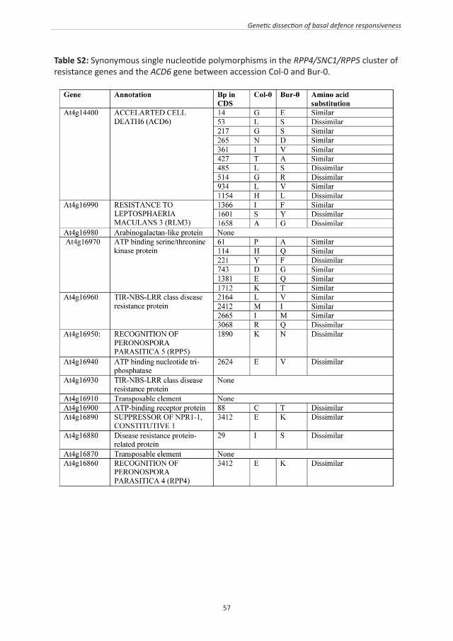
57
Genetic dissection of basal defence responsiveness
Table S2: Synonymous single nucleotide polymorphisms in the RPP4/SNC1/RPP5 cluster of resistance genes and the ACD6 gene between accession Col-0 and Bur-0.

58

59

60
CHAPTER 3
Benzoxazinoid Metabolites Regulate Innate Immunity against Aphids and Fungi in Maize.
Shakoor Ahmad1,5, Nathalie Veyrat1,2, Ruth Gordon-Weeks1, Yuhua Zhang1, Janet Martin1, Lesley Smart1, Gaetan Glauser2,6, Matthias Erb2, Victor Flors3, Monika Frey4,
and Jurriaan Ton1,7
1Rothamsted Research, Centre for Sustainable Pest and Disease Management, Harpenden, United Kingdom.
2Laboratory for Fundamental and Applied Research in Chemical Ecology (FARCE), University of Neuchâtel, Switzerland.
3Plant Physiology Section, Department of CAMN, University of Jaume I, Spain.4Technische Universität München, Lehrsuhl für Genetik, Freising, Germany.
5Plant-Microbe Interactions, Institute of Environmental Biology, Faculty of Science, Utrecht University, The Netherlands.
6Chemical Analytical Service of the Swiss Plant Science Web, University of Neuchâtel, Switzerland.
7Department of Animal and Plant Sciences, University of Sheffield, UK.
Plant Physiology (2011) 157: 317-327.

61
Role of benzoxazinoids in maize immunity
ABSTRACT
Benzoxazinoids (BXs), such as 2,4-dihydroxy-7-methoxy-2H-1,4-benzoxazin-3(4H)-one (DIMBOA), are secondary metabolites in grasses. The first step in BX biosynthesis converts indole-3-glycerol phosphate into indole. In maize, this reaction is catalysed by either Benzoxazineless 1 (BX1), or Indole glycerol phosphate lyase (IGL). The Bx1 gene is under developmental control and is mainly responsible for BX production, whereas the Igl gene is inducible by stress signals, such as wounding, herbivory, or jasmonates. To determine the role of BXs in defence against aphids and fungi, we compared basal resistance between Bx1 wild-type and bx1 mutant lines in the igl mutant background, thereby preventing BX production from IGL. Compared to Bx1 wild-type plants, BX-deficient bx1 mutant plants allowed better development of the cereal aphid Rhopalosiphum padi, and were affected in penetration resistance against the fungus Setosphaeria turtica. At stages preceding major tissue disruption, R. padi and S. turtica elicited increased accumulation of DIMBOA-glucoside, DIMBOA and HDMBOA-glucoside, which was most pronounced in apoplastic leaf extracts. Treatment with the defence elicitor chitosan similarly enhanced apoplastic accumulation of DIMBOA and 2-hydroxy-4,7-dimethoxy-1,4-benzoxazin-3-one (HDMBOA)-glucoside, but repressed transcription of genes controlling BX biosynthesis downstream of BX1. This repression was also obtained after treatment with the BX precursor indole and DIMBOA, but not with HDMBOA-glucoside. Furthermore, BX-deficient bx1 mutant lines deposited less chitosan-induced callose than Bx1 wild-type lines, whereas apoplast infiltration with DIMBOA, but not HDMBOA-glucoside, mimicked chitosan-induced callose. Hence, DIMBOA functions as a defence regulatory signal in maize innate immunity, which acts in addition to its well-characterised activity as a biocidal defence metabolite.

62
Chapter 3
INTRODUCTION
Induced plant defence against pests and diseases encompasses a wide variety of mechanisms, ranging from deposition of callose-rich papillae to accumulation of biocidal defence metabolites. Defensive metabolites can be synthesised de novo in response to microbe or insect attack, such as phytoalexins, but can also be produced constitutively and stored as an inactive form in the plant cell. These so-called phytoanticipins can be activated by β-glucosidase activity during herbivory, which allows for a very rapid release of biocidal aglycone metabolites (VanEtten et al., 1994; Morant et al., 2008). Well-characterised examples of phytoanticipins are glucosinolates, (GSs), which can be hydrolysed by endogenous β-thioglucoside glucohydrolases, called myrosinases. Although GSs have traditionally been associated with defence against herbivores, recent insights have revealed that they can also play an important role in resistance against microbes. For instance, Bedtnarek et al. (2009) demonstrated in Arabidopsis that early-acting penetration resistance against powdery mildew requires biosynthesis of the indolic GS 4-methoxyindol-3-ylmethylglucosinolate (4MI3G) and subsequent hydrolysis by the atypical myrosinase PEN2. Furthermore, penetration resistance of Arabidopsis against Phytophthora brassicae depends on the sequential action of the same class of indolic GSs and the indolic phytoalexin camalexin (Schlaeppi et al., 2010; Schlaeppi and Mauch, 2010). Interestingly, Clay et al. (2009) reported that callose deposition after treatment with the flagellin epitope flg22 requires intact biosynthesis and breakdown of 4MI3G. This finding uncovered a novel signalling role by indolic GSs in Arabidopsis immunity, but also raises the question of how callose deposition is regulated in non-Brassicaceous plants, which do not produce glucosinolates. Benzoxazinoids (BXs) are widely distributed phytoanticipins amongst Poaceae. It is commonly assumed that BX-glucosides are hydrolysed by plastid-targetted β-glucosidases upon tissue disruption, which results in the release of biocidal aglycone BXs (Morant et al., 2008). Since their discovery as plant secondary metabolites, many investigations have focussed on their role in plant defence against herbivorous insects and pathogens (Niemeyer, 1988, 2009). Most of these studies revealed positive correlations between resistance and BX levels in cereal varieties or inbred populations, or biocidal activity when added to artificial growth medium (for review, see Niemeyer, 2009). In maize, defence elicitation by pathogenic fungi or treatment with the defence regulatory hormone jasmonic acid (JA) influences BX metabolism by promoting the conversion of 2,4-dihydroxy-7-methoxy-2H-1,4-benzoxazin-3(4H)-one-glucoside (DIMBOA-glc) into N-O-methylated 2-hydroxy-4,7-dimethoxy-1,4-benzoxazin-3-one-glucoside (HDMBOA-glc) (Oikawa et al., 2002, 2004). When supplied to artificial growth medium, this di-methylated BX is more effective than DIMBOA-glc in reducing survival rates of the aphid Metopolophium dirhodum (Cambier et al., 2001). The biosynthesis of BXs is mostly under developmental control and leads to
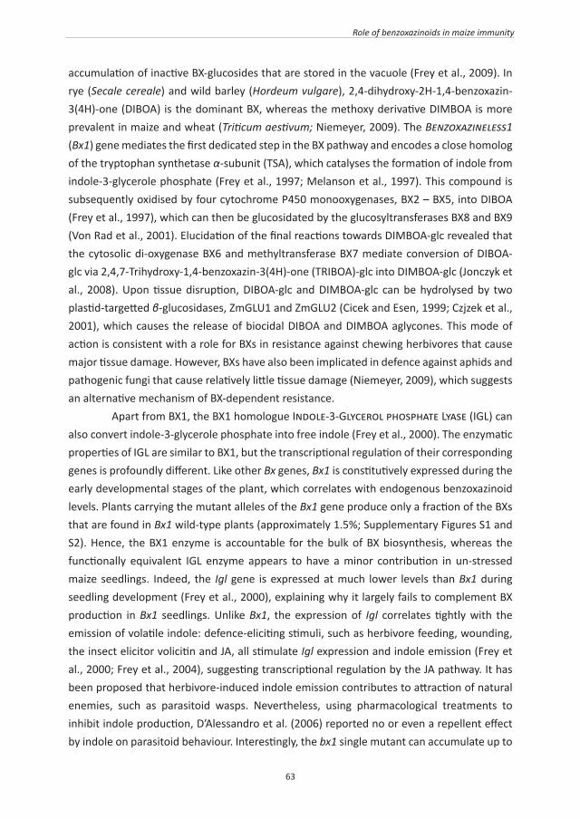
63
Role of benzoxazinoids in maize immunity
accumulation of inactive BX-glucosides that are stored in the vacuole (Frey et al., 2009). In rye (Secale cereale) and wild barley (Hordeum vulgare), 2,4-dihydroxy-2H-1,4-benzoxazin-3(4H)-one (DIBOA) is the dominant BX, whereas the methoxy derivative DIMBOA is more prevalent in maize and wheat (Triticum aestivum; Niemeyer, 2009). The Benzoxazineless1 (Bx1) gene mediates the first dedicated step in the BX pathway and encodes a close homolog of the tryptophan synthetase α-subunit (TSA), which catalyses the formation of indole from indole-3-glycerole phosphate (Frey et al., 1997; Melanson et al., 1997). This compound is subsequently oxidised by four cytochrome P450 monooxygenases, BX2 – BX5, into DIBOA (Frey et al., 1997), which can then be glucosidated by the glucosyltransferases BX8 and BX9 (Von Rad et al., 2001). Elucidation of the final reactions towards DIMBOA-glc revealed that the cytosolic di-oxygenase BX6 and methyltransferase BX7 mediate conversion of DIBOA-glc via 2,4,7-Trihydroxy-1,4-benzoxazin-3(4H)-one (TRIBOA)-glc into DIMBOA-glc (Jonczyk et al., 2008). Upon tissue disruption, DIBOA-glc and DIMBOA-glc can be hydrolysed by two plastid-targetted β-glucosidases, ZmGLU1 and ZmGLU2 (Cicek and Esen, 1999; Czjzek et al., 2001), which causes the release of biocidal DIBOA and DIMBOA aglycones. This mode of action is consistent with a role for BXs in resistance against chewing herbivores that cause major tissue damage. However, BXs have also been implicated in defence against aphids and pathogenic fungi that cause relatively little tissue damage (Niemeyer, 2009), which suggests an alternative mechanism of BX-dependent resistance. Apart from BX1, the BX1 homologue Indole-3-Glycerol phosphate Lyase (IGL) can also convert indole-3-glycerole phosphate into free indole (Frey et al., 2000). The enzymatic properties of IGL are similar to BX1, but the transcriptional regulation of their corresponding genes is profoundly different. Like other Bx genes, Bx1 is constitutively expressed during the early developmental stages of the plant, which correlates with endogenous benzoxazinoid levels. Plants carrying the mutant alleles of the Bx1 gene produce only a fraction of the BXs that are found in Bx1 wild-type plants (approximately 1.5%; Supplementary Figures S1 and S2). Hence, the BX1 enzyme is accountable for the bulk of BX biosynthesis, whereas the functionally equivalent IGL enzyme appears to have a minor contribution in un-stressed maize seedlings. Indeed, the Igl gene is expressed at much lower levels than Bx1 during seedling development (Frey et al., 2000), explaining why it largely fails to complement BX production in Bx1 seedlings. Unlike Bx1, the expression of Igl correlates tightly with the emission of volatile indole: defence-eliciting stimuli, such as herbivore feeding, wounding, the insect elicitor volicitin and JA, all stimulate Igl expression and indole emission (Frey et al., 2000; Frey et al., 2004), suggesting transcriptional regulation by the JA pathway. It has been proposed that herbivore-induced indole emission contributes to attraction of natural enemies, such as parasitoid wasps. Nevertheless, using pharmacological treatments to inhibit indole production, D’Alessandro et al. (2006) reported no or even a repellent effect by indole on parasitoid behaviour. Interestingly, the bx1 single mutant can accumulate up to
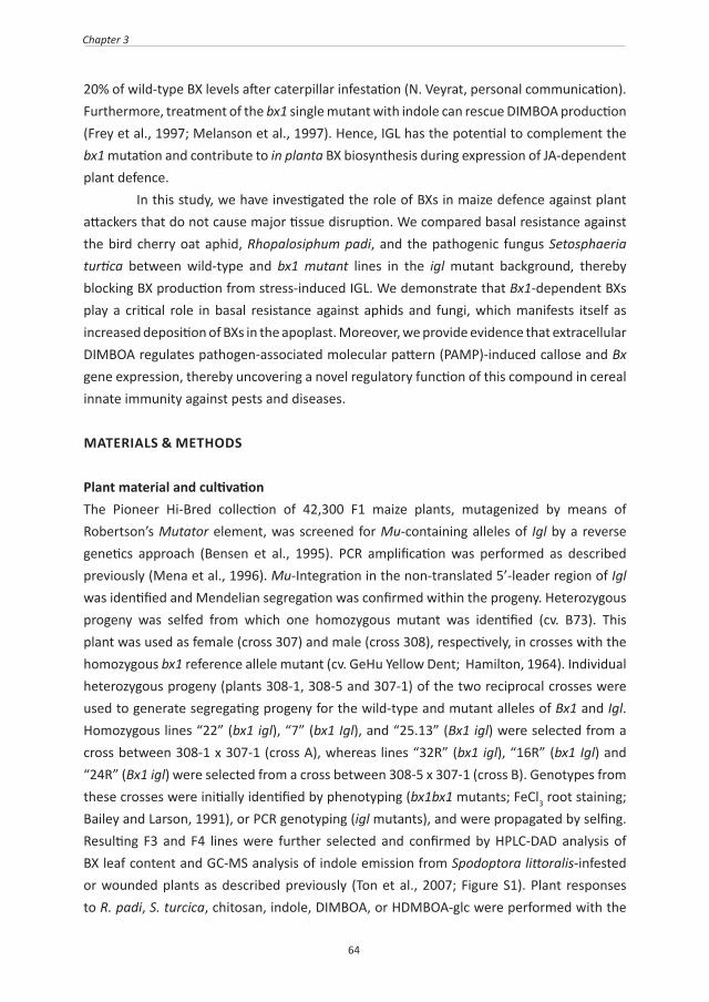
64
Chapter 3
20% of wild-type BX levels after caterpillar infestation (N. Veyrat, personal communication). Furthermore, treatment of the bx1 single mutant with indole can rescue DIMBOA production (Frey et al., 1997; Melanson et al., 1997). Hence, IGL has the potential to complement the bx1 mutation and contribute to in planta BX biosynthesis during expression of JA-dependent plant defence. In this study, we have investigated the role of BXs in maize defence against plant attackers that do not cause major tissue disruption. We compared basal resistance against the bird cherry oat aphid, Rhopalosiphum padi, and the pathogenic fungus Setosphaeria turtica between wild-type and bx1 mutant lines in the igl mutant background, thereby blocking BX production from stress-induced IGL. We demonstrate that Bx1-dependent BXs play a critical role in basal resistance against aphids and fungi, which manifests itself as increased deposition of BXs in the apoplast. Moreover, we provide evidence that extracellular DIMBOA regulates pathogen-associated molecular pattern (PAMP)-induced callose and Bx gene expression, thereby uncovering a novel regulatory function of this compound in cereal innate immunity against pests and diseases.
MATERIALS & METHODS
Plant material and cultivationThe Pioneer Hi-Bred collection of 42,300 F1 maize plants, mutagenized by means of Robertson’s Mutator element, was screened for Mu-containing alleles of Igl by a reverse genetics approach (Bensen et al., 1995). PCR amplification was performed as described previously (Mena et al., 1996). Mu-Integration in the non-translated 5’-leader region of Igl was identified and Mendelian segregation was confirmed within the progeny. Heterozygous progeny was selfed from which one homozygous mutant was identified (cv. B73). This plant was used as female (cross 307) and male (cross 308), respectively, in crosses with the homozygous bx1 reference allele mutant (cv. GeHu Yellow Dent; Hamilton, 1964). Individual heterozygous progeny (plants 308-1, 308-5 and 307-1) of the two reciprocal crosses were used to generate segregating progeny for the wild-type and mutant alleles of Bx1 and Igl. Homozygous lines “22” (bx1 igl), “7” (bx1 Igl), and “25.13” (Bx1 igl) were selected from a cross between 308-1 x 307-1 (cross A), whereas lines “32R” (bx1 igl), “16R” (bx1 Igl) and “24R” (Bx1 igl) were selected from a cross between 308-5 x 307-1 (cross B). Genotypes from these crosses were initially identified by phenotyping (bx1bx1 mutants; FeCl3 root staining; Bailey and Larson, 1991), or PCR genotyping (igl mutants), and were propagated by selfing. Resulting F3 and F4 lines were further selected and confirmed by HPLC-DAD analysis of BX leaf content and GC-MS analysis of indole emission from Spodoptora littoralis-infested or wounded plants as described previously (Ton et al., 2007; Figure S1). Plant responses to R. padi, S. turcica, chitosan, indole, DIMBOA, or HDMBOA-glc were performed with the
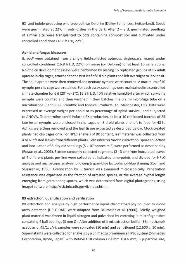
65
Role of benzoxazinoids in maize immunity
BX- and indole-producing wild-type cultivar Delprim (Delley Semences, Switzerland). Seeds were germinated at 22oC in petri-dishes in the dark. After 2 – 3 d, germinated seedlings of similar size were transplanted to pots containing compost soil and cultivated under controlled conditions (16:8 h L:D, 22°C).
Aphid and fungus bioassaysR. padi were obtained from a single field-collected apterous virginopara, reared under controlled conditions (16:8 h L:D, 22°C) on maize (cv. Delprim) for at least 10 generations. No-choice development assays were performed by placing 15 replicated groups of six adult apterae in clip cages, attached to the first leaf of 8 d old plants and left overnight to larviposit. The adult apterae were then removed and neonate nymphs were counted. A maximum of 10 nymphs per clip cage were retained. For each assay, seedlings were maintained in a controlled climate chamber for 6 d (20° +/- 2°C, 16:8 h L:D, 40% relative humidity) after which surviving nymphs were counted and then weighed in their batches in a 0.2 ml microfuge tube on a microbalance (Cahn C33, Scientific and Medical Products Ltd, Manchester, UK). Data were expressed as average weight per aphid or as percentage of aphid survival, and subjected to ANOVA. To determine aphid-induced BX production, at least 10 replicated batches of 25 late instar nymphs were enclosed in clip cages on 8 d old plants and left to feed for 48 h. Aphids were then removed and the leaf tissue extracted as described below. Mock-treated plants had clip cages only. For HPLC analysis of BX content, leaf material was collected from 4 to 6 infested leaves from different plants. Setosphaeria turcica cultivation, spore collection and inoculation of 8-day-old seedlings (5 x 10⁴ spores.ml-1) were performed as described by (Rostas et al., 2006). Sixteen randomly collected segments (2 - 3 cm) from inoculated leaves of 4 different plants per line were collected at indicated time-points and divided for HPLC analysis and microscopic analysis following trypan-blue lactophenol-blue staining (Koch and Slusarenko, 1990). Colonisation by S. turcica was examined microscopically. Penetration resistance was expressed as the fraction of arrested spores, or the average hyphal length emerging from germinating spores, which was determined from digital photographs, using ImageJ software (http://rsb.info.nih.gov/ij/index.html).
BX extraction, quantification and verificationBX extraction and analysis by high performance liquid chromatography coupled to diode array detection (HPLC-DAD) were adapted from Baumeler et al. (2000). Briefly, weighed plant material was frozen in liquid nitrogen and pulverised by vortexing in microfuge tubes containing 4 ball bearings (3 mm Ø). After addition of 1 mL extraction buffer (EB; methanol/acetic acid; 49/1; v/v), samples were sonicated (10 min) and centrifuged (12.600 g, 10 min). Supernatants were collected for analysis by a Shimadzu prominence HPLC system (Shimadzu Corporation, Kyoto, Japan) with BetaSil C18 column (250mm X 4.6 mm; 5 µ particle size;

66
Chapter 3
Thermo Scientific, USA) and diode array detector set at 254 nm. The mobile phase consisted of a mixture of pure water (solution A) and Methanol/Isopropanol/HAc (3800/200/1; v/v; solution B). The flow rate was maintained at 1 mL.min-1, starting with isocratic conditions at 10% B for 2 min, linear gradient to 50% B from 2 - 27 min, isocratic conditions at 50% B from 27 - 29 min, linear reverse gradient to 10% B from 29 - 31 min, and isocratic conditions at 10% B from 31 - 35 min. Retention times of the different BXs were established from synthetic standards (kindly provided by Prof. Dieter Sicker, University of Leipzig). BX tissue content (μg.g-1 FW) was estimated from standard curves, which showed linear relationships between peak area and concentration. Mass identities of DIMBOA-glc, DIMBOA and HDMBOA-glc were confirmed by ultra-high pressure liquid chromatography coupled to quadrupole time-of-flight mass spectrometry (UHPLC-QTOFMS; Glauser et al., 2011). Since DIMBOA and HDMBOA-glc elute closely together using the above HPLC separation protocol, we performed additional verification of both compounds by nuclear magnetic resonance (NMR) analysis after preparative HPLC purification. 1H, gradient correlation spectroscopy (GCOSY) and gradient heteronuclear single quantum coherence (GHSQC) spectra were recorded with a Varian VNMRS 500 MHz spectrometer; the chemical shifts were reported in ppm from tetramethylsilane with the residual solvent resonance taken as the internal standard. For DIMBOA, 1H NMR spectrum revealed five resonances (Figure S3A), which were readily assigned to the OCH3, aliphatic CH and three aromatic CH groups. For HDMBOA-glc, six proton resonances from the aglycone framework (Figure S3B) were detected whereas proton resonances from the glycoside moiety were observed as broad singlets or multiplets with chemical shifts comparable to closely related glycosides (Rashid et al., 1996). 1H-1H gCOSY and single bond 1H-13C gHSQC experiments were used to further confirm the identity of the HDMBOA-glc (data not shown). Although BX compounds can be unstable during extraction procedures, our extraction method yielded recovery rates of >98% when purified BX compounds were added to plant tissues before grinding.
Extraction of apoplastic fluidsThe method was adapted from Yu et al (1999) and Boudart et al. (2005). Briefly, collected leaf tissues were weighted and submerged into 14 µg.ml-1 proteinase K solution (Sigma) under a glass stopper in Greiner tubes. Vacuum infiltration was performed using a desiccator at -60 kPa for 5 min. After infiltration, leaf tissues were blotted dry, carefully rolled up, and placed in a 12-mL tubes, containing 20 ball bearings (3 mm Ø) and 0.5 mL EB supplemented with 14 µg.ml-1 proteinase K. After centrifugation for 5 min at 2.300 g (4°C), tissues were removed and the collected liquid comprising EB and apoplastic fluid was collected from beneath the ball bearings with a pipette and subjected to HPLC analysis. Leaf segments infiltrated with chitosan solution were incubated for 24 h in sealed petri-dishes before centrifugation.

67
Role of benzoxazinoids in maize immunity
Chemical treatments and callose quantificationIndole exposure was performed with 10-d old plants of similar size. Plants (n=9) per treatment were placed in air-tight glass chambers and exposed to indole that had been dissolved in dichloromethane, or dichloromethane only (mock), applied on filter paper discs. After 24 h of exposure, leaf segments (2 - 3 cm) from the second leaf were collected and divided for analysis of gene expression and BX content. Analyses of gene expression, BX content and callose deposition after chemical leaf infiltration were based on 9 - 18 randomly collected leaf segments (2 - 3 cm; second leaf) from at least 3 different 10-d old plants per treatment. Chitosan (Sigma) was dissolved to 1% (m/v) in 1% HAc initially, and diluted to 0.2% Chitosan (0.2% HAc) with water. Subsequent dilutions to 0.1 or 0.05% chitosan were performed with 0.2 % HAc, and adjusted to pH = 5.5 - 5.7. Chitosan solutions were infiltrated in leaf segments (2-3 cm) as described above, and left for 24 h under standard growth conditions before further analysis. Mock treatments were performed similarly with 0.2 % HAc (pH = 5.5 -5.7). DIMBOA and HDMBOA-glc were purified using a semi-preparative BetaSil C18 column (250mm X 10 mm; 5 µ particle size; Thermo Scientific, USA) at a flow rate of 4mL.min-1. Collected elutes were lyophilised and re-suspended in EB. After verification by HPLC-DAD and UHPLC-QTOFMS, compounds were diluted to indicated concentrations (1.96% methanol; 0.04% HAc; pH – 5.5 – 5.7) and infiltrated into leaf segments, as described above. Mock treatments were performed similarly using 1.96% methanol 0.04% HAc (pH = 5.5 – 5.7). Aniline-blue staining and quantification of callose by epi-fluorescence microscopy were performed, as described by Luna et al. (2011).
RNA isolation, cDNA preparation and RT-qPCR analysisGene expression analyses were based on three biologically replicated samples from the 2nd leaf of 10-d old plants. Total RNA was extracted as described previously (Matthes et al., 2010). Genomic DNA was digested according to manufacturer’s guidelines (RQ1 RNase-Free DNase; Promega, UK). Synthesis of cDNA was performed as described by Ton et al. (2007). Two technical replicates of each cDNA sample were subjected to RT-qPCR analysis, as described previously (Ahmad et al., 2011). Primer sequences are listed in Table S1. PCR efficiencies (E) of primer pairs were estimated from multiple amplification plots using the equation (1+E) = 10slope (Ramakers et al., 2003), and were confirmed to provide (1+E) values close to 2. Transcript levels were calculated relative to the reference genes GAPC or Actin-1 (Erb et al., 2009), using the 2ΔΔCt method, as described (Livak and Schmittgen, 2001; Schmittgen and Livak, 2008), or the 2ΔCt method, where ΔCt = Ct (reference gene) - Ct (gene of interest).
RESULTS
Selection of single and double mutants in lgl and Bx1

68
Chapter 3
The Igl gene is inducible by herbivory, tissue wounding, volicitin and methyl jasmonate (Frey et al., 2000, 2004), suggesting transcriptional regulation by the JA response pathway. It is, therefore, plausible that plants with a dysfunctional Bx1 gene can accumulate BX levels from the stress-inducible IGL enzyme. Consequently, the bx1 single mutant is unsuitable to assess the contribution of BXs to resistance against JA-eliciting attackers, such as R. padi and S. turtica (Delp et al., 2009; Erb et al., 2009). We, therefore, created Bx1 wild-type and bx1 mutant plants in the genetic background of a dysfunctional Igl gene. To this end, a Mutator (Mu)-induced mutant in the Igl gene was crossed with the original bx1bx1 mutant (Hamilton, 1964). Homozygous mutants in progenies from 2 independent crosses were selected and confirmed by high performance liquid chromatography (HPLC) analysis of BX leaf content, and gas chromatography (GC) analysis of wound-inducible indole emission (Figures S1 and S2). This selection resulted in three confirmed genotypes from each cross: the indole-producing bx1 single mutant (bx1 Igl), the indole-deficient igl single mutant (Bx1 igl), and the BX- and indole-deficient double mutant (bx1 igl).
Bx1 is required for basal resistance against aphidsTo establish the role of BXs in basal resistance against aphids, we compared growth and survival rates of R. padi between Bx1 wild-type lines (Bx1 igl) and bx1 mutant lines (bx1 igl and bx1 Igl) from both crosses. After 7 days of feeding from the first leaf, aphids gained significantly less weight when placed on BX-producing Bx1 igl lines compared to BX-deficient bx1 igl and bx1 Igl lines (Figure 1A). In addition, the percentage of aphid survival was less than 10% after feeding from the Bx1 wild-type lines, whereas over 70% remained alive after feeding from the bx1 mutant lines (Figure 1B). These differences were similar in progenies from both crosses and indicate a major contribution from the Bx1 gene to basal resistance against R. padi. Interestingly, aphids reared on the bx1 single mutant from the second cross showed marginally lower levels of survival than the corresponding bx1 igl double mutant (Student’s t-test; P = 0.048), suggesting a relatively small contribution from the Igl gene.
Figure 1: Contribution of Bx1 and Igl to basal resistance against the cereal aphid R. padi. Batches of neonate nymphs in clip cages were allowed to feed for 7 d from the first leaf of igl mutant lines, bx1 mutant lines, and bx1 igl double mutant lines, which had been selected from two independent crosses between the bx1 mutant and igl mutant of maize. A, Average weights (± SEM; n = 15) of neonate nymphs after 7 d. B, Average percentages of batch survival (± SEM) after 7 d. Different letters indicate statistically significant differences (ANOVA, followed by Fisher’s LSD test; α = 0.05). Wild-type alleles are indicated in black and mutant alleles in grey. The comparison between igl and bx1 igl mutant lines was repeated in two additional experiments with similar result.

69
Role of benzoxazinoids in maize immunity
Aphid infestation stimulates apoplastic BX accumulationTo examine which BX compounds contribute to Bx1-dependent resistance against R. padi, we quantified BX profiles after 2 d of feeding from BX-producing wild-type plants. Analysis of whole-leaf extracts by HPLC-diode array detection (HPLC-DAD) identified three major BX compounds: DIMBOA-glc, DIMBOA and HDMBOA-glc (Figure 2A), which were confirmed by ultra-high pressure liquid chromatography coupled to quadrupole time-of-flight mass spectrometry (UHPLCQTOFMS; Glauser et al., 2011) and nuclear magnetic resonance analysis (NMR; Supplementary Figure S3). Surprisingly, analysis of these compounds in whole-tissue extracts did not reveal major differences between mock- and aphid-infested plants (Figure 2). However, defence-related proteins and metabolites often accumulate in the apoplast of stressed tissues, and the aphid stylet must pass through the spaces between the epidermal and mesophyll cells in order to reach the phloem. We, therefore, considered the possibility that Bx1-dependent resistance against aphids depends on extracellular accumulation of active BX compounds. Indeed, targeted HPLC analysis of apoplastic leaf extracts revealed a statistically significant increase of DIMBOA-glc and DIMBOA at 48 h of feeding (Figure 2B). HDMBOA-glc levels were also increased in apoplastic leaf extracts from R. padi-infested plants, although this effect was more variable and borderline statistically significant (Student’s t-test; P = 0.051; Figure 2B). Together, these results indicate that infestation by R. padi boosts BX accumulation in the apoplast.
Figure 2: HPLC-DAD quantification of DIMBOA-glc, DIMBOA and HDMBOA-glc in whole-tissue extracts (A) and apoplastic extracts (B) from mock- and R. padi-infested maize leaves. Material was collected at 48 h after aphid feeding in clip cages. Mock treatments consisted of clip cages without aphids. Data represent mean values in μg.g-1 F.W. (± SEM) from 4 biologically replicated leaf samples. Asterisks indicate statistically significant differences compared to mock-treated leaves (Student’s t-test; α = 0.05). The experiment was repeated with similar results.
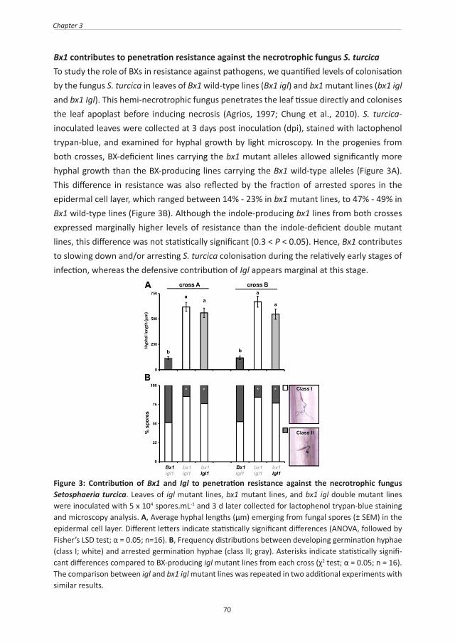
70
Chapter 3
Bx1 contributes to penetration resistance against the necrotrophic fungus S. turcicaTo study the role of BXs in resistance against pathogens, we quantified levels of colonisation by the fungus S. turcica in leaves of Bx1 wild-type lines (Bx1 igl) and bx1 mutant lines (bx1 igl and bx1 Igl). This hemi-necrotrophic fungus penetrates the leaf tissue directly and colonises the leaf apoplast before inducing necrosis (Agrios, 1997; Chung et al., 2010). S. turcica-inoculated leaves were collected at 3 days post inoculation (dpi), stained with lactophenol trypan-blue, and examined for hyphal growth by light microscopy. In the progenies from both crosses, BX-deficient lines carrying the bx1 mutant alleles allowed significantly more hyphal growth than the BX-producing lines carrying the Bx1 wild-type alleles (Figure 3A). This difference in resistance was also reflected by the fraction of arrested spores in the epidermal cell layer, which ranged between 14% - 23% in bx1 mutant lines, to 47% - 49% in Bx1 wild-type lines (Figure 3B). Although the indole-producing bx1 lines from both crosses expressed marginally higher levels of resistance than the indole-deficient double mutant lines, this difference was not statistically significant (0.3 < P < 0.05). Hence, Bx1 contributes to slowing down and/or arresting S. turcica colonisation during the relatively early stages of infection, whereas the defensive contribution of Igl appears marginal at this stage.
Figure 3: Contribution of Bx1 and Igl to penetration resistance against the necrotrophic fungus Setosphaeria turcica. Leaves of igl mutant lines, bx1 mutant lines, and bx1 igl double mutant lines were inoculated with 5 x 10⁴ spores.mL-1 and 3 d later collected for lactophenol trypan-blue staining and microscopy analysis. A, Average hyphal lengths (μm) emerging from fungal spores (± SEM) in the epidermal cell layer. Different letters indicate statistically significant differences (ANOVA, followed by Fisher’s LSD test; α = 0.05; n=16). B, Frequency distributions between developing germination hyphae (class I; white) and arrested germination hyphae (class II; gray). Asterisks indicate statistically signifi-cant differences compared to BX-producing igl mutant lines from each cross (χ2 test; α = 0.05; n = 16). The comparison between igl and bx1 igl mutant lines was repeated in two additional experiments with similar results.
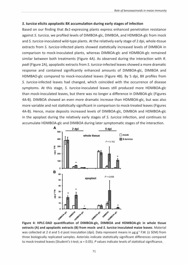
71
Role of benzoxazinoids in maize immunity
S. turcica elicits apoplastic BX accumulation during early stages of infectionBased on our finding that Bx1-expressing plants express enhanced penetration resistance against S. turcica, we profiled levels of DIMBOA-glc, DIMBOA, and HDMBOA-glc from mock and S. turcica-inoculated wild-type plants. At the relatively early stage of 2 dpi, whole-tissue extracts from S. turcica-infected plants showed statistically increased levels of DIMBOA in comparison to mock-inoculated plants, whereas DIMBOA-glc and HDMBOA-glc remained similar between both treatments (Figure 4A). As observed during the interaction with R. padi (Figure 2A), apoplastic extracts from S. turcica-infected leaves showed a more dramatic response and contained significantly enhanced amounts of DIMBOA-glc, DIMBOA and HDMBAO-glc compared to mock-inoculated leaves (Figure 4B). By 5 dpi, BX profiles from S. turcica-infected leaves had changed, which coincided with the occurrence of disease symptoms. At this stage, S. turcica-inoculated leaves still produced more HDMBOA-glc than mock-inoculated leaves, but there was no longer a difference in DIMBOA-glc (Figures 4A-B). DIMBOA showed an even more dramatic increase than HDMBOA-glc, but was also more variable and not statistically significant in comparison to mock-treated leaves (Figures 4A-B). Hence, maize deposits increased levels of DIMBOA-glc, DIMBOA and HDMBOA-glc in the apoplast during the relatively early stages of S. turcica infection, and continues to accumulate HDMBOA-glc and DIMBOA during later symptomatic stages of the interaction.
Figure 4: HPLC-DAD quantification of DIMBOA-glc, DIMBOA and HDMBOA-glc in whole tissue extracts (A) and apoplastic extracts (B) from mock- and S. turcica inoculated maize leaves. Material was collected at 2 d and 5 d post inoculation (dpi). Data represent means in μg.g-1 F.W. (± SEM) from three biologically replicated samples. Asterisks indicate statistically significant differences compared to mock-treated leaves (Student’s t-test; α = 0.05). P values indicate levels of statistical significance.
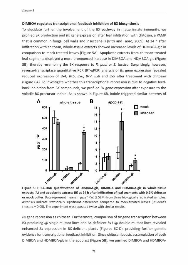
72
Chapter 3
DIMBOA regulates transcriptional feedback inhibition of BX biosynthesisTo elucidate further the involvement of the BX pathway in maize innate immunity, we profiled BX production and Bx gene expression after leaf infiltration with chitosan, a PAMP that is common in fungal cell walls and insect shells (Iritri and Faoro, 2009). At 24 h after infiltration with chitosan, whole-tissue extracts showed increased levels of HDMBOA-glc in comparison to mock-treated leaves (Figure 5A). Apoplastic extracts from chitosan-treated leaf segments displayed a more pronounced increase in DIMBOA and HDMBOA-glc (Figure 5B), thereby resembling the BX response to R. padi or S. turcica. Surprisingly, however, reverse-transcriptase quantitative PCR (RT-qPCR) analysis of Bx gene expression revealed reduced expression of Bx4, Bx5, Bx6, Bx7, Bx8 and Bx9 after treatment with chitosan (Figure 6A). To investigate whether this transcriptional repression is due to negative feed-back inhibition from BX compounds, we profiled Bx gene expression after exposure to the volatile BX precursor indole. As is shown in Figure 6B, indole triggered similar patterns of
Figure 5: HPLC-DAD quantification of DIMBOA-glc, DIMBOA and HDMBOA-glc in whole-tissue extracts (A) and apoplastic extracts (B) at 24 h after infiltration of leaf segments with 0.2% chitosan or mock buffer. Data represent means in μg.g-1 F.W. (± SEM) from three biologically replicated samples. Asterisks indicate statistically significant differences compared to mock-treated leaves (Student’s t-test; α = 0.05). The experiment was repeated twice with similar results.
Bx gene repression as chitosan. Furthermore, comparison of Bx gene transcription between BX-producing igl single mutant lines and BX-deficient bx1 igl double mutant lines revealed enhanced Bx expression in BX-deficient plants (Figures 6C-D), providing further genetic evidence for transcriptional feedback inhibition. Since chitosan boosts accumulation of both DIMBOA and HDMBOA-glc in the apoplast (Figure 5B), we purified DIMBOA and HDMBOA-

73
Role of benzoxazinoids in maize immunity
glc by preparative HPLC and examined which of both compounds is responsible for the feed-back response. At 24 h after infiltration of the purified compounds in the leaf apoplast, DIMBOA repressed Bx gene expression in a dose-dependent manner (Figures 6E-F), whereas HDMBOA-glc had no statistically significant effect (Figure 6G). We, therefore, conclude that DIMBOA acts as an extracellular signal for transcriptional feedback of BX biosynthesis.
Figure 6: Transcriptional feed-back regulation of the benzoxazinoid pathway by Bx1-dependent DIMBOA. Apoplastic infiltration with benzoxazinoid-inducing concentrations of chitosan (0.2 %) (A) and exposure to the volatile benzoxazinoid precursor indole (B) repressed Bx gene expression. benzoxazinoid-deficient bx igl double mutant lines displayed enhanced Bx gene transcription compared to benzoxazinoid-producing igl single mutant lines (C, D). Apoplastic leaf infiltration with DIMBOA (E, F) repressed Bx gene expression, whereas HDMBOA-glc had no effect (G), suggesting transcripitional feed-back regulation by apoplastic DIMBOA. Shown are average fold-change values (± SEM) of genes with a statistically significant level of induction (red) or repression (green) compared to mock treatments (A, B, E – G) or BX-producing igl single mutant lines (C, D). Differences in expression between three biologically replicated samples from independent experiments were tested for statistical significance, using Student’s t-tests or a non-parametric Wilcoxon Mann–Whitney test when values did not follow normal distributions (α = 0.05). n.d.: not determined.
DIMBOA regulates callose deposition PAMP-induced callose deposition is commonly used as a marker for plant innate immunity (Luna et al., 2011). Chitosan elicits callose deposition in a wide variety of plants (Iritri and Faoro, 2009). To optimise this PAMP response in maize, we quantified callose after leaf infiltration with increasing concentrations of chitosan. At 24 h after infiltration, maize

74
Chapter 3
leaves deposited callose in a dose-dependent manner (Figure S4), confirming that chitosan-induce defence of maize is marked by callose deposition. In Arabidopsis, PAMP-induced callose requires endogenous production of indolic GSs (Clay et al., 2009), suggesting a signalling function by these secondary metabolites. Since monocots do not produce indolic GSs, we tested whether BXs fulfil a similar role in maize innate immunity. To this end, we treated leaves with non-saturating concentrations of chitosan (0.05%) and assayed callose deposition in leaves of BX-producing Bx1 igl lines and BX-deficient bx1 igl lines revealed that the BX-deficient lines deposit significantly lower amounts of chitosan-induced callose than the BX-producing lines (Figure 7). Hence, PAMP-induced callose requires regulation by one or more Bx1-dependent metabolites. Since chitosan increases DIMBOA and HDMBOA-glc in the apoplast (Figure 5), we examined which of these two compounds are responsible for Bx1-dependent callose. To this end, we quantified callose intensities at 24 h after apoplast infiltration with either of both compounds. As observed for Bx gene repression (Figure 6), DIMBOA was active and triggered callose deposition in a dose-dependent manner, whereas HDMBOA-glc was inactive and failed to boost callose deposition (Figure 8). Hence, DIMBOA functions as an extracellular signal for PAMP-induced callose.
Figure 7: Bx1 regulates chitosan-induced callose deposition. Leaf segments from igl single mutant lines and bx1 igl double mutant lines were infiltrated with chitosan (0.05%) or mock solution. At 24 h after infiltration, leaf segments were collected for aniline-blue staining, UV-epifluoresence microscopy, and digital quantification of callose intensity. Shown are fold-induction values of callose (± SEM; n = 15), relative to the average callose intensity in mock-treated Bx1 igl lines from each cross. Photographs show representative differences in fluorescent callose signals under UV-epifluoresence microscopy.

75
Role of benzoxazinoids in maize immunity
Figure 8: DIMBOA-induced callose deposition. Infiltration with 20 μg.mL-1 DIMBOA elicits similar levels of callose deposition as infiltration with chitosan (0.1%), whereas infiltration with 20 μg.mL-1 HDMBOA-glc had no effect in comparison to the corresponding mock treatment. Shown are fold-induction values of callose deposition (± SEM; n = 15), relative to average callose intensities in mock treatments at 24 h after infiltration treatment. Different letters indicate statistically significant differences (ANOVA, followed by Fisher’s LSD test; α = 0.05). Photographs show representative differences in fluorescent callose signals by UV-epifluorescence microscopy.
DISCUSSION
The role of BXs in plant defence against pests and diseases has been studied for decades. Most of these studies are based on either in vitro evidence, where BX compounds had been supplemented to artificial growth medium, or on correlative evidence between resistance and BX levels among cereal varieties and/or inbred lines (for review, see Niemeyer, 1988, 2009). For instance, in vitro supplied DIMBOA has been demonstrated to affect a broad

76
Chapter 3
spectrum of herbivorous insects and microbes, including aphids (Cambier et al., 2001) and S. turcica (Rostás, 2007). Other studies revealed negative correlations between endogenous BX concentrations and aphid performance in populations of cereal varieties (Argandoña et al., 1980; Leszczynski and Dixon, 1990; Niemeyer and Perez, 1994). In the present study, we have investigated the defence function of BXs further, by comparing resistance phenotypes of wild-type and bx1 maize lines in the background of the igl mutant. Since IGL controls herbivore-induced indole production, the igl mutation prevents production of IGL-dependent BXs and, therefore, allows for a more accurate assessment of the defence contribution by BXs against IGL-inducing pests and diseases. Comparison between BX-producing and BX-deficient progenies from 2 independent crosses between the igl mutant and the bx1 mutant confirmed a major role for BXs in resistance against the cereal aphid R. padi (Figure 1). Using these lines, we also discovered a significant contribution of BXs to early-acting penetration resistance against the necrotophic fungus S. turcica (Figure 3). Hence, BXs play an important role in basal resistance against aphids and fungi. Interestingly, the expression of this BX-dependent resistence occurred before the occurrence of large-scale tissue disruption or symptom development. The involvement of BXs in penetration resistance against S. turcica (Figure 3B) suggests an alternative mode of action that contradicts the classical notion that BX defence requires tissue damage to hydrolyse vacuole-localised BX-glc compounds by plasmid-localised β-glucosidases (Morant et al., 2008). Chromatographic profiling of BX compounds revealed increased accumulation of apoplastic DIMBOA-glc, DIMBOA and HDMBOA-glc during the relatively early stages of infestation by either R. padi, or S. turcica (Figures 2 and 4). Notably, during both interactions, these effects preceded major tissue disruption or symptom development. Since aphid stylets and fungal hyphae must colonise the host apoplast before the host-parasite interaction can be established, enhanced deposition of biocidal BXs in this compartment is consistent with a role in penetration resistance. Based on our findings, we propose an alternative mode of BX-dependent defence, which depends on the accumulation of DIMBOA-glc and HDMBOA-glc into the apoplast, where they contribute to penetration resistance upon subsequent activation by plant- or attacker-derived β-glucosidases (Figure 9). In support of this, we observed increased accumulation of DIMBOA aglycone in the apoplast of challenged leaves (Figures 2 and 4). The lack of HDMBOA aglycone in this fraction can be explained by the highly unstable nature of this metabolite (Maresh et al., 2006). Although we cannot exclude that apoplastic BX accumulation during the early stages of aphid or fungal infestation occurs entirely without tissue damage, our experiments with chitosan demonstrate that this response can occur independently of tissue damage in maize. The mock treatments of these experiments involved a similar leaf infiltration as the chitosan treatments, but failed to increase apoplastic BX content (Figure 5). Hence, the difference in BX accumulation between both treatments is not triggered by

77
Role of benzoxazinoids in maize immunity
possible tissue damage during leaf infiltration. Future research will be necessary to identify the molecular transportation mechanisms underpinning PAMP-induced BX accumulation in the apoplast.
Figure 9: Model of BX-dependent innate immunity against aphids and fungi. Activation of maize innate immunity leads to apoplastic deposition of HDMBOA-glc and DIMBOA-glc. Subsequent hydrolysis into biocidal aglycones can provide chemical defence against pests and diseases (Cambier et al., 2001; Rostás, 2007) Both HDMBOA and DIMBOA are degraded into MBOA and BOA (Maresh et al., 2006), indicating that DIMBOA (red), and not HDMBOA, has an additional function in the regulation of Bx gene expression and callose deposition. I: intracellular space; A: apoplast.
In addition to the defensive contribution from Bx1, we were also able to determine the role of Igl by comparing resistance levels between indole-producing bx1 single mutants and indole-deficient bx1 igl double mutants. Although aphid performance and fungal colonization were consistently lower in Igl wild-type lines compared to igl mutant lines, this difference was only statistically significant for aphid survival in the progeny from one cross, and was not proportional to the relatively major contribution from the Bx1 allele (Figure 1B). Nonetheless, these relatively weak effects by Igl suggest a minor contribution from
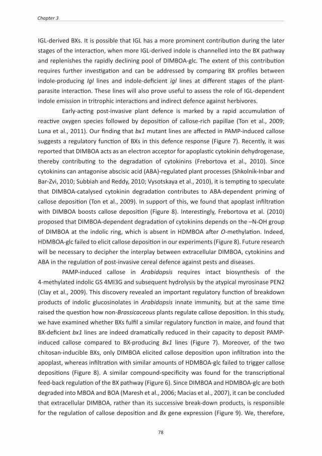
78
Chapter 3
IGL-derived BXs. It is possible that IGL has a more prominent contribution during the later stages of the interaction, when more IGL-derived indole is channelled into the BX pathway and replenishes the rapidly declining pool of DIMBOA-glc. The extent of this contribution requires further investigation and can be addressed by comparing BX profiles between indole-producing Igl lines and indole-deficient igl lines at different stages of the plant-parasite interaction. These lines will also prove useful to assess the role of IGL-dependent indole emission in tritrophic interactions and indirect defence against herbivores. Early-acting post-invasive plant defence is marked by a rapid accumulation of reactive oxygen species followed by deposition of callose-rich papillae (Ton et al., 2009; Luna et al., 2011). Our finding that bx1 mutant lines are affected in PAMP-induced callose suggests a regulatory function of BXs in this defence response (Figure 7). Recently, it was reported that DIMBOA acts as an electron acceptor for apoplastic cytokinin dehydrogenase, thereby contributing to the degradation of cytokinins (Frebortova et al., 2010). Since cytokinins can antagonise abscisic acid (ABA)-regulated plant processes (Shkolnik-Inbar and Bar-Zvi, 2010; Subbiah and Reddy, 2010; Vysotskaya et al., 2010), it is tempting to speculate that DIMBOA-catalysed cytokinin degradation contributes to ABA-dependent priming of callose deposition (Ton et al., 2009). In support of this, we found that apoplast infiltration with DIMBOA boosts callose deposition (Figure 8). Interestingly, Frebortova et al. (2010) proposed that DIMBOA-dependent degradation of cytokinins depends on the –N-OH group of DIMBOA at the indolic ring, which is absent in HDMBOA after O-methylation. Indeed, HDMBOA-glc failed to elicit callose deposition in our experiments (Figure 8). Future research will be necessary to decipher the interplay between extracellular DIMBOA, cytokinins and ABA in the regulation of post-invasive cereal defence against pests and diseases. PAMP-induced callose in Arabidopsis requires intact biosynthesis of the 4-methylated indolic GS 4MI3G and subsequent hydrolysis by the atypical myrosinase PEN2 (Clay et al., 2009). This discovery revealed an important regulatory function of breakdown products of indolic glucosinolates in Arabidopsis innate immunity, but at the same time raised the question how non-Brassicaceous plants regulate callose deposition. In this study, we have examined whether BXs fulfil a similar regulatory function in maize, and found that BX-deficient bx1 lines are indeed dramatically reduced in their capacity to deposit PAMP-induced callose compared to BX-producing Bx1 lines (Figure 7). Moreover, of the two chitosan-inducible BXs, only DIMBOA elicited callose deposition upon infiltration into the apoplast, whereas infiltration with similar amounts of HDMBOA-glc failed to trigger callose depositions (Figure 8). A similar compound-specificity was found for the transcriptional feed-back regulation of the BX pathway (Figure 6). Since DIMBOA and HDMBOA-glc are both degraded into MBOA and BOA (Maresh et al., 2006; Macias et al., 2007), it can be concluded that extracellular DIMBOA, rather than its successive break-down products, is responsible for the regulation of callose deposition and Bx gene expression (Figure 9). We, therefore,

79
Role of benzoxazinoids in maize immunity
hypothesize that this signalling function originates from a conserved detoxification response, which prevents auto-toxic build-up of BXs by translocation to the apoplast. Once deposited into the apoplast, BX-glucosides become hydrolysed and captured in a matrix of callose to provide targeted chemical defence against invading parasites. The striking analogy with indolic GS metabolites in Arabidopsis points to a conserved regulatory function of indole-derived secondary metabolites in innate immunity across the plant kingdom.
CONCLUSION
BX-deficient maize is more susceptible to aphids and is affected in penetration resistance against fungal pathogens. The difference in resistance between BX-deficient and BX-producing maize lines occurs before the onset of major tissue damage and manifests itself as increased accumulation of BX compounds in the apoplast. In addition to its contribution as a biocidal defence metabolite, extracellular DIMBOA regulates Bx gene expression and PAMP-induced callose, which reveals a novel regulatory function of BX metabolites in cereal innate immunity against pests and diseases.
ACKNOWLEDGEMENTS
The work described in this manuscript was supported by a BBSRC Institute Career Path Fellowship (no. BB/E023959/1) to Jurriaan Ton. We thank Ted Turlings for critically proofreading an earlier version of this manuscript. We also thank the technical support of the SCIC of Universitat Jaume I and Cristian Barrera for assistance during NMR analysis.

80
Chapter 3
SUPPLEMENTARY INFROMATION
Figure S1: Confirmation of mutant phenotypes of bx1 and igl carrying maize lines. Shown are typical HPLC-DAD chromatograms from un-stressed leaves of 7-d old seedlings (in pink), and GC-FID chromatograms of head-space collections from wounded leaves of 10-d old seedlings (in blue). Arrows in HPLC chromatograms indicate peaks corresponding to DIMBOA-glc or HDMBOA-glc; arrows in GC chromatograms indicate peaks corresponding to indole.

81
Role of benzoxazinoids in maize immunity
Table S1: Primers used for RT-qPCR analysis of gene expression
Gene Forward Primer Reverse Primer
GAPC 5’-GCATCAGGAACCCTGAGGAA-3’5’-CATGGGTGCATCTTTGCTTG-3’
ACTIN 5’-CCATGAGGCCACGTACAACT-3’ 5’-GGTAAAACCCCCACTGAGGA-3’
IGL 5’-GCCTCATAGTTCCCGACCTC-3’ 5’-GAATCCTCGTGAAGCTCGTG-3’
TSA 5’-TGAGGAGCGAGGCCATTATG-3’ 5’-GCGTTGTAAGCAGCACCAGC-3’
BX1 5’-CCCGAGCACGTAAAGCAGAT-3’ 5’-CTTCATGCCCCTGGCATACT-3’
BX2 5’-GACGAGGACGACGATAAGGACTT-3’ 5’-GGCCATACTCCTTCTGAAGAGACAG-3’
BX3 5’-ATGGCCGAGCTCATCAACAA-3’ 5’-TCGTCCTCACCTCCGTCTGT-3’
BX4 5’-TGTTCCTCCGGATCATCTGC-3’ 5’-AAGAGGCTGTCCCACCGCT-3’
BX5 5’-CCATTTCGACTGGGAGGTCC-3’ 5’-GTCCATGCTCACCTTCCAGC-3’
BX6 5’-AAGTTCAACACCATAGGACTCGATG-3’ 5’-CAGGTAGCTAGAGCCTGAAGTGGTC-3’
BX7 5’-GGCTGGGTTCCGTGACTACA-3’ 5’-GACCTCGATGATGGACGGG-3’
BX8 5’-GGAAGAGGATGAACGAGCTCAA-3′ 5’-GACCCAGCAGATTCATCGATG-3’
BX9 5’-GGGACCAGTTCGGCAACAT-3′ 5’-TGCCCACCTTCCACACGT-3’
GLU-1 5’-CCTCATGATGTGGGTGCAG-3′ 5’-ATGCATGACAAGGCCAGACT-3’
GLU-2 5’-AAAAACATGGGACCTCGTGA-3′ 5′-ATTGCATGACGACAATGCTAGA-3’

82
Chapter 3
Figure S2: HPLC-DAD quantification of basal and chitosan-induced DIMBOA-glc, DIMBOA and HDMBOA-glc in whole-leaf extracts from the igl single mutant, the bx1 single mutant, and the bx1 igl double mutant (cross B). Leaf samples were collected at 24 h after infiltration of leaf segments with mock buffer or 0.2% chitosan. Data represent means in μg.g-1 F.W. (± SEM) from three biologically replicated samples. Asterisks indicate statistically significant differences compared to mock-treated leaves (Student’s t-test; α = 0.05). nd: not detected.

83
Role of benzoxazinoids in maize immunity
Figure S3: 1H nuclear magnetic resonance (NMR) spectrogram of DIMBOA (A) and HDMBOA-glc (B). A, DIMBOA: 1H NMR (500 MHz, CD3OD): 3.76 (s, 3H), 5.67 (s, 1H), 6.61 (s, 1H), 6.67 (d, 8.5 Hz, 1H), 7.28 (d, 8.5 Hz, 1H). B, HDMBOA-glu: 1H NMR (500 MHz, CDCl3): 3.34 (broad singlet), 3.37 (broad singlet), 3.43 (broad singlet), 3.52 (broad singlet), 3.72 (3.72 s, 3H), 3.78 (broad multiplet, 2H), 3.90 (s, 3H), 4.72 (broad sinlet, 1H), 5.84 (s, 1H), 6.59 (d, 8 Hz, 1H), 6.66 (s, 1H), 7.05 (d, 8Hz, 1H).

84
Chapter 3
Figure S4: Dose-dependent callose deposition at 24 h after pressure infiltration with different con-centrations of chitosan. Shown are fold-induction values of callose deposition (± SEM; n = 15), relative to mock treatments at 24h after infiltration treatment.

85

86
CHAPTER 4
Benzoxazinoids in root exudates of maize attract Pseudomonas putida to the rhizosphere
Andrew L. Neal1, Shakoor Ahmad1, 2, Ruth Gordon-Weeks1, and Jurriaan Ton3
1Rothamsted Research, West Common, Harpenden, United Kingdom.2Plant-Microbe Interactions, Institute of Environmental Biology, Faculty of Science,
Utrecht University, P.O. Box 800.56, 3508 TB Utrecht, The Netherlands. 3Department of Animal and Plant Sciences, Western Bank, University of Sheffield, United
Kingdom.
PLoS ONE (2012) 7(4): e35498

87
Recruitment of beneficial bacteria by maize roots
ABSTRACT
Benzoxazinoids, such as 2,4-dihydroxy-7-methoxy-2H-1,4-benzoxazin-3(4H)-one (DIMBOA), are secondary metabolites in grasses. In addition to their function in plant defence against pests and diseases aboveground, benzoxazinoids (BXs) have also been implicated in defence belowground, where they can exert allelochemical or antimicrobial activities. We have studied the impact of BXs on the interaction between maize and Pseudomonas putida KT2440, a competitive coloniser of the maize rhizosphere with plant-beneficial traits. Chromatographic analyses revealed that DIMBOA is the main BX compound in root exudates of maize. In vitro analysis of DIMBOA stability indicated that DIMBOA tolerance of KT2440 bacteria is based on metabolism-dependent breakdown of DIMBOA. Transcriptome analysis of DIMBOA-exposed P. putida identified increased transcription of genes controlling benzoate catabolism and chemotaxis. Chemotaxis assays confirmed motility of P. putida towards DIMBOA. Moreover, colonisation essays with GREEN FLUORESCENT PROTEIN (GFP)-expressing P. putida showed that DIMBOA-producing roots of wild-type maize attract significantly higher numbers of P. putida cells than roots of the DIMBOA-deficient bx1 mutant. Our results demonstrate a central role for DIMBOA as a below-ground semiochemical for recruitment of plant-beneficial rhizobacteria during the relatively young and vulnerable growth stages of maize.
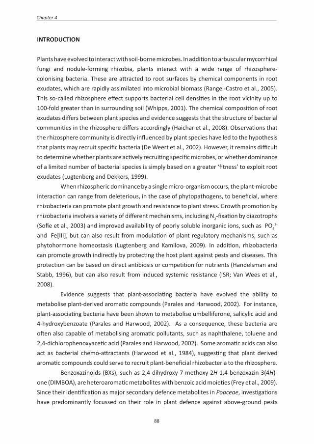
88
Chapter 4
INTRODUCTION
Plants have evolved to interact with soil-borne microbes. In addition to arbuscular mycorrhizal fungi and nodule-forming rhizobia, plants interact with a wide range of rhizosphere-colonising bacteria. These are attracted to root surfaces by chemical components in root exudates, which are rapidly assimilated into microbial biomass (Rangel-Castro et al., 2005). This so-called rhizosphere effect supports bacterial cell densities in the root vicinity up to 100-fold greater than in surrounding soil (Whipps, 2001). The chemical composition of root exudates differs between plant species and evidence suggests that the structure of bacterial communities in the rhizosphere differs accordingly (Haichar et al., 2008). Observations that the rhizosphere community is directly influenced by plant species have led to the hypothesis that plants may recruit specific bacteria (De Weert et al., 2002). However, it remains difficult to determine whether plants are actively recruiting specific microbes, or whether dominance of a limited number of bacterial species is simply based on a greater ‘fitness’ to exploit root exudates (Lugtenberg and Dekkers, 1999). When rhizospheric dominance by a single micro-organism occurs, the plant-microbe interaction can range from deleterious, in the case of phytopathogens, to beneficial, where rhizobacteria can promote plant growth and resistance to plant stress. Growth promotion by rhizobacteria involves a variety of different mechanisms, including N2-fixation by diazotrophs (Sofie et al., 2003) and improved availability of poorly soluble inorganic ions, such as PO4
3- and Fe[III], but can also result from modulation of plant regulatory mechanisms, such as phytohormone homeostasis (Lugtenberg and Kamilova, 2009). In addition, rhizobacteria can promote growth indirectly by protecting the host plant against pests and diseases. This protection can be based on direct antibiosis or competition for nutrients (Handelsman and Stabb, 1996), but can also result from induced systemic resistance (ISR; Van Wees et al., 2008). Evidence suggests that plant-associating bacteria have evolved the ability to metabolise plant-derived aromatic compounds (Parales and Harwood, 2002). For instance, plant-associating bacteria have been shown to metabolise umbelliferone, salicylic acid and 4-hydroxybenzoate (Parales and Harwood, 2002). As a consequence, these bacteria are often also capable of metabolising aromatic pollutants, such as naphthalene, toluene and 2,4-dichlorophenoxyacetic acid (Parales and Harwood, 2002). Some aromatic acids can also act as bacterial chemo-attractants (Harwood et al., 1984), suggesting that plant derived aromatic compounds could serve to recruit plant-beneficial rhizobacteria to the rhizosphere. Benzoxazinoids (BXs), such as 2,4-dihydroxy-7-methoxy-2H-1,4-benzoxazin-3(4H)-one (DIMBOA), are heteroaromatic metabolites with benzoic acid moieties (Frey et al., 2009). Since their identification as major secondary defence metabolites in Poaceae, investigations have predominantly focussed on their role in plant defence against above-ground pests

89
Recruitment of beneficial bacteria by maize roots
and pathogens (Niemeyer, 1988, 2009). BXs are typically produced during relatively early, vulnerable plant growth stages (Frey et al., 2009). In response to tissue damage, vacuolar reservoirs of BX-glucosides are hydrolysed by plastid-targeted β-glucosidases, causing rapid accumulation of aglucone BX biocidal metabolites (Morant et al., 2008). A recent study in maize revealed that Spodoptera larvae can detoxify DIMBOA by glycosylation and that the contribution of maize BXs to defence against these herbivores is based on an inducible conversion of DIMBOA-glc into 2-β-d-glucopyranosyloxy-4,7-dimethoxy-1,4-benzoxazin-3-one (HDMBOA-glc; Glauser et al., 2011). Interestingly, BXs are also active against attackers causing relatively minor tissue damage, such as aphids and pathogenic fungi. This function is based on an increased accumulation of DIMBOA in the apoplast before the onset of large-scale tissue damage, where it signals increased deposition of callose-rich papillae (Ahmad et al., 2011). Thus, the aboveground defence contribution of BXs is not only limited to their biocidal properties, but also includes a within-plant signalling function in the activation of plant innate immune responses against pests and diseases. BXs have also been implicated in plant defence below-ground. BXs are exuded in relatively large quantities from cereal roots, where they can act as allelochemicals against microbes, insects or competing plants (Niemeyer and Perez, 1994; Niemeyer, 2009). Once released, BXs degrade relatively quickly in aqueous environments with a half-life of less than 24 hours (Woodward et al., 1978). Upon hydrolysis, DIMBOA is converted into 6-methoxy-benzoxazolin-2-one (MBOA), a compound considerably more stable in sterile soil, but with significantly less toxicity than DIMBOA (Kumar et al., 1993). Biodegradation of MBOA leads to accumulation of phenoxazinones (Krogh et al., 2006), and requires activity by microbes, such as Acinetobacter calcoaceticus (Chase et al., 1991), soil-borne fungi (Friebe et al., 1998), or unidentified members in the rhizosphere community of oat (Friebe et al., 1996). Phenoxazinone products are typically more biocidal than benzoxazolinones and have antifungal (Anzai et al., 1960), antibacterial (Gerber and Lechevalier, 1964), and plant allelopathic properties (Gagliardo and Chilton, 1992). Hence, BX exudation by plants can have a major impact on rhizosphere communities in the soil. Plant-derived aromatic metabolites can act as chemo-attractants for Pseudomonas putida (Harwood et al., 1984; Parales and Harwood, 2002). We therefore hypothesised that BXs from root exudates of maize may attract and support P. putida cells. To address this hypothesis we studied the influence of BXs on P. putida KT2440, a competitive coloniser of the maize rhizosphere with plant-beneficial traits (Molina et al., 2000; Matilla et al., 2010). We identified DIMBOA as the dominant BX species in maize root exudates and found that exposure of P. putida to DIMBOA induces bacterial genes with putative functions in chemotactic responses. In vitro chemotaxis assays indeed revealed that P. putida KT2440 displays taxis towards DIMBOA. The ecological relevance of this response was confirmed by root colonisation assays in soil, using maize mutant lines impaired in BX biosynthesis.
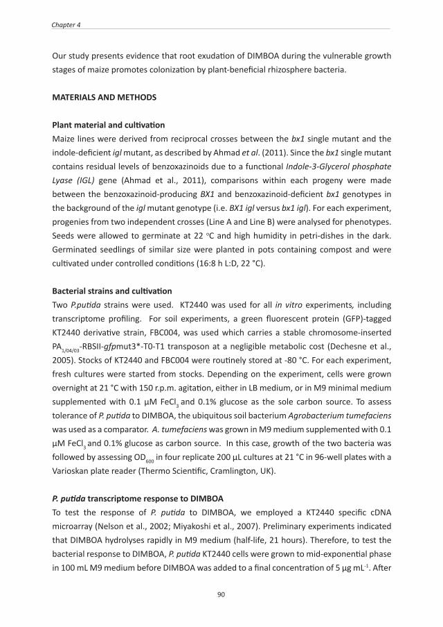
90
Chapter 4
Our study presents evidence that root exudation of DIMBOA during the vulnerable growth stages of maize promotes colonization by plant-beneficial rhizosphere bacteria.
MATERIALS AND METHODS
Plant material and cultivationMaize lines were derived from reciprocal crosses between the bx1 single mutant and the indole-deficient igl mutant, as described by Ahmad et al. (2011). Since the bx1 single mutant contains residual levels of benzoxazinoids due to a functional Indole-3-Glycerol phosphate Lyase (IGL) gene (Ahmad et al., 2011), comparisons within each progeny were made between the benzoxazinoid-producing BX1 and benzoxazinoid-deficient bx1 genotypes in the background of the igl mutant genotype (i.e. BX1 igl versus bx1 igl). For each experiment, progenies from two independent crosses (Line A and Line B) were analysed for phenotypes. Seeds were allowed to germinate at 22 oC and high humidity in petri-dishes in the dark. Germinated seedlings of similar size were planted in pots containing compost and were cultivated under controlled conditions (16:8 h L:D, 22 °C).
Bacterial strains and cultivationTwo P.putida strains were used. KT2440 was used for all in vitro experiments, including transcriptome profiling. For soil experiments, a green fluorescent protein (GFP)-tagged KT2440 derivative strain, FBC004, was used which carries a stable chromosome-inserted PA1/04/03-RBSII-gfpmut3*-T0-T1 transposon at a negligible metabolic cost (Dechesne et al., 2005). Stocks of KT2440 and FBC004 were routinely stored at -80 °C. For each experiment, fresh cultures were started from stocks. Depending on the experiment, cells were grown overnight at 21 °C with 150 r.p.m. agitation, either in LB medium, or in M9 minimal medium supplemented with 0.1 μM FeCl3 and 0.1% glucose as the sole carbon source. To assess tolerance of P. putida to DIMBOA, the ubiquitous soil bacterium Agrobacterium tumefaciens was used as a comparator. A. tumefaciens was grown in M9 medium supplemented with 0.1 μM FeCl3 and 0.1% glucose as carbon source. In this case, growth of the two bacteria was followed by assessing OD600 in four replicate 200 µL cultures at 21 °C in 96-well plates with a Varioskan plate reader (Thermo Scientific, Cramlington, UK).
P. putida transcriptome response to DIMBOATo test the response of P. putida to DIMBOA, we employed a KT2440 specific cDNA microarray (Nelson et al., 2002; Miyakoshi et al., 2007). Preliminary experiments indicated that DIMBOA hydrolyses rapidly in M9 medium (half-life, 21 hours). Therefore, to test the bacterial response to DIMBOA, P. putida KT2440 cells were grown to mid-exponential phase in 100 mL M9 medium before DIMBOA was added to a final concentration of 5 μg mL-1. After

91
Recruitment of beneficial bacteria by maize roots
1 hour of exposure, cells were harvested by centrifugation at 4 °C. RNA was extracted from three independently performed experiments. Cell pellets were treated with RNAprotect™ (Qiagen, Valencia, CA) immediately following centrifugation. Cell membrane lysis was achieved with 1 mg mL-1 lysozyme in buffer containing 10 mM TRIS and 1 mM EDTA at pH8 using Qiagen RNeasy® reagent kits following the manufacturer’s instructions. Extracted RNA was purified with TURBO DNA-free™ kits (Ambion, Applied Biosystems, Foster City, CA) and quantified on a NanoDrop 1000 spectrophotometer. cDNA was synthesised and labelled using the SuperScript™ indirect cDNA labelling system (Invitrogen, Carlsbad, CA). Synthesised cDNA paired samples (control or DIMBOA) were labelled with Cy3 or HyPer5 (Amersham, Little Chalfont, UK) fluorophores. To remove dye bias, the experimental design included dye-swap normalisation procedures, as described by Dabney & Storey (2007). Dye incorporation was verified to be more than 150 pM dye per sample. Equal amounts of Cy3-cDNA and HyPer5-cDNA, each representing a replicate comparison between control and DIMBOA-treated cells, were combined and dried in a speedvac before proceeding with array hybridisation. Microarrays were pre-treated with BlockIt™ Plus blocking buffer (Arrayit Corporation, Sunnyvale, CA) in order to inactivate reactive groups on the surface. Dried cDNA was rehydrated in buffer and hybridised to arrays (Progenika Biopharma S.A, Vizcaya, Spain) for 18 hours at 42 °C, according to the manufacturer’s instructions. Following hybridisation, arrays were washed, dried, and scanned with a GenePix® 4000B scanner (Molecular Devices, Sunnyvale, CA). Data were processed using TM4 microarray software (Saeed et al., 2003). Data from the three independent replicate experiments were combined and analysed together. Using Statistical Analysis for Microarrays procedures (Tusher et al., 2001), only genes that were consistently induced by DIMBOA in all three independent replicates were considered significant. Functional annotation of induced genes was performed using the supporting microarray documentation and the P. putida KT2440 KEGG genome database (www.genome.jp/kegg-bin/show_organism?org=ppu).
In vitro P. putida KT2440 chemotaxis assayChemo-attractiveness of DIMBOA was quantified using a modified capillary-based chemotaxis assay (Harwood et al., 1984), which relies on accumulation of bacterial cells in microcapillary tubes (1 μL volume, Drummond Scientific Company, Broomall, PA). Tubes containing glucose-free M9 medium (control), M9 medium with 5 μg ml-1 DIMBOA, or M9 medium with 0.01% casamino acids (positive control; Harwood et al., 1984) were incubated in individual wells of a 96-well plate. Each well contained 200 μL glucose-free M9 suspension with P. putida KT2440 bacteria (OD600=0.06). After 30 minutes, capillary contents were carefully collected and plated onto LB agar for cell enumeration.
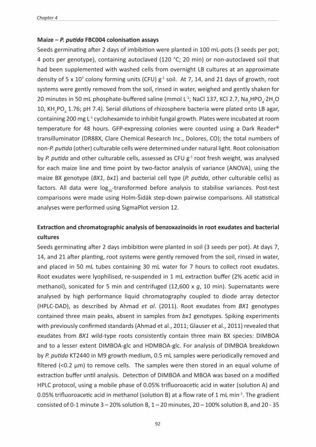
92
Chapter 4
Maize – P. putida FBC004 colonisation assaysSeeds germinating after 2 days of imbibition were planted in 100 mL-pots (3 seeds per pot; 4 pots per genotype), containing autoclaved (120 °C; 20 min) or non-autoclaved soil that had been supplemented with washed cells from overnight LB cultures at an approximate density of 5 x 10⁷ colony forming units (CFU) g-1 soil. At 7, 14, and 21 days of growth, root systems were gently removed from the soil, rinsed in water, weighed and gently shaken for 20 minutes in 50 mL phosphate-buffered saline (mmol L-1; NaCl 137, KCl 2.7, Na2HPO4·2H2O 10, KH2PO4 1.76; pH 7.4). Serial dilutions of rhizosphere bacteria were plated onto LB agar, containing 200 mg L-1 cyclohexamide to inhibit fungal growth. Plates were incubated at room temperature for 48 hours. GFP-expressing colonies were counted using a Dark Reader® transilluminator (DR88X, Clare Chemical Research Inc., Dolores, CO); the total numbers of non-P. putida (other) culturable cells were determined under natural light. Root colonisation by P. putida and other culturable cells, assessed as CFU g-1 root fresh weight, was analysed for each maize line and time point by two-factor analysis of variance (ANOVA), using the maize BX genotype (BX1, bx1) and bacterial cell type (P. putida, other culturable cells) as factors. All data were log10-transformed before analysis to stabilise variances. Post-test comparisons were made using Holm-Šidák step-down pairwise comparisons. All statistical analyses were performed using SigmaPlot version 12.
Extraction and chromatographic analysis of benzoxazinoids in root exudates and bacterial culturesSeeds germinating after 2 days imbibition were planted in soil (3 seeds per pot). At days 7, 14, and 21 after planting, root systems were gently removed from the soil, rinsed in water, and placed in 50 mL tubes containing 30 mL water for 7 hours to collect root exudates. Root exudates were lyophilised, re-suspended in 1 mL extraction buffer (2% acetic acid in methanol), sonicated for 5 min and centrifuged (12,600 x g, 10 min). Supernatants were analysed by high performance liquid chromatography coupled to diode array detector (HPLC-DAD), as described by Ahmad et al. (2011). Root exudates from BX1 genotypes contained three main peaks, absent in samples from bx1 genotypes. Spiking experiments with previously confirmed standards (Ahmad et al., 2011; Glauser et al., 2011) revealed that exudates from BX1 wild-type roots consistently contain three main BX species: DIMBOA and to a lesser extent DIMBOA-glc and HDMBOA-glc. For analysis of DIMBOA breakdown by P. putida KT2440 in M9 growth medium, 0.5 mL samples were periodically removed and filtered (<0.2 μm) to remove cells. The samples were then stored in an equal volume of extraction buffer until analysis. Detection of DIMBOA and MBOA was based on a modified HPLC protocol, using a mobile phase of 0.05% trifluoroacetic acid in water (solution A) and 0.05% trifluoroacetic acid in methanol (solution B) at a flow rate of 1 mL min-1. The gradient consisted of 0-1 minute 3 – 20% solution B, 1 – 20 minutes, 20 – 100% solution B, and 20 - 35

93
Recruitment of beneficial bacteria by maize roots
minutes isocratic conditions of 100% solution B. Chromatograms were recorded at 254 nm and retention times of DIMBOA and MBOA were established from standards.
RESULTS
Exudation of benzoxazinones from maize rootsRoots of BX1 wild-type and bx1 mutant lines were incubated for seven hours in water, after which the collected exudates were subjected to HPLC-DAD analysis of BXs. Root exudates from BX1 wild-type plants consistently contained three BX compounds, all of which were absent from exudates of bx1 mutant lines. The dominant compound was DIMBOA, with concentrations up to 31 μg g-1 fresh root weight (FW) in exudates from 7 days old roots (Figure 1). Levels of DIMBOA exudation showed a statistically significant linear decline in aging plants (Figure 1; linear regression, Line A, F1,13 = 17.74; p<0.001, Line B, F1,13=7.387; p=0.018). The other plant-derived BXs in root exudates from BX1 expressing plants were the BX glucosides DIMBOA-glc and HDMBOA-glc. Concentrations of these compounds did not exceed 3 μg g-1 FW and remained constant over time.
Figure 1: Root exudation of benzoxazinoids at different developmental stages in maize lines expressing a functional BX1 gene. The dominant BX compound in root exudates is the aglucone 2,4-dihydroxy-7- methoxy- 1,4-benzoxazin-3-one (DIMBOA), which shows a statistically significant linear decrease with plant age in both lines. Shown are average BX quantities, expressed in μg.g-1 root fresh weight (± SEM; n=3) exuded over a 7 hour time period.
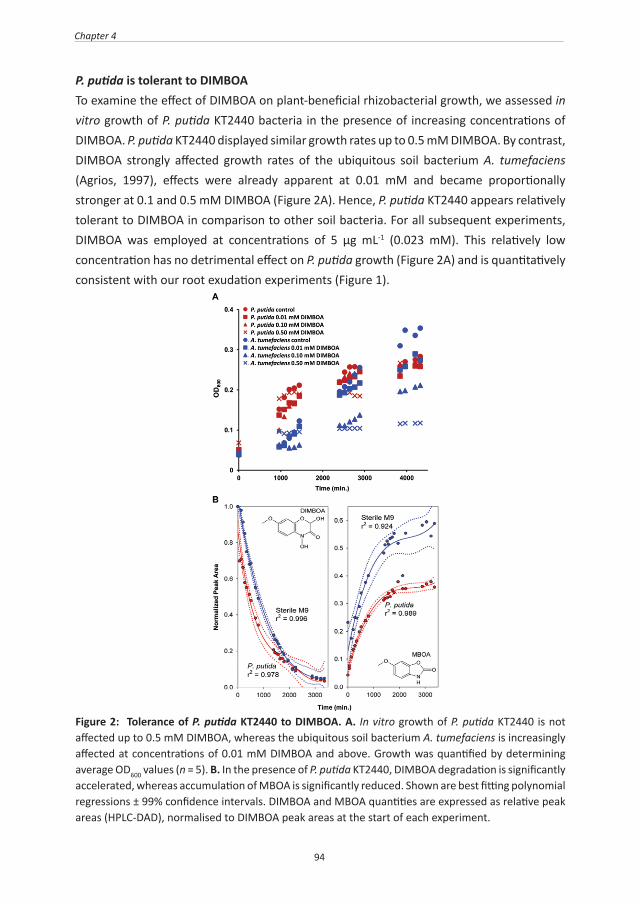
94
Chapter 4
P. putida is tolerant to DIMBOATo examine the effect of DIMBOA on plant-beneficial rhizobacterial growth, we assessed in vitro growth of P. putida KT2440 bacteria in the presence of increasing concentrations of DIMBOA. P. putida KT2440 displayed similar growth rates up to 0.5 mM DIMBOA. By contrast, DIMBOA strongly affected growth rates of the ubiquitous soil bacterium A. tumefaciens (Agrios, 1997), effects were already apparent at 0.01 mM and became proportionally stronger at 0.1 and 0.5 mM DIMBOA (Figure 2A). Hence, P. putida KT2440 appears relatively tolerant to DIMBOA in comparison to other soil bacteria. For all subsequent experiments, DIMBOA was employed at concentrations of 5 μg mL-1 (0.023 mM). This relatively low concentration has no detrimental effect on P. putida growth (Figure 2A) and is quantitatively consistent with our root exudation experiments (Figure 1).
Figure 2: Tolerance of P. putida KT2440 to DIMBOA. A. In vitro growth of P. putida KT2440 is not affected up to 0.5 mM DIMBOA, whereas the ubiquitous soil bacterium A. tumefaciens is increasingly affected at concentrations of 0.01 mM DIMBOA and above. Growth was quantified by determining average OD600 values (n = 5). B. In the presence of P. putida KT2440, DIMBOA degradation is significantly accelerated, whereas accumulation of MBOA is significantly reduced. Shown are best fitting polynomial regressions ± 99% confidence intervals. DIMBOA and MBOA quantities are expressed as relative peak areas (HPLC-DAD), normalised to DIMBOA peak areas at the start of each experiment.
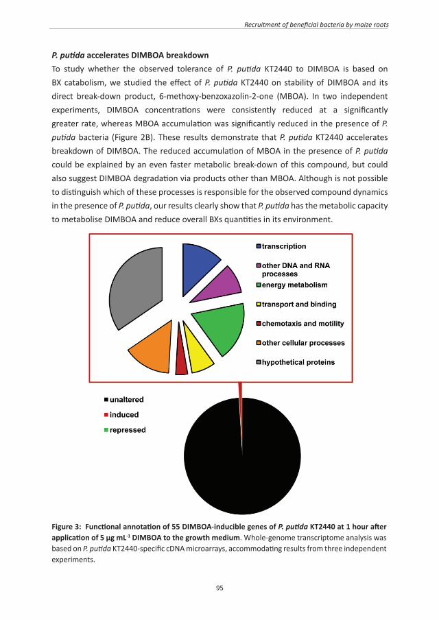
95
Recruitment of beneficial bacteria by maize roots
P. putida accelerates DIMBOA breakdownTo study whether the observed tolerance of P. putida KT2440 to DIMBOA is based on BX catabolism, we studied the effect of P. putida KT2440 on stability of DIMBOA and its direct break-down product, 6-methoxy-benzoxazolin-2-one (MBOA). In two independent experiments, DIMBOA concentrations were consistently reduced at a significantly greater rate, whereas MBOA accumulation was significantly reduced in the presence of P. putida bacteria (Figure 2B). These results demonstrate that P. putida KT2440 accelerates breakdown of DIMBOA. The reduced accumulation of MBOA in the presence of P. putida could be explained by an even faster metabolic break-down of this compound, but could also suggest DIMBOA degradation via products other than MBOA. Although is not possible to distinguish which of these processes is responsible for the observed compound dynamics in the presence of P. putida, our results clearly show that P. putida has the metabolic capacity to metabolise DIMBOA and reduce overall BXs quantities in its environment.
Figure 3: Functional annotation of 55 DIMBOA-inducible genes of P. putida KT2440 at 1 hour after application of 5 µg mL-1 DIMBOA to the growth medium. Whole-genome transcriptome analysis was based on P. putida KT2440-specific cDNA microarrays, accommodating results from three independent experiments.

96
Chapter 4
Impact of DIMBOA on the P. putida transcriptomeThe above in vitro analyses suggest that DIMBOA is metabolised by P. putida. To assess the global impact of DIMBOA on P. putida KT2440, whole-genome gene expression patterns were profiled at 1 hour of exposure to 5 μg mL-1 DIMBOA in M9 growth medium. Using KT2440-specific cDNA microarrays and a false discovery rate of 0.85% (Δ = 1.2; Tusher et al., 2001), we identified 55 genes showing consistently increased levels of transcription in response to DIMBOA treatment across three independent experiments. No genes were identified as significantly repressed by DIMBOA. A total of 36 genes could be ascribed to predicted functions, whereas 19 genes encoded hypothetical proteins of unknown function (Figure 3). Table I lists all 36 DIMBOA-inducible genes of identifiable function. Two groups of genes were of particular interest with respect to P. putida behaviour in the rhizosphere. One group of genes are typical of those associated with degradation of N-heteroaromatic compounds (Table I), and are consistent with the accelerated breakdown of DIMBOA by P. putida KT2440 (Figure 2B). A second group of genes are indicative of bacterial motility (Table I), thereby suggesting a chemotactic response of P. putida KT2440 to DIMBOA.
Table I: Functional annotation of P. putida KT2440 genes with a statistically significant induction at 1 h after exposure to 5 µg mL-1 DIMBOA. Shown are 36 genes with annotated functions out of a total of 55 DIMBOA-inducible genes are listed.
LOCUS GENE NAME
PRODUCT NAME CLASS
TRANSCRIPTION
PP4929 LysR family transcriptional regulator
Regulatory functions: DNA interactions
PP0387 rpoD RNA polymerase sigma factor RpoD Transcription: transcription factors
PP4546 hrpA ATP-dependent helicase, HrpA Transcription: other
PP4553 RNA polymerase sigma-70 factor, ECF subfamily
Transcription: transcription factors
PP4722 greA transcription elongation factor, GreA
Transcription: transcription factors
PP4602 AraC family transcriptional regulator
Regulatory functions: DNA interactions
PP3617 catR LysR family transcriptional regulator
Regulatory functions: DNA interactions
ENERGY METABOLISM
PP4124* nuoG NADH-quinone oxido-reductase, subunit G
Metabolism; Energy Metabolism; Oxidative
phosphorylation; Electron transport
97
Recruitment of beneficial bacteria by maize roots
PP4947 putA Tri-functional transcriptional regulator/ proline dehydrogenase/
pyrroline-5-carboxylate dehydrogenase
Metabolism; Amino Acid Metabolism; Arginine and
proline metabolism
PP4121* nuoCD Bi-functional NADH:ubiquinone oxido-reductase, subunit C/D
Metabolism; Energy Metabolism; Oxidative
phosphorylation; Electron transport
PP0256* molybdopterin oxidoreductase subunit alpha
Metabolism; Energy metabolism; electron
transportPP4690* Rieske (2Fe-2S) domain-containing
proteinMetabolism; Energy
metabolism; electron transport
PP5004 poly(3-hydroxyalkanoate) depolymerase
Metabolism; Energy metabolism; other
PP4434 dadA-1 D-amino acid dehydrogenase small subunit
Metabolism; Energy Metabolism; Nitrogen
metabolism
PP4551* α/β fold hydrolase family
PP4540* α/β fold hydrolase family
PP4661* NAD(P)H dehydrogenase (quinone)
TRANSPORT AND BINDING
PP5206 secretion protein HlyD family protein
Transport and binding proteins; unknown substrate
PP0281 Polar amino acid ABC transporter inner membrane subunit
Transport and binding proteins; amino acids, peptides and amines
PP2604* Major facilitator family transporter, shares homology with pcaK
Transport and binding proteins; unknown
substrate;
PP2241 Major facilitator family transporter Transport and binding proteins; unknown substrate
OTHER DNA AND RNA PROCESSES
PP4812 3-methyladenine DNA glycosylase Genetic Information Processing; Replication and Repair; Base excision repair
PP4708 pnp polynucleotide phosphorylase/ polyadenylase
Genetic Information Processing; Folding, Sorting
and Degradation; RNA degradation
PP4997 pyrR Bi-functional pyrimidine regulatory protein PyrR uracil phosphor-
ribosyltransferase
Metabolism; Nucleotide Metabolism; Pyrimidine
metabolism

98
Chapter 4
PP5286 dut deoxyuridine 5'-tri-phosphate nucleotido-hydrolase
Metabolism; Nucleotide Metabolism; Pyrimidine
metabolism
PP5296 gmk guanylate kinase Metabolism; Nucleotide Metabolism; Purine
metabolism
CHEMOTAXIS AND MOTILITY
PP4888 Methyl-accepting chemotaxis sensory transducer
PP4340 cheY two-component system, chemotaxis family, response
regulator, CheY
Environmental Information Processing; Signal
Transduction; Two-component system.Cellular Processes;
Cell Motility; Bacterial chemotaxis
OTHER CELLULAR PROCESSES
PP4315 PhzF family phenazine biosynthesis protein
PP5000 hslV ATP-dependent protease peptidase subunit: Heat shock protein
Cellular processes; adaptations to atypical
conditions
PP4874 rplI 50S ribosomal protein, L9 Genetic Information Processing; Translation;
Ribosome
PP4830 cobL Precorrin-6Y C5,15-methyl-transferase subunit, CbiE
Metabolism; Metabolism of Cofactors and Vitamins; Porphyrin and chlorophyll
metabolism
PP3446 ilvA-1 Threonine dehydratase Metabolism; Amino Acid Metabolism; Glycine, serine and threonine metabolism.
Metabolism; Amino Acid Metabolism; Valine, leucine and isoleucine biosynthesis
PP5110 ftsE cell division ATP-binding protein FtsE
Environmental Information Processing; Membrane
Transport; ABC transporters
PP5226 Lppl family lipoprotein
PP5398 ISPpu14 transposase Orf1 Transposition; DNA mediated. DNA binding.
Transposase activity
* DIMBOA-inducible genes with predicted functions in metabolism and transport of N-heteroaromatic compounds. See text for details.
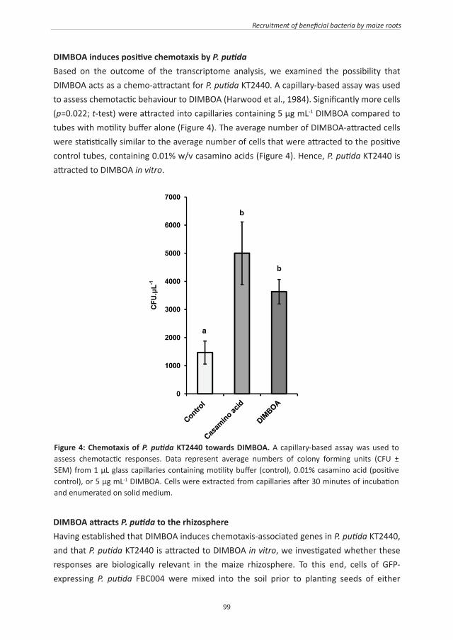
99
Recruitment of beneficial bacteria by maize roots
DIMBOA induces positive chemotaxis by P. putidaBased on the outcome of the transcriptome analysis, we examined the possibility that DIMBOA acts as a chemo-attractant for P. putida KT2440. A capillary-based assay was used to assess chemotactic behaviour to DIMBOA (Harwood et al., 1984). Significantly more cells (p=0.022; t-test) were attracted into capillaries containing 5 µg mL-1 DIMBOA compared to tubes with motility buffer alone (Figure 4). The average number of DIMBOA-attracted cells were statistically similar to the average number of cells that were attracted to the positive control tubes, containing 0.01% w/v casamino acids (Figure 4). Hence, P. putida KT2440 is attracted to DIMBOA in vitro.
Figure 4: Chemotaxis of P. putida KT2440 towards DIMBOA. A capillary-based assay was used to assess chemotactic responses. Data represent average numbers of colony forming units (CFU ± SEM) from 1 μL glass capillaries containing motility buffer (control), 0.01% casamino acid (positive control), or 5 μg mL-1 DIMBOA. Cells were extracted from capillaries after 30 minutes of incubation and enumerated on solid medium.
DIMBOA attracts P. putida to the rhizosphereHaving established that DIMBOA induces chemotaxis-associated genes in P. putida KT2440, and that P. putida KT2440 is attracted to DIMBOA in vitro, we investigated whether these responses are biologically relevant in the maize rhizosphere. To this end, cells of GFP-expressing P. putida FBC004 were mixed into the soil prior to planting seeds of either

100
Chapter 4
DIMBOA-producing wild-type plants (BX1), or BX-deficient bx1 mutant plants. After 7, 14 and 21 days of growth, roots of BX1 and bx1 plants from 2 independent genetic lines were collected and analysed for colonisation by P. putida FBC004 and other (non-GFP expressing) culturable rhizobacteria.
The first experiment was performed with soil that had been autoclaved once before to the start of the experiment (Figure 5A), presenting a relatively low competition environment for the introduced P. putida cells. Two-factor ANOVA of rhizosphere colonisation of plants from Line A revealed a statistically significant interaction between plant genotype (BX1 versus bx1) and bacterial cell type (P. putida versus others) at all three time-points (7 days: F1,14=9.151, p=0.009; 14 days: F1,14=43.432, p<0.001; 21 days: F1,14=7.977; p=0.014), even though a statistically significant main effect of BX genotype could not be detected. Inspection of the data revealed that more P. putida cells were recovered from roots of DIMBOA-producing BX1 plants than from roots of DIMBOA-deficient bx1 plants, whereas this was not the case for numbers of other culturable rhizobacteria. Holm-Šidák comparisons confirmed significantly higher P. putida cell numbers in rhizosphere extracts from BX1 roots compared to that from bx1 roots (statistical probabilities are presented in Figure 5A). For line B, a significant main effect of BX genotype was evident at day 7 (F1,16=18.163; p<0.001) and day 14 (F1,16=19.776; p<0.001), but not at day 21 (F1,16=3.775; p=0.070). However, a statistically significant interaction between BX genotype and rhizobacterial cell type was apparent at all three time-points, including day 21 (F1,16=6.122; p=0.025). Again, Holm-Šidák comparisons indicated significantly greater numbers of P. putida cells in the rhizosphere of BX1 plants compared to that of bx1 plants at all three time points (Figure 5A). Hence, BX1-dependent exudation of DIMBOA stimulates rhizosphere colonisation by P. putida bacteria. To investigate whether BX-dependent attraction of P. putida is also apparent in a more competitive soil environment, we repeated the experiment in non-autoclaved soil (Figure 5B). A significant main effect of BX genotype was observed at day 7 in seedlings from line A (F1,10=6.725; p=0.027), but not at any later growth stage. Holm-Šidák comparisons confirmed significantly higher numbers of P. putida cells in the rhizopshere of 7-day-old BX1 seedlings, but no statistically significant difference in the number of other culturable rhizobacteria (statistical probabilities are presented in Figure 5B). No main effect of BX genotype was identified for plants of line A at days 14 (F1,11=0.110; p=0.746) or 21 (F1,12=4.152; p=0.064). For line B, a significant main effect of BX genotype was observed at 7 days (F1,11=4.904; p=0.049). Although no significant main effect of BX genotype was observed at 14 days (F1,12=4.547; p=0.054), there was a significant interaction between BX genotype and rhizobacterial cell type at this time-point (F1,12=6.425; p=0.026). Subsequent Holm-Šidák comparisons confirmed significantly increased numbers of P. putida in the rhizosphere of BX1 plants at both 7 and 14 days (Figure 5B). At 21 days there was no longer a statistically significant effect by plant genotype, nor was there a statistically significant

101
Recruitment of beneficial bacteria by maize roots
interaction between plant genotype and rhizobacterial cell type (Figure 5B). Together, these data indicate that BX exudation in non-autoclaved soil stimulates rhizosphere colonisation by P. putida of relatively young seedlings. This BX effect becomes variable by 14 days and is absent in 21 day-old plants.
Figure 5: Rhizosphere colonisation of DIMBOA-producing (BX1) and DIMBOA-deficient (bx1) maize lines by green-fluorescent protein (GFP)-expressing P. putida and other culturable rhizobacteria in autoclaved (A) and non-autoclaved soil (B). P. putida cells were introduced into the soil prior to planting of maize seeds. Shown are average values (CFU g-1 root fresh weight ± SEM; n = 6-8), corresponding to P. putida or other rhizobacteria. Cells were enumerated after 7, 14 and 21 days of plant growth by plating root surface extractions onto solid agar medium. Probabilities indicate statistical differences between BX1 and bx1 plants within one line at each time-point (Holm-Šidák pairwise multiple comparisons) when two-factor ANOVA indicated a statistically significant main effect of BX genotype and/or a statistically significant interaction between BX genotype and bacterial cell type (α = 0.05).

102
Chapter 4
DISCUSSION
The rhizosphere is an energy-rich niche that is characterised by a rapid turnover of chemical compounds from plant root exudates (Jones et al., 2009). Before rhizobacteria can exploit these compounds in the rhizosphere, they must first locate their host and tolerate potentially toxic allelochemicals in root exudates. In this study, we provide evidence that rhizosphere-colonising P. putida cells are tolerant of the N-heteroaromatic allelochemical DIMBOA (Figure 2), which is exuded in relatively high quantities from roots of young maize seedlings (Figure 1). Since BXs are nitrogen-containing metabolites, it might be expected that constitutive DIMBOA exudation by seedlings provides significant ecological benefits, outweighing the metabolic cost. Apart from allelopathatic activity by DIMBOA (Niemeyer, 1988, 2009), our study revealed that DIMBOA can also acts as a below-ground semiochemical for recruitment of plant-beneficial rhizobacteria from a competitive soil environment (Figure 5B). Interestingly, mycorrhization of maize was recently reported to boost DIMBOA production (Song et al., 2011). Since mycorrhization is known to cause major qualitative changes in rhizobacterial communities (Linderman, 1988), it is tempting to speculate that increased DIMBOA exudation from mycorrhizal roots contributes to this so-called mycorrhizosphere effect. The BX content of maize roots has been studied extensively, because of their demonstrated roles as allelochemicals (Niemeyer and Perez, 1994; Niemeyer, 2009). Recent studies have identified the glucosides HDMBOA-glc and DIMBOA-glc as the principal BXs in roots and root exudates of maize (Glauser et al., 2011; Robert et al., 2012). DIMBOA was identified in both studies as only a minor component of the total root BX content. A possible explanation for this discrepancy lies in the different method of BXs extraction. In our study, entire root systems were incubated in water for 7 hours, whereas Robert et al. (2012) used a direct sampling method with a 50% (v/v) methanol extraction buffer from the root surface. Hence, the latter method analyzed root-exuded BXs directly, while our method assessed root-exuded BXs after prolonged incubation of the root system in water. Since BX glucosides are readily hydrolysed in water and DIMBOA is considerably more stable than HDMBOA (Maresh et al., 2006), it may not be surprising that our study identified the aglycone DIMBOA as the dominant BX from root exudates. Considering that soils constitute a mostly watery environment, we propose that the more breakdown-resilient DIMBOA compound functions as the long-distance BX signal that recruits beneficial rhizobacteria. The P. putida strain used in our studies was originally isolated from horticultural soil (Nakazawa, 2002), and is a competitive coloniser of rhizospheres of economically important crops (Molina et al., 2000). Using in vivo expression techniques (IVET), Ramos-González et al. (2005) identified 29 genes that are induced following 14 days of growth in the maize rhizosphere, including some with annotated functions in chemotaxis and
103
Recruitment of beneficial bacteria by maize roots
detoxification. However, despite the similarities in general cellular functions, there were no overlapping genes between this IVET study and our transcriptome analysis. A more recent transcriptome study of P. putida KT2440 identified gene induction as the dominant response after 6 days of colonisation in the maize rhizosphere (Matilla et al., 2007), which is in agreement with our finding that DIMBOA enhances P. putida gene expression. In total, Matilla et al. (2007) revealed enhanced expression of 93 genes in the maize rhizosphere, including genes with predicted functions in general metabolism, transcriptional regulation, transport, chemotaxis and DNA metabolism. With the exception of the ISPpu14 transposase Orf1 (PP5398), there is again no overlap between this study and our transcriptome analysis. This is not surprising, since our analysis was specifically focussed on the bacterial response to DIMBOA, and not to the multitude of responses that are required for rhizopshere competence, such as attachment to the maize root surface and metabolism of the wide range of compounds besides DIMBOA in root exudates. Furthermore, the transcriptional response reported in our study was expressed within 1 hour of exposure to DIMBOA. It is, therefore, likely that these gene expression patterns are specific to the initial stages of the interaction: the bacterial response to chemical cues from the host plant in the soil before they attach and establish themselves in the rhizosphere. Since our ultimate objective was to study the maize-bacterium interaction, rather than quantitative gene expression in P. putida KT2440 per se, we made no further attempts to confirm our in vitro transcription profiling with a complementary technique. It remains therefore difficult to establish unequivocally that specific genes identified as DIMBOA-inducible in vitro are in fact responsible for the biological interactions described in this study. Nevertheless, it is still instructive to consider these genes in the light of what is already known about environmentally responsive P. putida genes. Moreover, the DIMBOA-inducible gene expression patterns associated with tolerance to N-heteroaromatic compounds and bacterial motility led us to conduct follow-up experiments, which revealed a novel signalling mechanism during the initial phases of the maize - P. putida interaction. Motility is an essential trait for rhizosphere competence (Lugtenberg and Dekkers, 1999). Our transcriptome analysis identified two DIMBOA-inducible genes that can be associated with bacterial chemotaxis, and a third gene with a putative function in DIMBOA transport (Table I). The DIMBOA-responsive gene cheY (PP4340) is a chemotactic response regulator in bacteria (Wolanin et al., 2003). Furthermore, benzoate chemotaxis in P. putida PRS2000 depends on a methyl-accepting chemotaxis transducer (M-ACT) and the aromatic acid:H+ symporter (AAHS) PcaK (Harwood et al., 1994). Interestingly, our transcriptome analysis included the M-ACT homologue PP4888, and two genes, PP2241 and PP2604, belonging to the Major Facilitator Superfamily (MFS) of AAHS transporters (Pao et al., 1998). Of these two genes, PP2604 shares common features with pcaK (The STRING v.9 database; Szklarczyk et al., 2011). On the basis of these motality-related transcriptional

104
Chapter 4
patterns, we considered the possibility that DIMBOA act as a chemo-attractant for P.putida. This hypothesis was confirmed by our subsequent chemotaxis assays, which demonstrated positive taxis of P. putida KT2440 towards DIMBOA (Figure 4). P. putida KT2440 is tolerant of DIMBOA in comparison to other soil bacteria (Figure 2A). We subsequently found that P. putida KT2440 accelerates decomposition of DIMBOA and its direct break-down product MBOA (Figure 2B), indicating BX catabolism (Kumar et al., 1993). Such a mode of tolerance is supported by our transcriptome analysis, which revealed seven DIMBOA-inducible genes that can be associated with degradation of N-heteroaromatic compounds (Table I). These genes include nuoCD (PP4121) and nuoG (PP4124), which encode subunits of NADH dehydrogenase I, PP4690 encoding a Rieske 2Fe-2S family subunit of soluble dioxygenases, PP0256 encoding a molybdopterin oxidoreductase, PP4661 encoding a putative oxidoreductase, and the α/β hydrolase-fold superfamily genes PP4540 and PP4551, members of which catalyse 1H-3-hydroxy-4-oxoquinoline degradation by P. putida 33/1 (Fischer et al., 1999). We conclude that this mechanism of BX tolerance provides P. putida KT2440 with a competitive advantage over other micro-organisms in exploiting the maize rhizosphere. Our soil-based colonisation essays revealed that P. putida cells colonise maize roots of DIMBOA-producing lines in greater numbers than roots of DIMBOA-deficient lines. Although BX-dependent rhizosphere attraction of P. putida occurred in both autoclaved and non-autoclaved soil (Figure 5), the difference in P. putida colonisation between BX1 and bx1 lines in non-autoclaved soil was only consistent between both lines during the relatively young developmental stages of the plants (Figure 5B). This age-dependent decline in P. putida response to BXs concurs with our finding that DIMBOA root exudation declines steadily as seedlings age (Figure 1). In autoclaved soil, however, this age-dependency was unclear. Autoclaved soil provides a much less competitive environment for the introduced P. putida bacteria than non-autoclaved soil. It is, therefore, plausible that the lower exudation rates of DIMBOA from older plants are still sufficient to attract the bacteria from non-autoclaved soil to the rhizosphere. Alternatively, it is possible that HDMBOA-glc, which did not show a noticeable age-dependent decline in exudation rate (Figure 1), contributes to bacterial recruitment at later developmental stages of the host plant. In both autoclaved and non-autoclaved soil, numbers of other rhizosphere bacteria were similar between roots of BX1 and bx1 plants (Figure 5). The difference in response to BX-exuding roots between P. putida and other rhizobacteria indicates that the microbial composition of the rhizophere is strongly influenced by the presence of DIMBOA in root exudates of the host plant. Apart from direct anti-microbial effects, DIMBOA root exudation may have an additive effect considering that DIMBOA-exposed P. putida showed enhanced expression of the phzF gene (Table I), which encodes an enzyme in the biosynthesis of the broad-spectrum antibiotic phenazine (Blankenfeldt et al., 2004). Other studies have revealed bacterial attraction to

105
Recruitment of beneficial bacteria by maize roots
primary metabolites in plant roots: L-leucine and L-malate attract P. fluorescens to tomato roots (De Weert et al., 2002), while L-malate was found to promote attraction of Bacillus subtilis to the rhizosphere of Arabidopsis thaliana (De Weert et al., 2002). To our knowledge, DIMBOA is the first allelochemical shown to act as a chemo-attractant for beneficial rhizobacteria, which may explain why P. putida KT2440 is such a successful coloniser of the maize rhizosphere (Molina et al., 2000). Our discovery also strengthens the notion that certain bacteria have acquired the ability to detoxify aromatic plant compounds, allowing them to exploit the energy-rich rhizosphere of plant roots exuding allelochemical compounds. These same bacteria can be exploited for the remediation of aromatic pollutants and herbicides (Parales and Harwood, 2002). In summary, our study has shown that root exudation of BXs attracts plant beneficial rhizobacteria. Although BX biosynthesis is mostly developmentally regulated (Frey et al., 2009), recent evidence has revealed that BX production by maize seedlings is to a certain extent responsive to environmental stimuli (Erb et al., 2009; Erb et al., 2009). It would, therefore, be interesting to examine the BX-dependent effects on rhizobacteria during adaptive interactions between above- and belowground defences. Our study also provides important knowledge for agricultural programmes aiming at sustainable yield improvement of cereal crops. Management of soil-borne diseases has proved problematic, because plant roots are relatively inaccessible for fungicidal chemicals. Furthermore, growth promotion by excessive soil fertilisation can have detrimental environmental impacts. Selection for cereal varieties with an increased capacity for BX root exudation will lead to crops with an improved ability to recruit disease-suppressive and growth-promoting rhizosphere communities, thereby reducing the need for repeated applications of fungicides and fertilisers. However, there is evidence that the specialist Western Corn Rootworm (Diabrotica virgifera) uses root-exuded BXs, such as DIMBOA and MBOA, as feeding cues (Bjostad and Hibbard, 1992; Robert et al., 2012). The potential for crop improvement by selection for increased BX exudation should therefore be approached with caution. On the other hand, the accelerated degradation of DIMBOA and MBOA by P. putida (Figure 2B) may interfere with host location by D. virgifera and pose a potential opportunity for biocontrol of this pest.
ACKNOWLEDGEMENTS We thank Prof. Dieter Sicker, University of Leipzig, Germany, for providing us with various standards of BX compounds and Dr. Arnaud Dechesne, Technical University of Denmark, Lyngby for providing the GFP-tagged strain, FBC004. We would also like to thank two anonymous reviewers for their constructive comments on the original version of the manuscript, which helped to improve the manuscript greatly. None of the authors declare any conflicts of interest.

106

107

108
CHAPTER 5
General Discussion

109
General discussion
SCOPE
For a successful parasitic interaction, pathogens and insects have to cope with a plethora of plant defence mechanisms. Some of these defences are pre-existing, while others are inducible by the attacking organism. As was outlined in the General Introduction of this thesis (Figure 1; Chapter 1), induced defence in plants is based on sequentially activated defence layers that are active at different stages of the parasitic interaction. In general terms, these layers of the plant immune system can be separated between pre-invasive defence barriers, such as PAMP-induced closure of stomata (Melotto et al., 2006), early-acting post-invasive defence, such as localised deposition of ROS and callose (Luna et al., 2011), and late-acting post-invasive defences that are under control by de novo produced defence hormones, such as salicylic acid (SA; Heil and Ton, 2008). The overarching objective of the work presented in this thesis was to study the contribution of post-invasive defence barriers to basal resistance (Chapters 2 and 3) and their impact on the interactions with plant-beneficial microbes, such as rhizosphere-colonizing Pseudomonas putida bacteria (Chapter 4).
NATURAL OCCURRING VARIATION IN BASAL RESISTANCE
The vast majority of studies on natural variation in plant defence have focussed on ETI (De Meaux and Mitchell-Olds, 2003; Holub, 2007; Van Poecke et al., 2007), which is likely due to the robustness and reproducibility of the ETI phenotype. There are also numerous studies about natural variation in basal resistance against pathogens and herbivores, many of which are based on the genetic model plant species Arabidopsis (Koornneef et al., 2004). However, relatively few of these have linked this variation to actual resistance mechanisms. The natural variation in basal resistance of Arabidopsis to insects often originates from differences in pre-existing pools of glucosinolates (Kliebenstein et al., 2001; Koornneef et al., 2004). Glucosinolates enable a rapid production of biocidal isothiocyanates after herbivore attack and could therefore, be viewed as a constitutively primed defence mechanism. Natural variation in basal resistance of Arabidopsis against pathogens, on the other hand, seems to stem from more diverse mechanisms than from glucosinolates. For instance, Denby et al., (2004) reported that natural variation in basal resistance against the necrotroph Botrytis cinerea correlates with responsiveness of pathogen- and acifluorfen-induced camalexin, an indole-derived phytoalexin. Further genetic dissection of this basal resistance in a mapping population of recombinant inbred lines (RILs) revealed multiple small-to-medium-effect quantitative trait loci (QTLs), but it remained unclear to what extent these loci influence the responsiveness of camalexin induction itself. Similarly, Llorente et al. (2005) used a RIL population to dissect natural variation in basal resistance against the necrotrophic fungus Plectosphaerella cucumerina, which identified three different QTLs. The most influential

110
Chapter 5
QTL was caused by a polymorphism in the ERECTA gene, a LRR receptor-like kinase protein that influences responsiveness of pathogen-induced callose deposition. Natural variation in resistance against P. syringae pathogens has been reported by different groups (Ton et al., 1999; Kover and Schaal, 2002; Kover et al., 2005; Perchepied et al., 2006). Genetic dissection of variation in basal resistance against virulent P. syringae pv. tomato DC3000 between accessions Bayreuth and Shahdara revealed two major QTLs, of which one was found to regulate responsiveness of SA-inducible defence genes (Perchepied et al., 2006). The studies by Llorente et al. (2005) and Perchepied et al. (2006) illustrate that one gene can have a major contribution to natural variation in responsiveness of basal resistance mechanisms. In support of this, the QTL analysis presented in Chapter 2 revealed one locus on chromosome IV, which controls both responsiveness to exogenously applied SA, and basal resistance to P. syringae pv. tomato DC3000 (Chapter 2; Figures 6 & 7). The development of DNA arrays has made it possible to measure natural variation in the abundance of large numbers of gene transcripts, “expression level polymorphism” (ELP) analysis. If applied to a genetically characterised mapping population, ELP analysis can link natural variation in transcriptome responses to regulatory loci, called expression (e)QTLs. A first genomic comparison of the transcriptional response to exogenously applied SA between seven Arabidopsis accessions revealed that on average 2234 genes were differentially expressed in pair-wise comparisons (Kliebenstein et al., 2006). This variation correlated positively with genomic sequence diversity, suggesting that single nucleotide polymorphisms have relatively little influence on the genetic variation in SA-induced gene expression. Further in depth analysis of these data identified accession Mt-0 as hyper-responsive and accession Cvi-0 as hypo-responsive to SA (Van Leeuwen et al., 2007). Although these studies made an important step towards a better understanding of the evolution of basal resistance, it remains unknown which genomic regions are responsible for this variation, and in how far this variation actually impacts basal pathogen resistance. Further ELP analysis of well-characterised mapping populations would be necessary to identify the regulatory genes that are responsible for both natural variation in gene-network responses and basal defence responsiveness.
NATURAL VARIATION IN BASAL DEFENCE RESPONSIVENESS: ECOLOGICAL IMPLICATIONS
Priming of defence is a phenomenon that manifests itself as an increased responsiveness of inducible defences (Conrath et al., 2006). Since priming protects against a wide variety of diseases and pests, it is plausible that hostile environments select for constitutively primed defence responses. As a consequence, it can be expected that naturally occurring plant species display genetic variation in the responsiveness of inducible defences. Indeed, Chapter

111
General discussion
2 of this thesis shows natural variation between Arabidopsis accessions in the sensitivity of jasmonic acid (JA)- and SA-inducible marker genes, pointing to variation in responsiveness of relatively late-acting post-invasive defence barriers. The same set of accessions also showed natural variation in responsiveness of chitosan-induced callose deposition, suggesting variation in responsiveness of early-acting post-invasive defence barriers. Interestingly, there was a negative correlation between the responsiveness of early-acting defences (callose) and that of late-acting defences (SA-induced PR-1 gene induction). Although a sample size of six accessions is too small to reach definite conclusions about the ecological meaning of these findings, it is tempting to speculate that some of these accessions have de-sensitised their early defences in order to interact with plant beneficial microbes, such as plant-growth promoting rhizobacteria (PGPRs). In order to compensate for this immuno-compromised defence layer, these accessions have primed later-acting defences. Further experiments should be performed to test this hypothesis. Of the possibilities to consider, a HapMap population of 360 Arabidopsis accessions could be screened for PGPR colonization and PAMP-induced callose deposition in the roots (Buckler and Gore, 2007). Identification of genes promoting PGPR colonization but suppressing PAMP-induced callose would confirm this hypothesis.
CYP81D11: A SUPPRESSOR OF EARLY ACTING POST-INVASIVE DEFENCE?
The QTL analysis described in Chapter 2 identified two loci regulating PAMP-induced callose deposition. One major QTL was found on chromosome III, whereas a much weaker QTL was identified at the top of chromosome I. The direction of both QTLs show opposite effects: the major QTL at chromosome III mediates a suppressive effect from Bur-0 parent and stimulatory effect from Col-0 parent, whereas the weaker QTL at chromosome I mediates a stimulatory effect from Bur-0 parent and suppressive effect from Col-0 parent. Although both QTLs still spans a multitude of genes, it is interesting that the major locus on chromosome III maps onto the cis-jasmone inducible gene CYP81D11 (At3g28740). This gene was previously linked to the regulation of indirect plant defences against insects (Bruce et al., 2008; Matthes et al., 2010). Its role in direct plant defences, however, remains unclear. To further investigate a possible function of CYP81D11 in callose deposition, a T-DNA insertion mutant and a CYP81D11 over-expression line in the genetic background of Col-0 (line CYP81D11OE 5-5.3; Bruce et al., 2008; Matthes et al., 2010), were tested for flg22-induced callose deposition, as described by Luna et al., (2011). Interestingly, this experiment revealed that CYP81D11OE plants are severity affected in PAMP-induced callose deposition (Figure 1).
112
Chapter 5
Since the PEN2-dependent break-down product(s) of 4-methoxyindol-3-ylmethylglucosinolate (4MI3G) has been found to act as major regulator of PAMP-induced callose (Clay et al., 2009), and CYP81 enzymes have been shown to modulate the biosynthesis of indolic glucosinolates (Pfalz et al., 2011), it is tempting to hypothesize that CYP81D11 regulates callose by scavenging the callose-inducing break-down product of 4MI3G. To test this hypostasis, we analysed the CYP81D11OE line for endogenous IG levels and break-down products thereof. Although there were no statistically significant differences in total amounts of IGs, these preliminary results revealed increased accumulation of several IG breakdown products, of which 4-methoxyindole-3-carboxaldehyde was the most noticeable (Figure 2A). These findings point to the possibility that CYP81D11 inhibits callose formation through conversion of the callose-inducing PEN2 product into 4-methoxyindole-3-carboxaldehyde (Figure 2B). More experiments need to be carried out to get a more definite outcome concerning this matter. Furthermore, studying PAMP-induced callose phenotypes
Figure 1: CYP81D11 regulates flg22-induced callose depostion. Leaves from hydroponically cultivated 8-day- old seedlings of the wild-type (Col-0), a CYP81D11 T-DNA knock-out line (CYP81D11 KO), and an over-expression line (CYP81D11 OE 5-5.3) were treated with either flg22 (0.1 mM) or mock solution. At 24 h after treatment, cotelydons were collected for aniline blue staining, UV-epifluoresence microscopy, and digital quantification of callose intensity as described (Luna et al., 2011). Shown are relatively callose intensities (% callose-corresponding area). Photographs show representative differences in fluorescent callose signals under UV-epifluoresence microscopy.

113
General discussion
EXPLOITATION OF NATURAL VARIATION IN PLANT DEFENCE IN SUSTAINABLE CROP PROTECTION
How can natural variation be exploited to improve crop protection against diseases and pests? Although Arabidopsis cannot be considered to be a crop, the availability of genetically characterised mapping populations and genome-sequenced accessions provides easy access to the genetic basis of the defence strategies evolved within this species. A potential disadvantage of this approach is that certain defence traits of Arabidopsis are specific for Brassicaceae. For instance, glucosinolates play an important role in defence of Arabidopsis against unadapted insects and pathogens (Tierens et al., 2001; Arany et al., 2008; Bednarek
Figure 2: CYP81D11 as a negative regulator of glucosinolate metabolism and PAMP-induced callose deposition. A. Over-expression of CYP81D11 in CYP81D11 OE 5-5.3 plants results in increased levels of 4-methoxyindole-3-carboxyaldehde, which is a putative breakdown product of the callose-regulatory compound 4-methoxyindol-3-ylmethylglucosinolate (4MI3G). Leaf samples were collected from 5 weeks old plants, freeze-dried and analysed by HPLC following extraction. B. Biosynthesis of 4MI3G and break-down by the atypical myrosinase PEN2 controls PAMP-induced callose in Arabidopsis thaliana. 4MI3G is derived from tryptophan via a multi-step reaction that involves the mono-oxygenases CYP79B2, CYP79B3, CYP83B1 and CYP81F2. Hydrolisis of 4MI3G by PEN2 leads to the accumuation of an unknown product that is thought to promote apoplastic callose depostion (Clay et al., 2009). Shown in red is a putative mechanism by which CYP81D11 suppresses PAMP-induced callose depostion.
of Arabidopsis mutants in genes that are closely co-regulated with CYP81D11 at the gene expression level, e.g. At1g05560 (UGT1), At2g29420 (GSTU7), and At4g34138 (UGT73B1), might be an approach to identify other genes in the pathway.
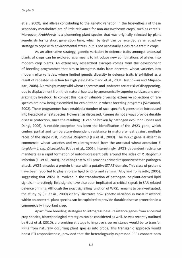
114
Chapter 5
et al., 2009), and alleles contributing to the genetic variation in the biosynthesis of these secondary metabolites are of little relevance for non-brassicaceous crops, such as cereals. Moreover, Arabidopsis is a pioneering plant species that was originally selected by plant geneticists for its short generation time, which by itself can be regarded as an adaptive strategy to cope with environmental stress, but is not necessarily a desirable trait in crops. As an alternative strategy, genetic variation in defence traits amongst ancestral plants of crops can be explored as a means to introduce new combinations of alleles into modern crop plants. An extensively researched example comes from the development of breeding programmes that aim to introgress traits from ancestral wheat varieties into modern elite varieties, where limited genetic diversity in defence traits is exhibited as a result of repeated selection for high yield (Skovmand et al., 2001; Trethowan and Mujeeb-Kazi, 2008). Alarmingly, many wild wheat ancestors and landraces are at risk of disappearing, due to displacement from their natural habitats by agronomically superior cultivars and over grazing by livestock. To combat this loss of valuable diversity, extensive collections of rare species are now being assembled for exploitation in wheat breeding programs (Skovmand, 2002). These programmes have enabled a number of race-specific R genes to be introduced into hexaploid wheat species. However, as discussed, R genes do not always provide durable disease protection, since the resulting ETI can be broken by pathogen evolution (Jones and Dangl, 2006). A notable exception has been the identification of the WKS1 gene, which confers partial and temperature-dependent resistance in mature wheat against multiple races of the stripe rust, Puccinia striiformis (Fu et al., 2009). The WKS1 gene is absent in commercial wheat varieties and was introgressed from the ancestral wheat accession T. turgidum L. ssp. Dicoccoides (Uauy et al., 2005). Interestingly, WKS1-dependent resistance manifests as a rapid formation of auto-fluorescent cells around the sides of P. striiformis infection (Fu et al., 2009), indicating that WKS1 provides primed responsiveness to pathogen attack. WKS1 encodes a protein kinase with a putative START domain. This class of proteins have been reported to play a role in lipid binding and sensing (Alpy and Tomasetto, 2005), suggesting that WKS1 is involved in the transduction of pathogen- or plant-derived lipid signals. Interestingly, lipid signals have also been implicated as critical signals in SAR-related defence priming. Although the exact signalling function of WKS1 remains to be investigated, the study by (Fu et al., 2009) clearly illustrates how genetic variation in basal resistance within an ancestral plant species can be exploited to provide durable disease protection in a commercially important crop. Apart from breeding strategies to introgress basal resistance genes from ancestral crop species, biotechnological strategies can be considered as well. As was recently outlined by Gust et al. (2010), a promising strategy to improve crop resistance would be to transfer PRRs from naturally occurring plant species into crops. This transgenic approach would boost PTI responsiveness, provided that the heterologously expressed PRRs connect onto

115
General discussion
the appropriate defence signalling pathways. Analogous to priming of basal resistance, a primed PTI response would provide broad-spectrum disease resistance if the augmented defence response precedes the opportunity to express ETS by the invading pathogen.
ROLE OF SECONDARY METABOLITES IN BASAL RESISTANCE
For basal defence responses involving secondary defence metabolites, considerable debate has surrounded the evolutionary mechanisms involved in creating the necessary pathways (Kwezi et al., 2007). It is emerging that, for many associated biosynthetic pathways, operons or clusters of co-regulated genes can be responsible for the sequential production of the necessary enzymes (Suzuki et al., 1990). These pathways typically result in the production of penultimate non-biocidal products, e.g. a glucosinolate (see above) or the glucoside of a benzoxazinoid, which provide a primed capacity to produce the biocide upon subsequent pathogen or herbivore attack (Suzuki et al., 1990; Papadopoulou et al., 1999). Interestingly, these so-called phytoanticipins can also exert a signalling role in defence responses (Liu et al., 1997). For instance, glucosinolate metabolites have been found to regulate PAMP-induced depositions of callose-rich papillae in Arabidopsis (Clay et al., 2009). Clusters of genes in the biosynthesis of secondary defence metabolites can comprise paralogous genes from gene duplication, but also non-homologous genes that are organised functionally with concomitant clustering and co-regulation. How such a pathway evolves before giving rise to an adaptive advantage is not fully understood (Kwezi et al., 2007), but it seems obvious that the final biochemical conversion from a non-biocidal storage product to a biocidal defence product is critical in the regulation of these chemical defences. It remains a future challenge in how far genetic variation in the timing, activity, and localisation of hydrolytic enzymes mediating these conversions contributes to differences in basal resistance against diseases and pests. If so, it will be equally interesting to investigate if these enzymes are targeted by pathogen and/or insect effectors.
THE ROLE OF MAIZE BENZOXAZINOIDS IN BASAL RESISTANCE AGAINST APHIDS AND FUNGI
Chapter 3 describes the role of indolic benzoxazinoids (BXs) in maize basal resistance against the bird cherry oat aphid, Rhopalosiphum padi and the pathogenic fungus Setosphaeria turtica. As discussed earlier, indolic glucosinolates play a crucial role in Arabidopsis immunity as they regulate callose deposition via activation of PEN2 (Clay et al., 2009). Maize does not produce indolic glucosinolates, but is equipped with benzoxazinoids (BXs). Since their discovery over 50 years ago, more than 500 papers have been published on the different aspects of their chemistry and biology. However, while the involvement of BXs in plant

116
Chapter 5
defence against different organisms has been well documented, the mechanisms underlying these effects are poorly explored. The objective of the study presented in Chapter 3 was to examine role of BXs in defence against aphids and fungi along with the underlying mechanisms. To this end, we have undertaken a detailed analysis of BX pathway regulation and BX localisation in response to different biotic stresses. The analysis critically depended on the construction of maize mutant lines that are impaired in the first dedicated step of the benzoxazinoid pathways: the conversion of indole-3-glycerol phosphate into indole. In maize, this reaction is catalysed by Indole glycerol phosphate lyase (IGL) or Benzoxazineless1 (BX1). The Igl gene is inducible by herbivory, wounding, insect elicitors or jasmonates (Frey et al., 2000; Frey et al., 2004; Ton et al., 2007; Erb et al., 2009), and is responsible for indole emission. The Bx1 gene is under developmental control and mediates indole production as a substrate for BX biosynthesis. To determine the role of BXs in defence against aphids and fungi, we determined basal resistance of wild-type and mutant bx1 plants in the igl mutant background, thereby preventing BX production from aphid- or fungus-induced IGL. These bx1 igl double mutant lines were derived from two independent reciprocal crosses between a Mutator (Mu)-induced mutant in the Igl gene, and the original bx1bx1 mutant (Hamilton, 1964). Since both parental plants have different genetic backgrounds, their progeny lines in the F3, F4 and F5 generation are still segregating for genetically different alleles from the parental plants. However, these segregation patterns can be expected to largely differ between the two independent progeny lines. Hence, defence phenotypes that are consistently expressed in both bx1 igl progeny lines are unlikely due to segregating genes from the parental backgrounds, but are rather caused by their inability to produce BXs. Moreover, apoplastic infiltration with the BX compound DIMBOA (2,4-dihydroxy-7-methoxy-2H-1,4-benzoxazin-3(4H)-one) stimulated callose deposition (Chapter 3; Figure 8). Together with the defence phenotypes of the BX-deficient bx1 igl lines, these results validate the conclusion that apoplastic DIMBOA stimulates callose deposition in maize. One major objective of this study was to establish whether there is a causal link between aphid resistance and the presence of an intact BX1 gene. Our results clearly demonstrate that this is the case; aphid survival rate and weight gain were higher on bx1 igl plant than Bx1 igl plants from both crosses (Figure 1; Chapter 3). Our results compliment numerous studies which report correlations between aphid performance and BX concentration in plants or in their feeding substrates. For example, rate of intrinsic weight increase of the grain aphid Sitobion avenae, the cherry-oat aphid Rhopalosiphum padi and mean relative growth rates of greenbug Schizaphis graminum and S.avenae were negatively correlated with BX levels in wheat seedlings(Leszczynski and Dixon, 1990; Thackray et al., 1990; Givovich and Niemeyer, 1994). In another study, 20 Hungarian wheat varieties differing in BX levels were tested for infestation rating by R.padi under field conditions and an inverse relationship was reported between infestation rate and BX concentration

117
General discussion
(Gianoli et al., 1996). Similarly, time ingesting from diets containing different quantities of BXs (DIMBOA and DIMBOA-glc) was negatively correlated with BX concentration for five cereal aphid species: M.dirhodum, R.padi, R.maidis, S.avenea and S.gramnium suggesting a role of BXs as feeding deterrent (Givovich and Niemeyer, 1994). Performance of the Russian wheat aphid (Diuraphis noxia) was lower on wheat cultivars with augmented levels of BXs than those having lower quantities. In this study, aphid performance was measured by the time aphids took to reach their feeding site and the number of aphids ultimately reaching phloem. Aphids took more time to reach the phloem and a lower proportion of them were able to reach the phloem of the cultivar with higher BXs contents and vice versa (Givovich and Niemeyer, 1996). The second objective of the work outlined in Chapter 3 was to examine the role of BXs in maize defence against the necrotrophic fungal pathogen S. turcica. Pathogen colonization was scored by measuring fungal hyphael lengths and the relative amount of arrested fungal spores. As expected, Bx1 igl lines from both crosses were more resistant to S. turcica than the corresponding bx1 igl lines (Chapter 3; Figure 3), demonstrating that BXs play a role in basal resistance against microbial pathogens. In wheat and maize, different studies have provided correlative evidence for the role of BXs in basal defense against different pathogens, such as Helminthosporium turticum, Ceplphalosporium maydis, and Puccinia graminis (Niemeyer, 2009). Our study has provided genetic and physiological evidence for a significant contribution of maize BXs to basal resistance against pathogenic fungi.
DIMBOA: A NOVEL APOPLASTIC REGULATOR OF POST-INVASIVE DEFENCE IN MAIZE
BXs are induced upon pathogen/insect attack (Leszczynski and Dixon, 1990; Weibull and Niemeyer, 1995; Gianoli and Niemeyer, 1997) and our data from Chapter 3 are in agreement with these findings. Interestingly, we found that most of these inductions took taking place in the extracellular spaces (apoplast) of the leaf tissue. We chose to quantify BX concentrations in the apoplast because the aphid stylet has to pass through this compartment to reach it feeding site, the phloem, and fungal hyphae colonize the apoplast. As we hypothesised, the most prominent increases in BXs upon aphid feeding, fungal infection and chitosan treatment were found in the apoplastic fluid. These findings provide novel insight into the role of BXs in fungal and aphid resistance. It was previously postulated that the release of toxic BXs are dependent on tissue disruption/damage. Our study reveals for the first time that BX dependent resistance in maize does not depend entirely on tissue disruption. Although chewing insects inflict major tissue disruption, thereby causing cellular release of vacuolar metabolites, the early stages of infestation by aphids and fungi are not associated with major tissue damage. Nevertheless, induction of BX accumulation by these pests

118
Chapter 5
occurred at time-points long preceding major tissue damage. Moreover, it was shown that apoplastic accumulation of DIMBOA signals increased callose deposition (Figure 8; Chapter 3), presumably to keep the accumulation of anti-microbial metabolites concentrated at the sites of colonization. Hence, apoplastic localisation of DIMBOA contributes to early-acting post-invasive defence, which is not only based on its biocidal activity, but also on its role as apoplastic callose-promoting signal. It would be interesting to investigate whether aphids and pathogenic fungi have co-evolved specific effectors that block apoplastic secretion of benzoxazinoid-glucosides and/or hydrolytic activity of corresponding beta-glucosidases. Further research needs to be carried out to answer unanswered questions about the cellular mechanisms regulating apoplastic accumulation of BXs. What are the mechanisms by which BX compounds are deposited into the apoplast? Are the BX compounds secreted in the glycosylated form, along with the corresponding beta-glucosidases? How is the apoplastic DIMBOA perceived by the plant cell? A better understanding of these processes may provide the means to manipulate these processes and enhance resistance against pests and diseases. Of the possibilities to be considered, the most obvious would involve a direct release of toxic BXs from the vacuole or cytoplasm to the apoplast, mediated via a plasma membrane transport system. Microscopic studies using specific antibodies against the BX-specific beta-glucosidases Glu1 and Glu2 (Cicek and Esen, 1999) could reveal whether hydrolysis of BX-glycosides takes place before or after translocation to the apoplast. Alternatively, a forward genetic analysis of PAMP-induced callose deposition could be considered. For instance, a nested association mapping (NAM) population, consisting of 25 recombinant inbred-line (RIL) populations has been developed in maize and been used to dissect resistance traits in maize (Poland et al., 2011). Using the same mapping population, similar studies can be performed to explore the molecular-genetic basis of callose-mediated defence in maize against aphids and fungi. Early acting post-invasive defence is marked by events such as reactive oxygen species accumulation and callose deposition (Luna et al., 2011). The work by Clay et al. (2009) revealed that indolic glucosinolates in Arabidopsis play a regulatory role in flagellin-induced callose deposition. This discovery pointed to the possibility that indole-derived secondary metabolites BXs in cereals could play a similar role in PAMP-induced callose deposition in maize. The results presented in Chapter 3 confirmed this hypothesis and suggest an evolutionary conserved signalling role of indolic metabolites in early post-invasive defence across the plant kingdom.
SIGNALLING ROLE OF DIMBOA IN THE RHIZOSPHERE
Apart from their role in aboveground plant defence, BXs also function in belowground plant defence. BXs are exuded from roots into the rhizosphere, where they act as allelochemicals,

119
General discussion
or exert antimicrobial activities (Niemeyer, 2009). The study described in Chapter 4 describes the belowground impact of BXs in maize-rhizobacteria interactions. The plant root system not only provides support and intake of nutrient and water, but is also a source of secondary metabolites. Plant roots produce and store a blend of chemicals/metabolites, and can release them in the form of root exudates into their immediate vicinity: the rhizosphere. The presence of chemicals in root exudates has a well-documented role in manipulating and dictating the plant’s relationship with pathogenic and symbiotic microbes in the rhizosphere. A well know class of chemicals that are present in root exudates are the isoflavones. These compounds have been identified from root exudates of soybean, which attract nitrogen fixing bacteria Bradyrhizobium japonicum and a pathogen Phytopthora sojae towards roots (Bais et al., 2006). Similarly L-malic acid has been reported to be released by Arabidopsis thaliana in their root exudates, which selectively attracts the plant-beneficial rhizobacterial strain Bacillus subitilis FB17 (Rudrappa et al., 2008). In support of this, the results presented in Chapter 4 provides evidence that maize roots release DIMBOA, which stimulates chemotaxis-related gene expression and in vitro chemotaxis in P. putida KT2440 (Chapter 4; Figures 3 & 4). However, in order for rhizobacteria to establish a plant-beneficial interaction, they not only need to locate their host, but they also need to be capable of tolerating toxic chemicals that are present in the exudates. The ability to tolerate and detoxify these chemicals will provide a competitive advantage to these microbes over other strains that are incapable of tolerating these toxic chemicals in the rhizosphere. Indeed, P. putida KT2440 were tolerant to DIMBOA in comparison to other soil-borne bacteria and its exposure to DIMBOA elicited gene expression that is associated with benzoate breakdown (Chapter 4; Table I). Moreover, in vitro analysis of DIMBOA stability indicated that DIMBOA tolerance of KT2440 bacteria is based on metabolism-dependent breakdown of DIMBOA and 6-methoxy-benzoxazolin-2-one (MBOA), a product of DIMBOA degradation (Chapter 4; Figure 2). Chapter 4 finally revealed that DIMBOA-exuding roots of Bx1 igl plants allowed higher levels of P. putida KT2440 colonized than did DIMBOA-deficient bx1 igl plants. It is, therefore, likely that the increased bacterial colonization of BX-producing roots is the additive result of positive chemotaxis and tolerance to DIMBOA. Root colonization of Arabidopsis by P. putida KT2440 elicits an induced systemic resistance response against P. syringae pv. tomato DC3000 (Matilla et al., 2010). Moreover, preliminary data from our laboratory have revealed that colonization of maize roots by KT2440 bacteria primes emission of wound-inducible volatiles (Figure 3). Hence, P. putida KT2440 is a rhizobacterial strain with plant-beneficial characteristics. Together with the data presented in Chapter 4, these results suggest that BXs fulfil a signalling role in the rhizosphere to recruit and select for plant-beneficial bacteria. Interestingly, however, a recent study by Robert et al., (2012) revealed that these BX signals can also be used by the specialised root herbivore (Diabrotica virgifera) to locate their hosts. Hence, BXs are potent
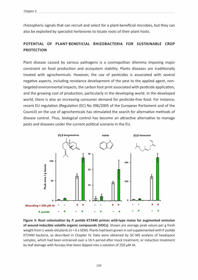
120
Chapter 5
rhizospheric signals that can recruit and select for a plant-beneficial microbes, but they can also be exploited by specialist herbivores to locate roots of their plant hosts.
POTENTIAL OF PLANT-BENEFICIAL RHIZOBACTERIA FOR SUSTAINABLE CROP PROTECTION
Plant disease caused by various pathogens is a cosmopolitan dilemma imposing major constraint on food production and ecosystem stability. Plants diseases are traditionally treated with agrochemicals. However, the use of pesticides is associated with several negative aspects, including resistance development of the pest to the applied agent, non-targeted environmental impacts, the carbon foot print associated with pesticide application, and the growing cost of production, particularly in the developing world. In the developed world, there is also an increasing consumer demand for pesticide-free food. For instance, recent EU regulation (Regulation (EC) No 396/2005 of the European Parliament and of the Council) on the use of agrochemicals has stimulated the search for alternative methods of disease control. Thus, biological control has become an attractive alternative to manage pests and diseases under the current political scenario in the EU.
Figure 3: Root colonization by P. putida KT2440 primes wild-type maize for augmented emission of wound-inducible volatile organic compounds (VOCs). Shown are average peak values per g fresh weight from 1-week-old plants (n = 6 ± SEM). Plants had been grown in soil supplemented with P. putida KT2440 bacteria, as described in Chapter IV. Data were obtained by GC-MS analysis of headspace samples, which had been entrained over a 16 h period after mock treatment, or induction treatment by leaf damage with forceps that been dipped into a solution of 250 µM JA.

121
General discussion
There are different ways by which rhizobacteria can protect plants. These include competition for nutrients (Kamilova et al., 2005), siderophore-mediated competition for iron (Schippers et al., 1987), signal interference (Lin et al., 2003), antibiosis (Haas and Keel, 2003) and ISR (Zamioudis and Pieterse, 2011). Interestingly, transcriptome analysis of DIMBOA-exposed P. putida revealed enhanced expression of the phzF gene (Chapter 4, Table I), which encodes an enzyme in the biosynthesis of the broad-spectrum antibiotic phenazine (Blankenfeldt et al., 2004). These results suggest that DIMBOA exudation from cereal roots has the potential to boost phenazine production by natural Pseudomonas bacteria in the rhizosphere, which has been linked to suppression of take-all disease (Thomashow and Weller, 1988). Moreover, preliminary evidence suggests that root colonization by P. putida KT2440 primes emission of wounding-inducible volatiles aboveground (Figure 3). These volatiles have well-known functions in the recruitment of natural enemies of herbivores (Turlings and Ton, 2006), and some can also serve as long-distance signals to prime defence in neighbouring plants (Heil and Ton, 2008). The study described in Chapter 4 demonstrates that BXs can act as belowground signals to recruit and select for plant-beneficial P. putida bacteria. This discovery is exploitable for plant breeder and biotech companies to sustainably manage pests and diseases by selecting for cereal crops that exude increased amounts of DIMBOA from their roots. Furthermore, it was recently reported that mycorrhization increases DIMBOA levels in maize roots (Song et al., 2011). It is known that mycorrhization causes major changes in the rhizobacterial community (Linderman, 1988). Considering the results in Chapter 4, DIMBOA may be a driving factor behind this plant-beneficial mycorrhizoshere effect. Classical breeding strategies can be used to select varieties with an enhanced capacity to exudate DIMBOA in the roots. If these breeding schemes can be combined with the selection for increased BX content aboveground, these varieties will not only be able to better in recruiting a disease-suppressing rhizosphere, but they will also be more resistant to pests and diseases aboveground. Less targeted approaches can be used to identify similar traits in non-cereal crops. With the next-generation sequencing technologies becoming more cost-efficient, it will become possible to carry out large-scale QTL analyses in order to associate crop performance in terms of growth and stress resistance with metagenomic profiles in the rhizosphere (Poland et al., 2011; Bisseling et al., 2009). Such an approach will lead to the identification of novel plant genes that promote the establishment of a disease-suppressing and growth-promoting rhizosphere.
AKNOWLEDGEMENTS
We thank Georg Jander and Adewale Martins Adio from the Boyce Thompson Institute for Plant Research (Cornell University; New Yourk, United States of America) for analysing

122
Chapter 5
glucosinolate profiles.

123
124
References
References
Abramovitch RB, Janjusevic R, Stebbins CE, Martin GB (2006) Type III effector AvrPtoB requires intrinsic E3 ubiquitin ligase activity to suppress plant cell death and immunity. Proceedings of the National Academy of Sciences of the United States of America 103: 2851-2856
Adam L, Ellwood S, Wilson I, Saenz G, Xiao S, Oliver RP, Turner JG, Somerville S (1999) Comparison of Erysiphe cichoracearum and E. cruciferarum and a Survey of 360 Arabidopsis thaliana Accessions for Resistance to These Two Powdery Mildew Pathogens. Molecular Plant-Microbe Interactions 12: 1031-1043
Agrios GN (1997) Plant Pathology, Ed 4. Academic Press Inc., San Diego, USAAhmad S, Gordon-Weeks R, Pickett J, Ton J (2010) Natural variation in priming of basal resistance:
from evolutionary origin to agricultural exploitation. Molecular Plant Pathology 11: 817-827Ahmad S, Van Hulten M, Martin J, Pieterse CM, Van Wees SCM, Ton J (2011) Genetic dissection
of basal defence responsiveness in accessions of Arabidopsis thaliana. Plant Cell and Environment 34: 1191-1206
Ahmad S, Veyrat N, Gordon-Weeks R, Zhang Y, Martin J, Smart L, Glauser G, Erb M, Flors V, Frey M, Ton J (2011) Benzoxazinoid Metabolites Regulate Innate Immunity against Aphids and Fungi in Maize. Plant Physiology 157: 317-327
Ahn I-P, Kim S, Lee Y-H, Suh S-C (2007) Vitamin B1-Induced Priming Is Dependent on Hydrogen Peroxide and the NPR1 Gene in Arabidopsis. Plant Physiology 143: 838-848
Ahuja I, Kissen R, Bones AM (2012) Phytoalexins in defense against pathogens. Trends in Plant Science 17: 73-90
Alpy F, Tomasetto C (2005) Give lipids a start: The Star-related lipid transfer (START) domain in mammals. Journal of Cell Science 118: 2791-2801
Anderson JP, Badruzsaufari E, Schenk PM, Manners JM, Desmond OJ, Ehlert C, Maclean DJ, Ebert PR, Kazan K (2004) Antagonistic interaction between abscisic acid and jasmonate-ethylene signaling pathways modulates defense gene expression and disease resistance in Arabidopsis. The Plant Cell 16: 3460-3479
Anzai K, Isono K, Okuma K, Suzuki S (1960) The new antibiotics, questiomycins A and B. The Journal of antibiotics 13: 125-132
Arany A, de Jong T, Kim H, van Dam N, Choi Y, Verpoorte R, van der Meijden E (2008) Glucosinolates and other metabolites in the leaves of Arabidopsis thaliana from natural populations and their effects on a generalist and a specialist herbivore. Chemoecology 18: 65-71
Argandoña VH, Luza JG, Niemeyer HM, Corcuera LJ (1980) Role of hydroxamic acids in the resistance of cereals to aphids. Phytochemistry 19: 1665-1668
Attaran E, Zeier TE, Griebel T, Zeier J (2009) Methyl salicylate production and jasmonate signaling are not essential for systemic acquired resistance in Arabidopsis. The Plant Cell 21: 954-971
Atwell S, Huang YS, Vilhjalmsson BJ, Willems G, Horton M, Li Y, Meng D, Platt A, Tarone AM, Hu TT, Jiang R, Muliyati NW, Zhang X, Amer MA, Baxter I, Brachi B, Chory J, Dean C, Debieu M, de Meaux J, Ecker JR, Faure N, Kniskern JM, Jones JDG, Michael T, Nemri A, Roux F, Salt DE, Tang C, Todesco M, Traw MB, Weigel D, Marjoram P, Borevitz JO, Bergelson J, Nordborg M (2010) Genome-wide association study of 107 phenotypes in Arabidopsis thaliana inbred lines. Nature 465: 627-631
Bailey BA, Larson RL (1991) Maize microsomal benzoxazinone N-monooxygenase. Plant Physiology 95: 792-796
Bais HP, Weir TL, Perry LG, Gilroy S, Vivanco JM (2006) The role of root exudates in rhizosphere interations with plants and other organisms. In Annual Review of Plant Biology, Vol 57. Annual Reviews, Palo Alto, pp 233-266
Baumeler A, Hesse M, Werner C (2000) Benzoxazinoids-cyclic hydroxamic acids, lactams and their corresponding glucosides in the genus Aphelandra (Acanthaceae). Phytochemistry 53: 213-222
Beckers GJM, Jaskiewicz M, Liu Y, Underwood WR, He SY, Zhang S, Conrath U (2009) Mitogen-Activated Protein Kinases 3 and 6 Are Required for Full Priming of Stress Responses in Arabidopsis thaliana. The Plant Cell 21: 944-953
Bednarek P, Pislewska-Bednarek M, Svatos A, Schneider B, Doubsky J, Mansurova M, Humphry

125
References
M, Consonni C, Panstruga R, Sanchez-Vallet A, Molina A, Schulze-Lefert P (2009) A Glucosinolate Metabolism Pathway in Living Plant Cells Mediates Broad-Spectrum Antifungal Defense. Science 323: 101-106
Bensen RJ, Johal GS, Crane VC, Tossberg JT, Schnable PS, Meeley RB, Briggs SP (1995) Cloning and characterization of the maize An1 gene. The Plant Cell 7: 75-84
Berrocal-Lobo M, Molina A, Solano R (2002) Constitutive expression of ETHYLENE-RESPONSE-FACTOR1 in Arabidopsis confers resistance to several necrotrophic fungi. The Plant Journal 29: 23-32
Bisseling T, Dangl JL, Schulze-Lefert P (2009) Next-Generation Communication. Science 324: 691Bjostad LB, Hibbard BE (1992) 6-Methoxy-2-benzoxazolinone: A semiochemical for host location by
western corn rootworm larvae. Journal of Chemical Ecology 18: 931-944Blankenfeldt W, Kuzin AP, Skarina T, Korniyenko Y, Tong L, Bayer P, Janning P, Thomashow LS,
Mavrod DV (2004) Structure and function of the phenazine biosynthetic protein PhzF from Pseudomonas fluorescens. Proceedings of the National Academy of Sciences of the United States of America 101: 16431-16436
Blée E (2002) Impact of phyto-oxylipins in plant defense. Trends in Plant Science 7: 315-322Bodenhausen N (2007) Arabidopsis thaliana response to insect feeding: new components controlling
defense gene expression and plant resistance. Ph.D. thesis, University of Lausanne Bodenhausen N, Reymond P (2007) Signaling Pathways Controlling Induced Resistance to Insect
Herbivores in Arabidopsis. Molecular Plant-Microbe Interactions 20: 1406-1420Bouchabke O, Chang F, Simon M, Voisin R, Pelletier G, Durand-Tardif M (2008) Natural Variation in
Arabidopsis thaliana as a tool for highlighting differential drought responses. PLoS ONE 3: e1705
Boudart G, Jamet E, Rossignol M, Lafitte C, Borderies G, Jauneau A, Esquerre-Tugaye MT, Pont-Lezica R (2005) Cell wall proteins in apoplastic fluids of Arabidopsis thaliana rosettes: identification by mass spectrometry and bioinformatics. Proteomics 5: 212-221
Boyle C, Walters DR (2006) Saccharin-induced protection against powdery mildew in barley: effects on growth and phenylpropanoid metabolism. Plant Pathology 55: 82-91
Bruce TJA, Matthes MC, Chamberlain K, Woodcock CM, Mohib A, Webster B, Smart LE, Birkett MA, Pickett JA, Napier JA (2008) cis-Jasmone induces Arabidopsis genes that affect the chemical ecology of multitrophic interactions with aphids and their parasitoids. Proceedings of the National Academy of Sciences of the United States of America 105: 4553-4558
Bruce TJA, Matthes MC, Napier JA, Pickett JA (2007) Stressful “memories” of plants: Evidence and possible mechanisms. Plant Science 173: 603-608
Buckler E, Gore M (2007) An Arabidopsis haplotype map takes root. Nature Genetics 39: 1056-1057Burow M, Wittstock U, Gershenzon J (2008) Sulfur-containing secondary metabolites and their role
in plant defense sulfur metabolism in phototrophic organisms. In R Hell, C Dahl, D Knaff, T Leustek, eds, Vol 27. Springer Netherlands, pp 201-222
Cambier V, Hance T, De Hoffmann E (2001) Effects of 1,4-benzoxazin-3-one derivatives from maize on survival and fecundity of Metopolophium dirhodum (Walker) on artificial diet. Journal of Chemical Ecology 27: 359-370
Chanda B, Xia Y, Mandal MK, Yu K, Sekine K, Gao Q-m, Selote D, Hu Y, Stromberg A, Navarre D, Kachroo A, Kachroo P (2011) Glycerol-3-phosphate is a critical mobile inducer of systemic immunity in plants. Nature Genetics 43: 421-427
Chase WR, Nair MG, Putnam AR, Mishra SK (1991) Azobenzene: microbial transformation of rye (secale cereale L.) allelochemical in field soils by Acinetobacter calcoaceticus Journal of Chemical Ecology 17: 1575-1584
Chaturvedi R, Krothapalli K, Makandar R, Nandi A, Sparks AA, Roth MR, Welti R, Shah J (2008) Plastid omega3-fatty acid desaturase-dependent accumulation of a systemic acquired resistance inducing activity in petiole exudates of Arabidopsis thaliana is independent of jasmonic acid. The Plant Journal 54: 106-117
Chen X, Steed A, Harden C, Nicholson P (2006) Characterization of Arabidopsis thaliana–Fusarium graminearum interactions and identification of variation in resistance among ecotypes. Molecular Plant Pathology 7: 391-403
Choi J, Choi D, Lee S, Ryu C-M, Hwang I (2011) Cytokinins and plant immunity: old foes or new friends? Trends in Plant Science 16: 388-394
Chung C-L, Longfellow J, Walsh E, Kerdieh Z, Van Esbroeck G, Balint-Kurti P, Nelson R (2010) Resistance loci affecting distinct stages of fungal pathogenesis: use of introgression lines for

126
References
QTL mapping and characterization in the maize - Setosphaeria turcica pathosystem. BMC Plant Biology 10: 103
Cicek M, Esen A (1999) Expression of soluble and catalytically active plant (monocot) beta-glucosidases in E. coli. Biotechnology and Bioengineering 63: 392-400
Clay NK, Adio AM, Denoux C, Jander G, Ausubel FM (2009) Glucosinolate metabolites required for an Arabidopsis innate immune response. Science 323: 95-101
Cohen YR (2002) beta-aminobutyric acid-induced resistance against plant pathogens. Plant Disease 86: 448-457
Conrath U (2011) Molecular aspects of defence priming. Trends in Plant Science 16: 524-531Conrath U, Beckers GJ, Flors V, Garcia-Agustin P, Jakab G, Mauch F, Newman MA, Pieterse CM,
Poinssot B, Pozo MJ, Pugin A, Schaffrath U, Ton J, Wendehenne D, Zimmerli L, Mauch-Mani B (2006) Priming: getting ready for battle. Molecular Plant-Microbe Interactions 19: 1062-1071
Cordier C, Pozo MJ, Barea JM, Gianinazzi S, Gianinazzi-Pearson V (1998) Cell defense responses associated with localized and systemic resistance to Phytophthora induced in tomato by an arbuscular mycorrhizal fungus. Molecular Plant Microbe-Interactions 11: 1017-1028
Cui H, Xiang T, Zhou JM (2009) Plant immunity: a lesson from pathogenic bacterial effector proteins. Cell Microbiology 11: 1453-1461
Czechowski T, Bari RP, Stitt M, Scheible WR, Udvardi MK (2004) Real-time RT-PCR profiling of over 1400 Arabidopsis transcription factors: unprecedented sensitivity reveals novel root- and shoot-specific genes. The Plant Journal 38: 366-379
Czechowski T, Stitt M, Altmann T, Udvardi MK, Scheible W-R (2005) Genome-wide identification and testing of superior reference genes for transcript normalization in Arabidopsis. Plant Physiology 139: 5-17
Czjzek M, Cicek M, Zamboni V, Burmeister WP, Bevan DR, Henrissat B, Esen A (2001) Crystal structure of a monocotyledon (maize ZMGlu1) beta-glucosidase and a model of its complex with p-nitrophenyl beta-D-thioglucoside. Biochemical Journal 354: 37-46
D'Alessandro M, Held M, Triponez Y, Turlings TC (2006) The role of indole and other shikimic acid derived maize volatiles in the attraction of two parasitic wasps. Journal of Chemical Ecology 32: 2733-2748
Dabney AR, Storey JD (2007) A new approach to intensity-dependent normalization of two-channel microarrays. Biostatistics 8: 128-139
De Almeida LFR, Frei F, Mancini E, De Martino L, De Feo V (2010) Phytotoxic Activities of Mediterranean Essential Oils. Molecules 15: 4309-4323
De Meaux J, Mitchell-Olds T (2003) Evolution of plant resistance at the molecular level: ecological context of species interactions. Heredity 91: 345-352
De Weert S, Vermeiren H, Mulders IHM, Kuiper I, Hendrickx N, Bloemberg GV, Vanderleyden J, De Mot R, Lugtenberg BJJ (2002) Flagella-Driven Chemotaxis Towards Exudate Components Is an Important Trait for Tomato Root Colonization by Pseudomonas fluorescens. Molecular Plant-Microbe Interactions 15: 1173-1180
Dechesne A, Pallud C, Bertolla F, Grundmann GL (2005) Impact of the microscale distribution of a Pseudomonas strain introduced into soil on potential contacts with indigenous bacteria. Applied and Environmental Microbiology 71: 8123-8131
Delp G, Gradin T, Ahman I, Jonsson LM (2009) Microarray analysis of the interaction between the aphid Rhopalosiphum padi and host plants reveals both differences and similarities between susceptible and partially resistant barley lines. Molecular Genetics & Genomics 281: 233-248
Denby KJ, Jason LJM, Murray SL, Last RL (2005) ups1, an Arabidopsis thaliana camalexin accumulation mutant defective in multiple defence signalling pathways. The Plant Journal 41: 673-684
Denby KJ, Kumar P, Kliebenstein DJ (2004) Identification of Botrytis cinerea susceptibility loci in Arabidopsis thaliana. The Plant Journal 38: 473-486
Dervinis C, Frost CJ, Lawrence SD, Novak NG, Davis JM (2010) Cytokinin Primes Plant Responses to Wounding and Reduces Insect Performance. Journal of Plant Growth Regulation 29: 289-296
Durrant WE, Dong X (2004) Systemic acquired resistance. Annual Review of Phytopathology 42: 185-209
Engelberth J, Alborn HT, Schmelz EA, Tumlinson JH (2004) Airborne signals prime plants against insect herbivore attack. Proceedings of the National Academy of Sciences of the United States of America 101: 1781-1785

127
References
Erb M, Flors V, Karlen D, de Lange E, Planchamp C, D'Alessandro M, Turlings TC, Ton J (2009) Signal signature of aboveground-induced resistance upon belowground herbivory in maize. The Plant Journal 59: 292-302
Erb M, Gordon-Weeks R, Flors V, Camanes G, Turlings TC, Ton J (2009) Belowground ABA boosts aboveground production of DIMBOA and primes induction of chlorogenic acid in maize. Plant Signaling & Behavior 4: 636-638
Eulgem T, Rushton PJ, Schmelzer E, Hahlbrock K, Somssich IE (1999) Early nuclear events in plant defence signalling: rapid gene activation by WRKY transcription factors. Embo Journal 18: 4689-4699
Facchini PJ (2001) Alkaloid biosysnthesis in plants: biochemistry, cell Biology, molecular regulation, and metabolic engineering applications. Annual Review of Plant Physiology and Plant Molecular Biology 52: 29-66
Faessel L, Nassr N, Lebeau T, Walter B (2010) Chemically-induced resistance on soybean inhibits nodulation and mycorrhization. Plant and Soil 329: 259-268
Fan J, Crooks C, Lamb C (2008) High-throughput quantitative luminescence assay of the growth in planta of Pseudomonas syringae chromosomally tagged with Photorhabdus luminescens luxCDABE. The Plant Journal 53: 393-399
Fischer F, Kunne S, Fetzner S (1999) Bacterial 2,4-dioxygenases: new members of the alpha/beta hydrolase-fold superfamily of enzymes functionally related to serine hydrolases. Journal of Bacteriology 181: 5725-5733
Flors V, Ton J, Jakab G, Mauch-Mani B (2005) Abscisic acid and callose: Team players in defence against pathogens? Journal of Phytopathology 153: 377-383
Forsyth A, Mansfield JW, Grabov N, de Torres M, Sinapidou E, Grant MR (2010) Genetic Dissection of Basal Resistance to Pseudomonas syringae pv. phaseolicola in Accessions of Arabidopsis. Molecular Plant-Microbe Interactions 23: 1545-1552
Frebortova J, Novak O, Frebort I, Jorda R (2010) Degradation of cytokinins by maize cytokinin dehydrogenase is mediated by free radicals generated by enzymatic oxidation of natural benzoxazinones. The Plant Journal 61: 467-481
Frey M, Chomet P, Glawischnig E, Stettner C, Grun S, Winklmair A, Eisenreich W, Bacher A, Meeley RB, Briggs SP, Simcox K, Gierl A (1997) Analysis of a chemical plant defense mechanism in grasses. Science 277: 696-699
Frey M, Schullehner K, Dick R, Fiesselmann A, Gierl A (2009) Benzoxazinoid biosynthesis, a model for evolution of secondary metabolic pathways in plants. Phytochemistry 70: 1645-1651
Frey M, Spiteller D, Boland W, Gierl A (2004) Transcriptional activation of Igl, the gene for indole formation in Zea mays: a structure-activity study with elicitor-active N-acyl glutamines from insects. Phytochemistry 65: 1047-1055
Frey M, Stettner C, Pare PW, Schmelz EA, Tumlinson JH, Gierl A (2000) An herbivore elicitor activates the gene for indole emission in maize. Proceedings of the National Academy of Sciences of the United States of America 97: 14801-14806
Friebe A, Vilich V, Hennig L, Kluge M, Sicker D (1998) Detoxification of Benzoxazolinone Allelochemicals from Wheat by Gaeumannomyces graminis var. tritici, G. graminis var. graminis, G. graminis var.avenae, and Fusarium culmorum. Applied and Environmental Microbiology 64: 2386-2391
Friebe A, Wieland I, Schulz M (1996) Tolerance of Avena sativa to the allelochemical benzoxazolinone. Degradation of BOA by root-colonizing bacteria. Journal of Applied Botany-Angewandte Botanik 70: 150-154
Frost CJ, Mescher MC, Carlson JE, De Moraes CM (2008) Plant Defense Priming against Herbivores: Getting Ready for a Different Battle. Plant Physiology 146: 818-824
Fu D, Uauy C, Distelfeld A, Blechl A, Epstein L, Chen X, Sela H, Fahima T, Dubcovsky J (2009) A Kinase-START Gene Confers Temperature-Dependent Resistance to Wheat Stripe Rust. Science 323: 1357-1360
Fu ZQ, Guo M, Jeong BR, Tian F, Elthon TE, Cerny RL, Staiger D, Alfano JR (2007) A type III effector ADP-ribosylates RNA-binding proteins and quells plant immunity. Nature 447: 284-288
Gagliardo R, Chilton W (1992) Soil transformation of 2(3H)-benzoxazolone of rye into phytotoxic 2-amino-3H-phenoxazin-3-one. Journal of Chemical Ecology 18: 1683-1691
Gerber NN, Lechevalier MP (1964) Phenazines and Phenoxaziones from Waksmania aerata sp. nov. and Pseudomonas iodina. Biochemistry 3: 598-602

128
References
Gianoli E, Niemeyer HM (1997) Characteristics of Hydroxamic Acid Induction in Wheat Triggered by Aphid Infestation. Journal of Chemical Ecology 23: 2695-2705
Gianoli E, Papp M, Niemeyer HM (1996) Costs and benefits of hydroxamic acids-related resistance in winter wheat against the bird cherry-oat aphid, Rhopalosiphum padi L. Annals of Applied Biology 129: 83-90
Givovich A, Niemeyer HM (1994) Effect of hydroxamic acids on feeding-behavior and performance of cereal aphids (Hemiptera, Aphididea) on wheat. European Journal of Entomology 91: 371-374
Givovich A, Niemeyer HM (1996) Role of hydroxamic acids in the resistance of wheat to the Russian Wheat Aphid, Diuraphis noxia (Mordvilko) (Hom., Aphididae). Journal of Applied Entomology 120: 537-539
Glauser G, Marti G, Villard N, Doyen GA, Wolfender J-L, Turlings TCJ, Erb M (2011) Induction and detoxification of maize 1,4-benzoxazin-3-ones by insect herbivores. The Plant Journal 68: 901-911
Glazebrook J (2005) Contrasting mechanisms of defense against biotrophic and necrotrophic pathogens. Annual Review of Phytopathology 43: 205-227
Glazebrook J, Rogers EE, Ausubel FM (1997) Use of Arabidopsis for genetic dissection of plant defense responses. Annual Review of Genetics 31: 547-569
Gomez-Gomez L, Boller T (2000) FLS2: An LRR receptor-like kinase involved in the perception of the bacterial elicitor flagellin in Arabidopsis. Molecular Cell 5: 1003-1012
González-Lamothe R, Mitchell G, Gattuso M, Diarra M, Malouin F, Bouarab K (2009) Plant Antimicrobial Agents and Their Effects on Plant and Human Pathogens. International Journal of Molecular Sciences 10: 3400-3419
Großkinsky DK, Naseem M, Abdelmohsen UR, Plickert N, Engelke T, Griebel T, Zeier J, Novák O, Strnad M, Pfeifhofer H, van der Graaff E, Simon U, Roitsch T (2011) Cytokinins Mediate Resistance against Pseudomonas syringae in Tobacco through Increased Antimicrobial Phytoalexin Synthesis Independent of Salicylic Acid Signaling. Plant Physiology 157: 815-830
Guo M, Tian F, Wamboldt Y, Alfano JR (2009) The Majority of the Type III Effector Inventory of Pseudomonas syringae pv. tomato DC3000 Can Suppress Plant Immunity. Molecular Plant-Microbe Interactions 22: 1069-1080
Gust AA, Brunner F, Nürnberger T (2010) Biotechnological concepts for improving plant innate immunity. Current Opinion in Biotechnology 21: 204-210
Haas D, Keel C (2003) Regulation of antibiotic production in root-colonizing Pseudomonas spp. and relevance for biological control of plant disease. Annual Review of Phytopathology 41: 117-153
Haichar FZ, Marol C, Berge O, Rangel-Castro JI, Prosser JI, Balesdent J, Heulin T, Achouak W (2008) Plant host habitat and root exudates shape soil bacterial community structure. International Society for Microbial Ecology Journal 2: 1221-1230
Halkier BA, Gershenzon J (2006) Biology and biochemistry of glucosinolates. In Annual Review of Plant Biology, Vol 57, pp 303-333
Hamiduzzaman MM, Jakab G, Barnavon L, Neuhaus JM, Mauch-Mani B (2005) beta-Aminobutyric acid-induced resistance against downy mildew in grapevine acts through the potentiation of callose formation and jasmonic acid signaling. Molecular Plant-Microbe Interactions 18: 819-829
Hamilton RH (1964) A corn mutant deficient in 2,4-Dihydroxy-7-Methoxy-1,4-Benzoxazin-3-One with an altered tolerance of atrazine. Weeds 12: 27-30
Handelsman J, Stabb EV (1996) Biocontrol of soilborne plant pathogens. The Plant Cell 8: 1855-1869Harwood CS, Nichols NN, Kim MK, Ditty JL, Parales RE (1994) Identification of the pcaRKF gene cluster
from Pseudomonas putida: involvement in chemotaxis, biodegradation, and transport of 4-hydroxybenzoate. Journal of Bacteriology 176: 6479-6488
Harwood CS, Rivelli M, Ornston LN (1984) Aromatic acids are chemoattractants for Pseudomonas putida. Journal of Bacteriololgy 160: 622-628
Heck S, Grau T, Buchala A, Metraux JP, Nawrath C (2003) Genetic evidence that expression of NahG modifies defence pathways independent of salicylic acid biosynthesis in the Arabidopsis-Pseudomonas syringae pv. tomato interaction. The Plant Journal 36: 342-352
Heil M (2002) Ecological costs of induced resistance. Current Opinion in Plant Biology 5: 345-350Heil M (2009) Damaged-self recognition in plant herbivore defence. Trends in Plant Science 14: 356-

129
References
363Heil M, Baldwin IT (2002) Fitness costs of induced resistance: emerging experimental support for a
slippery concept. Trends in Plant Science 7: 61-67Heil M, Ibarra-Laclette E, Adame-Alvarez RM, Martinez O, Ramirez-Chavez E, Molina-Torres J,
Herrera-Estrella L (2012) How plants sense wounds: damaged-self recognition is based on plant-derived elicitors and induces octadecanoid signaling. PLoS ONE 7: e30537
Heil M, Silva Bueno JC (2007) Within-plant signaling by volatiles leads to induction and priming of an indirect plant defense in nature. Proceedings of the National Academy of Sciences of the United States of America 104: 5467-5472
Heil M, Ton J (2008) Long-distance signalling in plant defence. Trends in Plant Science 13: 264-272Hoffland E, Hakulinen J, Van Pelt JA (1996) Comparison of systemic resistance induced by avirulent
and nonpathogenic Pseudomonas species. Phytopathology 86: 757-762Holub EB (2007) Natural variation in innate immunity of a pioneer species. Current Opinion in Plant
Biology 10: 415-424Houterman PM, Ma L, Van Ooijen G, De Vroomen MJ, Cornelissen BJC, Takken FLW, Rep M (2009)
The effector protein Avr2 of the xylem-colonizing fungus Fusarium oxysporum activates the tomato resistance protein I-2 intracellularly. The Plant Journal 58: 970-978
Huckelhoven R (2007) Cell wall-associated mechanisms of disease resistance and susceptibility. Annual Review of Phytopathology 45: 101-127
Huffaker A, Kaplan F, Vaughan MM, Dafoe NJ, Ni X, Rocca JR, Alborn HT, Teal PEA, Schmelz EA (2011) Novel Acidic Sesquiterpenoids Constitute a Dominant Class of Pathogen-Induced Phytoalexins in Maize. Plant Physiology 156: 2082-2097
Huffaker A, Pearce G, Ryan CA (2006) An endogenous peptide signal in Arabidopsis activates components of the innate immune response. Proceedings of the National Academy of Sciences of the United States of America 103: 10098-10103
Iritri M, Faoro F (2009) Chitosan as a MAMP, searching for a PRR. Plant , Signaling & Behavior 4: 66-68Iwai T, Seo S, Mitsuhara I, Ohashi Y (2007) Probenazole-Induced Accumulation of Salicylic Acid Confers
Resistance to Magnaporthe grisea in Adult Rice Plants. Plant and Cell Physiology 48: 915-924Jakab G, Cottier V, Toquin V, Rigoli G, Zimmerli L, Metraux JP, Mauch-Mani B (2001) beta-Aminobutyric
acid-induced resistance in plants. European Journal of Plant Pathology 107: 29-37Jaskiewicz M, Conrath U, Peterhaensel C (2011) Chromatin modification acts as a memory for systemic
acquired resistance in the plant stress response. Embo Reports 12: 50-55Jia Z, Zou B, Wang X, Qiu J, Ma H, Gou Z, Song S, Dong H (2010) Quercetin-induced H2O2 mediates
the pathogen resistance against Pseudomonas syringae pv. Tomato DC3000 in Arabidopsis thaliana. Biochemical and Biophysical Research Communications 396: 522-527
Jin L, Wang S, Wang X, Shen Y (2009) Seed size influences arbuscular mycorrhizal symbiosis across leguminous host-plant species at the seedling stage. Symbiosis 49: 111-116
Jonczyk R, Schmidt H, Osterrieder A, Fiesselmann A, Schullehner K, Haslbeck M, Sicker D, Hofmann D, Yalpani N, Simmons C, Frey M, Gierl A (2008) Elucidation of the final reactions of DIMBOA-glucoside biosynthesis in maize: characterization of Bx6 and Bx7. Plant Physiology 146: 1053-1063
Jones DL, Nguyen C, Finlay RD (2009) Carbon flow in the rhizosphere: carbon trading at the soil-root interface. Plant and Soil 321: 5-33
Jones JDG, Dangl JL (2006) The plant immune system. Nature 444: 323-329Jung HW, Tschaplinski TJ, Wang L, Glazebrook J, Greenberg JT (2009) Priming in Systemic Plant
Immunity. Science 324: 89-91Kagan IA, Hammerschmidt R (2002) Arabidopsis ecotype variability in camalexin production and
reaction to Infection by Alternaria brassicicola. Journal of Chemical Ecology 28: 2121-2140Kamilova F, Validov S, Azarova T, Mulders I, Lugtenberg B (2005) Enrichment for enhanced competitive
plant root tip colonizers selects for a new class of biocontrol bacteria. Environmental Microbiology 7: 1809-1817
Kauss H, Jeblick W, Ziegler J, Krabler W (1994) Pretreatment of parsley (Petroselinum crispum L.) suspension cultures with methyl jasmonate enhances elicitation of activated oxygen species. Plant Physiology 105: 89-104
Kennedy DO, Wightman EL (2011) Herbal Extracts and Phytochemicals: Plant Secondary Metabolites and the Enhancement of Human Brain Function. Advances in Nutrition: An International Review Journal 2: 32-50

130
References
Kliebenstein DJ, Kroymann J, Brown P, Figuth A, Pedersen D, Gershenzon J, Mitchell-Olds T (2001) Genetic control of natural variation in Arabidopsis glucosinolate accumulation. Plant Physiology 126: 811-825
Kliebenstein DJ, Rowe HC (2008) Ecological costs of biotrophic versus necrotrophic pathogen resistance, the hypersensitive response and signal transduction. Plant Science 174: 551-556
Kliebenstein DJ, West MA, van Leeuwen H, Kim K, Doerge RW, Michelmore RW, St Clair DA (2006) Genomic survey of gene expression diversity in Arabidopsis thaliana. Genetics 172: 1179-1189
Koch E, Slusarenko A (1990) Arabidopsis is susceptible to infection by a downy mildew fungus. The Plant Cell 2: 437-445
Kodama O, Suzuki T, Miyakawa J, Akatsuka T (1988) Ultraviolet-induced accumulation of phytoalexins in rice leaves. Agricultural and Biological Chemistry 52: 2469-2473
Kohler A, Schwindling S, Conrath U (2002) Benzothiadiazole-induced priming for potentiated responses to pathogen infection, wounding, and infiltration of water into leaves requires the NPR1/NIM1 gene in Arabidopsis. Plant Physiololy 128: 1046-1056
Koornneef A, Leon-Reyes A, Ritsema T, Verhage A, Den Otter FC, Van Loon LC, Pieterse CM (2008) Kinetics of salicylate-mediated suppression of jasmonate signaling reveal a role for redox modulation. Plant Physiology 147: 1358-1368
Koornneef A, Pieterse CMJ (2008) Cross talk in defense signaling. Plant Physiology 146: 839-844Koornneef M, Alonso-Blanco C, Vreugdenhil D (2004) Naturally occurring genetic variation in
Arabidopsis thaliana. Annual Review of Plant Biology 55: 141-172Kover PX, Schaal BA (2002) Genetic variation for disease resistance and tolerance among Arabidopsis
thaliana accessions. Proceedings of the National Academy of Sciences of the United States of America 99: 11270-11274
Kover PX, Wolf JB, Kunkel BN, Cheverud JM (2005) Genetic architecture of Arabidopsis thaliana response to infection by Pseudomonas syringae. Heredity 94: 507-517
Krogh SS, Mensz SJ, Nielsen ST, Mortensen AG, Christophersen C, Fomsgaard IS (2006) Fate of benzoxazinone allelochemicals in soil after incorporation of wheat and rye sprouts. Journal of Agricultural and Food Chemistry 54: 1064-1074
Kumar P, Gagliardo RW, Chilton WS (1993) Soil Transformation of Wheat and Corn Metabolites Mboa and Dim2boa into Aminophenoxazinones. Journal of Chemical Ecology 19: 2453-2461
Kwezi L, Meier S, Mungur L, Ruzvidzo O, Irving H, Gehring C (2007) The Arabidopsis thaliana brassinosteroid receptor (AtBRI1) contains a domain that functions as a guanylyl cyclase in vitro. PLoS ONE 2: e449
Leszczynski B, Dixon AFG (1990) Resistance of cereals to aphids: Interaction between hydroxamic acids and the aphid Sitobion avenae (Homoptera: Aphididae). Annals of Applied Biology 117: 21-30
Li L, Li CY, Howe GA (2001) Genetic analysis of wound signaling in tomato. Evidence for a dual role of jasmonic acid in defense and female fertility. Plant Physiology 127: 1414-1417
Liavonchanka A, Feussner I (2006) Lipoxygenases: Occurrence, functions and catalysis. Journal of Plant Physiology 163: 348-357
Liljeroth E, Bengtsson T, Wiik L, Andreasson E (2010) Induced resistance in potato to <i>Phytphthora infestans</i>—effects of BABA in greenhouse and field tests with different potato varieties. European Journal of Plant Pathology 127: 171-183
Lin Y-H, Xu J-L, Hu J, Wang L-H, Ong SL, Leadbetter JR, Zhang L-H (2003) Acyl-homoserine lactone acylase from Ralstonia strain XJ12B represents a novel and potent class of quorum-quenching enzymes. Molecular Microbiology 47: 849-860
Linderman RG (1988) Mycorrhizal interactions with the rhizosphere microflora - The mycorrhizopsphere effect. Phytopathology 78: 366-371
Lipka V, Dittgen J, Bednarek P, Bhat R, Wiermer M, Stein M, Landtag J, Brandt W, Rosahl S, Scheel D, Llorente F, Molina A, Parker J, Somerville S, Schulze-Lefert P (2005) Pre- and postinvasion defenses both contribute to nonhost resistance in Arabidopsis. Science 310: 1180-1183
Liu P-P, von Dahl CC, Klessig DF (2011) The Extent to Which Methyl Salicylate Is Required for Signaling Systemic Acquired Resistance Is Dependent on Exposure to Light after Infection. Plant Physiology 157: 2216-2226
Liu Y, Ruoho A, Rao V, Hurley J (1997) Catalytic mechanism of the adenylyl and guanylyl cyclases: modeling and mutational analysis. Proceedings of the National Academy of Sciences of the

131
References
United States of America 94: 13414 - 13419Livak KJ, Schmittgen TD (2001) Analysis of Relative Gene Expression Data Using Real-Time Quantitative
PCR and the 2−ΔΔCT Method. Methods 25: 402-408Llorente F, Alonso-Blanco C, Sánchez-Rodriguez C, Jorda L, Molina A (2005) ERECTA receptor-like
kinase and heterotrimeric G protein from Arabidopsis are required for resistance to the necrotrophic fungus Plectosphaerella cucumerina. The Plant Journal 43: 165-180
Lorenzo O, Chico JM, Sanchez-Serrano JJ, Solano R (2004) JASMONATE-INSENSITIVE1 encodes a MYC transcription factor essential to discriminate between different jasmonate-regulated defense responses in Arabidopsis. The Plant Cell 16: 1938-1950
Lorenzo O, Piqueras R, Sánchez-Serrano JJ, Solano R (2003) ETHYLENE RESPONSE FACTOR1 integrates signals from ethylene and jasmonate pathways in plant defense. The Plant Cell 15: 165-178
Lorenzo O, Solano R (2005) Molecular players regulating the jasmonate signalling network. Current Opinion in Plant Biology 8: 532-540
Lugtenberg B, Kamilova F (2009) Plant-Growth-Promoting Rhizobacteria. Annual Review of Microbiology 63: 541-556
Lugtenberg BJJ, Dekkers LC (1999) What makes Pseudomonas bacteria rhizosphere competent? Environmental Microbiology 1: 9-13
Lukasik E, Takken FL (2009) STANDing strong, resistance proteins instigators of plant defence. Current Opinion in Plant Biology 12: 427-436
Luna E, Bruce TJA, Roberts MR, Flors V, Ton J (2012) Next-Generation Systemic Acquired Resistance. Plant Physiology 158: 844-853
Luna E, Pastor V, Robert J, Flors V, Mauch-Mani B, Ton J (2011) Callose Deposition: A Multifaceted Plant Defense Response. Molecular Plant-Microbe Interactions 24: 183-193
Macias FA, Molinillo JM, Varela RM, Galindo JC (2007) Allelopathy--a natural alternative for weed control. Pest Management Science 63: 327-348
Mao G, Meng X, Liu Y, Zheng Z, Chen Z, Zhang S (2011) Phosphorylation of a WRKY Transcription Factor by Two Pathogen-Responsive MAPKs Drives Phytoalexin Biosynthesis in Arabidopsis. The Plant Cell 23: 1639-1653
Maresh J, Zhang J, Lynn DG (2006) The Innate Immunity of Maize and the Dynamic Chemical Strategies Regulating Two-Component Signal Transduction in Agrobacterium tumefaciens. ACS Chemical Biology 1: 165-175
Martino LD, Mancini E, Almeida LFRd, Feo VD (2010) The Antigerminative Activity of Twenty-Seven Monoterpenes. Molecules 15: 6630-6637
Matilla MA, Espinosa-Urgel M, Rodriguez-Herva JJ, Ramos JL, Ramos-Gonzalez MI (2007) Genomic analysis reveals the major driving forces of bacterial life in the rhizosphere. Genome Biology 8
Matilla MA, Ramos JL, Bakker PAHM, Doornbos R, Badri DV, Vivanco JM, Ramos-González MI (2010) Pseudomonas putida KT2440 causes induced systemic resistance and changes in Arabidopsis root exudation. Environmental Microbiology Reports 2: 381-388
Matthes MC, Bruce TJA, Ton J, Verrier PJ, Pickett JA, Napier JA (2010) The transcriptome of cis-jasmone-induced resistance in Arabidopsis thaliana and its role in indirect defence. Planta 232: 1163-1180
McDowell JM, Woffenden BJ (2003) Plant disease resistance genes: recent insights and potential applications. Trends in Biotechnology 21: 178-183
Melanson D, Chilton MD, Masters-Moore D, Chilton WS (1997) A deletion in an indole synthase gene is responsible for the DIMBOA-deficient phenotype of bxbx maize. Proceedings of the National Academy of Sciences of the United States of America 94: 13345-13350
Melotto M, Underwood W, He SY (2008) Role of stomata in plant innate immunity and foliar bacterial diseases. Annual Review of Phytopathology 46: 101-122
Melotto M, Underwood W, Koczan J, Nomura K, He SY (2006) Plant stomata function in innate immunity against bacterial invasion. Cell 126: 969-980
Mena M, Ambrose BA, Meeley RB, Briggs SP, Yanofsky MF, Schmidt RJ (1996) Diversification of C-function activity in maize flower development. Science 274: 1537-1540
Mewis I, Appel HM, Hom A, Raina R, Schultz JC (2005) Major Signaling Pathways Modulate Arabidopsis Glucosinolate Accumulation and Response to Both Phloem-Feeding and Chewing Insects. Plant Physiology 138: 1149-1162
Michael W (2000) Interference of alkaloids with neuroreceptors and ion channels. In R Atta ur, ed,
132
References
Studies in Natural Products Chemistry, Vol Volume 21, Part B. Elsevier, pp 3-122Michael W (2003) Evolution of secondary metabolites from an ecological and molecular phylogenetic
perspective. Phytochemistry 64: 3-19Mishina TE, Zeier J (2007) Pathogen-associated molecular pattern recognition rather than development
of tissue necrosis contributes to bacterial induction of systemic acquired resistance in Arabidopsis. The Plant Journal 50: 500-513
Mitchell-Olds T, Schmitt J (2006) Genetic mechanisms and evolutionary significance of natural variation in Arabidopsis. Nature 441: 947-952
Mithoefer A, Boland W (2008) Recognition of herbivory-associated molecular patterns. Plant Physiology 146: 825-831
Miya A, Albert P, Shinya T, Desaki Y, Ichimura K, Shirasu K, Narusaka Y, Kawakami N, Kaku H, Shibuya N (2007) CERK1, a LysM receptor kinase, is essential for chitin elicitor signaling in Arabidopsis. Proceedings of the National Academy of Sciences of the United States of America 104: 19613-19618
Miyakoshi M, Shintani M, Terabayashi T, Kai S, Yamane H, Nojiri H (2007) Transcriptome Analysis of Pseudomonas putida KT2440 Harboring the Completely Sequenced IncP-7 Plasmid pCAR1. Journal of Bacteriology 189: 6849-6860
Molina L, Ramos C, Duque E, Ronchel MC, Garcıa JM, Wyke L, Ramos JL (2000) Survival of Pseudomonas putida KT2440 in soil and in the rhizosphere of plants under greenhouse and environmental conditions. Soil Biology and Biochemistry 32: 315-321
Morant AV, Jorgensen K, Jorgensen C, Paquette SM, Sanchez-Perez R, Moller BL, Bak S (2008) beta-Glucosidases as detonators of plant chemical defense. Phytochemistry 69: 1795-1813
Mukherjee M, Larrimore KE, Ahmed NJ, Bedick TS, Barghouthi NT, Traw MB, Barth C (2010) Ascorbic Acid Deficiency in Arabidopsis Induces Constitutive Priming That is Dependent on Hydrogen Peroxide, Salicylic Acid, and the NPR1 Gene. Molecular Plant-Microbe Interactions 23: 340-351
Mur LAJ, Naylor G, Warner SAJ, Sugars JM, White RF, Draper J (1996) Salicylic acid potentiates defence gene expression in tissue exhibiting acquired resistance to pathogen attack. The Plant Journal 9: 559-571
Mysore KS, Ryu C-M (2004) Nonhost resistance: how much do we know? Trends in Plant Science 9: 97-104
Nakazawa T (2002) Travels of a Pseudomonas, from Japan around the world. Environmental Microbiology 4: 782-786
Nelson KE, Weinel C, Paulsen IT, Dodson RJ, Hilbert H, Martins dos Santos VA, Fouts DE, Gill SR, Pop M, Holmes M, Brinkac L, Beanan M, DeBoy RT, Daugherty S, Kolonay J, Madupu R, Nelson W, White O, Peterson J, Khouri H, Hance I, Chris Lee P, Holtzapple E, Scanlan D, Tran K, Moazzez A, Utterback T, Rizzo M, Lee K, Kosack D, Moestl D, Wedler H, Lauber J, Stjepandic D, Hoheisel J, Straetz M, Heim S, Kiewitz C, Eisen JA, Timmis KN, Dusterhoft A, Tummler B, Fraser CM (2002) Complete genome sequence and comparative analysis of the metabolically versatile Pseudomonas putida KT2440. Environmental Microbiology 4: 799-808
Nielsen R (2005) Molecular signatures of natural selection. Annual Review of Genetics 39: 197-218Niemeyer HM (1988) Hydroxamic acids (4-hydroxy-1,4-benzoxazin-3-ones), defence chemicals in the
gramineae. Phytochemistry 27: 3349-3358Niemeyer HM (2009) Hydroxamic Acids Derived from 2-Hydroxy-2H-1,4-Benzoxazin-3(4H)-one: Key
Defense Chemicals of Cereals. Journal of Agricultural and Food Chemistry 57: 1677-1696Niemeyer HM, Perez FJ (1994) Potential of Hydroxamic Acids in the Control of Cereal Pests, Diseases,
and Weeds. In Allelopathy, Vol 582. American Chemical Society, pp 260-270Nikus J, Jonsson LMV (1999) Tissue localization of β-glucosidase in rye, maize and wheat seedlings.
Physiologia Plantarum 107: 373-378Nomura K, Melotto M, He S-Y (2005) Suppression of host defense in compatible plant–Pseudomonas
syringae interactions. Current Opinion in Plant Biology 8: 361-368Nordborg M, Weigel D (2008) Next-generation genetics in plants. Nature 456: 720-723Oikawa A, Ishihara A, Iwamura H (2002) Induction of HDMBOA-Glc accumulation and DIMBOA-Glc
4-O-methyltransferase by jasmonic acid in poaceous plants. Phytochemistry 61: 331-337Oikawa A, Ishihara A, Tanaka C, Mori N, Tsuda M, Iwamura H (2004) Accumulation of HDMBOA-Glc
is induced by biotic stresses prior to the release of MBOA in maize leaves. Phytochemistry 65: 2995-3001

133
References
Oostendorp M, Kunz W, Dietrich B, Staub T (2001) Induced Disease Resistance in Plants by Chemicals. European Journal of Plant Pathology 107: 19-28
Ossowski S, Schneeberger K, Clark RM, Lanz C, Warthmann N, Weigel D (2008) Sequencing of natural strains of Arabidopsis thaliana with short reads. Genome Research 18: 2024-2033
Pao SS, Paulsen IT, Saier MH, Jr. (1998) Major facilitator superfamily. Microbiology and Molecular Biology Reviews 62: 1-34
Papadopoulou K, Melton RE, Leggett M, Daniels MJ, Osbourn AE (1999) Compromised disease resistance in saponin-deficient plants. Proceedings of the National Academy of Sciences of the United States of America 96: 12923-12928
Parales RE, Harwood CS (2002) Bacterial chemotaxis to pollutants and plant-derived aromatic molecules. Current Opinion in Microbiology 5: 266-273
Parker JE, Coleman MJ, Szabo V, Frost LN, Schmidt R, vanderBiezen EA, Moores T, Dean C, Daniels MJ, Jones JDG (1997) The Arabidopsis downy mildew resistance gene RPP5 shares similarity to the toll and interleukin-1 receptors with N and L6. The Plant Cell 9: 879-894
Pedras MSC, Adio AM (2008) Phytoalexins and phytoanticipins from the wild crucifers Thellungiella halophila and Arabidopsis thaliana: Rapalexin A, wasalexins and camalexin. Phytochemistry 69: 889-893
Perchepied L, Kroj T, Tronchet M, Loudet O, Roby D (2006) Natural variation in partial resistance to Pseudomonas syringae is controlled by two major QTLs in Arabidopsis thaliana. PLoS ONE 1: e123
Pfalz M, Mikkelsen MD, Bednarek P, Olsen CE, Halkier BA, Kroymann J (2011) Metabolic Engineering in Nicotiana benthamiana Reveals Key Enzyme Functions in Arabidopsis Indole Glucosinolate Modification. The Plant Cell 23: 716-729
Pieterse CMJ, Leon-Reyes A, Van der Ent S, Van Wees SCM (2009) Networking by small-molecule hormones in plant immunity. Nature Chemical Biology 5: 308-316
Pieterse CMJ, van Wees SC, van Pelt JA, Knoester M, Laan R, Gerrits H, Weisbeek PJ, van Loon LC (1998) A novel signaling pathway controlling induced systemic resistance in Arabidopsis. Plant Cell 10: 1571-1580
Pieterse CMJ, Van Wees SCM, Hoffland E, Van Pelt JA, Van Loon LC (1996) Systemic resistance in Arabidopsis induced by biocontrol bacteria is independent of salicylic acid accumulation and pathogenesis-related gene expression. Plant Cell 8: 1225-1237
Poland JA, Bradbury PJ, Buckler ES, Nelson RJ (2011) Genome-wide nested association mapping of quantitative resistance to northern leaf blight in maize. Proceedings of the National Academy of Sciences of the United States of America 108: 6893-6898
Pozo MJ, Azcón-Aguilar C (2007) Unraveling mycorrhiza-induced resistance. Current Opinion in Plant Biology 10: 393-398
Pozo MJ, Van Der Ent S, Van Loon LC, Pieterse CM (2008) Transcription factor MYC2 is involved in priming for enhanced defense during rhizobacteria-induced systemic resistance in Arabidopsis thaliana. New Phytologist 180: 511-523
Pozo MJ, Van Loon LC, Pieterse CMJ (2005) Jasmonates - signals in plant-microbe interactions. Journal of Plant Growth Regulation 23: 211-222
Pozo MJ, Verhage A, García-Andrade J, García JM, Azcón-Aguilar C (2009) Priming plant defence against pathogens by arbuscular mycorrhizal fungi mycorrhizas - Functional processes and ecological impact. In C Azcón-Aguilar, JM Barea, S Gianinazzi, V Gianinazzi-Pearson, eds. Springer Berlin Heidelberg, pp 1-13
Pre M, Atallah M, Champion A, De Vos M, Pieterse CMJ, Memelink J (2008) The AP2/ERF domain transcription factor ORA59 integrates jasmonic acid and ethylene signals in plant defense. Plant Physiology 147: 1347-1357
Ramakers C, Ruijter JM, Deprez RH, Moorman AF (2003) Assumption-free analysis of quantitative real-time polymerase chain reaction (PCR) data. Neuroscience Letters 339: 62-66
Ramos-Gonzalez MI, Campos MJ, Ramos JL (2005) Analysis of Pseudomonas putida KT2440 gene expression in the maize rhizosphere: In vitro expression technology capture and identification of root-activated promoters. Journal of Bacteriology 187: 4033-4041
Rangel-Castro JI, Killham K, Ostle N, Nicol GW, Anderson IC, Scrimgeour CM, Ineson P, Meharg A, Prosser JI (2005) Stable isotope probing analysis of the influence of liming on root exudate utilization by soil microorganisms. Environmental Microbiology 7: 828-838
Rashid MA, Gustafson KR, Cardellina JH, 2nd, Boyd MR (1996) A benzoic acid glycoside from

134
References
Geniostoma antherotrichum. Phytochemistry 41: 1205-1207Rasmann S, De Vos M, Casteel CL, Tian D, Halitschke R, Sun JY, Agrawal AA, Felton GW, Jander G
(2012) Herbivory in the Previous Generation Primes Plants for Enhanced Insect Resistance. Plant Physiology 158: 854-863
Rasmann S, Kollner TG, Degenhardt J, Hiltpold I, Toepfer S, Kuhlmann U, Gershenzon J, Turlings TCJ (2005) Recruitment of entomopathogenic nematodes by insect-damaged maize roots. Nature 434: 732-737
Ren D, Liu Y, Yang K-Y, Han L, Mao G, Glazebrook J, Zhang S (2008) A fungal-responsive MAPK cascade regulates phytoalexin biosynthesis in Arabidopsis. Proceedings of the National Academy of Sciences of the United States of America 105: 5638-5643
Robert CAM, Veyrat N, Glauser G, Marti G, Doyen GR, Villard N, Gaillard MDP, Köllner TG, Giron D, Body M, Babst BA, Ferrieri RA, Turlings TCJ, Erb M (2012) A specialist root herbivore exploits defensive metabolites to locate nutritious tissues. Ecology Letters 15: 55-64
Rohmer M (1999) The discovery of a mevalonate-independent pathway for isoprenoid biosynthesis in bacteria, algae and higher plants[dagger]. Natural Product Reports 16
Romeis J, Zebitz CPW (1997) Searching behaviour of Encarsia formosa as mediated by colour and honeydew. Entomologia Experimentalis et Applicata 82: 299-309
Ross AF (1961) Systemic acquired resistance induced by localized virus infections in plants. Virology 14: 340-358
Rostás M (2007) The effects of 2,4-dihydroxy-7-methoxy-1,4-benzoxazin-3-one on two species of Spodoptera and the growth of Setosphaeria turcica in vitro. Journal of Pest Science 80: 35-41
Rostas M, Ton J, Mauch-Mani B, Turlings TCJ (2006) Fungal infection reduces herbivore-induced plant volatiles of maize but does not affect naive parasitoids. Journal of Chemical Ecology 32: 1897-1909
Rowe HC, Walley JW, Corwin J, Chan EKF, Dehesh K, Kliebenstein DJ (2010) Deficiencies in Jasmonate-Mediated Plant Defense Reveal Quantitative Variation in Botrytis cinerea Pathogenesis. PLoS Pathogens 6
Rudrappa T, Czymmek KJ, Paré PW, Bais HP (2008) Root-Secreted Malic Acid Recruits Beneficial Soil Bacteria. Plant Physiology 148: 1547-1556
Saeed AI, Sharov V, White J, Li J, Liang W, Bhagabati N, Braisted J, Klapa M, Currier T, Thiagarajan M, Sturn A, Snuffin M, Rezantsev A, Popov D, Ryltsov A, Kostukovich E, Borisovsky I, Liu Z, Vinsavich A, Trush V, Quackenbush J (2003) TM4: A free, open-source system for microarray data management and analysis. Biotechniques 34: 374-378
Sanchez-Vallet A, Ramos B, Bednarek P, López G, Piślewska-Bednarek M, Schulze-Lefert P, Molina A (2010) Tryptophan-derived secondary metabolites in Arabidopsis thaliana confer non-host resistance to necrotrophic Plectosphaerella cucumerina fungi. The Plant Journal 63: 115-127
Scheer JM, Ryan CA (2002) The systemin receptor SR160 from Lycopersicon peruvianum is a member of the LRR receptor kinase family. Proceedings of the National Academy of Sciences of the United States of America 99: 9585-9590
Schippers B, Bakker AW, Bakker P (1987) Interactions of deleterious and benficial rhizoshere microorgansims and the the effect of cropping practices. Annual Review of Phytopathology 25: 339-358
Schlaeppi K, Abou-Mansour E, Buchala A, Mauch F (2010) Disease resistance of Arabidopsis to Phytophthora brassicae is established by the sequential action of indole glucosinolates and camalexin. The Plant Journal 62: 840-851
Schlaeppi K, Mauch F (2010) Indolic secondary metabolites protect Arabidopsis from the oomycete pathogen Phytophthora brassicae. Plant Signaling and Behavior 5: 1099-1101
Schmelz EA, Kaplan F, Huffaker A, Dafoe NJ, Vaughan MM, Ni X, Rocca JR, Alborn HT, Teal PE (2011) Identity, regulation, and activity of inducible diterpenoid phytoalexins in maize. Proceedings of the National Academy of Sciences of the United States of America 108: 5455-5460
Schmittgen TD, Livak KJ (2008) Analyzing real-time PCR data by the comparative C-T method. Nature Protocols 3: 1101-1108
Schwessinger B, Zipfel C (2008) News from the frontline: recent insights into PAMP-triggered immunity in plants. Current Opinion in Plant Biology 11: 389-395
Shindo C, Bernasconi G, Hardtke CS (2007) Natural Genetic Variation in Arabidopsis: Tools, Traits and Prospects for Evolutionary Ecology. Annals of Botany 99: 1043-1054
Shkolnik-Inbar D, Bar-Zvi D (2010) ABI4 mediates abscisic acid and cytokinin inhibition of lateral root

135
References
formation by reducing polar auxin transport in Arabidopsis. The Plant Cell 22: 3560-3573Shorey HH, Hale RL (1965) Mass-rearing of the larvae of nine noctoid species on a simple artificial
medium. Journal of Economic Entomology 58: 522-524Simon M, Loudet O, Durand S, Berard A, Brunel D, Sennesal F-X, Durand-Tardif M, Pelletier G,
Camilleri C (2008) Quantitative trait loci mapping in five new large recombinant inbred line populations of Arabidopsis thaliana genotyped with consensus single-nucleotide polymorphism markers. Genetics 178: 2253-2264
Skovmand B, Reynolds MP, DeLacy IH (2001) Mining wheat germplasm collections for yield enhancing traits. Euphytica 119: 25-32
Skovmand BS, Rajaram, S.J.M., Ribaut, J.M. and Hede, A.R. (2002) Wheat genetic resources. In: Bread Wheat: improvement and production,production,FAO Plant Production and Protection Series, Rome(Curtis, B.C.,Rajaram, S. and Gomez Macpherson, H., eds), 30, 567. ISBN:9251048096
Slaughter A, Daniel X, Flors V, Luna E, Hohn B, Mauch-Mani B (2012) Descendants of Primed Arabidopsis Plants Exhibit Resistance to Biotic Stress. Plant Physiology 158: 835-843
Sofie D, Jos V, Yaacov O (2003) Plant Growth-Promoting Effects of Diazotrophs in the Rhizosphere. Critical Reviews in Plant Sciences 22: 107-149
Song YY, Cao M, Xie LJ, Liang XT, Zeng RS, Su YJ, Huang JH, Wang RL, Luo SM (2011) Induction of DIMBOA accumulation and systemic defense responses as a mechanism of enhanced resistance of mycorrhizal corn (Zea mays L.) to sheath blight. Mycorrhiza 21: 721-731
Srivastava P, George S, Marois JJ, Wright DL, Walker DR (2011) Saccharin-induced systemic acquired resistance against rust (Phakopsora pachyrhizi) infection in soybean: Effects on growth and development. Crop Protection 30: 726-732
Staal J, Kaliff M, Dewaele E, Persson M, Dixelius C (2008) RLM3, a TIR domain encoding gene involved in broad-range immunity of Arabidopsis to necrotrophic fungal pathogens. The Plant Journal 55: 188-200
Stacey G, McAlvin CB, Kim S-Y, Olivares J, Soto MJ (2006) Effects of endogenous salicylic acid on nodulation in the model legumes Lotus japonicus and Medicago truncatula. Plant Physiology 141: 1473-1481
Subbiah V, Reddy KJ (2010) Interactions between ethylene, abscisic acid and cytokinin during germination and seedling establishment in Arabidopsis. Journal of Biosciences 35: 451-458
Suzuki N, Choe H, Nishida Y, Yamawaki-Kataoka Y, Ohnishi S, Tamaoki T, Kataoka T (1990) Leucine-rich repeats and carboxyl terminus are required for interaction of yeast adenylate cyclase with RAS proteins. Proceedings of the National Academy of Sciences of the United States of America 87: 8711 - 8715
Szklarczyk D, Franceschini A, Kuhn M, Simonovic M, Roth A, Minguez P, Doerks T, Stark M, Muller J, Bork P, Jensen LJ, von Mering C (2011) The STRING database in 2011: functional interaction networks of proteins, globally integrated and scored. Nucleic Acids Research 39: D561-568
Talke I, Blaudez D, Maathuis F, Sanders D (2003) CNGCs: prime targets of plant cyclic nucleotide signalling? Trends in Plant Science 8: 286 - 293
Tautz J, Rostas M (2008) Honeybee buzz attenuates plant damage by caterpillars. Current Biology 18: R1125-R1126
Thackray DJ, Wrattent SD, Edwards PJ, Niemeyer HM (1990) Resistance to the aphids Sitobion avenae and Rhopalosiphum padi in Gramineae in relation to hydroxamic acid levels. Annals of Applied Biology 116: 573-582
Thomashow LS, Weller DM (1988) Role of a phenazine antibiotic from Pseudimonas fluorescens in biological control of Gaeumannomyces graminis var tritici. Journal of Bacteriology 170: 3499-3508
Thomma BPHJ, Eggermont K, Penninckx IAMA, Mauch-Mani B, Vogelsang R, Cammue BPA, Broekaert WF (1998) Separate jasmonate-dependent and salicylate-dependent defense-response pathways in Arabidopsis are essential for resistance to distinct microbial pathogens. Proceedings of the National Academy of Sciences of the United States of America 95: 15107-15111
Tierens KFM, Thomma BPHJ, Brouwer M, Schmidt J, Kistner K, Porzel A, Mauch-Mani B, Cammue BPA, Broekaert WF (2001) Study of the role of antimicrobial glucosinolate-derived isothiocyanates in resistance of Arabidopsis to microbial pathogens. Plant Physiology 125: 1688-1699

136
References
Todesco M, Balasubramanian S, Hu TT, Traw MB, Horton M, Epple P, Kuhns C, Sureshkumar S, Schwartz C, Lanz C, Laitinen RAE, Huang Y, Chory J, Lipka V, Borevitz JO, Dangl JL, Bergelson J, Nordborg M, Weigel D (2010) Natural allelic variation underlying a major fitness trade-off in Arabidopsis thaliana. Nature 465: 632-636
Ton J, D'Alessandro M, Jourdie V, Jakab G, Karlen D, Held M, Mauch-Mani B, Turlings TC (2007) Priming by airborne signals boosts direct and indirect resistance in maize. The Plant Journal 49: 16-26
Ton J, Davison S, Van Wees SC, Van Loon L, Pieterse CM (2001) The arabidopsis ISR1 locus controlling rhizobacteria-mediated induced systemic resistance is involved in ethylene signaling. Plant Physiology 125: 652-661
Ton J, De Vos M, Robben C, Buchala A, Métraux J-P, Van Loon LC, Pieterse CMJ (2002) Characterization of Arabidopsis enhanced disease susceptibility mutants that are affected in systemically induced resistance. The Plant Journal 29: 11-21
Ton J, Flors V, Mauch-Mani B (2009) The multifaceted role of ABA in disease resistance. Trends in Plant Science 14: 310-317
Ton J, Jakab G, Toquin V, Flors V, Iavicoli A, Maeder MN, Metraux JP, Mauch-Mani B (2005) Dissecting the beta-aminobutyric acid-induced priming phenomenon in Arabidopsis. Plant Cell 17: 987-999
Ton J, Mauch-Mani B (2004) ß-amino-butyric acid-induced resistance against necrotrophic pathogens is based on ABA-dependent priming for callose. The Plant Journal 38: 119-130
Ton J, Pieterse CM, Van Loon LC (1999) Identification of a locus in arabidopsis controlling both the expression of rhizobacteria-mediated induced systemic resistance (ISR) and basal resistance against Pseudomonas syringae pv. tomato. Molecular Plant-Microbe Interactions 12: 911-918
Ton J, Van Pelt JA, Van Loon LC, Pieterse CM (2002) Differential effectiveness of salicylate-dependent and jasmonate/ethylene-dependent induced resistance in Arabidopsis. Molecular Plant-Microbe Interactions 15: 27-34
Torres MA, Jones JDG, Dangl JL (2006) Reactive oxygen species signaling in response to pathogens. Plant Physiology 141: 373-378
Trethowan RM, Mujeeb-Kazi A (2008) Novel Germplasm Resources for Improving Environmental Stress Tolerance of Hexaploid Wheat. Crop science 48: 1255-1265
Treutter D (2006) Significance of flavonoids in plant resistance: a review. Environmental Chemistry Letters 4: 147-157
Truman W, Bennett MH, Kubigsteltig I, Turnbull C, Grant M (2007) Arabidopsis systemic immunity uses conserved defense signaling pathways and is mediated by jasmonates. Proceedings of the National Academy of Sciences of the United States of America 104: 1075-1080
Truman WM, Bennett MH, Turnbull CGN, Grant MR (2010) Arabidopsis Auxin Mutants Are Compromised in Systemic Acquired Resistance and Exhibit Aberrant Accumulation of Various Indolic Compounds. Plant Physiology 152: 1562-1573
Turlings TC, Ton J (2006) Exploiting scents of distress: the prospect of manipulating herbivore-induced plant odours to enhance the control of agricultural pests. Current Opinion in Plant Biology 9: 421-427
Tusher VG, Tibshirani R, Chu G (2001) Significance analysis of microarrays applied to the ionizing radiation response. Proceedings of the National Academy of Sciences of the United States of America 98: 5116-5121
Uauy C, Brevis JC, Chen XM, Khan I, Jackson L, Chicaiza O, Distelfeld A, Fahima T, Dubcovsky J (2005) High-temperature adult-plant (HTAP) stripe rust resistance gene Yr36 from Triticum turgidum ssp dicoccoides is closely linked to the grain protein content locus Gpc-B1. Theoretical and Applied Genetics 112: 97-105
Van Baarlen P, Woltering EJ, Staats M, Van Kan JAL (2007) Histochemical and genetic analysis of host and non-host interactions of Arabidopsis with three Botrytis species: an important role for cell death control. Molecular Plant Pathology 8: 41-54
Van Der Biezen EA, Freddie CT, Kahn K, Parker JE, Jones JDG (2002) Arabidopsis RPP4 is a member of the RPP5 multigene family of TIR-NB-LRR genes and confers downy mildew resistance through multiple signalling components. The Plant Journal 29: 439-451
Van der Ent S, Van Hulten M, Pozo MJ, Czechowski T, Udvardi MK, Pieterse CM, Ton J (2009) Priming

137
References
of plant innate immunity by rhizobacteria and beta-aminobutyric acid: differences and similarities in regulation. New Phytologist 183: 419-431
Van der Ent S, Verhagen BW, Van Doorn R, Bakker D, Verlaan MG, Pel MJ, Joosten RG, Proveniers MC, Van Loon LC, Ton J, Pieterse CM (2008) MYB72 is required in early signaling steps of rhizobacteria-induced systemic resistance in Arabidopsis. Plant Physiology 146: 1293-1304
Van Hulten M, Pelser M, van Loon LC, Pieterse CM, Ton J (2006) Costs and benefits of priming for defense in Arabidopsis. Proceedings of the National Academy of Sciences of the United States of America 103: 5602-5607
Van Leeuwen H, Kliebenstein DJ, West MA, Kim K, van Poecke R, Katagiri F, Michelmore RW, Doerge RW, St Clair DA (2007) Natural variation among Arabidopsis thaliana accessions for transcriptome response to exogenous salicylic acid. The Plant Cell 19: 2099-2110
Van Loon LC, Bakker PAHM, van der Heijdt WHW, Wendehenne D, Pugin A (2008) Early Responses of Tobacco Suspension Cells to Rhizobacterial Elicitors of Induced Systemic Resistance. Molecular Plant-Microbe Interactions 21: 1609-1621
Van Oosten VR, Bodenhausen N, Reymond P, Van Pelt JA, Van Loon LC, Dicke M, Pieterse CMJ (2008) Differential effectiveness of microbially induced resistance against herbivorous insects in Arabidopsis. Molecular Plant-Microbe Interactions 21: 919-930
Van Poecke RM, Sato M, Lenarz-Wyatt L, Weisberg S, Katagiri F (2007) Natural variation in RPS2-mediated resistance among Arabidopsis accessions: correlation between gene expression profiles and phenotypic responses. The Plant Cell 19: 4046-4060
Van Verk MC, Bol JF, Linthorst HJM (2011) WRKY Transcription Factors Involved in Activation of SA Biosynthesis Genes. Bmc Plant Biology 11
Van Wees SC, Van der Ent S, Pieterse CM (2008) Plant immune responses triggered by beneficial microbes. Current Opinion in Plant Biology 11: 443-448
Van Wees SCM, De Swart EAM, Van Pelt JA, Van Loon LC, Pieterse CMJ (2000) Enhancement of induced disease resistance by simultaneous activation of salicylate- and jasmonate-dependent defense pathways in Arabidopsis thaliana. Proceedings of the National Academy of Sciences of the United States of America 97: 8711-8716
Van Wees SCM, Luijendijk M, Smoorenburg I, Van Loon LC, Pieterse CMJ (1999) Rhizobacteria-mediated induced systemic resistance (ISR) in Arabidopsis is not associated with a direct effect on expression of known defense-related genes but stimulates the expression of the jasmonate-inducible gene Atvsp upon challenge. Plant Molecular Biology 41: 537-549
VanEtten HD, Mansfield JW, Bailey JA, Farmer EE (1994) Two Classes of Plant Antibiotics: Phytoalexins versus "Phytoanticipins". The Plant Cell 6: 1191-1192
Verhagen BWM, Glazebrook J, Zhu T, Chang HS, van Loon LC, Pieterse CMJ (2004) The transcriptome of rhizobacteria-induced systemic resistance in arabidopsis. Molecular Plant-Microbe Interactions 17: 895-908
Verhagen BWM, Trotel-Aziz P, Couderchet M, Höfte M, Aziz A (2010) Pseudomonas spp.-induced systemic resistance to Botrytis cinerea is associated with induction and priming of defence responses in grapevine. Journal of Experimental Botany 61: 249-260
Vicente J, Cascón T, Vicedo B, García-Agustín P, Hamberg M, Castresana C (2011) Role of 9-Lipoxygenase and α-Dioxygenase Oxylipin Pathways as Modulators of Local and Systemic Defense. Molecular Plant: 1-15
Vlot AC, Liu PP, Cameron RK, Park SW, Yang Y, Kumar D, Zhou FS, Padukkavidana T, Gustafsson C, Pichersky E, Klessig DF (2008) Identification of likely orthologs of tobacco salicylic acid-binding protein 2 and their role in systemic acquired resistance in Arabidopsis thaliana. The Plant Journal 56: 445-456
Von Dahl C, Baldwin I (2007) Deciphering the Role of Ethylene in Plant–Herbivore Interactions. Journal of Plant Growth Regulation 26: 201
Von Rad U, Huttl R, Lottspeich F, Gierl A, Frey M (2001) Two glucosyltransferases are involved in detoxification of benzoxazinoids in maize. The Plant Journal 28: 633-642
Vysotskaya LB, Veselov SY, Kudoyarova GR (2010) Effect on shoot water relations, and cytokinin and abscisic acid levels of inducing expression of a gene coding for isopentenyltransferase in roots of transgenic tobacco plants. Journal of Experimental Botany 61: 3709-3717
Walters DR, Boyle C (2005) Induced resistance and allocation costs: What is the impact of pathogen challenge? Physiological and Molecular Plant Pathology 66: 40

138
References
Walters DR, Paterson L, Walsh DJ, Havis ND (2008) Priming for plant defense in barley provides benefits only under high disease pressure. Physiological and Molecular Plant Pathology 73: 95-100
Weibull J, Niemeyer HM (1995) Changes in dihydroxymethoxybenzoxazinone glycoside content in wheat plants infected by three plant pathogenic fungi. Physiological and Molecular Plant Pathology 47: 201-212
Weller DM, Raaijmakers JM, McSpadden Gardener BB, Thomashow LS (2002) Microbial populations responsible for specific soil suppressiveness to pathogens. Annual Review of Phytopathology 40: 309-348
Whalen MC, Innes RW, Bent AF, Staskawicz BJ (1991) Identification of Pseudomonas syringae pathogens of Arabidopsis and a bacterial locus determining avirulence on both Arabidopsis and soybean. The Plant Cell 3: 49-59
Whipps JM (2001) Microbial interactions and biocontrol in the rhizosphere. Journal of Experimental Botany 52: 487-511
Wolanin PM, Webre DJ, Stock JB (2003) Mechanism of phosphatase activity in the chemotaxis response regulator CheY. Biochemistry 42: 14075-14082
Woodward MD, Corcuera LJ, Helgeson JP, Upper CD (1978) Decomposition of 2,4-Dihydroxy-7-methoxy-2H-1,4-benzoxazin-3(4H)-one in Aqueous Solutions. Plant Physiology 61: 796-802
Xiang T, Zong N, Zhang J, Chen J, Chen M, Zhou J-M (2010) BAK1 Is Not a Target of the Pseudomonas syringae Effector AvrPto. Molecular Plant-Microbe Interactions 24: 100-107
Xu J, Li Y, Wang Y, Liu H, Lei L, Yang H, Liu G, Ren D (2008) Activation of MAPK kinase 9 induces ethylene and camalexin biosynthesis and enhances sensitivity to salt stress in Arabidopsis. Journal of Biological Chemistry 283: 26996-27006
Yoshioka K, Nakashita H, Klessig DF, Yamaguchi I (2001) Probenazole induces systemic acquired resistance in Arabidopsis with a novel type of action. The Plant Journal 25: 149-157
Yu Q, Tang C, Chen Z, Kuo J (1999) Extraction of apoplastic sap from plant roots by centrifugation. New Phytologist 143: 299-304
Zamioudis C, Pieterse CMJ (2011) Modulation of Host Immunity by Beneficial Microbes. Molecular Plant-Microbe Interactions 25: 139-150
Zhang J, Lu H, Li X, Li Y, Cui H, Wen C-K, Tang X, Su Z, Zhou J-M (2010) Effector-Triggered and Pathogen-Associated Molecular Pattern-Triggered Immunity Differentially Contribute to Basal Resistance to Pseudomonas syringae. Molecular Plant-Microbe Interactions 23: 940-948
Zhang J, Shao F, Cui H, Chen L, Li H, Zou Y, Long C, Lan L, Chai J, Chen S, Tang X, Zhou J-M (2007) A Pseudomonas syringae effector inactivates MAPKs to suppress PAMP-Induced immunity in plants. Cell Host & Microbe 1: 175-185
Zhang S, Yang X, Sun M, Sun F, Deng S, Dong H (2009) Riboflavin-induced Priming for Pathogen Defense in Arabidopsis thaliana. Journal of Integrative Plant Biology 51: 167-174
Zhang YL, Goritschnig S, Dong XN, Li X (2003) A gain-of-function mutation in a plant disease resistance gene leads to constitutive activation of downstream signal transduction pathways in suppressor of npr1-1, constitutive 1. The Plant Cell 15: 2636-2646
Zimmerli L, Jakab G, Métraux J-P, Mauch-Mani B (2000) Potentiation of pathogen-specific defense mechanisms in Arabidopsis by β-aminobutyric acid. Proceedings of the National Academy of Sciences of the United States of America 97: 12920-12925
Zipfel C, Robatzek S, Navarro L, Oakeley EJ, Jones JDG, Felix G, Boller T (2004) Bacterial disease resistance in Arabidopsis through flagellin perception. Nature 428: 764-767

139

140
Summary
Summary
Basal resistance involves a multitude of pathogen- and herbivore-inducible defence mechanisms, ranging from localized callose deposition to systemic defence gene induction by salicylic acid (SA) and jasmonic acid (JA). It is commonly assumed that the speed and intensity of these inducible defences determines the effectiveness of basal resistance. To examine the genetic basis of basal resistance, the genetic basis of responsiveness of basal defence mechanisms was investigated within a selection of Arabidopsis accessions. As is described in Chapter 2 of this thesis, responsiveness of the PDF1.2 gene to exogenously applied JA correlated positively with basal resistance against the necrotrophic fungus Plectosphaerella cucumerina and the generalist herbivore Spodoptera littoralis. Conversely, accessions with increased sensitivity of the PR-1 gene upon application of SA were more resistant to the hemi-biotrophic pathogen Pseudomonas syringae, and expressed higher levels of defence-related transcription factor (TF) genes. Unexpectedly, however, accessions with primed responsiveness to SA deposited comparatively little callose after treatment with microbe-associated molecular patterns (PAMPs). Subsequent quantitative trait locus (QTL) analysis identified two loci regulating PAMP-induced callose and one locus regulating SA-induced PR-1 expression. The latter QTL was found to contribute to basal resistance against P. syringae. None of the defence regulatory QTLs influenced plant growth, suggesting that the constitutive defence priming conferred by these loci is not associated with major costs on plant growth. This study shows that natural variation in basal resistance can be exploited to identify genetic loci that prime the plant’s basal defence arsenal. Chapter 3 of this thesis describes the contribution of benzoxazinoids (BXs) in basal resistance of maize. BXs, such as 2,4-dihydroxy-7-methoxy-2H-1,4-benzoxazin-3(4H)-one (DIMBOA), are pharmaceutically active secondary metabolites in Poaceae. The first dedicated reaction of the BX biosynthesis pathway converts indole-3-glycerol phosphate into indole. In maize, this reaction is catalysed by either Benzoxazineless 1 (BX1), or Indole glycerol phosphate lyase (IGL). The Bx1 gene of maize is under developmental control and is mainly responsible for BX production, whereas the Igl gene is inducible by stress signals, such as wounding, herbivory, or jasmonates. To determine the role of BXs in defence against aphids and fungi, basal resistance was compared between Bx1 wild-type and bx1 mutant lines in the igl mutant background, thereby preventing BX production from IGL. Compared to Bx1 wild-type plants, BX-deficient bx1 mutant plants were more susceptible to the cereal aphid Rhopalosiphum padi, and the necrotrophic fungus Setosphaeria turtica. Furthermore infestation by R. padi and S. turtica elicited increased accumulation of DIMBOA-glucoside, DIMBOA and HDMBOA-glucoside, which was most pronounced in apoplastic leaf extracts. Treatment with the defence elicitor chitosan similarly enhanced apoplastic accumulation of DIMBOA and 2-hydroxy-4,7-dimethoxy-1,4-benzoxazin-3-one (HDMBOA)-glucoside,

141
Summary
but repressed transcription of genes controlling BX biosynthesis downstream of BX1. This repression was also obtained after treatment with the BX precursor indole and DIMBOA, but not with HDMBOA-glucoside. Hence, apoplastic DIMBOA acts as a negative regulator of its own biosynthesis genes. To further examine the regulatory role of apoplastic DIMBOA in plant defence, BX-deficient bx1 mutant lines were examined for callose deposition upon treatment with the fungus- and insect-derived PAMP chitosan. This experiment revealed that bx1 mutant lines deposited considerably less chitosan-induced callose than did Bx1 wild-type lines. Moreover, apoplastic leaf infiltration with DIMBOA, but not HDMBOA-glucoside, mimicked chitosan-induced callose. Together, these results strongly suggest that apoplastic DIMBOA not only functions as a biocidal defence compound, but also act as a within-plant signal to control PAMP-induce callose deposition. BXs have also been implicated in basal defences belowground, where they can exert allelochemical or antimicrobial activities. Chapter 4 of this thesis describes a study into the impact of BXs on the interaction between maize and Pseudomonas putida KT2440, a competitive coloniser of the maize rhizosphere with plant-beneficial traits. Chromatographic analyses revealed that DIMBOA is the dominant BX compound in root exudates of maize during the early developmental stages of the plant. In vitro analyses of DIMBOA stability indicated that DIMBOA tolerance of P. putida KT2440 bacteria is based on metabolism-dependent breakdown of DIMBOA. Transcriptome analysis of DIMBOA-exposed P. putida confirmed increased transcription of genes controlling benzoate catabolism. This transcriptome analysis also revealed DIMBOA-inducible expression of genes that is involved in bacterial motility. Subsequent chemotaxis assays verified motility of P. putida towards DIMBOA. Moreover, colonisation essays with GREEN FLUORESCENT PROTEIN (GFP)-expressing P. putida showed that DIMBOA-producing roots of Bx1 wild-type lines attract significantly higher numbers of P. putida cells than roots of DIMBOA-deficient bx1 mutant lines. In combination with Chapter 3, the results described in Chapter 4 demonstrate a central signalling role for DIMBOA during the regulation of basal plant defence. Aboveground, DIMBOA acts as a defence regulatory signal of maize basal resistance, while belowground this compound acts as a semiochemical for recruitment of beneficial rhizobacteria during the relatively young and vulnerable growth stages of maize. Preliminary results presented in the general Discussion (Chapter 5) indicate that root colonization by Pseudomonas putida primes aboveground basal defences against herbivores, thereby further highlighting the central and multifaceted function of DIMBOA in maize basal resistance.
142
Samenvatting
Samenvatting
Basale resistentie in planten is gebaseerd op de effectiviteit van verscheidene pathogeen- en herbivoor-induceerbare verdedigingsmechanismen. Deze variëren van gelokaliseerde callose depositie in de epidermale celwand tot systemische gen-inductie door de plantenhormonen salicylzuur (SA) en jasmonzuur (JA). Het wordt algemeen aangenomen dat de snelheid en intensiteit van deze induceerbare verdedigingsmechanismen bepalend zijn voor de effectiviteit van basale resistentie in planten. Hoofdstuk 2 beschrijft een genetische analyse van basale resistentie in Arabidopsis thaliana (Arabidopsis). Hiertoe werd allereerst de natuurlijke variatie in gevoeligheid van basale afweermechanismen bepaald in een selectie van zes Arabidopsis accessies. De responsiviteit van het PDF1.2 gen na toediening van JA correleerde met een verhoogde basale resistentie tegen de necrotrofe schimmel Plectosphaerella cucumerina en de generalistische herbivoor Spodoptera littoralis. Omgekeerd, accessies met een verhoogde inductie van het PR-1 gen na toediening van SA, bleken beter bestand tegen het hemi-biotrofe pathogeen Pseudomonas syringae. Bovendien lieten deze accessies een hogere constitutieve expressie zien van afweer-gerelateerde transcriptiefactor (TF) genen. Een opmerkelijke bijkomstige waarneming was dat de accessies met een gesensitiseerde SA respons een relatief lage afzetting van callose lieten zien aan de epidermale celwand na toediening van het “pathogen-associated molecular pattern” (PAMP) chitosan. Een quantitative trait locus (QTL) analyse identificeerde vervolgens twee loci op chromosoom III, welke beide een regulerende werking uitoefenen op PAMP-geïnduceerde callose depositie. Een andere locus op chromosoom IV liet een duidelijke invloed zien op de regulering van SA-geïnduceerde PR-1 expressie. Deze laatste QTL bleek bij te dragen aan de basale resistentie tegen P. syringae. Geen van de afweer-gerelateerde QTLs beïnvloedde de groei van planten, hetgeen suggereert dat constitutieve sensitisering van afweer door deze loci niet gepaard gaat met grote fitness kosten op plantengroei. Hoofdstuk 3 beschrijft een onderzoek naar de bijdrage van aromatische metabolieten in de basale resistentie van maïs. Deze zogenaamde “benzoxazinoids” (BXs), zoals 2,4-dihydroxy-7-methoxy-2H-1,4-benzoxazin-3(4H)-one (DIMBOA), zijn biologisch actieve secundaire metabolieten in grassen (Poaceae). De eerste specifieke reactie in de BX biosynthese route zet indole-3-fosfaat om in glycerol indole. In maïs wordt deze reactie gekatalyseerd door BENZOXAZINELESS 1 (BX1) of INDOOL GLYCEROL fosfaat lyase (IGL). Het corresponderende Bx1 gen is onder ontwikkelings-afhankelijke controle en is voornamelijk verantwoordelijk voor BX productie gedurende de vroege ontwikkelingsstadia van de plant, terwijl het corresponderende Igl gen induceerbaar is door stress-signalen, zoals verwonding, herbivorie, of jasmonaten. Om de rol van BXs in basale resistentie tegen bladluizen en schimmels te bepalen, werd de basale weerstand vergeleken tussen Bx1 wild-

143
Samenvatting
type en gemuteerde bx1 lijnen in de genetische achtergrond van de Igl mutant. Hiermee kon de stress-induceerbare inductie van BXs door IGL uitgesloten worden. In vergelijking met Bx1 wild-type planten, waren BX-deficiënte bx1 planten meer vatbaar voor het de bladluis Rhopalosiphum padi, en de necrotrofe schimmel Setosphaeria turtica. Verder induceerden R. padi en S. turtica een verhoogde de accumulatie van DIMBOA-glucoside, DIMBOA en HDMBOA-glucoside, welke het meest uitgesproken was in apoplastische blad extracten. Behandeling met de PAMP chitosan verhoogde eveneens de apoplastisch accumulatie van DIMBOA en 2-hydroxy-4,7-dimethoxy-1,4-benzoxazine-3-on (HDMBOA)-glucoside, maar onderdrukte de transcriptie van genen in BX biosynthese. Deze transcriptionele onderdrukking werd ook waargenomen na behandeling met de BX precursor indole en DIMBOA, maar niet na toedining van HDMBOA-glucoside. Onderzoek naar de regulerende capaciteit van DIMBOA op callose depositie wees uit dat de BX-deficiënte bx1 mutant minder chitosan-geïnduceerde callose liet zien dan de corresponderende Bx1 wild-type lijnen. Verder leidde apoplast infiltratie met DIMBOA, maar niet HDMBOA-glucoside, tot callose depositie. Deze resultaten suggereren dat DIMBOA functioneert als een extracellulair signaal in de regulatie van PAMP-geinduceerde basale resistentie in mais. Benzoxazinoids (BXS) zijn ook betrokken in ondergrondse basale resistentie, waar ze allelopathische of antimicrobiële activiteiten kunnen uitoefenen. Hoofdstuk 4 beschrijft de impact van BXs op de interactie tussen maïs en Pseudomonas putida KT2440, een competatieve kolonisator van de maïs rhizosfeer met plant-heilzame eigenschappen, zoals groeibevordeinrg en verhoging van basale resistentie. Chromatografische analyses wezen uit dat DIMBOA de belangrijkste BX is in wortelexudaten van maïs. In vitro analyse van de stabiliteit van DIMBOA lieten zien dat DIMBOA-tolerantie van P. putida KT2440 bacteriën is gebaseerd op een metabolisme-afhankelijke afbraak van DIMBOA. Transcriptoom analyse van DIMBOA-behandelde P. putida suggereerde vervolgens een toegenomen transcriptie van genen in de afbraak van aromatische verbindingen, hetgeen de DIMBOA tolerantie verklaart. Verder wees dezelfde transcriptoom analyse op een verhoogde expressie van genen die de motiliteit van bacteriën beinvloeden. In vitro chemotaxis experimenten bevestigde dat P. putida cellen zich inderdaad naar een bron van DIMBOA toebewegen. Bovendien lieten wortelkolonisatie assays zien dat DIMBOA-producerende wortels van wild-type maïs aanzienlijk grotere aantallen van P. putida cellen aantrokken dan wortels van de DIMBOA-deficiënte bx1 mutant. De resultaten uit hoofdstuk 3 en hoofdstuk 4 tonen aan dat DIMBOA een centrale rol speelt in de regulatie van basale resistentie in zowel boven- als ondergrondse delen van maïs. In de bladeren van maïs functioneert DIMBOA als een afweer-regulerend signaal. In de wortel-exudaten functioneert DIMBOA als een signaalstof, die een belangrijke motor is achter de aantrekking van nuttige rhizobacteriën tijdens de relatief jonge en kwetsbare groeistadia van maïs. Het onderzoek wat beschreven staat in dit proefschrift heeft belangrijke

144
Samenvatting
inzichten opgeleverd in de rol van specifieke moleculen, genen en mechanismen in basale ziekteresistentie van planten en kan een bijdrage leveren aan het verbeteren van de weerbaarheid van planten.

145
146
Acknowledgments
Acknowledgements
In the name of God, most Gracious, most Merciful. Pursuing a PhD project is a mixed experience of pain and joy. It is just like climbing a high peak, step by step, accompanied with bitterness, hardships, frustration, encouragements and trust with so many kind people around. When I found myself approaching the top of this peak and enjoying the beautiful scenery, I realized that it was in fact teamwork that got me there. I know it will not be enough to express my gratitude in words to all those people who have helped me, Nonetheless, I would still like to give many, many thanks to all these people. First and foremost, I would like to thank my PhD supervisor, Dr. Jurriaan Ton. I will never forget the tremendous help and support that I received from him during my PhD project, ranging from recruiting me on the project, practical help in the lab, support with the writing to moral support. Without his inspirational guidance, his enthusiasm, his encouragement, his patience, and his unselfish help, I could never have finished my doctoral work. For me, he is not only a great teacher, but also a lifetime friend and advisor. I could not have asked for a better role model. I could not be prouder of my academic roots and hope that I can pass on the research values and dreams that he has given me. Many thanks to my co-adviser Dr. Ruth Gordon-Weeks, for her great support throughout my stay in Rothamsted. Before starting my PhD project, Ruth took me on as an intern to assist her with her research on the role of hydroxamic acids in wheat resistance. I will remain thankful throughout my life, since that project has given a new dimension, opportunity, and direction to my professional career. I would like to show my gratitude to my academic supervisor Professor Corne Pieterse, for accepting me as a PhD student at Utrecht University in the first place and allowing me to work in his laboratory. He is the gentlest person I have ever met and I will always remember his positive “can do” attitude. I would like to thank his entire team for their warm welcome and friendly hospitality during my extended stays at Utrecht. In particular, Christos Zamioudis, who was always there when I needed help. Chirstos, many thanks for your unselfish help and suppourt and for being my paranimf!. Many thanks to Dr Tieme Zeilmaker and Dr Marcel van Verk, for thier help during the layout of my thesis booklet. This thesis was funded by the UK Biotechnology and Biological Sciences Research Council (BBSRC), and I would like to thank BBSRC for their generous support. Most of the research described in the thesis was conducted at the department of Biological Chemistry and Crop Protection at Rothamsted Research. It has been a great privilege to spend several years in this Department, and its members will always remain dear to me. I would like to thank all my Rothamsted colleagues for their help and support. I am grateful to my current head of department Professor Linda Field, former head of department Professor John

147
Acknowledgments
Pickett and Rothamsted scientific director Professor Maurice Moloney for their help and support. I owe my deepest gratitude to Dr Toby Bruce for his constant help during my stay at Rothamsted and keeping me in touch with my creator by giving me lifts to Friday prayers. Many thanks also to all my colleagues of the Rothamsted Cricket club for providing me the opportunity to play and enjoy my beloved sport. I am grateful to Dr Andrew Neil, Dr Yuhua Zhang, Estrella Luna-Diez, Nathalie Veyrat, Dr Emma Weeks, Dr John Caulfield, Dr Sarah Dewhirst, Dr Mary Hamilton, Dr Tony Hooper, Dr Keith Chamberlain, Lesley Smart, Barry Pye, Christine Woodcock, Janet Martin, James Cook, Dr Michael Birkett, Angela Cornford, Sheila Bishop, Dr Jing-Jiang Zhou, Dr Chris Bass, Dr Joanna Amey, Xiaoli He, Martin Williamson, Liam Donnellan, Dr Allison van de Meene, Professor Kim Hammond-Kosack, Dr Jason Rudd, Dr Hans Cools, Nelly Kinns, Dr Michaela Matthes and Henriett Elek for their help and support. Estrella, thanks for helping me out all the time, the fun time in and outside the lab, and the good discussions. I am sincerely thankful to all the co-authors of the publications included in this thesis. I will forever be in debt to my former University advisor, Professor Subhan Qureshi, who has been most helpful in providing active advice during my career and, in fact, persuading me to pursue a career in research. His enthusiasm and love for teaching is contagious. I would like to acknowledge the most important person in my life – my wife Shahnaz. She has been a constant source of strength and inspiration. I realise that we have lost a lot of valuable time together due to my research abroad. Without her encouragement, patience and understanding, it would have been impossible for me to finish the work described in this thesis. My special gratitude extends to my brothers, sisters and their families, my best friend Dr Jawad Shah Khalil, and my beloved late Uncle Dr Bashir Qureshi for their loving and unconditional support. Lastly, I wish to thank my parents. They bore me, raised me, supported me, taught me, and loved me. I lost my Father during my PhD traineeship and to him i dedicate this thesis.
148
Curriculum vitae
Shakoor was born on 14th of April 1976 in the remote village “Bharat Khas” of the Bannu District in the Khyber Pakhtoonkhwa Province of Pakistan. Soon after his birth, his parents moved to the capital city of the Khyber Pakhtoonkhwa province, Peshawar, where he obtained his primary and secondary education. Subsequently, he obtained his 1st University degree in veterinary sciences from Sindh Agriculture University Tandojam, Pakistan. During his studies, he developed a special interest in the subject of Immunology and Pharmacology. After having worked for several years as a veterinarian in Pakistan, he moved to the United Kingdom and obtained an MSc degree with distinction in pharmaceutical sciences from Kingston University London. Immediately after his graduation in 2007, he was selected for a six-month internship under supervision of Dr. Ruth Gordon-Weeks at Rothamsted Research (UK) to investigate the role of secondary metabolites in resistance of wheat against pests and diseases. During this internship, he qualified himself for a scientific assistantship in the laboratory of Dr. Jurriaan Ton to study the genetic and biochemical basis of basal resistance in Arabidopsis and maize, with the possibility to obtain a PhD degree in the department of Professor Corné Pieterse at Utrecht University (The Netherlands). The work described in this thesis is the result of that research project.

149

150
List of Publications
Published articles
Ahmad S, Veyrat N, Gordon-Weeks R, Lasley S , Martin J,Fray M, Erb M, Gaëtan G and Ton J. 2011. Benzoxazinoid Metabolites Regulate Innate Immunity against Aphids and Fungi in Maize. Plant Physiology 157: 317-327.
Ahmad S, Van Hulten M, Martin J, Pieterse CMJ, Van Wees SCM, Ton J. 2011. Genetic dissection of basal defence responsiveness in accessions of Arabidopsis thaliana. Plant, Cell & Environment 34(7): 1191-1206.
Ahmad S, Gordon-Weeks R, Pickett J, Ton J. 2010. Natural variation in priming of basal resistance: from evolutionary origin to agricultural exploitation. Molecular Plant Pathology 11(6), 817-827.
Neil A, Ahmad S, Gordon-Weeks R, Ton J. 2012. Benzoxazinoids in root exudates of maize attract Pseudomonas putida to the rhizosphere. PLoS ONE 7(4): e35498.
Van Hulten M, Ahmad S, Luna-Diez E, Weeks RG, Pieterse CM, Ton J. 2009. Natural variation in defence responsiveness amongst Arabidopsis acessions. Commun Agric Appl Biol Sci 74(3), 801-807. Gordon-Weeks R, Smart L, Ahmad S, Elek H, Zhang Y, Martin J and Pickett J. 2010. The regulation of a natural plant defence pathway in wheat and its role in aphid resistance. Biology of plant-Microbe Interactions 7, Proceedings of the XIV Molecular Plant-Microbe Interactions Congress, Quebec, Canada 2009.
Gordon-Weeks R, Smart L, Ahmad S, Zhang, Y, Elek, H, Jing H-C, Martin J and Pickett J. 2010. The role of the benzoxazinone pathway in aphid resistance in wheat. HGCA Project Report No. PR473 October 2010.
Submitted article Elek H, Smart L, Martin J, Ahmad S, Werner P, Nadasy M, Pickett J and Gordon-Weeks R. 2012. The potential of hydroxamic acids in tetraploid and hexaploid wheat varieties as resistance factors against the bird-cherry oat aphid, Rhopalosiphum padi (submitted).
