plant immune responses against viruses: how does a virus ... · plant immune responses against...
TRANSCRIPT

REVIEW
Plant Immune Responses Against Viruses: How Does a VirusCause Disease?OA
Kranthi K. Mandadi and Karen-Beth G. Scholthof1
Department of Plant Pathology and Microbiology, Texas A&M University, College Station, Texas 77843
Plants respond to pathogens using elaborate networks of genetic interactions. Recently, significant progress has been madein understanding RNA silencing and how viruses counter this apparently ubiquitous antiviral defense. In addition, plants alsoinduce hypersensitive and systemic acquired resistance responses, which together limit the virus to infected cells and impartresistance to the noninfected tissues. Molecular processes such as the ubiquitin proteasome system and DNA methylationare also critical to antiviral defenses. Here, we provide a summary and update of advances in plant antiviral immuneresponses, beyond RNA silencing mechanisms—advances that went relatively unnoticed in the realm of RNA silencing andnonviral immune responses. We also document the rise of Brachypodium and Setaria species as model grasses to studyantiviral responses in Poaceae, aspects that have been relatively understudied, despite grasses being the primary source ofour calories, as well as animal feed, forage, recreation, and biofuel needs in the 21st century. Finally, we outline critical gaps,future prospects, and considerations central to studying plant antiviral immunity. To promote an integrated model of plantimmunity, we discuss analogous viral and nonviral immune concepts and propose working definitions of viral effectors,effector-triggered immunity, and viral pathogen-triggered immunity.
INTRODUCTION
Plant viruses are superb entities for the elucidation of host–microbe interactions as they encode relatively few proteins andare exclusively dependent on host cellular metabolism for multi-plication and movement. Virologists have had many “firsts” in thestudy of plant immune responses, including the description of thehypersensitive response (HR), systemic acquired resistance (SAR),and elaboration of the gene-for-gene resistance response—contemporary immune response paradigms that were discoveredmore than 50 years ago studying plant-infecting viruses (Samuel,1934; Holmes, 1937, 1938, 1954; Ross, 1961a, 1961b). Para-doxically, in the molecular genetics era, critical advances in ourmechanistic understanding of innate immunity have been madeprimarily by studying plant pathogenic bacteria and fungi andprimarily using dicotyledonous (dicot) hosts. Although there havebeen studies with viruses such as Tobacco mosaic virus (TMV),Tomato bushy stunt virus (TBSV), Potato virus X (PVX), potyvi-ruses, cucumoviruses, and bromoviruses, almost without excep-tion dicotyledonous plants, primarily Nicotiana benthamiana andArabidopsis thaliana, were used as experimental hosts (Whithamet al., 2003, 2006; Ascencio-Ibáñez et al., 2008; García-Marcoset al., 2009; Hanssen et al., 2011; Postnikova and Nemchinov,2012). Far fewer studies pertain to viruses that infect grasses (thePoaceae). In this review, our intent is fivefold: (1) provide a briefhistorical precedence to the origins of contemporary immuneresponse concepts in host–pathogen interactions, (2) summarize
recent developments and the current state of knowledge of virus–host interactions, (3) present a primer on the emergence of monocothost–virus pathosystems toward understanding antiviral immuneresponses in grasses, (4) outline areas that may be particularly fruitfulfor study in the coming years with an overarching goal of unifyingplant biology and virology, and (5) discuss contentious semantics (orlack thereof) in describing antiviral immunity in contrast with thenonviral immune responses.
ANTIVIRAL IMMUNE RESPONSES
The initial reports by Francis O. Holmes in 1929, working withTMV infection of Nicotiana glutinosa, that local necrotic lesionswere a sign of plant virus infection rapidly opened up theprospects to determine virus titer, isolate viruses, dissect anti-viral defenses, and most importantly to quantify viruses usingbioassays (Holmes, 1929). This method ultimately led to break-throughs in understanding the nature of the virus, extendingfrom the crystallization of TMV by Wendell M. Stanley in 1935 tobuilding an infectious cDNA construct (Dawson et al., 1986;Scholthof et al., 1999; Creager, 2002; Scholthof, 2004, 2011,2013). Both Stanley and Holmes were at the Rockefeller Institutefor Medical Research at the Princeton, NJ research facility.Holmes made the interesting discovery that the necrotic locallesions that developed on TMV-inoculated N. glutinosa leaveswere due to a resistance response associated with a gene, N (fornecrotic lesion response). Within a decade, Holmes had movedthe N gene from N. glutinosa to economically important tobacco(Nicotiana tabacum) and had become the first scientist todemonstrate that a dominant gene was associated with the re-sistance response against TMV infection. As such, Holmes
1Address correspondence to [email protected] Access articles can be viewed online without a subscription.www.plantcell.org/cgi/doi/10.1105/tpc.113.111658
The Plant Cell, Vol. 25: 1489–1505, May 2013, www.plantcell.org ã 2013 American Society of Plant Biologists. All rights reserved.

provided the first genetic and physiological evidence for a phe-nomenon that a virus infection could be limited to the infectionsites through the action of host defense machinery. Holmes isalso credited with identifying several resistance (R) genes intomato (Solanum lycopersicum) and pepper (Capsicum annuum)and for moving them into different cultivars for protection of fieldand greenhouse-grown plants against TMV (Holmes, 1929, 1937,1938, 1954).
Holmes’ findings were seminal to the field of plant pathologyand led to the emergence of core concepts in plant diseaseresistance. For example, the TMV:N-gene response showed thata gene-for-gene interaction induced a local lesion or HR in theinfected plant. Interestingly, Frank Ross was also at theRockefeller Institute for Medical Research in the late 1930s withStanley and in the same laboratory where Holmes was working.Ross is credited with discovering SAR. In 1961, Ross found thatthe zone surrounding TMV-induced local lesions on some to-bacco species was completely resistant to subsequent TMVinfection, as well as to unrelated viruses, including Tobacconecrosis virus and Tobacco ringspot virus (Ross, 1961a, 1961b).However, in beans (Phaseolus vulgaris), the zone around theTMV-induced lesions protected the plant only from subsequentchallenges by TMV, not against infection by heterologous vi-ruses such as Tobacco necrosis virus or Alfalfa mosaic virus.From these results, Ross suggested that the differences in these“local acquired resistance” responses were indicative of differ-ential host responses to virus infections. In subsequent experi-ments, Ross used Holmes’ N gene–expressing N. tabacumSamsun NN plants and inoculated a half-leaf with TMV. Necroticlocal lesions were observed within a few days. Subsequent chal-lenge of the opposite half-leaf or upper leaves of the same plantwith TMV resulted in no detectable virus. He described this as“systemic acquired resistance” and determined that it was acti-vated within 2 to 3 d of TMV inoculation (and local lesion formation).This immune response persisted for >20 d with fewer and smallerlesions observed on the upper leaves as well, as summarized byRussell (1978). Ross’s observations led to further studies towardelucidating the nature of such immune response in plants.
As we now know, virus-associated chlorotic lesions or spots,ringspots, and necrotic lesions on leaves, stems, and fruits arevarious symptomatic manifestations of host immune responsestriggered in the infected cells. In the instances of HR and ne-crosis, virus accumulation is limited to a few hundred infectedcells. Classically, HR-mediated resistance is known to be trig-gered when a pathogen-encoded avirulence factor (Avr) is rec-ognized in plants by a host R gene product (Albar et al., 2006;Moffett, 2009). According to current plant immunity descrip-tions, there are two layers of plant immune responses againstmicrobial pathogens. First, the recognition of certain conservedpathogen- or microbe-associated molecular patterns (P/MAMPs)by plant pattern recognition receptors (PRRs) initiates the so calledP/MAMP-triggered immune (PTI) response, which may occasion-ally result in HR (Jones and Dangl, 2006; Bent and Mackey, 2007;Boller and Felix, 2009; Dodds and Rathjen, 2010; Schwessingerand Ronald, 2012). As a counter-response to plant PTI defenses,adapted microbes deliver specific effector proteins into plant cells,which compromise PTI defenses and interfere with host defensesignaling. To further defend the action of the microbial effectors,
plants evolved specific surveillance systems involving receptor-like proteins (R proteins) that directly or indirectly recognize themicrobial effectors or monitor their activities in the cell to triggerthe so-called effector-triggered immune (ETI) response. Paradox-ically, an effector protein can also be the elicitor of ETI defense.Whether the effector or elicitor role of an effector protein prevails isprimarily predicated on the presence of the complementing R genein the plant. The ETI responses, and to a somewhat lesser extentthe PTI responses, are closely associated with or even culminate inHR, thus imparting resistance against the invading pathogen(Jones and Dangl, 2006).Based on current definitions of microbial P/MAMPs and effec-
tors (Jones and Dangl, 2006; Bent and Mackey, 2007; Boller andFelix, 2009; Dodds and Rathjen, 2010; Schwessinger and Ronald,2012; Spoel and Dong, 2012), viruses are not generally viewed asencoding P/MAMPs or effectors, and antiviral immune responsestriggered via the R proteins are not typically classified as ETI re-sponses. In fact, antiviral immune concepts are generally excludedfrom plant innate immunity models (Jones and Dangl, 2006; Bentand Mackey, 2007; Boller and Felix, 2009; Hogenhout et al., 2009;Dodds and Rathjen, 2010; Schwessinger and Ronald, 2012; Spoeland Dong, 2012). One intent of this review is to present a discus-sion of such analogous plant antiviral immune responses. Fur-thermore, we attempt to unify the semantics of plant immuneresponses through integration of antiviral immune concepts anddefinitions in the current plant immunity models.
HYPERSENSITIVE AND NECROTIC RESISTANCERESPONSES TO VIRUS INFECTION
HR and necrotic responses impart resistance against diverse plantpathogenic fungi, bacteria, and viruses, and, to some extent, usesimilar mechanisms. During a viral infection, in a manner similar tononviral infections, an HR response is initiated by Avr/R proteininteractions that lead to metabolic changes in defense hormonelevels, such as salicylic acid (SA), jasmonic acid (JA), and nitricoxide (NO), and the accumulation of reactive oxygen species(ROS), such as O22 and hydrogen peroxide, both in the infectedand noninfected tissues (Culver and Padmanabhan, 2007; Carret al., 2010; Pallas and García, 2011; Mandadi and Scholthof,2012). At the cellular level, HR affects calcium (Ca2+) ion homeo-stasis and alters membrane potential and permeability (Mur et al.,2008). For example, TMV and turnip crinkle virus (TCV) infectionsboth induce callose deposition at the plasmodesmata and altermembrane permeability permitting electrolyte leakage in tobaccoand Arabidopsis, respectively (Weststeijn, 1978; Ueki and Citovsky,2005; Culver and Padmanabhan, 2007; Carr et al., 2010; Pallas andGarcía, 2011; Zavaliev et al., 2011). Furthermore, during HR, severalcaspase-like proteinases, such as the vacuolar processing en-zymes, are activated. Vacuolar processing enzymes primarily actas effectors of cell death or necrosis during HR (Mur et al., 2008).Although necrosis is observed concomitantly with HR-mediatedresistance, necrosis can be uncoupled from the resistance re-sponse. For example, the potato (Solanum tuberosum) resistanceprotein (Rx1) recognizes the PVX capsid protein (CP) and inhibitsPVX replication, thus imparting resistance independent of theHR-associated necrosis triggered subsequently upon PVX CP
1490 The Plant Cell

accumulation (Bendahmane et al., 1999). Another example is thedistinct resistance responses exhibited by N. glutinosa and Nico-tiana clevelandii against cauliflower mosaic virus (CaMV) strainW260 (Cole et al., 2001). N. clevelandii triggers resistance re-sponses against CaMV W260 by producing discernible necroticlesions, while N. glutinosa produces only mild non-necrotic chlo-rotic lesions (Cole et al., 2001). Furthermore, these two manifes-tations of resistance segregate independently, as evidenced fromhybridization experiments between N. glutinosa and N. clevelandii,reminiscent of Holmes’s experiments with TMV and solanaceousplants that showed that symptoms and immunity appeared to beseparate phenomena (Holmes, 1932). Similarly, mutations in theTBSV p19 protein, within amino acid residues 43 through 85,abolished the typical HR-associated necrotic lesions on N. taba-cum during TBSV infection, instead producing mild chlorotic le-sions, suggesting that the cell death/necrosis phenotype can beuncoupled from resistance response (Chu et al., 2000). Finally, thetomato resistance protein (Tm-1) imparts resistance against To-mato mosaic virus (ToMV) by inactivating the ToMV replicaseprotein, without eliciting HR-associated cell death (Ishibashiet al., 2007, 2009). Taken together, multiple studies with PVX,TBSV, CaMV, and ToMV underscore that HR-associated ne-crosis and resistance responses, although related, are distinctprocesses and that the interactions among viral and host pro-teins could impose differential constraints on the manifestationof resistance phenotypes.
At the molecular and biochemical level, several genetic signalingcascades are activated during HR to induce multiple proteins, in-cluding mitogen-activated protein (MAP) kinase proteins, and arereviewed in detail by others (Mur et al., 2008). Downstream ofthese primary signaling cascades, expression of several defense-related proteins, such as glucanases, chitinases, and defensins, inthe pathogenesis-related protein family are upregulated (Mur et al.,2008). Remarkably, the genetic components that mediate HRagainst diverse viral and nonviral pathogens are conserved acrossplant genera. For example, of the three major signaling modulesthat function early in HR signaling (Figure 1), two modules mediateHR against viral and nonviral pathogens alike.
The first functional module is comprised of an adaptor protein,SUPPRESSOR OF THE G2 ALLELE OF SKP1 (SGT1), whichphysically interacts with REQUIRED FOR MLA12 RESISTANCE1(RAR1), HEAT SHOCK PROTEIN90 (HSP90), and the R proteins(Austin et al., 2002; Takahashi et al., 2003; Bieri et al., 2004).Together, the SGT1/RAR1/HSP90/R protein complex mediatesdownstream MAP kinase activation and changes in defensegene expression and hormone levels (Dodds and Rathjen, 2010).As a molecular chaperone, the SGT1/RAR1/HSP90 complexalso ensures correct folding and stability of R proteins and fa-cilitates R protein recognition of specific pathogen elicitors. In-terestingly, SGT1 also interacts with multiple E3-ubiquitin ligasecomponents, such as S PHASE KINASE-ASSOCIATED PRO-TEIN1 (SKP1) and CULLIN1 in the CULLIN-RING LIGASE (CRL)complex and CSN4 and CSN5 in the COP9 signalosome (CSN)(Azevedo et al., 2002). During TMV infection of tobacco, bothSGT1 and RAR1 interact with CSN3 and CSN8 to mediate the Ngene resistance against TMV; silencing of either CSN3 or CSN8compromises the N gene resistance (Liu et al., 2002b; Shirasu,2009). HSP90 is also a key regulator of N gene resistance, as it
both physically interacts with the N protein and is indispensablefor N gene resistance (Liu et al., 2004). Together, these inter-actions suggest a role for ubiquitin-mediated proteolysis in Rprotein–mediated resistances and are discussed in further detailin subsequent sections of the review. Although the dynamics ofinteractions between the viral and host proteins needs furtherelucidation, the functions of the SGT1/RAR1/HSP90 complexappear strikingly conserved to impart resistance against diversepathogen types.A second functional module that mediates HR against viral
and nonviral pathogens alike requires the interaction of twolipases, ENHANCED DISEASE SUSCEPTIBILITY1 (EDS1) (Aartset al., 1998; Falk et al., 1999) and PHYTOALEXIN DEFICIENT4(PAD4) (Feys et al., 2001) with SENESCENCE-ASSOCIATEDGENE101 (SAG101) (Feys et al., 2005). In Arabidopsis, the EDS1/PAD4/SAG101 complex regulates HRT-mediated resistanceagainst TCV (Zhu et al., 2011). Typically, R proteins with a Toll-interleukin1-like (TIR) domain in their N terminus require EDS1 tomediate the resistance response (Aarts et al., 1998), while Rproteins with a coiled coil (CC) domain require NON-RACE-SPECIFIC DISEASE RESISTANCE1 (NDR1), a plasma mem-brane glycophosphatidyl-inositol–anchored protein (Day et al.,2006), to mediate the R gene resistance response (Aarts et al.,1998). An exception to this is the Arabidopsis HRT protein,which possesses a CC domain and mediates resistance againstTCV through interactions with EDS1 and not NDR1 (Chandra-Shekara et al., 2004). HRT-mediated resistance also requiresa functional SA-mediated signaling pathway (Chandra-Shekaraet al., 2004). Reduction of endogenous SA through mutations inSA biosynthesis genes such as SALICYLIC ACID INDUCTION-DEFICIENT2/ISOCHORISMATE SYNTHASE1 (ICS1) (Wildermuthet al., 2001) compromises HRT-mediated resistance (Chandra-Shekara et al., 2004), without affecting the HRT-mediated HR.When both EDS1 and SA signaling were disrupted, HRT-medi-ated HR and resistance against TCV were abolished, suggestingthat both molecules are required for HRT-mediated resistance(Chandra-Shekara et al., 2004). This finding also supports theaforementioned notion that HR and resistance responses, al-though closely related, are unique processes.A third functional module thus far only described for bacterial
infection mediates HR and resistance response. It comprisesNDR1 and RPM1 INTERACTING PROTEIN4 (RIN4) proteins(Century et al., 1997; Coppinger et al., 2004; Day et al., 2006). RIN4functions as a broad-spectrum molecular switch for R protein–mediated resistances against bacterial pathogens. For example,in Arabidopsis and Pseudomonas syringae pv tomato DC3000(PstDC3000) infections, RIN4 physically interacts with R proteins,RESISTANCE TO P. SYRINGAE PV MACULICOLA1 (RPM1) andRESISTANCE TO P. SYRINGAE2 (RPS2), respectively, only in theabsence of the bacterial elicitors, AvrRpm1 and AvrRpt2. Thisinteraction maintains the R proteins in an inactive conformation(Mackey et al., 2002, 2003; Axtell and Staskawicz, 2003). How-ever, in the presence of AvrRpm1 and AvrRpt2, RIN4 protein iseither phosphorylated or targeted for AvrRpt2-mediated pro-teolysis, resulting in the activation of the R proteins (Mackeyet al., 2002; Coaker et al., 2005). RIN4 also physically interactswith NDR1 (Day et al., 2006). As a surveillance protein, NDR1interacts with RIN4, thus depleting the total pool of RIN4
Viruses and Host Immune Responses 1491
available for interaction with RPM1 and RPS2. This, in turn,activates R protein–mediated resistance and HR. NDR1 hastopologies similar to integrin proteins and functions in plantstress signaling by promoting plasma membrane–cell wall ad-hesions (Knepper et al., 2011). Whether NDR1 and RIN4
signaling is critical for virus-triggered immune responses isyet to be determined.In summary, though diverse plant pathogenic bacteria, fungi,
and viruses encode different Avr proteins, possess different elic-itors, and use distinct infection strategies, they appear to similarly
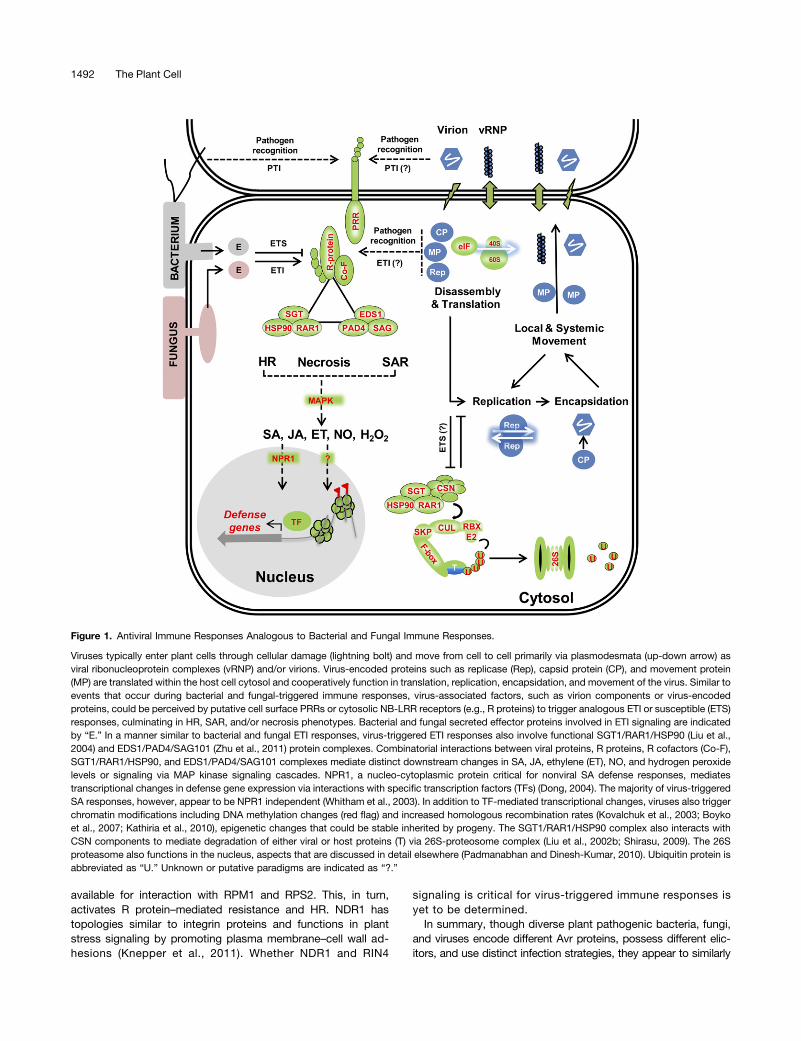
Figure 1. Antiviral Immune Responses Analogous to Bacterial and Fungal Immune Responses.
Viruses typically enter plant cells through cellular damage (lightning bolt) and move from cell to cell primarily via plasmodesmata (up-down arrow) asviral ribonucleoprotein complexes (vRNP) and/or virions. Virus-encoded proteins such as replicase (Rep), capsid protein (CP), and movement protein(MP) are translated within the host cell cytosol and cooperatively function in translation, replication, encapsidation, and movement of the virus. Similar toevents that occur during bacterial and fungal-triggered immune responses, virus-associated factors, such as virion components or virus-encodedproteins, could be perceived by putative cell surface PRRs or cytosolic NB-LRR receptors (e.g., R proteins) to trigger analogous ETI or susceptible (ETS)responses, culminating in HR, SAR, and/or necrosis phenotypes. Bacterial and fungal secreted effector proteins involved in ETI signaling are indicatedby “E.” In a manner similar to bacterial and fungal ETI responses, virus-triggered ETI responses also involve functional SGT1/RAR1/HSP90 (Liu et al.,2004) and EDS1/PAD4/SAG101 (Zhu et al., 2011) protein complexes. Combinatorial interactions between viral proteins, R proteins, R cofactors (Co-F),SGT1/RAR1/HSP90, and EDS1/PAD4/SAG101 complexes mediate distinct downstream changes in SA, JA, ethylene (ET), NO, and hydrogen peroxidelevels or signaling via MAP kinase signaling cascades. NPR1, a nucleo-cytoplasmic protein critical for nonviral SA defense responses, mediatestranscriptional changes in defense gene expression via interactions with specific transcription factors (TFs) (Dong, 2004). The majority of virus-triggeredSA responses, however, appear to be NPR1 independent (Whitham et al., 2003). In addition to TF-mediated transcriptional changes, viruses also triggerchromatin modifications including DNA methylation changes (red flag) and increased homologous recombination rates (Kovalchuk et al., 2003; Boykoet al., 2007; Kathiria et al., 2010), epigenetic changes that could be stable inherited by progeny. The SGT1/RAR1/HSP90 complex also interacts withCSN components to mediate degradation of either viral or host proteins (T) via 26S-proteosome complex (Liu et al., 2002b; Shirasu, 2009). The 26Sproteasome also functions in the nucleus, aspects that are discussed in detail elsewhere (Padmanabhan and Dinesh-Kumar, 2010). Ubiquitin protein isabbreviated as “U.” Unknown or putative paradigms are indicated as “?.”
1492 The Plant Cell

alter the metabolic, physiological, and cellular states of the hosts.Furthermore, signaling components such as the SGT1/RAR1/HSP90 and EDS1/PAD4/SAG101 complexes, similarly serve tomediate resistance responses against diverse viral and nonviralpathogens, suggesting that plants have evolved such broad-spectrum disease resistances to simultaneously defend againstdiverse pathogen types.
SYSTEMIC NECROSIS RESPONSES
In contrast with the resistant (or incompatible) host–virus inter-actions, most susceptible (or compatible) virus infections do nottrigger HR and do not produce localized necrotic lesion phe-notypes to limit the virus spread in the host plants. However,a similar yet distinct form of necrosis, termed systemic necrosis,is observed in susceptible interactions. For example, systemicnecrosis was reported in young Nicotiana rustica plants in-oculated with TMV (Holmes, 1936); N. benthamiana with mixedinfections of potexviruses, PVX, or Plantago asiatica mosaic vi-rus (PLAMV) isolate Li1 and Potato virus Y (González-Jara et al.,2004; Ozeki et al., 2006); TBSV-infected N. benthamiana and N.clevelandii (Chu et al., 2000); Cucumber mosaic virus (CMV) andsatellite RNA-D infected tomato (Xu and Roossinck, 2000); andPanicum mosaic virus (PMV) and its satellite virus (SPMV) in-fected Brachypodium distachyon and millet species (Panicummiliaceum, Pennisetum glaucum, and Setaria italica; Scholthof,1999; Mandadi and Scholthof, 2012).
Systemic necrosis resembles necrosis commonly observed inlesion mimic mutants, resulting either from constitutive or un-controlled cell death (Lorrain et al., 2003; Moeder and Yoshioka,2008). Unlike HR-associated necrosis, systemic necrosis ismanifested much later in the infection and is primarily observedin the upper noninoculated tissues. Another distinction betweensystemic necrosis and HR-associated necrosis is that systemicnecrosis is a lethal response that can kill the host plant, while HRdoes not. Moreover, systemic necrosis is thought not to pre-clude virus multiplication or its systemic movement, therebyresulting in a susceptible infection. In contrast with the relativelywell understood mechanisms leading to HR and associatednecrosis, we are just beginning to understand the molecularprocesses that underlie systemic necrosis responses and howsystemic necrosis responses relate to antiviral immunity. Recentfindings revealed that despite the differing roles or outcomes,systemic necrosis and HR-associated necrosis share remark-able similarities at the biochemical and molecular level. For ex-ample, both systemic necrosis and HR-associated necrosisinvolve programmed cell death, alter expression of similardefense-related genes, and trigger ROS accumulation (Xu andRoossinck, 2000; Kim et al., 2008; Komatsu et al., 2010; Xuet al., 2012).
Komatsu et al. (2010) investigated the molecular determinantsleading to systemic necrosis elicited by infection with PLAMV,a potexvirus, in N. benthamiana. Using a candidate gene ap-proach, they determined that genes mediating HR in an incom-patible interaction also mediate systemic necrosis in compatibleinfections. For example, systemic necrosis responses dependon a functional SGT1/RAR1 complex and also require MAPKKKa/MEK2 signaling (Komatsu et al., 2010). Unexpectedly, silencing of
SGT1 and RAR1 promoted the accumulation of PLAMV (Komatsuet al., 2010) in the infected N. benthamiana plants. This resultconflicts with a prevailing assumption that systemic necrosisdoes not impede viral accumulation and suggests that sys-temic necrosis may indeed promote antiviral immunity duringa compatible plant–virus interaction.Systemic necrosis is also elicited in N. benthamiana plants
infected with a recombinant PVX vector expressing the potyviralhelper component-proteinase (HC-Pro) (González-Jara et al.,2004). To decipher systemic necrosis responses, Pacheco et al.(2012) performed global analysis of gene expression of N.benthamiana plants after systemic necrosis was triggered by therecombinant PVX-HC-Pro virus. Comparisons of transcriptionalchanges associated with HR-associated necrosis during an in-compatible interaction revealed striking similarities among thealtered gene expression profiles in systemic necrosis and HR-associated necrosis. Together, these studies support an emergingtheme that systemic necrosis and HR-associated necrosis involvesimilar physiological, molecular, and biochemical features. How-ever, the biological relevance of systemic necrosis in compatibleinfections remains ambiguous. Perhaps systemic necrosis ismerely an uncontrolled or incomplete HR-associated necrosisresponse that is triggered in distal tissues when the localized HRfails to limit virus spread. As a consequence, it may serve asa last attempt of tissue-level suicide to save the remainder of theplant. Further studies are needed to understand the biologicalrelevance of systemic necrosis in antiviral immunity and themolecular constraints that distinguish systemic necrosis fromHR-associated necrosis.
SAR
Similar to HR, SAR is triggered during an incompatible interactioninvolving Avr and R proteins in the primary infected cells. However,the resistance is transduced to the noninfected distant tissues.Although the exact mechanisms of SAR are not defined, it is ini-tiated through a local interaction among Avr and R proteins andresults in accumulation of phytohormones such as SA and JA inthe distant tissues (reviewed in Vlot et al., 2008a). The NON-EXPRESSOR OF PR1 (NPR1) protein functions downstream of SAto mediate changes in expression of defense genes (Dong, 2004).Depending on the pathogen type and stage of infection, NPR1also interacts with components of JA signaling (Figure 1). UnlikeHR, SAR is a long-lasting immune response primed to providedistant tissue resistance against subsequent infections. In the caseof TMV-triggered SAR, the response persists up to 3 weeks (Ross,1961b). How SAR can be sustained for so long is not clear.However, epigenetic modifications, such as DNA methylation andchromatin remodeling, may be critical to maintain a stable SARsignal (reviewed in Spoel and Dong, 2012). Recent studies ofArabidopsis infected with PstDC3000 demonstrated that SAR canbe stably inherited to the next generation, even when the progenywas not exposed to the pathogens—possibly via PstDC3000-triggered hypomethylation of host chromatin (Luna et al., 2012).Interestingly, the transgenerational stability of SAR requires NPR1,as progeny of the SA-insensitive npr1-1 mutant plants failed topossess SAR in the next generation (Luna et al., 2012). This in-duced resistance phenomena is also triggered in the progeny of
Viruses and Host Immune Responses 1493
plants exposed to caterpillar herbivory (Rasmann et al., 2012). Inthis case, the stable resistance response is dependent on intact JAsignaling and requires the biogenesis of short interfering RNA thatcould mediate the epigenetic chromatin modifications (Rasmannet al., 2012).
With respect to viral responses, tobacco plants infected withTMV or Oilseed rape mosaic virus were shown to display in-creased frequency of host DNA homologous recombination inboth infected and distant noninoculated leaves (Kovalchuk et al.,2003). The increased homologous recombination rate persisted inthe progeny of the TMV-infected plants, but not Oilseed rapemosaic virus–infected plants, and resulted in increased DNA re-arrangements and hypomethylation of the Leucine-rich repeat(LRR) gene loci homologous to the N gene (Boyko et al., 2007). Inaddition, progeny of TMV-infected plants with higher recom-bination rates exhibited broad-spectrum tolerance to TMV, P.syringae, and Phytophthora nicotianae infections (Kathiria et al.,2010). Whether these apparently stable resistance responsesmediated by increased homologous recombination frequencyare comparable to next-generation SAR is unclear. Additionalstudies of virus-induced homologous recombination frequencyand the molecular determinants of viral SAR are needed to un-derstand if and how SAR and its associated (epi)genetic alter-ations are established and maintained during a viral infection.
Another intriguing aspect of SAR is the nature of the systemicsignal that mediates SAR in noninfected tissues. Althoughthe exact nature of the SAR signal remains unclear, severalmetabolites have been proposed as putative signals that me-diate SAR in viral and nonviral infections. These include glycerol-3-phosphate (Chanda et al., 2011), indole derivatives (Trumanet al., 2010), azeleic acid (Jung et al., 2009), methyl salicylate(MeSA) (Park et al., 2007; Vlot et al., 2008b), and glycerolipids(Chaturvedi et al., 2008). SAR likely involves interaction amongmultiple SAR signals, such as MeSA, lipid-transfer proteins, andglycerolipids (Liu et al., 2011). Significant crosstalk among theSAR signals and environmental factors, such as light, adds yetanother layer of complexity to SAR signaling (Vlot et al., 2008a).Since the first discovery of SAR in 1961 by Ross, the molecularresponses involved in virus-triggered SAR have been investigatedusing the TMV pathosystem. Multiple signals including MeSAappear to perpetuate SAR during TMV infection (Park et al.,2007; Vlot et al., 2008a; Dempsey and Klessig, 2012). Whetherthese signals also perpetuate SAR in other viral pathosystemsrequires further investigation. Nevertheless, SAR is yet anotherconserved plant defense response triggered against diversepathogenic bacteria, fungi, and viruses. Moreover, in contrastwith the HR, SAR renders a broader and long-lasting resistanceto diverse pathogen types simultaneously.
R GENE–MEDIATED RESPONSES TO VIRUS INFECTION
Over the past decade, several R genes that mediate resistanceagainst viruses have been identified (Collier and Moffett, 2009;Gururani et al., 2012). The majority of the cloned dominant Rgenes encode the conserved nucleotide binding (NB) and LRRfamily proteins (Collier and Moffett, 2009; Moffett, 2009;Gururani et al., 2012). NB-LRR proteins also contain additional
N-terminal domains such as the TIR homology domain, a CCdomain, a Solanaceae domain, or a predicted BED zinc fingerdomain. Until recently, the LRR domain was thought to be themajor domain critical for R protein function. However, growingevidence indicates that both the LRR and the N terminus do-mains (TIR or CC) are critical for proper resistance responses.The two domains function through intramolecular interactionsand interactions with other proteins (R cofactors) to mediaterecognition of pathogen elicitors (Collier and Moffett, 2009;Moffett, 2009). R cofactors are also essential for host-mediatedHR and SAR responses. For example, the tobacco N proteinthat recognizes TMV replicase (p50) to trigger the resistanceresponse requires a chloroplast-localized sulfurtransferase, NRECEPTOR-INTERACTING PROTEIN1 (NRIP1) (Caplan et al.,2008). NRIP1 physically interacts with the N-terminal TIR do-main of the N protein and TMV p50 protein (Caplan et al., 2008).Interestingly, p50 alters the endogenous localization of NRIP1from the chloroplast to the cytosol and nucleus where it inter-acts with the N protein (Caplan et al., 2008).Similarly, the potato resistance proteins Rx, Rx2, and GREEN
PEACH APHID2 interact with RAN GTPASE-ACTIVATING PRO-TEIN (RanGAP2) (Sacco et al., 2007). The interaction of RanGAP2with the N-terminal CC domain of Rx and related proteins is re-quired for specific recognition of PVX coat protein and resistanceagainst PVX (Sacco et al., 2007). Recently, it was shown thatRanGAP2 directs the Rx protein from the nucleus to the cytosoland contributes to the resistance response (Tameling et al., 2010).Such interactions of R proteins and cofactor proteins are ob-served for fungal, bacterial, and other viral resistance responses(Moffett, 2009). In summary, a plethora of plant:pathogen re-sistance scenarios often require combinatorial interactions amongmultiple host and pathogen factors such as the Avr, R, and Rcofactors. In turn, the resistance responses triggered by suchinteractions (variously described as the guard hypothesis [Dangland Jones, 2001], the decoy model [van der Hoorn and Kamoun,2008], or the bait and switch model [Collier and Moffett, 2009])culminate in HR and SAR responses via the action of hormoneand signaling molecules such as SA, JA, ethylene, NO, and ROS,as reviewed recently (Culver and Padmanabhan, 2007; Trunigerand Aranda, 2009; Carr et al., 2010).In viral infections, in addition to the dominant R gene–related
resistance responses, another form of recessive resistance ex-ists that is typically derived by a loss of function in host proteinscritical for the establishment of disease (Robaglia and Caranta,2006; Iyer-Pascuzzi and McCouch, 2007; Truniger and Aranda,2009; Gururani et al., 2012). For example, amino acid mutationsin the eukaryotic translation initiation factor, eIF4E, mediatesresistance against several viruses in Arabidopsis, tomato, pep-per, pea (Pisum sativum), melon (Cucumis melo), and barley(Hordeum vulgare) (Lellis et al., 2002; Ruffel et al., 2002, 2005;Nicaise et al., 2003; Gao et al., 2004; Kanyuka et al., 2005; Pironet al., 2010). In addition, mutations in another subunit of eu-karyotic translation initiation factor, eIF4G, imparts recessiveresistance against Rice yellow mottle virus (RYMV) in rice (Oryzasativa) (Albar et al., 2006) and to CMV and TCV in Arabidopsis(Yoshii et al., 2004). In these situations, the host cells are chal-lenged in their ability to efficiently translate viral proteins, thusaffecting viral replication and/or local movement (Collier and
1494 The Plant Cell

Moffett, 2009; Gururani et al., 2012). Other host proteins alsomediate recessive resistance, such as the Arabidopsis tonoplast-localized transmembrane proteins, TOM1 and TOM2A, which in-teract with each other and with the TMV-encoded 130K and 180Kreplicase proteins (Ishikawa et al., 1993; Yamanaka et al., 2000;Tsujimoto et al., 2003). The rate of intracellular accumulation ofTMV and its associated subgenomic RNAs was significantly de-creased in the loss-of-function mutants tom1 and tom2a. Fur-thermore, the resistance imparted is specific against TMV but notagainst CMV (a cucumovirus) or TCV (a carmovirus) (Ishikawaet al., 1993; Ohshima et al., 1998; Yamanaka et al., 2000; Hagiwaraet al., 2003; Tsujimoto et al., 2003).
In addition to the aforementioned resistance proteins, otherhost proteins can contribute to viral resistance responses. In thiscategory, lectin proteins are novel and intriguing resistance-imparting proteins. In animals, lectins activate immune respon-ses against diverse pathogens. Certain lectin receptors, such asthe C-type lectins, recognize fungal PAMPS (Willment andBrown, 2008). In plants, lectins bind to mono- or oligosaccharidemolecules to discriminate self- from non-self-originated carbo-hydrates, thus rendering them ideal for pathogen perception(Van Damme et al., 2004). For example, the soybean (Glycinemax) lectin b-glucan binding protein binds with high affinity toa 1,3-branched heptaglucoside, a PAMP found in the cell wallsof Phytophthora sojae (Mithöfer et al., 2000; Fliegmann et al.,2004), and triggers defense reactions. In this context, the dis-covery of an Arabidopsis jacalin-type lectin, RESTRICTED TEVMOVEMENT1 (RTM1), that mediates resistance against To-bacco etch virus (TEV), was surprising (Chisholm et al., 2000), asthe TEV CP is not glycosylated.
More recently, another jacalin-type lectin, JAX1, was shown toconfer resistance against multiple potexviruses (PVX, PLAMV,White clover mosaic virus, and Asparagus virus 3; Yamaji et al.,2012). Although JAX1 and RTM1 are closely related lectin pro-teins, they impart distinct forms of resistance. JAX1 functions atthe cellular level, inhibiting replication of PLAMV (Yamaji et al.,2012), and RTM1 inhibits systemic movement of TEV (Chisholmet al., 2000). An intriguing aspect of lectin-mediated resistance(LMR) compared with the NB-LRR resistance is that it does notinvoke HR and SAR responses, nor does it alter SA levels, sig-naling, or other typical defense gene expression changes com-monly modulated in immune resistance responses (Yamaji et al.,2012). This suggests that LMR engages an as yet unidentified anddistinct mechanism of antiviral immunity. According to the pro-posed working model of LMR function (Chisholm et al., 2000;Yamaji et al., 2012), lectin proteins such as JAX1 and RTM1recognize certain viral proteins that are glycosylated within plantcells to trigger resistance. In the case of JAX1, LMR responsescould inhibit viral replication by promoting aggregation ofreplicase-associated proteins, while RTM1 could inhibit viralmovement through interference with viral movement–associatedproteins. In addition to RTM1, a small heat shock-like protein(RTM2) (Whitham et al., 2000) and a meprin and TRAF homologydomain-containing protein (RTM3) that physically interacts withRTM1 (Cosson et al., 2010) also restrict viral movement. RTM2possesses a transmembrane domain, but unlike other HSPs, itsexpression is not heat inducible and does not contribute tothermotolerance (Whitham et al., 2000). Notably, both RTM1 and
RTM2 are expressed in phloem-associated tissues and sieveelements, correlating with their function in impeding virusmovement. Interestingly, the N terminus of the potyvirus coatprotein contains features that can overcome RTM resistance,suggesting that interactions of RTM proteins with viral CPs maymediate resistance responses (Decroocq et al., 2009). Furtherstudies on LMR should reveal the mechanism of this intriguingantiviral resistance response.In another study, complementary proteomic approaches were
used to identify a class of endoplasmic reticulum (ER)–residingchaperones that influence antiviral immune responses (Caplanet al., 2009). In N. benthamiana, the ER proteins ERp57, P5,calreticulin2, and CRT3 are strongly expressed within 2 h of TMVinfection in plants expressing the N gene (Caplan et al., 2009).Virus-induced gene silencing (VIGS) of the ER chaperones re-sulted in loss of N-mediated resistance against TMV, althoughthe cell death/necrosis symptoms and TMV movement in theupper noninoculated leaves was only partially abolished, prob-ably due to incomplete knockdown of the respective transcriptsby VIGS. Since ER chaperones function in folding and accu-mulation of other membrane proteins, the authors tested theeffects on other membrane proteins. Loss of Nb-CRT3 and Nb-CRT5 resulted in reduced accumulation of a plasma membrane–localized induced receptor-like kinase, IRK (Caplan et al., 2009).Similar to the expression of calreticulins, IRK is strongly upre-gulated early in TMV infection, and VIGS knockdown of IRKexpression resulted in loss of N-mediated resistance againstTMV. Although the mechanism of how an ER chaperone altersIRK accumulation is not clear, these results reveal yet anotherintricate interplay among ER-residing chaperones and plasmamembrane receptor proteins in N-mediated immune responses.Taken together, resistance genes, particularly those encoding
the NB-LRR proteins, have well-conserved roles in plants, whichare to guard the host cells against diverse viral and nonviralpathogens and to trigger disease resistance. Moreover, the generalmechanism of the recognition of R proteins and Avr factorsappears to be similar for viral and nonviral pathogens, wherebyR cofactors play crucial roles to guide or modulate R/Avr inter-actions, ultimately activating HR and resistance responses. Fi-nally, while the predominant antiviral resistance responses aremediated by the dominant R genes, other host proteins, such asthe elongation initiation factors, TOM proteins, ER chaperones,calreticulins, and lectin proteins, also influence host resistanceagainst diverse viral infections.
UBIQUITIN PROTEASOME SYSTEM
The ubiquitin proteasome system (UPS) has emerged as a prom-inent player in influencing virus–host interactions at almost everystage of antiviral defense, both in plants and animals (Gao andLuo, 2006; Citovsky et al., 2009). In turn, viruses use a plethora ofstrategies to modulate UPS processes. UPS regulates cellularactivities including cell cycle, transcription, and signal transduction(Hershko and Ciechanover, 1998). The plant UPS primarily in-volves the activity of ubiquitin-activating enzyme (E1), ubiquitin-conjugating enzyme (E2), and ubiquitin-ligase (E3) (Hua andVierstra, 2011). These three proteins form the E3 ubiquitin ligase
Viruses and Host Immune Responses 1495

complex that specifically polyubiquitinates cellular proteins thatare subsequently targeted for degradation by the 26S proteasome.SKP1 is another critical component of the SCF (for SKP1, CULLIN,and F-box) class of E3 ubiquitin ligases and, through interactionswith CULLIN and F-box proteins, recruits proteins for poly-ubiquitination (Hua and Vierstra, 2011).
Whether UPS processes are employed by the plants to de-fend against virus infections or viruses use the UPS to promotevirulence is ambiguous. For example, the tobacco N gene–mediated resistance response against TMV requires the functionof the RAR1/SGT1 complex. RAR1/SGT1 physically interactswith SKP1 and the CSN (Liu et al., 2002b), another E3 ubiquitinligase that targets cellular proteins for turnover in plant growthand development (Nezames and Deng, 2012). VIGS of RAR1,SGT1, SKP1, or the CSN gene products abolishes the N gene–mediated resistance (Liu et al., 2002b) and supports a role forUPS in mediating the N-mediated HR and resistance responses.Movement proteins of TMV and Turnip yellow mosaic virus arealso specifically targeted for degradation by the host UPS ma-chinery, resulting in decreased virulence and pathogenicity(Reichel and Beachy, 2000; Drugeon and Jupin, 2002), high-lighting the role of the UPS in antiviral immune responses.
Viruses also can use UPS processes to promote virulence.The TBSV replication component, p33, interacts with a host E2ubiquitin-conjugase, Cdc34 (Li et al., 2008). Although the p33protein is a substrate for UPS-mediated turnover in vivo,an optimal level of p33 polyubiquitination appears to mediatepositive interaction among the TBSV replicase components anda host ENDOSOMAL SORTING COMPLEX REQUIRED FORTRANSPORT protein, Vps23p, which is critical for effectivereplication of TBSV (Li et al., 2008). This result suggests thathost UPS processes may also aid the virus by facilitating in-fection. Similarly, downregulation of a 26S proteasome subunitRPN1 in N. benthamiana inhibits systemic transport of TMV andPVX, two taxonomically distinct viruses (Jin et al., 2006), againsupporting the conclusion that UPS can promote virulence. Inthe case of RPN1 silencing, the virulence-promoting effect oc-curs in part through modulation of auxin transport and brassi-nosteroid signaling, both of which are required for efficientvascular development (Jin et al., 2006). As the vasculature is themeans by which viruses systemically infect plant tissues, thealtered vasculature in RPN1-silenced plants prohibits viralmovement. Given these contradictory outcomes of UPS func-tions in different virus–host responses, it appears that virusesand hosts employ a variety of strategies to enhance virulence orpromote host immunity.
Among other examples of viruses that target UPS processesto promote virulence is Beet severe curly top virus (BSCTV),a geminivirus. BSCTV encodes a pathogenicity factor, C2, whichinteracts and interferes with the UPS-mediated degradation ofa host S-adenosyl-methionine decarboxylase 1 (SAMDC1)enzyme (Zhang et al., 2011). SAMDC1 mediates the decar-boxylation of S-adenosyl-methionine (SAM) to dcSAM. BecauseSAM serves as a donor for methyl groups in various methylationreactions, altering the levels of SAM and dcSAM (as a result ofmodulating SAMDC1 activity) may have an effect on host DNAmethylation status. As predicted, loss of function of eitherSAMDC1 or the BSCTV C2 protein results in enhanced de
novo methylation of the dsDNA replicative form of the BSCTVgenome, thus reducing BSCTV replication and dampeningBSCTV infectivity (Zhang et al., 2011). Together, these resultssuggest that the BSCTV C2 protein acquired its remarkablefunction of modulating host DNA methylation-mediatedgene silencing via attenuation of UPS-mediated degradation ofSAMDC1.In plants, the CSN complex regulates the activity of another
class of ubiquitin ligases, the CRLs, primarily through the ad-dition or removal of a RELATED TO UBIQUITIN modifier to theCRLs, a process dubbed rubylation (Hua and Vierstra, 2011).The appropriate function of CRLs is thus determined by its ru-bylation status. Similar to BSCTV, Tomato yellow leaf curl Sar-dinia virus (TYLCSV), another geminivirus, also encodes a C2protein. TYLCSV C2 protein physically interacts with a CSNsubunit, CSN5, in N. benthamiana. This interaction interfereswith the derubylation activity of CSN and, consequently, itsfunction (Lozano-Durán et al., 2011). Compromised CSN func-tion also suppresses JA-mediated defense responses and severalother host processes in the infected plants. In line with theseresults, treating plants with JAs reduced the virulence of TYLCSV,suggesting that TYLCSV C2 protein targeting of CSN (andthereby CSNs polyubiquitination activity) has biologically relevantconsequences (Lozano-Durán et al., 2011).Finally, among other strategies viruses employ to modulate
host ubiquitin proteasome systems, some viruses encode pro-teins that resemble the UPS components. For example, P0, anRNA silencing suppressor encoded by poleroviruses, is a bi-ological mimic of an F-box protein (Pazhouhandeh et al., 2006).F-box proteins are also UPS components and associate withCULLIN proteins to mediate protein polyubiquitination. The P0protein forms a complex with the Arabidopsis SKP1 homologs,ASK1 and ASK2 (Pazhouhandeh et al., 2006), and then as-sembles into a CULLIN-based ubiquitin ligase to specificallytarget ARGONAUTE1 (AGO1) for degradation (Baumbergeret al., 2007). Intriguingly, P0-mediated AGO1 degradation is in-sensitive to proteasome inhibitors such as MG132, suggestingthat the 26S proteasome may not be involved in the turnover ofAGO1. Further studies to understand host UPS componentsand interactions with viral proteins, as well as the identificationof UPS protein targets during a viral infection, will shed morelight on this fascinating cellular process associated with antiviralimmune responses. Given the diversity of outcomes and severalviruses targeting multiple UPS components, a critical role forUPS-mediated resistance or susceptibility in virus–host inter-actions and antiviral immune responses can be inferred.
MONOCOT ANTIVIRAL RESPONSES: A KNOWLEDGE GAP
As discussed above, studies of virus–host interactions haveunequivocally advanced our understanding of plant immuneresponses and revealed strategies utilized by viruses to targethost defense components. However, there is a gap in ourknowledge of monocotyledonous plant antiviral responses. Forinstance, of the 31 cloned plant antiviral resistance genes, onlyone is from a monocot (Gururani et al., 2012). This is a rice re-cessive resistance gene, eIF(iso)4G1, that confers resistance
1496 The Plant Cell

against RYMV (Albar et al., 2006) by interacting with the viralgenome-linked protein (VPg) (Hébrard et al., 2010). In rice,a mutation in eIF(iso)4G1 disrupts its interaction with VPg, thusimparting resistance against RYMV.
Several economically important monocot-infecting viruses suchas Barley stripe mosaic virus (BSMV), Wheat streak mosaic virus,and Brome mosaic virus, which also infect N. benthamiana, havebeen studied using dicot host plants to dissect viral processes andpathogenicity determinants causing disease (Bragg and Jackson,2004; French and Stenger, 2004; Mise and Pocsai, 2004). How-ever, little experimental data are available pertaining to the alteredhost immune responses in their respective grass host species.Moreover, although there have been studies in identification ofseveral resistance gene loci for monocot crops, including rice,barley, maize (Zea mays), and wheat (Triticum aestivum) (Trottetand Gouis, 2004; Redinbaugh and Pratt, 2009), most of thesestudies were primarily aimed toward breeding for resistanceagainst diverse monocot-infecting viruses. The underlying mech-anisms of immunity imparted by the resistance loci remain largelyunknown. We also know little of the host molecular processesaltered in compatible monocot plant virus infections and whetherthe antiviral responses are conserved among dicot and monocothost–virus interactions.
The lack of a tractable monocot genetic model system, akin toArabidopsis or N. benthamiana, has been a major hindrance toresearch aimed at understanding the antiviral immune responsesin monocots. However, emerging monocot model plants such asBrachypodium distachyon (Brachypodium) are bridging this gap.Brachypodium is a temperate grass with all the essential qualitiesof a genetic model system (Vogel and Bragg, 2009; Brkljacic et al.,2011). Its fully sequenced genome is highly syntenic to the ge-nomes of important monocot crops such as wheat, rice, barley,millet, and maize, thus allowing functional and comparative ge-nomic studies with potential for direct translation to field crops(International Brachypodium Initiative, 2010). Many genomic andfunctional resources have been developed around Brachypodium,including microarrays, yeast two-hybrid libraries, ;200 inbredaccessions, EMS/TILLING lines, and annotated T-DNA knockoutlines (Filiz et al., 2009; Fox et al., 2010; International Brachypo-dium Initiative, 2010; Brkljacic et al., 2011; Cao et al., 2011; Bragget al., 2012). Brachypodium has become an accepted model plantfor research related to plant–pathogen interactions, as evidencedby the growing list of publications (Routledge et al., 2004; Parkeret al., 2008; Peraldi et al., 2011; Cui et al., 2012; Lee et al., 2012;Mandadi and Scholthof, 2012). The reported bacterial, fungal, andviral pathogens that infect Brachypodium species (Table 1) willlikely prompt further studies in understanding the molecular basisof disease in monocots.
THE PANICOVIRUS COMPLEX: NEW FINDINGS IN PLANTVIROLOGY USING BRACHYPODIUM
PMV is a single-stranded positive-sense RNA that is the typemember of genus Panicovirus in the family Tombusviridae(Turina et al., 1998, 2000). PMV infects turfgrass, forage grasses,switchgrass, and food crops, such as millets. A peculiar featureof PMV is that it supports the replication of a satellite virus,SPMV, resulting in a viral synergism (Scholthof, 1999; Mandadi
and Scholthof, 2012). As a complex, PMV+SPMV causes dis-ease in the host plants characterized by exacerbated symptomsrelative to those produced by PMV alone (Scholthof, 1999;Omarov et al., 2005). We are using this unique panicovirus com-plex as a foundational tool for fundamental advances in host–virusinteractions, as well as potential translation to the field due to thedistribution of PMV on agriculturally important crop hosts.The utility of Brachypodium in studying monocot virus–host
responses is evident from work done by our group and others. Inour laboratory, we used transcriptomic approaches to identifyBrachypodium molecular responses to compatible infections ofPMV and SPMV. By comparison to the reported dicot virus hostresponses, we identified both conserved and unique molecular
TABLE 1. Pathogens infecting Brachypodium distachyon and B.sylvaticum. a
VirusesBarley stripe mosaic virus (BSMV)Brome mosaic virus (BMV)Panicum mosaic virus (PMV) + SPMV b
Maize mild mottle virus (MMMV)Sorghum yellow banding virus (SYBV)Foxtail mosaic virus (FoMV)
Fungi: BasidiomycetesUstilago bromivora - loose smutTilletia olidea - smutPuccinia brachypodii - leaf rustPuccinia striiformis - stripe rustPuccinia coronata - crown rust
Fungi: AscomycetesClaviceps purpurea - ergotBlumeria graminis - powdery mildewEpichloë sylvatica - endophyteNeotyphodium sp. - endophyteStagonospora nodorum - glume blotchSetosphaeria turcica - northern leaf blightPyrenophora teres - net blotchPyrenophora erythrospila - leaf spotCochliobolus sativus - root rot/leaf spotAlternaria sp. - leaf spotAscochyta sp. - leaf spotSclerotinia homoeocarpa - dollar spotCladosporium sp. - leaf moldEpicoccum sp. - glume spotFusarium graminearum - head blightFusarium culmorum - head blightMagnaporthe grisea - rice blast
Fungal-like organismsPythium ultimum - root rot
BacteriaAgrobacterium tumefaciens - crown gall
aPlant viruses infecting Brachypodium distachyon were compiled fromCui et al., 2012; Lee et al., 2012; Mandadi and Scholthof, 2012; and, K.K.Mandadi, J.D. Pyle, and K.-B.G. Scholthof, unpublished data. Fungi,fungal-like organisms, and bacteria (with their common names) thatinfect Brachpodium species were compiled from Parker et al., 2008;Brkljacic et al., 2011; and, Halbritter et al., 2012.bThis reflects the infection of PMV alone or the mixed infection of PMVplus its satellite virus, SPMV (Mandadi and Scholthof, 2012).
Viruses and Host Immune Responses 1497

pathways altered in monocot host–virus responses (Mandadiand Scholthof, 2012). Among the conserved responses, themonocot antiviral defenses involve classical defense hormonessuch as SA, JA, and ethylene. Multiple genes in SA biosynthesisand signaling, such as ICS1, ABERRANT GROWTH AND DEATH2,ALTERNATIVE OXIDASE, pathogenesis-related proteins, andWRKY transcription factors, were upregulated in Brachypodiuminfected with PMV alone and PMV+SPMV (Mandadi andScholthof, 2012). By contrast, several genes in JA and ET sig-naling, such as LIPOXYGENASE2, ALLENE OXIDE SYNTHASE,VEGETATIVE STORAGE PROTEIN1, FATTY ACID DESATUR-ASE7, ETHYLENE RESPONSE FACTOR1 (ERF1), ERF3-like,ERF4, and ERF/Related to AP2.2, were downregulated (Mandadiand Scholthof, 2012). These results also support an existingnotion that SA signaling and JA/ET signaling are antagonistic toeach other in response to pathogen infection (An and Mou,2011; Van der Does et al., 2013). In addition, several genes inbiological processes, such as carbon metabolism, metabolitetransport, cell wall remodeling, protein synthesis and degrada-tion, and photosynthesis, were altered in a manner similar tothose described for dicot virus interactions (Whitham et al.,2003, 2006; Ascencio-Ibáñez et al., 2008; García-Marcos et al.,2009; Hanssen et al., 2011; Postnikova and Nemchinov, 2012).These include genes encoding glutathione S-transferases, cy-tochrome P450s, glucosyl hydrolases, heat shock proteins,ribosomal components, UPS proteins, and chlorophyll a/b binding and photosystem-related proteins (Mandadi andScholthof, 2012). However, unlike other known dicot antiviralresponses, we found ;30 protein kinases, particularly those inthe receptor-like kinase subfamily, which are uniquely altered inboth PMV and PMV+SPMV triggered responses in Brachy-podium (Mandadi and Scholthof, 2012). This overrepre-sentation of protein kinases appears to be a unique feature ofmonocots, as our transcriptome comparisons with the fewother reported monocot virus infections, such as maize in-fected with Rice black-streaked dwarf virus (genus Phytor-eovirus) (Jia et al., 2012) and rice infected with Rice stripe virus(genus Tenuivirus) (Satoh et al., 2010), also revealed a higherproportion of upregulated kinases in the respective monocotvirus infections. Together, these results suggest that althoughmonocot and dicot plants share some commonalities,they have discrete and unique responses to plant virusinfections.
Recently, through a screen for BSMV resistance amongdiverse Brachypodium accessions using BSMV strain NorthDakota (ND18) followed by genetic fine-mapping of a resistanceaccession Bd3-1, the first dominant R gene (BSR1) for monocotvirus resistance was identified (Cui et al., 2012). Similar to knowndominant R proteins in dicots, the putative Brachypodium BSR1gene encodes a CC-NB-LRR protein (Cui et al., 2012; Lee et al.,2012). Interestingly, genome reassortment experiments usingthe avirulent BSMV ND18 strain and a virulent BSMV Norwich(NW) strain in the Bd3-1 accession revealed that the BSMV NWtriple gene block 1 (TGB1NW) movement protein is the virulencetrigger. Site-directed mutagenesis assays further revealed thatthe TGB1NW amino acid residues at position 390 to 392 arecritical for elicitation of HR and necrosis-like symptoms, pre-sumably through interactions of this region with BSR1 (Lee et al.,
2012). More research to understand these and other monocotantiviral defenses will continue to provide crucial insights intothe currently poorly understood monocot antiviral immuneresponses.
FUTURE PROSPECTS AND CONSIDERATIONS
As mentioned above, one obvious area of future research focusis studying monocot antiviral immune responses. Comparativestudies of antiviral responses in dicot and monocots will likelyopen new areas of investigation owing to the divergent evolu-tionary relationships of dicots and monocots. Moreover, be-cause monocot and dicot plants have significantly differentmorphological and anatomical characteristics, including shoot-root architecture and cell wall composition and vasculature, aswell as the genome-level differences, research aimed to un-derstand how these differences affect antiviral immunities hasintrinsic merit. To this end, Brachypodium and other emerginggrass models such as Setaria viridis (Brutnell et al., 2010) aregaining reputations as outstanding experimental plants to studymonocot-infecting viruses such as BSMV (Cui et al., 2012; Leeet al., 2012) and PMV (Mandadi and Scholthof, 2012). Brachy-podium is also an excellent model for studying nonviral patho-gens (Table 1). Brachypodium will continue to play a critical rolein addressing questions about monocot pathobiology, asa “working grass hero” (Garvin, 2007).Much more research is needed toward elaboration of the
molecular pathways that lead to SAR in virus–host interactions.For example, the identity of the exact SAR signal in host–virusinteractions is elusive. The precise roles of MeSA, azeleic acid,and glycerolipids in antiviral SAR need further investigation.Whether virus-triggered SAR also involves combinatorial inter-actions among the SAR signaling molecules and environmentalfactors remains to be tested. Finally, contribution of epigeneticfactors in triggering and perpetuating SAR signals and whethervirus-triggered SAR can be stably inherited to the next genera-tion need to be determined, particularly in cereals and othergrasses.Although the antiviral HR mechanisms and the downstream
defense hormone signaling are known for some viruses, ingeneral, antiviral signal transduction is a black box, especiallyfor monocots. Moreover, relative to the well understood earlyevents in the plant immune responses triggered in fungal andbacterial infections, much work remains before we understandthe early signaling events during the perception of virus or virus-encoded factors by the host receptor proteins, particularly at thecell membrane. For example, we do not know if any plasmamembrane or cytosolic receptor proteins can directly perceiveviral features analogous to PRRs to trigger viral resistanceresponses (Figure 1) nor do we know if any endogenous danger/damage-associated molecular patterns, which function in elic-iting nonviral immune responses (Brutus et al., 2010), have anyrole in eliciting antiviral immune responses. These questionsnecessarily transition into an area of semantics that we suggestwould likely benefit from some convergence, particularly whendescribing antiviral immune responses that are analogous tononviral immune responses. For example, the R gene–mediatedvirus resistance involves specific recognition of virus-encoded Avr
1498 The Plant Cell

proteins that culminate in HR and SAR responses, as shown forTMV N gene resistance and others (Whitham et al., 1994; Liuet al., 2002a; Schoelz, 2006). However, it is unclear whether theycan be classified as ETI responses because of the existing defi-nition (or lack thereof) of what constitutes an effector protein fordiverse pathogen types (i.e., viruses versus bacteria). To resolvethese irregularities, we propose working definitions of viral ef-fectors, ETI, and PTI responses in viral infections (Table 2).
Typical bacterial and fungal effector proteins are encoded bythese microbes and delivered into the plant cells, wherein theyinterfere with PTI or other immune regulators (Jones and Dangl,2006; Bent and Mackey, 2007; Dodds and Rathjen, 2010; Spoeland Dong, 2012). Plant viruses do not encode effector proteinsper se if the definition is limited to proteins that are deliveredinside the host cells via microbial secretion systems. Yet, virusesencode proteins that are translated in the host cells and promotevirulence by interfering with host defense pathways using avariety of strategies as discussed earlier. For example, theTBSV-encoded P19 protein promotes TBSV accumulation andvirulence by suppressing host RNA silencing defenses (Scholthof,2006). The BSCTV-encoded C2 protein is a silencing suppressorprotein that also usurps host UPS processes to promote sus-ceptibility (Zhang et al., 2011). The CaMV-encoded P6 proteinsuppresses SA-mediated defenses, as well as RNA silencingdefenses (Love et al., 2007, 2012). Oftentimes, these viral pro-teins are recognized by R genes to trigger immune responses.For example, the CaMV P6 protein is an avirulence determinantrecognized by R genes in several plants, including Datura stra-monium, Nicotiana bigelovii, N. glutinosa, Nicotiana edwardso-nii, and Arabidopsis ecotype Tsu-0. Similarly, the TBSV P19silencing suppressor is also an elicitor of HR in certain Nicotianaspecies (Scholthof et al., 1995; Hsieh et al., 2009), includingNicotiana sylvestris, N. tabacum, and Nicotiana bonariensis(Angel and Schoelz, 2013). Furthermore, in the case of tobaccoN-mediated resistance, the TMV-encoded p50 protein is rec-ognized by the N protein and its cofactor NRIP1 to elicit the N-mediated host immune response (Caplan et al., 2008). Theseand several other instances reviewed in detail elsewhere(Schoelz, 2006) beg the following question: Should thesevirulence-promoting factors be referred to as effectors and theimmune responses they trigger be classified as ETI responses?Based on the analogous functions of nonviral effector proteins,we propose a working definition for viral effectors: Viral effectorsare virus-encoded proteins that when present in host cells in-terfere with host defense signaling components to promotevirulence.
Earlier reviews discussing microbial effectors and their func-tional definitions noted that several bacterial and fungal avir-ulence factors are indeed “double agents” that can promotepathogenesis while eliciting ETI responses, suggesting thatsome avirulence proteins are also effectors (Alfano and Collmer,2004; Schoelz, 2006). For example, the AvrPtoB protein ofPstDC3000 can simultaneously suppress HR, as well as elicitETI responses in the presence of the appropriate R gene, Pto(Abramovitch et al., 2003). As discussed above, several virus-encoded Avr factors, such as CaMV P6 protein, can interferewith plant defense pathways while eliciting R-mediated re-sistance responses (Schoelz, 2006). According to our proposed
definition for viral effectors, virus Avr factors that interfere withhost defenses should thus be referred as effectors. Logically,the immune responses they trigger should be classified as ETIresponses. From this, we propose a working definition: ETI inviral infection is a form of host immune response triggered by Rproteins that recognize, either directly or indirectly, virus-encoded effectors or their activities within the host cells.Another example pertains to P/MAMPs and PTI responses. By
definition, viruses do not possess conserved P/MAMP-like fea-tures such as flagellin or chitin. However, structures such as, butnot limited to, the virion (or capsid), viral ribonucleoproteincomplexes, and viral-encoded glycoproteins embedded on thehost-derived lipid membranes of plant rhabdoviruses (Goldberget al., 1991; Jackson et al., 2005) are analogous to P/MAMPs.Importantly, these structures are conserved among members ofrelated virus taxa. Thus, we hypothesize that such viral patternsare analogous to P/MAMPs and are accessible for recognitionby membrane-bound receptor-like proteins (PRRs or WAKs) totrigger PTI-like responses. As a working definition for viral PTI,we propose the following: PTI in viral infection is a form of basalhost immune response triggered upon recognition of conservedviral molecular features by specific membrane-bound receptor-like proteins.The above descriptions of viral effectors and virus-triggered
immune responses are functionally synonymous with plant im-mune responses triggered in other microbial infections. Con-silience of an integrated view of plant–pathogen interactions willbe predicated on including viruses that are often overlooked inleading plant innate immunity models (Jones and Dangl, 2006;Bent and Mackey, 2007; Boller and Felix, 2009; Hogenhoutet al., 2009; Dodds and Rathjen, 2010; Schwessinger andRonald, 2012; Spoel and Dong, 2012).
CONCLUSION: MORE QUESTIONS THAN ANSWERS
In 1958, Sam Wildman, in a summary of the state-of-the-art ofplant virology noted “our knowledge of what the virus is doinginside the host is not nearly as ‘sophisticated’ as our knowledgeof what a plant virus is” (Wildman, 1959). By the early 1980s,technical difficulties associated with studying virus–host mo-lecular interactions coupled with the virologists greater fasci-nation with structural biology, mutagenesis, transgenic crops,and being able to do reverse genetics on plant viruses resulted invirologists taking a very different path from plant biologists and
Table 2. Working Definitions for Plant–Virus Interactions andImmune Responses
Term Working Definition
Viral effectors Virus-encoded proteins that when present in host cellsinterfere with host defense signaling components topromote virulence
Viral ETI A type of host immune response triggered by R proteinsthat recognize, directly or indirectly, virus-encodedeffectors or their activities within the host cells
Viral PTI A type of basal host immune response triggered uponrecognition of conserved viral molecular features byspecific membrane-bound receptor-like proteins
Viruses and Host Immune Responses 1499

plant pathologists working with bacterial or fungal pathosystems.In recent years, despite having all the tools in place, the focus ofmany virologists was viral physicochemistry and/or identification ofvirulence determinants. This has led, in our opinion, to a gap inunderstanding the mechanisms of infection and host immune re-sponses at the cellular level—especially when compared with thesuccesses of those working on phytopathogenic fungi, bacteria,nematodes, and vector-borne host interactions (Jones and Dangl,2006; Dodds and Rathjen, 2010; Schwessinger and Ronald, 2012;Spoel and Dong, 2012). As also mentioned above, several ques-tions pertaining to plant viruses and their interactions within thehost cells still remain unanswered: What are the major viral elicitorsthat induce HR and SAR?Which host proteins (e.g., PRRs) mediateviral recognition and by what mechanism? Unlike nonviral patho-gens that can be recognized in the extracellular or periplasticspaces, how does an obligate viral pathogen elicit an intracellularhost immune response? How is this response reiterated or main-tained as the virus moves from cell to cell through the plasmo-desmata and subsequently to the noninoculated tissues? Is thisresponse cell autonomous? Wildman in 1958 also asked a verysimilar question: Is the “behavior” of the virus in the inoculated cellthe same as that in a noninoculated systemically infected cell?Unfortunately, the question remains unanswered. Furthermore,despite the direct relevance with production agriculture and ournearly complete reliance on grasses (Poaceae) for food, feed, for-age, recreation, and biofuel needs, critical aspects of grass–virusinteractions largely remain understudied. In recent years, somework has been performed with laboratory dicotylendous plants asalternate hosts, including Arabidopsis and N. benthamiana; how-ever, we are just beginning to disentangle the fundamental mo-lecular interactions between plant viruses and grasses that result indisease or resistance. As such, by outlining here the status quo andgaps in our knowledge of virus–host interactions, we hope toprovide the direction and impetus for a new generation of plantbiologists and plant pathologists to explore the mystery of host–virus interactions within the broader context of host-pathogeninteractions.
ACKNOWLEDGMENTS
This work was supported in part by a Texas Higher Education Co-ordinating Board’s Norman Hackerman Advanced Research ProgramGrant (00517-002-2009) and the Texas A&M University Program toEnhance Scholarly and Creative Activities. We thank Veria Alvarado,Christopher Lyons, Jesse Pyle, and Herman Scholthof for helpfulcomments.
AUTHOR CONTRIBUTIONS
All authors contributed to writing this article.
Received March 19, 2013; revised April 23, 2013; accepted May 1, 2013;published May 24, 2013.
REFERENCES
Aarts, N., Metz, M., Holub, E., Staskawicz, B.J., Daniels, M.J., andParker, J.E. (1998). Different requirements for EDS1 and NDR1 bydisease resistance genes define at least two R gene-mediated
signaling pathways in Arabidopsis. Proc. Natl. Acad. Sci. USA 95:10306–10311.
Abramovitch, R.B., Kim, Y.-J., Chen, S., Dickman, M.B., andMartin, G.B. (2003). Pseudomonas type III effector AvrPtoB inducesplant disease susceptibility by inhibition of host programmed celldeath. EMBO J. 22: 60–69.
Albar, L., Bangratz-Reyser, M., Hébrard, E., Ndjiondjop, M.-N.,Jones, M., and Ghesquière, A. (2006). Mutations in the eIF(iso)4Gtranslation initiation factor confer high resistance of rice to Riceyellow mottle virus. Plant J. 47: 417–426.
Alfano, J.R., and Collmer, A. (2004). Type III secretion systemeffector proteins: Double agents in bacterial disease and plantdefense. Annu. Rev. Phytopathol. 42: 385–414.
An, C., and Mou, Z. (2011). Salicylic acid and its function in plantimmunity. J. Integr. Plant Biol. 53: 412–428.
Angel, C.A., and Schoelz, J.E. (2013). A survey of resistance toTomato bushy stunt virus in the genus Nicotiana reveals that thehypersensitive response is triggered by one of three different viralproteins. Mol. Plant Microbe Interact. 26: 240–248.
Ascencio-Ibáñez, J.T., Sozzani, R., Lee, T.-J., Chu, T.-M.,Wolfinger, R.D., Cella, R., and Hanley-Bowdoin, L. (2008). Globalanalysis of Arabidopsis gene expression uncovers a complex arrayof changes impacting pathogen response and cell cycle duringgeminivirus infection. Plant Physiol. 148: 436–454.
Austin, M.J., Muskett, P., Kahn, K., Feys, B.J., Jones, J.D.G., andParker, J.E. (2002). Regulatory role of SGT1 in early R gene-mediatedplant defenses. Science 295: 2077–2080.
Axtell, M.J., and Staskawicz, B.J. (2003). Initiation of RPS2-specifieddisease resistance in Arabidopsis is coupled to the AvrRpt2-directed elimination of RIN4. Cell 112: 369–377.
Azevedo, C., Sadanandom, A., Kitagawa, K., Freialdenhoven, A.,Shirasu, K., and Schulze-Lefert, P. (2002). The RAR1 interactorSGT1, an essential component of R gene-triggered disease resistance.Science 295: 2073–2076.
Baumberger, N., Tsai, C.-H., Lie, M., Havecker, E., and Baulcombe,D.C. (2007). The polerovirus silencing suppressor P0 targets ARGONAUTEproteins for degradation. Curr. Biol. 17: 1609–1614.
Bendahmane, A., Kanyuka, K., and Baulcombe, D.C. (1999). The Rxgene from potato controls separate virus resistance and cell deathresponses. Plant Cell 11: 781–792.
Bent, A.F., and Mackey, D. (2007). Elicitors, effectors, and R genes:The new paradigm and a lifetime supply of questions. Annu. Rev.Phytopathol. 45: 399–436.
Bieri, S., Mauch, S., Shen, Q.-H., Peart, J., Devoto, A., Casais, C.,Ceron, F., Schulze, S., Steinbiss, H.H., Shirasu, K., and Schulze-Lefert, P. (2004). RAR1 positively controls steady state levels ofbarley MLA resistance proteins and enables sufficient MLA6accumulation for effective resistance. Plant Cell 16: 3480–3495.
Boller, T., and Felix, G. (2009). A renaissance of elicitors: Perceptionof microbe-associated molecular patterns and danger signals bypattern-recognition receptors. Annu. Rev. Plant Biol. 60: 379–406.
Boyko, A., Kathiria, P., Zemp, F.J., Yao, Y., Pogribny, I., and Kovalchuk, I.(2007). Transgenerational changes in the genome stability andmethylationin pathogen-infected plants: (virus-induced plant genome instability).Nucleic Acids Res. 35: 1714–1725.
Bragg, J.N., and Jackson, A.O. (2004). Barley stripe mosaic. In Vi-ruses and Virus Diseases of Poaceae (Gramineae). H. Lapierre andP. Signoret, eds (Paris: INRA), pp. 456–457.
Bragg, J.N., Wu, J., Gordon, S.P., Guttman, M.E., Thilmony, R.,Lazo, G.R., Gu, Y.Q., and Vogel, J.P. (2012). Generationand characterization of the Western Regional Research CenterBrachypodium T-DNA insertional mutant collection. PLoS ONE 7:e41916.
1500 The Plant Cell

Brkljacic, J., et al. (2011). Brachypodium as a model for the grasses:Today and the future. Plant Physiol. 157: 3–13.
Brutnell, T.P., Wang, L., Swartwood, K., Goldschmidt, A., Jackson,D., Zhu, X.-G., Kellogg, E., and Van Eck, J. (2010). Setaria viridis: Amodel for C4 photosynthesis. Plant Cell 22: 2537–2544.
Brutus, A., Sicilia, F., Macone, A., Cervone, F., and De Lorenzo, G.(2010). A domain swap approach reveals a role of the plant wall-associated kinase 1 (WAK1) as a receptor of oligogalacturonides.Proc. Natl. Acad. Sci. USA 107: 9452–9457.
Cao, S., Siriwardana, C.L., Kumimoto, R.W., and Holt, B.F., III.,(2011). Construction of high quality Gateway™ entry libraries andtheir application to yeast two-hybrid for the monocot model plantBrachypodium distachyon. BMC Biotechnol. 11: 53.
Caplan, J.L., Mamillapalli, P., Burch-Smith, T.M., Czymmek, K.,and Dinesh-Kumar, S.P. (2008). Chloroplastic protein NRIP1mediates innate immune receptor recognition of a viral effector. Cell132: 449–462.
Caplan, J.L., Zhu, X., Mamillapalli, P., Marathe, R., Anandalakshmi,R., and Dinesh-Kumar, S.P. (2009). Induced ER chaperonesregulate a receptor-like kinase to mediate antiviral innate immuneresponse in plants. Cell Host Microbe 6: 457–469.
Carr, J.P., Lewsey, M.G., and Palukaitis, P. (2010). Signaling ininduced resistance. Adv. Virus Res. 76: 57–121.
Century, K.S., Shapiro, A.D., Repetti, P.P., Dahlbeck, D., Holub,E., and Staskawicz, B.J. (1997). NDR1, a pathogen-inducedcomponent required for Arabidopsis disease resistance. Science278: 1963–1965.
Chanda, B., Xia, Y., Mandal, M.K., Yu, K., Sekine, K.T., Gao, Q.-M.,Selote, D., Hu, Y., Stromberg, A., Navarre, D., Kachroo, A., andKachroo, P. (2011). Glycerol-3-phosphate is a critical mobileinducer of systemic immunity in plants. Nat. Genet. 43: 421–427.
Chandra-Shekara, A.C., Navarre, D., Kachroo, A., Kang, H.-G.,Klessig, D., and Kachroo, P. (2004). Signaling requirements androle of salicylic acid in HRT- and rrt-mediated resistance to Turnipcrinkle virus in Arabidopsis. Plant J. 40: 647–659.
Chaturvedi, R., Krothapalli, K., Makandar, R., Nandi, A., Sparks,A.A., Roth, M.R., Welti, R., and Shah, J. (2008). Plastid v3-fattyacid desaturase-dependent accumulation of a systemic acquired resistanceinducing activity in petiole exudates of Arabidopsis thaliana is independentof jasmonic acid. Plant J. 54: 106–117.
Chisholm, S.T., Mahajan, S.K., Whitham, S.A., Yamamoto, M.L.,and Carrington, J.C. (2000). Cloning of the Arabidopsis RTM1gene, which controls restriction of long-distance movement of Tobaccoetch virus. Proc. Natl. Acad. Sci. USA 97: 489–494.
Chu, M., Desvoyes, B., Turina, M., Noad, R., and Scholthof, H.B.(2000). Genetic dissection of Tomato bushy stunt virus p19-protein-mediated host-dependent symptom induction and systemic invasion.Virology 266: 79–87.
Citovsky, V., Zaltsman, A., Kozlovsky, S.V., Gafni, Y., andKrichevsky, A. (2009). Proteasomal degradation in plant-pathogeninteractions. Semin. Cell Dev. Biol. 20: 1048–1054.
Coaker, G., Falick, A., and Staskawicz, B. (2005). Activation ofa phytopathogenic bacterial effector protein by a eukaryotic cyclophilin.Science 308: 548–550.
Cole, A.B., Király, L., Ross, K., and Schoelz, J.E. (2001). Uncouplingresistance from cell death in the hypersensitive response ofNicotiana species to Cauliflower mosaic virus infection. Mol. PlantMicrobe Interact. 14: 31–41.
Collier, S.M., and Moffett, P. (2009). NB-LRRs work a “bait andswitch” on pathogens. Trends Plant Sci. 14: 521–529.
Coppinger, P., Repetti, P.P., Day, B., Dahlbeck, D., Mehlert, A., andStaskawicz, B.J. (2004). Overexpression of the plasma membrane-
localized NDR1 protein results in enhanced bacterial disease resistancein Arabidopsis thaliana. Plant J. 40: 225–237.
Cosson, P., Sofer, L., Le, Q.H., Léger, V., Schurdi-Levraud, V.,Whitham, S.A., Yamamoto, M.L., Gopalan, S., Le Gall, O.,Candresse, T., Carrington, J.C., and Revers, F. (2010). RTM3, whichcontrols long-distance movement of potyviruses, is a member of a newplant gene family encoding a meprin and TRAF homology domain-containing protein. Plant Physiol. 154: 222–232.
Creager, A.N.H. (2002). The Life of a Virus: Tobacco mosaic virus asan Experimental Model, 1930–1965. (Chicago: University of Chi-cago Press).
Cui, Y., et al. (2012). Fine mapping of the Bsr1 Barley stripe mosaicvirus resistance gene in the model grass Brachypodium distachyon.PLoS ONE 7: e38333.
Culver, J.N., and Padmanabhan, M.S. (2007). Virus-induced disease:Altering host physiology one interaction at a time. Annu. Rev. Phytopathol.45: 221–243.
Dangl, J.L., and Jones, J.D.G. (2001). Plant pathogens andintegrated defence responses to infection. Nature 411: 826–833.
Dawson, W.O., Beck, D.L., Knorr, D.A., and Grantham, G.L. (1986).cDNA cloning of the complete genome of Tobacco mosaic virus andproduction of infectious transcripts. Proc. Natl. Acad. Sci. USA 83:1832–1836.
Day, B., Dahlbeck, D., and Staskawicz, B.J. (2006). NDR1 interaction withRIN4 mediates the differential activation of multiple disease resistancepathways in Arabidopsis. Plant Cell 18: 2782–2791.
Decroocq, V., Salvador, B., Sicard, O., Glasa, M., Cosson, P.,Svanella-Dumas, L., Revers, F., García, J.A., and Candresse, T.(2009). The determinant of potyvirus ability to overcome the RTMresistance of Arabidopsis thaliana maps to the N-terminal region ofthe coat protein. Mol. Plant Microbe Interact. 22: 1302–1311.
Dempsey, D.A., and Klessig, D.F. (2012). SOS - Too many signalsfor systemic acquired resistance? Trends Plant Sci. 17: 538–545.
Dodds, P.N., and Rathjen, J.P. (2010). Plant immunity: Towards anintegrated view of plant-pathogen interactions. Nat. Rev. Genet. 11:539–548.
Dong, X. (2004). NPR1, all things considered. Curr. Opin. Plant Biol. 7:547–552.
Drugeon, G., and Jupin, I. (2002). Stability in vitro of the 69Kmovement protein of Turnip yellow mosaic virus is regulated by theubiquitin-mediated proteasome pathway. J. Gen. Virol. 83: 3187–3197.
Falk, A., Feys, B.J., Frost, L.N., Jones, J.D.G., Daniels, M.J., andParker, J.E. (1999). EDS1, an essential component of R gene-mediateddisease resistance in Arabidopsis has homology to eukaryotic lipases.Proc. Natl. Acad. Sci. USA 96: 3292–3297.
Feys, B.J., Moisan, L.J., Newman, M.-A., and Parker, J.E. (2001).Direct interaction between the Arabidopsis disease resistancesignaling proteins, EDS1 and PAD4. EMBO J. 20: 5400–5411.
Feys, B.J., Wiermer, M., Bhat, R.A., Moisan, L.J., Medina-Escobar, N.,Neu, C., Cabral, A., and Parker, J.E. (2005). Arabidopsis SENESCENCE-ASSOCIATED GENE101 stabilizes and signals within an ENHANCEDDISEASE SUSCEPTIBILITY1 complex in plant innate immunity. Plant Cell17: 2601–2613.
Filiz, E., Ozdemir, B.S., Budak, F., Vogel, J.P., Tuna, M., and Budak,H. (2009). Molecular, morphological, and cytological analysis ofdiverse Brachypodium distachyon inbred lines. Genome 52:876–890.
Fliegmann, J., Mithöfer, A., Wanner, G., and Ebel, J. (2004). An ancientenzyme domain hidden in the putative b-glucan elicitor receptor ofsoybean may play an active part in the perception of pathogen-
Viruses and Host Immune Responses 1501

associated molecular patterns during broad host resistance. J. Biol.Chem. 279: 1132–1140.
Fox, S.E., Priest, H.D., Bryant, D.W., Wilhelm, L.J., Michael, T.P.,and Mockler, T.C. (2010). Universal genome array and tran-scriptome atlas for Brachypodium distachyon (abstract no. W095).In Plant & Animal Genomes XVIII Conference, January 9–13, (SanDiego, CA).
French, R., and Stenger, D.C. (2004). Wheat streak mosaic. In Vi-ruses and Virus Diseases of Poaceae (Gramineae). H. Lapierre andP. Signoret, eds (Paris INRA), pp. 602–604.
Gao, G., and Luo, H. (2006). The ubiquitin-proteasome pathway inviral infections. Can. J. Physiol. Pharmacol. 84: 5–14.
Gao, Z., Johansen, E., Eyers, S., Thomas, C.L., Noel Ellis, T.H., andMaule, A.J. (2004). The potyvirus recessive resistance gene, sbm1,identifies a novel role for translation initiation factor eIF4E in cell-to-cell trafficking. Plant J. 40: 376–385.
García-Marcos, A., Pacheco, R., Martiáñez, J., González-Jara, P.,Díaz-Ruíz, J.R., and Tenllado, F. (2009). Transcriptional changesand oxidative stress associated with the synergistic interactionbetween Potato virus X and Potato virus Y and their relationship withsymptom expression. Mol. Plant Microbe Interact. 22: 1431–1444.
Garvin, D.F. (2007). Brachypodium: A new monocot model plantsystem emerges. J. Sci. Food Agric. 87: 1177–1179.
Goldberg, K.-B., Modrell, B., Hillman, B.I., Heaton, L.A., Choi, T.-J.,and Jackson, A.O. (1991). Structure of the glycoprotein gene ofSonchus yellow net virus, a plant rhabdovirus. Virology 185: 32–38.
González-Jara, P., Tenllado, F., Martínez-García, B., Atencio, F.A.,Barajas, D., Vargas, M., Díaz-Ruiz, J., and Díaz-Ruíz, J.R. (2004).Host-dependent differences during synergistic infection byPotyviruses with Potato virus X. Mol. Plant Pathol. 5: 29–35.
Gururani, M.A., Venkatesh, J., Upadhyaya, C.P., Nookaraju, A.,Pandey, S.K., and Park, S.W. (2012). Plant disease resistancegenes: Current status and future directions. Physiol. Mol. PlantPathol. 78: 51–65.
Hagiwara, Y., Komoda, K., Yamanaka, T., Tamai, A., Meshi, T.,Funada, R., Tsuchiya, T., Naito, S., and Ishikawa, M. (2003).Subcellular localization of host and viral proteins associated withtobamovirus RNA replication. EMBO J. 22: 344–353.
Halbritter, A.H., Carroll, G.C., Güsewell, S., and Roy, B.A. (2012).Testing assumptions of the enemy release hypothesis: Generalistversus specialist enemies of the grass Brachypodium sylvaticum.Mycologia 104: 34–44.
Hanssen, I.M., van Esse, H.P., Ballester, A.-R., Hogewoning, S.W.,Parra, N.O., Paeleman, A., Lievens, B., Bovy, A.G., and Thomma,B.P.H.J. (2011). Differential tomato transcriptomic responses inducedby Pepino mosaic virus isolates with differential aggressiveness. PlantPhysiol. 156: 301–318.
Hébrard, E., Poulicard, N., Gérard, C., Traoré, O., Wu, H.-C., Albar,L., Fargette, D., Bessin, Y., and Vignols, F. (2010). Directinteraction between the Rice yellow mottle virus (RYMV) VPg andthe central domain of the rice eIF(iso)4G1 factor correlates with ricesusceptibility and RYMV virulence. Mol. Plant Microbe Interact. 23:1506–1513.
Hershko, A., and Ciechanover, A. (1998). The ubiquitin system.Annu. Rev. Biochem. 67: 425–479.
Hogenhout, S.A., Van der Hoorn, R.A.L., Terauchi, R., andKamoun, S. (2009). Emerging concepts in effector biology of plant-associated organisms. Mol. Plant Microbe Interact. 22: 115–122.
Holmes, F.O. (1929). Local lesions in tobacco mosaic. Bot. Gaz. 87:39–55.
Holmes, F.O. (1932). Symptoms of tobacco mosaic disease. Contrib.Boyce Thompson Inst. 4: 323–357.
Holmes, F.O. (1936). Interspecific transfer of a gene governing type ofresponse to tobacco-mosaic infection. Phytopathology 26: 1007–1014.
Holmes, F.O. (1937). Inheritance of resistance to tobacco-mosaicdisease in the pepper. Phytopathology 27: 637–642.
Holmes, F.O. (1938). Inheritance of resistance to tobacco-mosaicdisease in tobacco. Phytopathology 28: 553–561.
Holmes, F.O. (1954). Inheritance of resistance to viral diseases inplants. Adv. Virus Res. 2: 1–30.
Hsieh, Y.-C., Omarov, R.T., and Scholthof, H.B. (2009). Diverse andnewly recognized effects associated with short interfering RNAbinding site modifications on the Tomato bushy stunt virus p19silencing suppressor. J. Virol. 83: 2188–2200.
Hua, Z., and Vierstra, R.D. (2011). The cullin-RING ubiquitin-proteinligases. Annu. Rev. Plant Biol. 62: 299–334.
International Brachypodium Initiative (2010). Genome sequencingand analysis of the model grass Brachypodium distachyon. Nature463: 763–768.
Ishibashi, K., Naito, S., Meshi, T., and Ishikawa, M. (2009). Aninhibitory interaction between viral and cellular proteins underliesthe resistance of tomato to nonadapted tobamoviruses. Proc. Natl.Acad. Sci. USA 106: 8778–8783.
Ishibashi, K., Masuda, K., Naito, S., Meshi, T., and Ishikawa, M.(2007). An inhibitor of viral RNA replication is encoded by a plantresistance gene. Proc. Natl. Acad. Sci. USA 104: 13833–13838.
Ishikawa, M., Naito, S., and Ohno, T. (1993). Effects of the tom1mutation of Arabidopsis thaliana on the multiplication of Tobaccomosaic virus RNA in protoplasts. J. Virol. 67: 5328–5338.
Iyer-Pascuzzi, A.S., and McCouch, S.R. (2007). Recessiveresistance genes and the Oryza sativa-Xanthomonas oryzae pv.oryzae pathosystem. Mol. Plant Microbe Interact. 20: 731–739.
Jackson, A.O., Dietzgen, R.G., Goodin, M.M., Bragg, J.N., andDeng, M. (2005). Biology of plant rhabdoviruses. Annu. Rev.Phytopathol. 43: 623–660.
Jia, M.-A., Li, Y., Lei, L., Di, D., Miao, H., and Fan, Z. (2012). Alteration ofgene expression profile in maize infected with a double-stranded RNAfijivirus associated with symptom development. Mol. Plant Pathol. 13:251–262.
Jin, H., Li, S., and Villegas, A., Jr.,. (2006). Down-regulation of the26S proteasome subunit RPN9 inhibits viral systemic transport andalters plant vascular development. Plant Physiol. 142: 651–661.
Jones, J.D.G., and Dangl, J.L. (2006). The plant immune system.Nature 444: 323–329.
Jung, H.W., Tschaplinski, T.J., Wang, L., Glazebrook, J., andGreenberg, J.T. (2009). Priming in systemic plant immunity. Science324: 89–91.
Kanyuka, K., Druka, A., Caldwell, D.G., Tymon, A., McCallum, N.,Waugh, R., and Adams, M.J. (2005). Evidence that the recessivebymovirus resistance locus rym4 in barley corresponds to the eukaryotictranslation initiation factor 4E gene. Mol. Plant Pathol. 6: 449–458.
Kathiria, P., Sidler, C., Golubov, A., Kalischuk, M., Kawchuk, L.M.,and Kovalchuk, I. (2010). Tobacco mosaic virus infection results inan increase in recombination frequency and resistance to viral,bacterial, and fungal pathogens in the progeny of infected tobaccoplants. Plant Physiol. 153: 1859–1870.
Kim, B., Masuta, C., Matsuura, H., Takahashi, H., and Inukai, T.(2008). Veinal necrosis induced by Turnip mosaic virus infection inArabidopsis is a form of defense response accompanying HR-likecell death. Mol. Plant Microbe Interact. 21: 260–268.
Knepper, C., Savory, E.A., and Day, B. (2011). Arabidopsis NDR1 is anintegrin-like protein with a role in fluid loss and plasma membrane-cellwall adhesion. Plant Physiol. 156: 286–300.
Komatsu, K., Hashimoto, M., Ozeki, J., Yamaji, Y., Maejima, K.,Senshu, H., Himeno, M., Okano, Y., Kagiwada, S., and Namba, S.
1502 The Plant Cell

(2010). Viral-induced systemic necrosis in plants involves bothprogrammed cell death and the inhibition of viral multiplication,which are regulated by independent pathways. Mol. Plant MicrobeInteract. 23: 283–293.
Kovalchuk, I., Kovalchuk, O., Kalck, V., Boyko, V., Filkowski, J.,Heinlein, M., and Hohn, B. (2003). Pathogen-induced systemicplant signal triggers DNA rearrangements. Nature 423: 760–762.
Lee, M.Y., et al. (2012). Brachypodium distachyon line Bd3-1resistance is elicited by the Barley stripe mosaic virus triple geneblock 1 movement protein. J. Gen. Virol. 93: 2729–2739.
Lellis, A.D., Kasschau, K.D., Whitham, S.A., and Carrington, J.C.(2002). Loss-of-susceptibility mutants of Arabidopsis thaliana revealan essential role for eIF(iso)4E during potyvirus infection. Curr. Biol.12: 1046–1051.
Li, Z., Barajas, D., Panavas, T., Herbst, D.A., and Nagy, P.D. (2008).Cdc34p ubiquitin-conjugating enzyme is a component of thetombusvirus replicase complex and ubiquitinates p33 replicationprotein. J. Virol. 82: 6911–6926.
Liu, Y., Burch-Smith, T., Schiff, M., Feng, S., and Dinesh-Kumar,S.P. (2004). Molecular chaperone Hsp90 associates with resistanceprotein N and its signaling proteins SGT1 and Rar1 to modulate aninnate immune response in plants. J. Biol. Chem. 279: 2101–2108.
Liu, P.-P., von Dahl, C.C., Park, S.-W., and Klessig, D.F. (2011).Interconnection between methyl salicylate and lipid-based long-distance signaling during the development of systemic acquiredresistance in Arabidopsis and tobacco. Plant Physiol. 155: 1762–1768.
Liu, Y., Schiff, M., Marathe, R., and Dinesh-Kumar, S.P. (2002a).Tobacco Rar1, EDS1 and NPR1/NIM1 like genes are required for N-mediated resistance to Tobacco mosaic virus. Plant J. 30: 415–429.
Liu, Y., Schiff, M., Serino, G., Deng, X.-W., and Dinesh-Kumar, S.P.(2002b). Role of SCF ubiquitin-ligase and the COP9 signalosome inthe N gene-mediated resistance response to Tobacco mosaic virus.Plant Cell 14: 1483–1496.
Lorrain, S., Vailleau, F., Balagué, C., and Roby, D. (2003). Lesionmimic mutants: Keys for deciphering cell death and defensepathways in plants? Trends Plant Sci. 8: 263–271.
Love, A.J., Laird, J., Holt, J., Hamilton, A.J., Sadanandom, A., andMilner, J.J. (2007). Cauliflower mosaic virus protein P6 is a suppressorof RNA silencing. J. Gen. Virol. 88: 3439–3444.
Love, A.J., Geri, C., Laird, J., Carr, C., Yun, B.-W., Loake, G.J.,Tada, Y., Sadanandom, A., and Milner, J.J. (2012). Cauliflowermosaic virus protein P6 inhibits signaling responses to salicylic acidand regulates innate immunity. PLoS ONE 7: e47535.
Lozano-Durán, R., Rosas-Díaz, T., Gusmaroli, G., Luna, A.P.,Taconnat, L., Deng, X.W., and Bejarano, E.R. (2011). Geminivirusessubvert ubiquitination by altering CSN-mediated derubylation of SCFE3 ligase complexes and inhibit jasmonate signaling in Arabidopsisthaliana. Plant Cell 23: 1014–1032.
Luna, E., Bruce, T.J.A., Roberts, M.R., Flors, V., and Ton, J. (2012).Next-generation systemic acquired resistance. Plant Physiol. 158:844–853.
Mackey, D., Belkhadir, Y., Alonso, J.M., Ecker, J.R., and Dangl,J.L. (2003). Arabidopsis RIN4 is a target of the type III virulenceeffector AvrRpt2 and modulates RPS2-mediated resistance. Cell112: 379–389.
Mackey, D., Holt, B.F., III., Wiig, A., and Dangl, J.L. (2002). RIN4interacts with Pseudomonas syringae type III effector molecules andis required for RPM1-mediated resistance in Arabidopsis. Cell 108:743–754.
Mandadi, K.K., and Scholthof, K.-B.G. (2012). Characterization ofa viral synergism in the monocot Brachypodium distachyon revealsdistinctly altered host molecular processes associated with disease.Plant Physiol. 160: 1432–1452.
Mise, K., and Pocsai, E. (2004). Brome mosaic. In Viruses and VirusDiseases of Poaceae (Gramineae). H. Lapierre and P. Signoret, eds(Paris INRA), pp. 735–739.
Mithöfer, A., Fliegmann, J., Neuhaus-Url, G., Schwarz, H., and Ebel, J.(2000). The hepta-b-glucoside elicitor-binding proteins from legumesrepresent a putative receptor family. Biol. Chem. 381: 705–713.
Moeder, W., and Yoshioka, K. (2008). Lesion mimic mutants: Aclassical, yet still fundamental approach to study programmed celldeath. Plant Signal. Behav. 3: 764–767.
Moffett, P. (2009). Mechanisms of recognition in dominant R genemediated resistance. Adv. Virus Res. 75: 1–33.
Mur, L.A.J., Kenton, P., Lloyd, A.J., Ougham, H., and Prats, E.(2008). The hypersensitive response; the centenary is upon us buthow much do we know? J. Exp. Bot. 59: 501–520.
Nezames, C.D., and Deng, X.-W. (2012). The COP9 signalosome:Its regulation of cullin-based E3 ubiquitin ligases and role inphotomorphogenesis. Plant Physiol. 160: 38–46.
Nicaise, V., German-Retana, S., Sanjuán, R., Dubrana, M.-P.,Mazier, M., Maisonneuve, B., Candresse, T., Caranta, C., andLeGall, O. (2003). The eukaryotic translation initiation factor 4Econtrols lettuce susceptibility to the potyvirus Lettuce mosaic virus.Plant Physiol. 132: 1272–1282.
Ohshima, K., Taniyama, T., Yamanaka, T., Ishikawa, M., and Naito,S. (1998). Isolation of a mutant of Arabidopsis thaliana carryingtwo simultaneous mutations affecting Tobacco mosaic virusmultiplication within a single cell. Virology 243: 472–481.
Omarov, R.T., Qi, D., and Scholthof, K.-B.G. (2005). The capsidprotein of satellite panicum mosaic virus contributes to systemicinvasion and interacts with its helper virus. J. Virol. 79: 9756–9764.
Ozeki, J., Takahashi, S., Komatsu, K., Kagiwada, S., Yamashita,K., Mori, T., Hirata, H., Yamaji, Y., Ugaki, M., and Namba, S.(2006). A single amino acid in the RNA-dependent RNA polymeraseof Plantago asiatica mosaic virus contributes to systemic necrosis.Arch. Virol. 151: 2067–2075.
Pacheco, R., García-Marcos, A., Manzano, A., de Lacoba, M.G.,Camañes, G., García-Agustín, P., Díaz-Ruíz, J.R., and Tenllado,F. (2012). Comparative analysis of transcriptomic and hormonalresponses to compatible and incompatible plant-virus interactionsthat lead to cell death. Mol. Plant Microbe Interact. 25: 709–723.
Padmanabhan, M.S., and Dinesh-Kumar, S.P. (2010). All hands ondeck—The role of chloroplasts, endoplasmic reticulum, and thenucleus in driving plant innate immunity. Mol. Plant MicrobeInteract. 23: 1368–1380.
Pallas, V., and García, J.A. (2011). How do plant viruses inducedisease? Interactions and interference with host components. J.Gen. Virol. 92: 2691–2705.
Park, S.-W., Kaimoyo, E., Kumar, D., Mosher, S., and Klessig, D.F.(2007). Methyl salicylate is a critical mobile signal for plant systemicacquired resistance. Science 318: 113–116.
Parker, D., Beckmann, M., Enot, D.P., Overy, D.P., Rios, Z.C.,Gilbert, M., Talbot, N., and Draper, J. (2008). Rice blast infectionof Brachypodium distachyon as a model system to study dynamichost/pathogen interactions. Nat. Protoc. 3: 435–445.
Pazhouhandeh, M., Dieterle, M., Marrocco, K., Lechner, E., Berry,B., Brault, V., Hemmer, O., Kretsch, T., Richards, K.E., Genschik,P., and Ziegler-Graff, V. (2006). F-box-like domain in thepolerovirus protein P0 is required for silencing suppressor function.Proc. Natl. Acad. Sci. USA 103: 1994–1999.
Peraldi, A., Beccari, G., Steed, A., and Nicholson, P. (2011). Brachypodiumdistachyon: A new pathosystem to study Fusarium head blight and otherFusarium diseases of wheat. BMC Plant Biol. 11: 100.
Piron, F., Nicolaï, M., Minoïa, S., Piednoir, E., Moretti, A., Salgues,A., Zamir, D., Caranta, C., and Bendahmane, A. (2010). An
Viruses and Host Immune Responses 1503

induced mutation in tomato eIF4E leads to immunity to twopotyviruses. PLoS ONE 5: e11313.
Postnikova, O.A., and Nemchinov, L.G. (2012). Comparative analysisof microarray data in Arabidopsis transcriptome during compatibleinteractions with plant viruses. Virol. J. 9: 101.
Rasmann, S., De Vos, M., Casteel, C.L., Tian, D., Halitschke, R.,Sun, J.Y., Agrawal, A.A., Felton, G.W., and Jander, G. (2012).Herbivory in the previous generation primes plants for enhancedinsect resistance. Plant Physiol. 158: 854–863.
Redinbaugh, M., and Pratt, R. (2009). Virus resistance. In Handbookof Maize: Its Biology, J. Bennetzen and S. Hake, eds (New York:Springer), pp. 251–270.
Reichel, C., and Beachy, R.N. (2000). Degradation of Tobaccomosaic virus movement protein by the 26S proteasome. J. Virol. 74:3330–3337.
Robaglia, C., and Caranta, C. (2006). Translation initiation factors: Aweak link in plant RNA virus infection. Trends Plant Sci. 11: 40–45.
Ross, A.F. (1961a). Localized acquired resistance to plant virusinfection in hypersensitive hosts. Virology 14: 329–339.
Ross, A.F. (1961b). Systemic acquired resistance induced bylocalized virus infections in plants. Virology 14: 340–358.
Routledge, A.P., Shelley, G., Smith, J.V., Talbot, N.J., Draper, J.,and Mur, L.A. (2004). Magnaporthe grisea interactions with themodel grass Brachypodium distachyon closely resemble those withrice (Oryza sativa). Mol. Plant Pathol. 5: 253–265.
Ruffel, S., Dussault, M.-H., Palloix, A., Moury, B., Bendahmane, A.,Robaglia, C., and Caranta, C. (2002). A natural recessiveresistance gene against Potato virus Y in pepper corresponds to theeukaryotic initiation factor 4E (eIF4E). Plant J. 32: 1067–1075.
Ruffel, S., Gallois, J.L., Lesage, M.L., and Caranta, C. (2005). Therecessive potyvirus resistance gene pot-1 is the tomato orthologue of thepepper pvr2-eIF4E gene. Mol. Genet. Genomics 274: 346–353.
Russell, G.E. (1978). Plant Breeding for Pest and Disease Resistance.(London: Butterworths).
Sacco, M.A., Mansoor, S., and Moffett, P. (2007). A RanGAP proteinphysically interacts with the NB-LRR protein Rx, and is required forRx-mediated viral resistance. Plant J. 52: 82–93.
Samuel, G. (1934). The movement of tobacco mosaic virus within theplant. Ann. Appl. Biol. 21: 90–111.
Satoh, K., Kondoh, H., Sasaya, T., Shimizu, T., Choi, I.-R., Omura,T., and Kikuchi, S. (2010). Selective modification of rice (Oryzasativa) gene expression by Rice stripe virus infection. J. Gen. Virol.91: 294–305.
Schoelz, J.E. (2006). Viral determinants of resistance versus susceptibility.In Natural Resistance Mechanisms of Plants to Viruses. G. Loebensteinand J. Carr, eds (Dordrecht, The Netherlands: Springer), pp. 13–43.
Scholthof, H.B. (2006). The Tombusvirus-encoded P19: Fromirrelevance to elegance. Nat. Rev. Microbiol. 4: 405–411.
Scholthof, H.B., Scholthof, K.B., and Jackson, A.O. (1995).Identification of Tomato bushy stunt virus host-specific symptomdeterminants by expression of individual genes from a Potato virusX vector. Plant Cell 7: 1157–1172.
Scholthof, K.-B.G. (1999). A synergism induced by satellite panicummosaic virus. Mol. Plant Microbe Interact. 12: 163–166.
Scholthof, K.-B.G. (2004). Tobacco mosaic virus: A model system forplant biology. Annu. Rev. Phytopathol. 42: 13–34.
Scholthof, K.-B.G. (2011). TMV in 1930: Francis O. Holmes and thelocal lesion assay. Microbe 6: 221–225.
Scholthof, K.-B.G. (March 15, 2013). Making a virus visible: FrancisO. Holmes and a biological assay for Tobacco mosaic virus. J. Hist.Biol. http://dx.doi.org/10.1007/s10739-013-9353-0.
Scholthof, K.-B.G., Shaw, J.G., and Zaitlin, M. (1999). TobaccoMosaic Virus: One Hundred Years of Contributions to Virology. (St.Paul, MN: APS Press).
Schwessinger, B., and Ronald, P.C. (2012). Plant innate immunity:Perception of conserved microbial signatures. Annu. Rev. PlantBiol. 63: 451–482.
Shirasu, K. (2009). The HSP90-SGT1 chaperone complex for NLRimmune sensors. Annu. Rev. Plant Biol. 60: 139–164.
Spoel, S.H., and Dong, X. (2012). How do plants achieve immunity?Defence without specialized immune cells. Nat. Rev. Immunol. 12:89–100.
Takahashi, A., Casais, C., Ichimura, K., and Shirasu, K. (2003).HSP90 interacts with RAR1 and SGT1 and is essential for RPS2-mediated disease resistance in Arabidopsis. Proc. Natl. Acad. Sci.USA 100: 11777–11782.
Tameling, W.I.L., Nooijen, C., Ludwig, N., Boter, M., Slootweg, E.,Goverse, A., Shirasu, K., and Joosten, M.H.A.J. (2010). RanGAP2mediates nucleocytoplasmic partitioning of the NB-LRR immunereceptor Rx in the Solanaceae, thereby dictating Rx function. PlantCell 22: 4176–4194.
Trottet, M., and Gouis, J.L. (2004). Manipulation of resistance genes.In Viruses and Virus Diseases of Poaceae (Gramineae). H. Lapierreand P. Signoret, eds (Paris INRA), pp. 180–186.
Truman, W.M., Bennett, M.H., Turnbull, C.G.N., and Grant, M.R.(2010). Arabidopsis auxin mutants are compromised in systemicacquired resistance and exhibit aberrant accumulation of variousindolic compounds. Plant Physiol. 152: 1562–1573.
Truniger, V., and Aranda, M.A. (2009). Recessive resistance to plantviruses. Adv. Virus Res. 75: 119–159.
Tsujimoto, Y., Numaga, T., Ohshima, K., Yano, M.-A., Ohsawa, R., Goto,D.B., Naito, S., and Ishikawa, M. (2003). Arabidopsis TOBAMOVIRUSMULTIPLICATION (TOM) 2 locus encodes a transmembrane protein thatinteracts with TOM1. EMBO J. 22: 335–343.
Turina, M., Desvoyes, B., and Scholthof, K.-B.G. (2000). A genecluster encoded by Panicum mosaic virus is associated with virusmovement. Virology 266: 120–128.
Turina, M., Maruoka, M., Monis, J., Jackson, A.O., and Scholthof,K.-B.G. (1998). Nucleotide sequence and infectivity of a full-lengthcDNA clone of Panicum mosaic virus. Virology 241: 141–155.
Ueki, S., and Citovsky, V. (2005). Identification of an interactor ofcadmium ion-induced glycine-rich protein involved in regulation ofcallose levels in plant vasculature. Proc. Natl. Acad. Sci. USA 102:12089–12094.
Van Damme, E.J.M., Barre, A., Rougé, P., and Peumans, W.J.(2004). Cytoplasmic/nuclear plant lectins: A new story. Trends PlantSci. 9: 484–489.
Van der Does, D., Leon-Reyes, A., Koornneef, A., Van Verk, M.C.,Rodenburg, N., Pauwels, L., Goossens, A., Körbes, A.P.,Memelink, J., Ritsema, T., Van Wees, S.C.M., and Pieterse, C.M.J. (2013). Salicylic acid suppresses jasmonic acid signaling downstreamof SCFCOI1-JAZ by targeting GCC promoter motifs via transcriptionfactor ORA59. Plant Cell 25: 744–761.
van der Hoorn, R.A.L., and Kamoun, S. (2008). From guard to decoy:A new model for perception of plant pathogen effectors. Plant Cell20: 2009–2017.
Vlot, A.C., Klessig, D.F., and Park, S.-W. (2008a). Systemic acquiredresistance: The elusive signal(s). Curr. Opin. Plant Biol. 11: 436–442.
Vlot, A.C., Liu, P.-P., Cameron, R.K., Park, S.-W., Yang, Y., Kumar,D., Zhou, F., Padukkavidana, T., Gustafsson, C., Pichersky, E.,and Klessig, D.F. (2008b). Identification of likely orthologs oftobacco salicylic acid-binding protein 2 and their role in systemicacquired resistance in Arabidopsis thaliana. Plant J. 56: 445–456.
1504 The Plant Cell

Vogel, J., and Bragg, J. (2009). Brachypodium distachyon, a newmodel for the Triticeae. In Genetics and Genomics of the Triticeae,C. Feuillet and G.J. Muehlbauer, eds (Berlin: Springer), pp. 427–449.
Weststeijn, E.A. (1978). Permeability changes in the hypersensitivereaction of Nicotiana tabacum cv. Xanthi nc. after infection withTobacco mosaic virus. Physiol. Plant Pathol. 13: 253–258.
Whitham, S., Dinesh-Kumar, S.P., Choi, D., Hehl, R., Corr, C., andBaker, B. (1994). The product of the Tobacco mosaic virusresistance gene N: similarity to toll and the interleukin-1 receptor.Cell 78: 1101–1115.
Whitham, S.A., Anderberg, R.J., Chisholm, S.T., and Carrington,J.C. (2000). Arabidopsis RTM2 gene is necessary for specificrestriction of Tobacco etch virus and encodes an unusual small heatshock-like protein. Plant Cell 12: 569–582.
Whitham, S.A., Quan, S., Chang, H.-S., Cooper, B., Estes, B., Zhu,T., Wang, X., and Hou, Y.-M. (2003). Diverse RNA viruses elicit theexpression of common sets of genes in susceptible Arabidopsisthaliana plants. Plant J. 33: 271–283.
Whitham, S.A., Yang, C., and Goodin, M.M. (2006). Global impact:Elucidating plant responses to viral infection. Mol. Plant MicrobeInteract. 19: 1207–1215.
Wildermuth, M.C., Dewdney, J., Wu, G., and Ausubel, F.M. (2001).Isochorismate synthase is required to synthesize salicylic acid forplant defence. Nature 414: 562–565.
Wildman, S. (1959). Discussion of Chapters XLIII and XLIV. In PlantPathology: Problems and Progress 1908–1958. C.S. Holton, G.W.Fischer, R.W. Fulton, H. Hart, and S.E.A. McCallan, eds (Madison,WI: University of Wisconsin Press), pp. 501–502.
Willment, J.A., and Brown, G.D. (2008). C-type lectin receptors inantifungal immunity. Trends Microbiol. 16: 27–32.
Xu, P., and Roossinck, M.J. (2000). Cucumber mosaic virus Dsatellite RNA-induced programmed cell death in tomato. Plant Cell12: 1079–1092.
Xu, P., Wang, H., Coker, F., Ma, J.Y., Tang, Y., Taylor, M., andRoossinck, M.J. (2012). Genetic loci controlling lethal cell death intomato caused by viral satellite RNA infection. Mol. Plant MicrobeInteract. 25: 1034–1044.
Yamaji, Y., Maejima, K., Komatsu, K., Shiraishi, T., Okano, Y.,Himeno, M., Sugawara, K., Neriya, Y., Minato, N., Miura, C.,Hashimoto, M., and Namba, S. (2012). Lectin-mediated resistanceimpairs plant virus infection at the cellular level. Plant Cell 24: 778–793. Erratum. Plant Cell 24: 3482.
Yamanaka, T., Ohta, T., Takahashi, M., Meshi, T., Schmidt, R., Dean, C.,Naito, S., and Ishikawa, M. (2000). TOM1, an Arabidopsis gene requiredfor efficient multiplication of a tobamovirus, encodes a putativetransmembrane protein. Proc. Natl. Acad. Sci. USA 97: 10107–10112.
Yoshii, M., Nishikiori, M., Tomita, K., Yoshioka, N., Kozuka, R.,Naito, S., and Ishikawa, M. (2004). The Arabidopsis cucumovirusmultiplication 1 and 2 loci encode translation initiation factors 4Eand 4G. J. Virol. 78: 6102–6111.
Zavaliev, R., Ueki, S., Epel, B.L., and Citovsky, V. (2011). Biology ofcallose (b-1,3-glucan) turnover at plasmodesmata. Protoplasma248: 117–130.
Zhang, Z., et al. (2011). BSCTV C2 attenuates the degradation ofSAMDC1 to suppress DNA methylation-mediated gene silencing inArabidopsis. Plant Cell 23: 273–288.
Zhu, S., Jeong, R.-D., Venugopal, S.C., Lapchyk, L., Navarre, D.,Kachroo, A., and Kachroo, P. (2011). SAG101 forms a ternarycomplex with EDS1 and PAD4 and is required for resistancesignaling against Turnip crinkle virus. PLoS Pathog. 7: e1002318.
Viruses and Host Immune Responses 1505

DOI 10.1105/tpc.113.111658; originally published online May 24, 2013; 2013;25;1489-1505Plant Cell
Kranthi K. Mandadi and Karen-Beth G. ScholthofPlant Immune Responses Against Viruses: How Does a Virus Cause Disease?
This information is current as of June 3, 2020
References /content/25/5/1489.full.html#ref-list-1
This article cites 168 articles, 58 of which can be accessed free at:
Permissions https://www.copyright.com/ccc/openurl.do?sid=pd_hw1532298X&issn=1532298X&WT.mc_id=pd_hw1532298X
eTOCs http://www.plantcell.org/cgi/alerts/ctmain
Sign up for eTOCs at:
CiteTrack Alerts http://www.plantcell.org/cgi/alerts/ctmain
Sign up for CiteTrack Alerts at:
Subscription Information http://www.aspb.org/publications/subscriptions.cfm
is available at:Plant Physiology and The Plant CellSubscription Information for
ADVANCING THE SCIENCE OF PLANT BIOLOGY © American Society of Plant Biologists