plant structure and function - weebly · 2018-09-06 · roots •anchors a plant in soil, absorbs...
TRANSCRIPT

Plant Structure and Function

Plant Hierarchy
• Plants are composed of different tissues which are made from different types of cells.
• Tissue – a group of cells with a common function, structure, or both
• Organ – consists of several types of tissues that together carry out particular functions.

Basic plant organs
• Plants have three basic organs: roots, stems, and leave.
• These organs form a root system, and a shoot system (stem and leaves).

Roots
• Anchors a plant in soil, absorbs water, and often stores carbohydrates.
• Many roots are taproots, such as carrots having one main root and smaller branches outward
• Most of the absorption of water and minerals occurs primarily near the tips of roots where tiny root hairs increase the surface area.

Stems
• Organ consisting of alternating systems of notes (points where leaves attach) and internodes (stem segments).
• Axillary bud – a structure that can form a lateral root, or branch, mostly dormant in a young plant
• Apical bud, or terminal bud – near the shoot tip
• Inhibition of axillary buds by an apical bud is called apical dominance.

stems
• Apical dominance increases a plant’s exposure to light.
• Removal of the apical bud, usually stimulates the growth of axillary buds.

Leaves
• The leaf is the main photosynthetic organ (green stems as well)
• Leaves vary extensively but generally consist of a flattened blade, a stalk and a petiole (joins the leaf to the stem at a node)

Dermal, Vascular, and Ground Tissues
• Each plant organ has dermal, vascular, and ground tissues.
• The dermal tissue is the plant’s out protective covering (like skin) it forms the first line of defense against physical damage and pathogens

Fig. 35-8
Dermaltissue
Groundtissue Vascular
tissue

• In non-woody plants dermal tissue consists of the epidermis
• A waxy coating called the cuticle helps prevent water loss from the epidermis
• In woody plants, a protective tissue called periderm replaces the epidermis in older regions

Vascular tissue
• Vascular tissue carries out long-distance transport of materials between roots and shoots.
• The two vascular tissues are xylem and phloem
• Xylem moves water and dissolved minerals upward from roots into the shoots.
• Phloem transports organic nutrients from where they were made to where they are needed.

Ground Tissue
• Tissues that are neither dermal nor vascular are the ground tissue.
• Ground tissue internal to the vascular tissue is pith.
• Ground tissue external to the vascular tissue is cortex.
• Ground tissue includes specialized cells for storage, photosynthesis, and support.

Plant cell types: parenchyma cells
• Have thin flexible walls
• Are the least specialized
• Perform most metabolic functions
• Retain the ability to divide and differentiate

Fig. 35-10a
Parenchyma cells in Elodea leaf,
with chloroplasts (LM)60 µm

Collenchyma cells
• Are grouped into strands and help support the young parts of the plant shoot
• Have thicker uneven cell walls
• Provide flexible support without restraining growth

Fig. 35-10b
Collenchyma cells (in Helianthus stem) (LM)
5 µm

Schlerenchyma cells
• Are rigid because of thick secondary walls that are strengthened
• They are dead at functional maturity

Fig. 35-10c
5 µm
25 µm
Sclereid cells in pear (LM)
Fiber cells (cross section from ash tree) (LM)
Cell wall

Water conducting cells of the xylem
• The two types of water-conducting cells, tracheidsand vessel elements are dead at maturity

Fig. 35-10d1
Vessel Tracheids 100 µm
Tracheids and vessels(colorized SEM)

Sugar-conducting cells of the phloem
• Sieve-tube elements are alive at functional maturity, but lack organelles
• Sieve plates are the porous end walls that allow fluid to flow between cells along the sieve tube
• Each sieve-tube element has a companion cell whose nucleus and ribosomes serve both cells
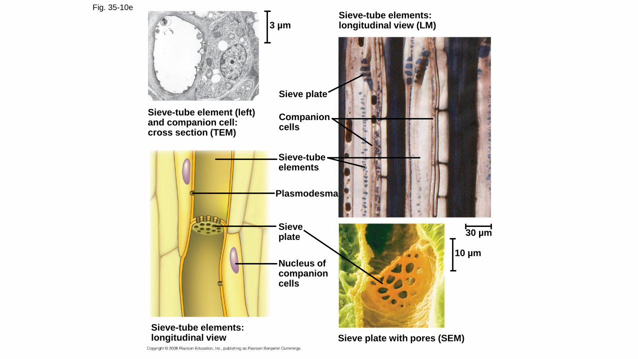
Fig. 35-10e
Sieve-tube element (left)and companion cell:cross section (TEM)
3 µmSieve-tube elements:longitudinal view (LM)
Sieve plate
Companioncells
Sieve-tubeelements
Plasmodesma
Sieveplate
Nucleus ofcompanioncells
Sieve-tube elements:longitudinal view Sieve plate with pores (SEM)
10 µm
30 µm

Fig. 35-10e3
Sieve-tubeelement
Plasmodesma
Sieveplate
Nucleus ofcompanioncells
Sieve-tube elements:longitudinal view Sieve plate with pores (SEM)
10 µm

meristems
• Meristems generate cells for new organs.
• A plant can grow throughout its life
• Annuals – complete their life cycle in a year or less
• Biennials – require two growing seasons
• Perennials – live for many years

Apical meristems
• Are located at the tips of roots and shoots and at the axillary buds of shoots
• Apical meristems elongate shoots and roots, called primary growth
Lateral meristems
• Lateral meristems add thickness to woody plants, a process called secondary growth.
• There are two lateral meristems: the vascular cambium and the cork cambium
• The vascular cambium adds layers of vascular tissue called secondary xylem (wood) and secondary phloem
• The cork cambium replaces the epidermis with periderm, which is thicker and tougher

Fig. 35-11
Shoot tip (shootapical meristemand young leaves)
Lateral meristems:
Axillary budmeristem
Vascular cambium
Cork cambium
Root apicalmeristems
Primary growth in stems
Epidermis
Cortex
Primary phloem
Primary xylem
Pith
Secondary growth in stems
Periderm
Corkcambium
Cortex
Primaryphloem
Secondaryphloem
Pith
Primaryxylem
Secondaryxylem
Vascular cambium

Fig. 35-12Apical bud
This year’s growth
(one year old)
Bud scale
Axillary buds
Leaf
scar
Bud
scar
Node
Internode
One-year-old side
branch formed
from axillary bud
near shoot tip
Last year’s growth
(two years old) Leaf scar
Stem
Bud scar left by apical
bud scales of previous
winters
Leaf scar
Growth of two
years ago
(three years old)

Fig. 35-13
Ground
Dermal
Key
to labels
Vascular
Root hair
Epidermis
Cortex Vascular cylinder
Zone of
differentiation
Zone of
elongation
Zone of cell
division
Apical
meristem
Root cap
100 µm

Roots and Root Growth

Fig. 35-14Epidermis
Cortex
Endodermis
Vascular
cylinder
Pericycle
Core of
parenchyma
cells
Xylem
Phloem100 µm
Root with xylem and phloem in the center
(typical of eudicots)
(a)
Root with parenchyma in the center (typical of
monocots)
(b)
100 µm
Endodermis
Pericycle
Xylem
Phloem
50 µm
Key
to labels
Dermal
Ground
Vascular

Fig. 35-14a1
Root with xylem and phloem in the center
(typical of eudicots)
(a)
100 µm
Epidermis
Cortex
Endodermis
Vascular
cylinder
Pericycle
Xylem
Phloem
Dermal
Ground
Vascular
Key
to labels

Fig. 35-14a2
Vascular
Ground
Dermal
Key
to labels
Root with xylem and phloem in the center
(typical of eudicots)
(a)
Endodermis
Pericycle
Xylem
Phloem
50 µm

Arrangement of stem tissues
• In plants called monocots, the vascular bundles are spread out throughout the ground tissue.
• In plants called dicots, the vascular bundles are in a ring around the inner ground tissue, with a thin layer of ground tissue surrounding.

Fig. 35-17a
Sclerenchyma
(fiber cells)
Phloem Xylem
Ground tissue
connecting
pith to cortex
Pith
CortexEpidermis
Vascular
bundle
1 mm
Cross section of stem with vascular bundles forming
a ring (typical of eudicots)
(a)
Dermal
Ground
Vascular
Key
to labels

Fig. 35-17b
Ground
tissue
Epidermis
Key
to labels
Cross section of stem with scattered vascular bundles
(typical of monocots)
Dermal
Ground
Vascular
(b)
Vascular
bundles
1 mm

Tissue organization of leaves
• The epidermis in leaves has breaks in it called stomata, which allow CO2 exchange between the air and the photosynthetic cells in a leaf.
• Each stomatal pore is surrounded by two guard cells, which regulate its opening and closing.
• The ground tissue in a leaf, called mesophyll, is sandwiched between the upper and lower epidermis

Tissue organization in leaves
• The upper layer of mesophyll is called palisade mesophyll where photosynthesis mostly occurs.
• Below this layer is a loosely arranged layer called the spongy mesophyll, where gas exchange occurs
• The veins or vascular tissue connect with those of the stem.
• Each vein is surrounded by a protective bundle sheath.

Fig. 35-18a
Key
to labels
Dermal
Ground
VascularCuticle Sclerenchyma
fibersStoma
Bundle-
sheath
cell
Xylem
Phloem
(a) Cutaway drawing of leaf tissues
Guard
cells
Vein
Cuticle
Lower
epidermis
Spongy
mesophyll
Palisade
mesophyll
Upper
epidermis

Fig. 35-18b
Guard
cells
Stomatal
pore
Surface view of a spiderwort
(Tradescantia) leaf (LM)
Epidermal
cell
(b)
50
µm

Fig. 35-18c
Upperepidermis
Palisademesophyll
Keyto labels
Dermal
Ground
Vascular
Spongymesophyll
Lowerepidermis
Vein Air spaces Guard cells
Cross section of a lilac(Syringa) leaf (LM)
(c)
10
0 µ
m

Secondary Growth
• Secondary growth adds girth to stems and roots in woody plants, (it doesn’t occur in leaves)
• The secondary tissues are produced by the vascular cambium and cork cambium

Fig. 35-19a1
Epidermis
Cortex
Primary phloem
Vascular cambium
Primary xylem
Pith
Primary and secondary growth
in a two-year-old stem
(a)
Periderm (mainly
cork cambia
and cork)
Secondary phloem
Secondary
xylem
Epidermis
Cortex
Primary phloem
Vascular cambium
Primary xylem
Pith

Fig. 35-19a2
Epidermis
Cortex
Primary phloem
Vascular cambium
Primary xylem
Pith
Primary and secondary growth
in a two-year-old stem
(a)
Periderm (mainly
cork cambia
and cork)
Secondary phloem
Secondary
xylem
Epidermis
Cortex
Primary phloem
Vascular cambium
Primary xylem
Pith
Vascular ray
Secondary xylem
Secondary phloem
First cork cambium
Cork

Fig. 35-19a3
Epidermis
Cortex
Primary phloem
Vascular cambium
Primary xylem
Pith
Primary and secondary growth
in a two-year-old stem
(a)
Periderm (mainly
cork cambia
and cork)
Secondary phloem
Secondary
xylem
Epidermis
Cortex
Primary phloem
Vascular cambium
Primary xylem
Pith
Vascular ray
Secondary xylem
Secondary phloem
First cork cambium
Cork
Cork
Bark
Most recent corkcambium
Layers ofperiderm
