plasma microrna-133a is a new marker for both acute myocardial infarction and underlying coronary...
TRANSCRIPT

RESEARCH Open Access
Plasma microRNA-133a is a new marker for bothacute myocardial infarction and underlyingcoronary artery stenosisFeng Wang1†, Guangwen Long1†, Chunxia Zhao1, Huaping Li1, Sandip Chaugai1, Yan Wang1, Chen Chen1,2*
and Dao Wen Wang1
Abstract
Background: Previous study demonstrated that miR-133a was released into blood from injured myocardium incardiovascular diseases. However, the dynamic change of circulating miR-133a level in the early phase of acutemyocardial infarction (AMI) and the correlation between miR-133a and severity of coronary stenosis in coronaryheart disease (CHD) patients are not clear.
Methods and results: Three different cohorts (including 13 AMI patients, 176 angina pectoris patients and127 control subjects) were enrolled to investigate the expression levels of circulating miR-133a in patients withmyocardial ischemia and also the relationship between plasma miR-133a and severity of coronary stenosis.Plasma miR-133a levels of participants were examined by real-time quantitative PCR. Simultaneously, plasmacardiac troponin I (cTnI) concentrations were measured by ELISA assays. The results showed that circulatingmiR-133a level was significantly increased in AMI patients in time-dependent manner, and achieved a 72.1 foldpeak at 21.6 ± 4.5 hours after the onset of AMI symptoms and exhibited a similar trend to plasma cTnI level. Wealso found that plasma miR-133a levels were higher in CHD patients than control group. Importantly, the levelsof circulating miR-133a positively correlated with the severities of the coronary artery stenosis. Receiver operatingcharacteristic (ROC) analysis revealed that circulating miR-133a had considerable diagnostic accuracy for CHDwith an AUC of 0.918 (95% confidence interval 0.877-0.960).
Conclusions: Circulating miR-133a may be a new biomarker for AMI and as a potential diagnostic tool. Andincreased miR-133a level may be used to predict both the presence and severity of coronary lesions inCHD patients.
Keywords: Biomarker, CHD, Circulating miRNA
IntroductionAcute myocardial infarction (AMI) is the worst acutesyndrome of coronary heart diseases (CHD) with highmorbidity and mortality. An early and correct diagnosisis critical for providing appropriate therapy to improvethe survival rate and prognosis [1]. Blood biomarker
cardiac troponin I (cTnI) is widely used in clinical practiceas the gold standard for diagnosing acute myocardialinfarction [2], but plasma cTnI concentrations may befalsely elevated in certain cardiac as well as non cardiacdiseases such as severe heart failure, atrial fibrillation,chronic kidney disease, severe sepsis and septic shock[3-6]. Therefore, it is necessary to search novel biomarkerswith high sensitivity and specificity for early diagnosisof AMI.MicroRNAs (miRNAs) are endogenous small non-coding
RNAs with 21-25 nucleotides in length. By pairing withthe 3’ UTR of target mRNAs, miRNAs can regulateprotein-coding genes at the posttranscriptional level via
* Correspondence: [email protected]†Equal contributors1The Institute of Hypertension and Department of Internal Medicine, TongjiHospital, Tongji Medical College, Huazhong University of Science andTechnology, Wuhan, People’s Republic of China2Department of Internal Medicine, Tongji Hospital, Tongji Medical College,Huazhong University of Science and Technology, 1095# Jiefang Ave, Wuhan430030, PR China
© 2013 Wang et al.; licensee BioMed Central Ltd. This is an Open Access article distributed under the terms of the CreativeCommons Attribution License (http://creativecommons.org/licenses/by/2.0), which permits unrestricted use, distribution, andreproduction in any medium, provided the original work is properly cited.
Wang et al. Journal of Translational Medicine 2013, 11:222http://www.translational-medicine.com/content/11/1/222

degradation of mRNAs or repression of protein translation[7]. At present, About 700 human miRNAs have beenidentified, and most of them were found to be tissue-/cell-specific [8]. Mounting evidences suggest that miRNAsplay crucial roles in various physiological and pathologicprocesses, and the dysfunctions of miRNAs are associatedwith various diseases and pathophysiologies [9-11]. Re-cently, studies showed that miRNAs are abundantlypresent in body fluid and can be used as biomarkers forsome diseases [12-14]. MiR-133a is a muscle specific-miRNA and is expressed abundantly in myocardial cells[15-17]. It has been established that miR-133a playsimportant roles in myogenesis, cardiac development andhypertrophy [18-23]. Previous studies demonstrated thatmiR-133a had a low level presence in the plasma ofhealthy people [15], and it was expressed differentiallyin different cardiovascular diseases [15,24]. Recently, ithas been reported that the elevated miR-133a is releasedinto peripheral circulation from the injured myocardiumafter Ca2+ stimulation [25]. Although these studies demon-strated that the expression of circulating miR-133a in-creased in patients with CHD (including AMI and anginapectoris) and circulating miR-133a can be used as amarker for cardiomyocyte death, few clinical studieshave reported on the dynamic change in circulating miR-133a level in the early phase of AMI, and also the correl-ation between miR-133a concentration and the severityof coronary stenosis in CHD patients is not clear.In the present work, we aimed to confirm the role of
plasma miR-133a as a biomarker for CHD, especiallyfor AMI. Furthermore, we investigated the correlationbetween the levels of circulating miR-133a and theGensini score (a numerical value for assessment theseverity of coronary artery stenosis) in coronary heartdisease patients.
Materials and methodsCharacteristics of patientsExperiments were conducted in accordance with theDeclaration of Helsinki. Three cohorts participated in thisstudy.The first cohort included 13 patients of AMI and 27
healthy volunteers. The inclusion criteria for AMI patientswere based on the third Universal Definition of Myocar-dial Infarction [26]. Briefly, AMI patients were clinicallydiagnosed by the following criteria: 1) acute ischemicchest pain within 24 hours; 2) electrocardiogram changeof acute myocardial infarction (pathological Q wave,ST-segment elevation or depression); 3) plasma cTnI >0.1 ng/mL. The initial blood sample (denoted by T0)was collected immediately after the AMI patient wasadmitted to Tongji hospital. Other 5 subsequent bloodsamples were obtained at 4, 12, 24, 48, 72 hours afterthe first collection, denoted by 4 h, 12 h, 24 h, 48 h and
72 h, respectively. The second cohort included 22 CHDpatients with chest pain having single lesion of the leftanterior descending coronary artery and 8 non-CHDpatients with negative results of coronary angiography.The third cohort contained 246 patients with acutechest pain. Further, coronary angiography showed that154 of them were CHD patients with complex lesionsof coronary artery, and the remaining 92 patients werenon-CHD patients with no coronary artery stenosis. Asingle blood sample from each participant in bothsecond and third cohorts was obtained immediatelyafter admission, and coronary angiography was used toconfirm CHD and define the coronary artery lesions.Blood samples were collected via venous puncture. Afterisolation by centrifugation, the plasma were transferred toRNase-free tubes and stored at -80°C until furtherprocessing.Participants were selected from inpatients or outpatients
departments of Tongji hospital between October 2009and June 2011 in Wuhan, China. The study was approvedby the Medical Ethics Committee in Tongji Hospitaland written informed consents were obtained from allthe participants.
RNA isolationTotal RNAs were isolated by TRIzol LS Reagent (Invi-trogen) according to the manufacturer’s protocol as de-scribed previously [27]. In brief, total RNA was purifiedfrom 500 μL of plasma and ultimately eluted into 25 μLof RNase-free water.
Detection and quantification of miRNAs by real-time PCRTwo microgram of total RNA was reverse-transcribedusing Transcript First-strand cDNA synthesis SuperMix(TransGen Biotech, Beijing, China) according to themanufacturer’s protocol. The Bulge-Loop™ miRNA qRT-PCR Detection Kit (Ribobio Co., Guangzhou, China) andSYBR Green PCR SuperMix Kit (TransGen Biotech,Beijing, China) were used in real-time PCR for examiningthe relative quantification of miR-133a according to themanufacturer’s protocol with the Rotor-Gene 6000 system(Corbett Life Science, QIAGEN, Hilden, Germany), andU6 was measured as endogenous control for normalizingthe data of experimental qRT-PCR. Each specimen wasmeasured in triplicate. The threshold cycle (Ct) was de-fined as the fractional cycle number at which fluorescenceexceed the threshold. In our experiment the detectionlimit of Ct value was defined as 40. The Ct values fromqRT-PCR assays over 40 were treated as 40 [15,25,28,29].
Cardiac troponin I determinationThe concentrations of cardiac Troponin I (cTnI) weremeasured by the Human Troponin I ELISA kit (Abnova,Taiwan) according to manufacturer’s protocol.
Wang et al. Journal of Translational Medicine 2013, 11:222 Page 2 of 9http://www.translational-medicine.com/content/11/1/222
Statistical analysisReal-time PCR assays were analyzed by 2-ΔΔct method,which is a widely used method to present relative geneexpression by comparative Ct. All the data of patients’clinical characteristics are described as mean ± SD, andthe other data are described as mean ± SEM. The dataof miR-133a and cTnI were analyzed by the Kolmogorov-Smirnov test to examine whether they followed the nor-mal distribution. If the data fit the normal distribution,then student’s t test and the ANOVA are used. Otherwise,Mann–Whitney U test and two-tailed Kruskal-Wallis testsare performed. In this study, both the data of miR-133aand cTnI were found to follow the normal distributionby the Kolmogorov-Smirnov test and hence student’s ttest and ANOVA were used. Categorical variables werecompared by χ2 test. The correlation analyses weredetermined by linear regression analysis. The receiveroperating characteristic (ROC) curve was used to assessthe predictive power for diagnosing CHD. Multiple logisticregression analysis was carried out to investigate whethermiRNA-133a was an independent predictor of CHD afteradjustment for relevant co-variants (including age, sex,smoking and cardiovascular risk factors hypertension,diabetes, hyperlipidemia etc.) as previously described[30]. All statistical analyses were accomplished by using
SPSS 17.0 software, and the cutoff point of statisticalsignificance was set at p < 0.05 (two-sided).
ResultsThe real-time RT-PCR (qRT-PCR) assay formiRNA quantificationTo ensure the method of qRT-PCR assay for miR-133aquantification is viable and suitable, the amplification curvesfor both miR-133a and U6 were provided (Additional file 1:Figure S1A and B). To verify primer specificities, meltingcurve analyses (Additional file 1: Figure S1C and D) andagarose gel electrophoresis images (Additional file 1: FigureS2A and B) were performed, the RNAs extracted frommouse heart and brain were treated as positive andnegative control, respectively.
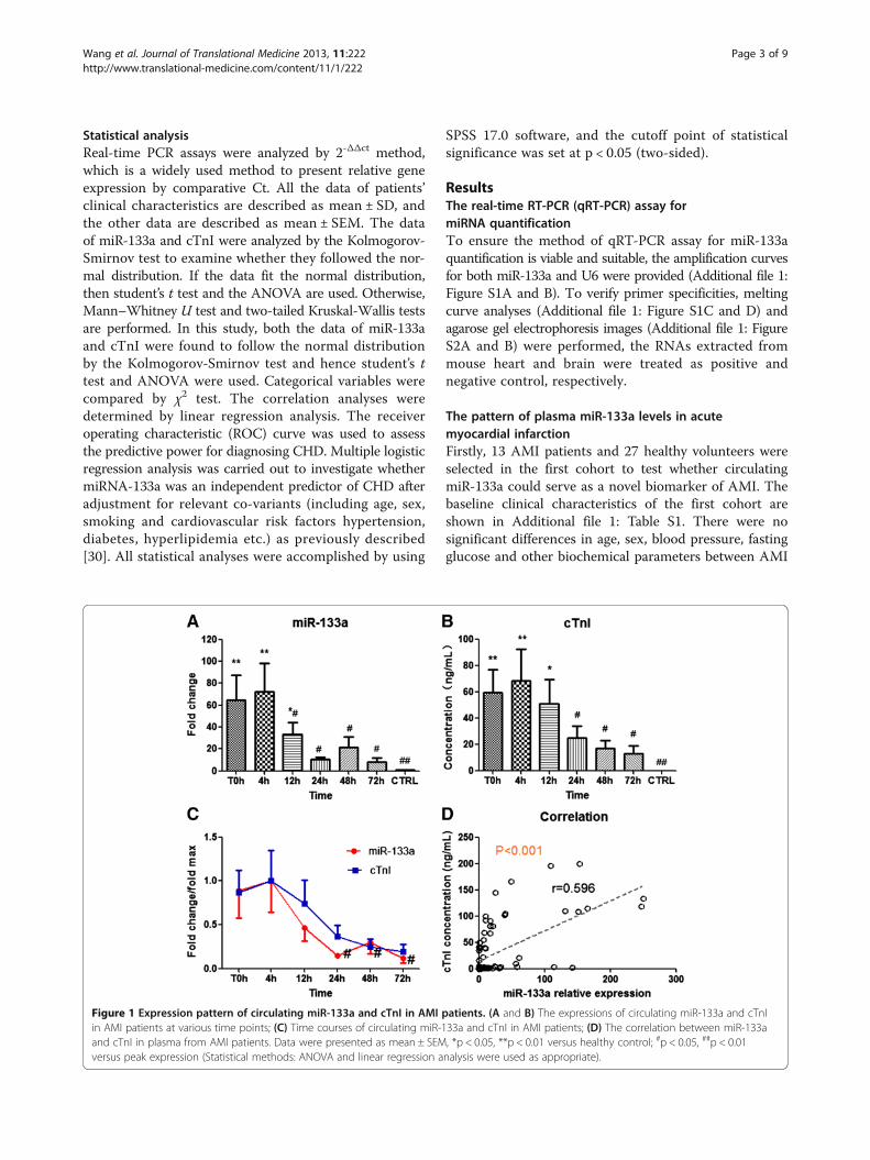
The pattern of plasma miR-133a levels in acutemyocardial infarctionFirstly, 13 AMI patients and 27 healthy volunteers wereselected in the first cohort to test whether circulatingmiR-133a could serve as a novel biomarker of AMI. Thebaseline clinical characteristics of the first cohort areshown in Additional file 1: Table S1. There were nosignificant differences in age, sex, blood pressure, fastingglucose and other biochemical parameters between AMI
Figure 1 Expression pattern of circulating miR-133a and cTnI in AMI patients. (A and B) The expressions of circulating miR-133a and cTnIin AMI patients at various time points; (C) Time courses of circulating miR-133a and cTnI in AMI patients; (D) The correlation between miR-133aand cTnI in plasma from AMI patients. Data were presented as mean ± SEM, *p < 0.05, **p < 0.01 versus healthy control; #p < 0.05, ##p < 0.01versus peak expression (Statistical methods: ANOVA and linear regression analysis were used as appropriate).
Wang et al. Journal of Translational Medicine 2013, 11:222 Page 3 of 9http://www.translational-medicine.com/content/11/1/222

patients and healthy volunteers. Six blood samples wereobtained from each AMI patient at various time points(T0h, 4 h, 12 h, 24 h, 48 h, and 72 h) to investigate thedynamic change trend in circulating miR-133a level inthe early phase of AMI. The first plasma sample wascollected at 17.6 ± 4.5 hours after the onset of AMIsymptoms (T0h), and other 5 collecting time points were4 h, 12 h, 24 h, 48 h, and 72 h after T0. As shown inFigure 1A, circulating miR-133a concentrations weresignificantly increased in the early phase (the first 3time points) after the occurrence of AMI in all patientscompared with controls. To get a more intuitive look atthe data, we also presented data using scatter plots(Additional file 1: Figure S3A). In those patients, circulatingmiR-133a achieved a peak (~72.1 fold) at 4 h, and showeda tendency to gradually return close to its control levelover the next 3 days. The concentrations of cTnI weremeasured in the same blood samples from AMI patients,simultaneously. Interestingly, cTnI remarkably increasedin the early phase of AMI and achieved a peak (~2445.2fold) at 4 h resembling miR-133a (Figure 1B). They bothexhibited the same trend in the early phase of AMIwith the elevated peak at 4 h, and then gradually
declined close to their normal level over the next 3 days(Figure 1C). Furthermore, correlation analysis showed apositive correlation between circulating levels of miR-133aand cTnI concentrations in AMI patients (Figure 1D). Thesedata suggested that circulating miR-133a may be regardedas a novel biomarker of acute myocardial infarction.
The correlation between plasma miR-133a levels and theseverities of coronary lesion in CHD patientsTo investigate whether circulating miR-133a expressioncorrelates with the severity of coronary artery stenosisin CHD patients, 22 CHD patients with single stenoticlesion in the proximal left anterior descending coronaryartery and 8 non-CHD patients with negative results ofcoronary angiography were recruited in the second cohort.Based on the severity of coronary artery stenosis, theparticipants were divided into 4 groups (Additional file1: Table S2). The first group consisted of 8 CHD patientswith severe coronary artery stenosis (81%~ 100%, LAD 1);the second group contained 7 CHD patients with moder-ate coronary artery stenosis (51%~ 80%, LAD 2); the thirdgroup consisted of 7 CHD patients with mild coronaryartery stenosis (30% ~ 50%, LAD 3); the fourth group
Figure 2 Expression pattern of circulating miR-133a and cTnI in CHD patients with single stenosis in the proximal of left anteriordescending coronary artery. (A and B) The expressions of circulating miR-133a and cTnI in CHD patients with single stenosis of coronary artery;(C) The correlation between plasma miR-133a and the degree of coronary atherosclerotic stenosis in CHD patients with single stenosis in theproximal of left anterior descending coronary artery. Data were presented as mean ± SEM, *p < 0.05, **p < 0.01, NS, not significant versus non-CHD chest pain patient (Statistical methods: Student’s t test and linear regression analysis were used as appropriate).
Wang et al. Journal of Translational Medicine 2013, 11:222 Page 4 of 9http://www.translational-medicine.com/content/11/1/222
included 8 non-CHD patients with negative results ofcoronary angiography (non-CHD). Their characteristicsare summarized in Additional file 1: Table S3. MiR-133aquantitative analysis showed that circulating miR-133alevels were significantly elevated in both LAD 1 and LAD2 groups, especially in LAD 1 (~5-fold), compared to non-CHD patients, and the levels of miR-133a in LAD 3showed no significant difference compared with non-CHD patients (Figure 2A and Additional file 1: FigureS3B). The concentration of plasma cTnI was not sig-nificantly different among the subgroups, although itwas slightly higher in the first and second groups(Figure 2B). Linear regression analysis showed that thelevels of plasma miR-133a positively correlated withthe severity of coronary artery stenosis in CHD patientswith single left anterior descending coronary athero-sclerosis (Figure 2C), but there was no association be-tween plasma cTnI and the degree of coronary stenosis.These results indicated that elevated miR-133a inplasma was better than cTnI for reflecting the severityof coronary artery stenosis in non-AMI CHD patientswith single stenotic lesion of left anterior descendingcoronary artery.
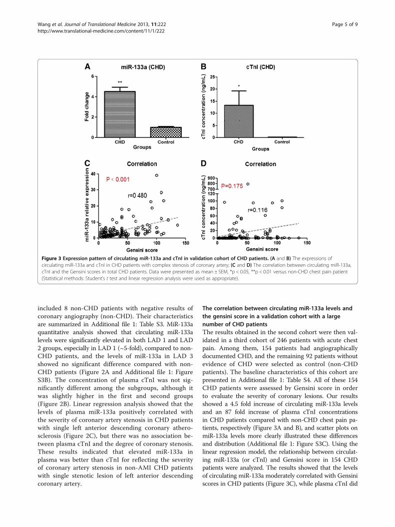
The correlation between circulating miR-133a levels andthe gensini score in a validation cohort with a largenumber of CHD patientsThe results obtained in the second cohort were then val-idated in a third cohort of 246 patients with acute chestpain. Among them, 154 patients had angiographicallydocumented CHD, and the remaining 92 patients withoutevidence of CHD were selected as control (non-CHDpatients). The baseline characteristics of this cohort arepresented in Additional file 1: Table S4. All of these 154CHD patients were assessed by Gensini score in orderto evaluate the severity of coronary lesions. Our resultsshowed a 4.5 fold increase of circulating miR-133a levelsand an 87 fold increase of plasma cTnI concentrationsin CHD patients compared with non-CHD chest pain pa-tients, respectively (Figure 3A and B), and scatter plots onmiR-133a levels more clearly illustrated these differencesand distribution (Additional file 1: Figure S3C). Using thelinear regression model, the relationship between circulat-ing miR-133a (or cTnI) and Gensini score in 154 CHDpatients were analyzed. The results showed that the levelsof circulating miR-133a moderately correlated with Gensiniscores in CHD patients (Figure 3C), while plasma cTnI did
Figure 3 Expression pattern of circulating miR-133a and cTnI in validation cohort of CHD patients. (A and B) The expressions ofcirculating miR-133a and cTnI in CHD patients with complex stenosis of coronary artery; (C and D) The correlation between circulating miR-133a,cTnI and the Gensini scores in total CHD patients. Data were presented as mean ± SEM, *p < 0.05, **p < 0.01 versus non-CHD chest pain patient(Statistical methods: Student’s t test and linear regression analysis were used as appropriate).
Wang et al. Journal of Translational Medicine 2013, 11:222 Page 5 of 9http://www.translational-medicine.com/content/11/1/222

not show any correlation with Gensini scores (Figure 3D).Further, the plasma cTnI concentrations from 140 of 154CHD patients were detected simultaneously. 72 of these140 CHD patients had high levels of plasma cTnI, approxi-mately 169-fold increase as compared with non-CHD pa-tients (Figure 4A); but the remaining 68 CHD patients hadvery low levels of cTnI (≤ 0.05 ng/mL), which were notconsistent with the severity of coronary artery stenosis.Further, in order to determine whether the expression ofcirculating miR-133a in CHD patients is superior to cTnIin the detection of the severity of coronary artery stenosis,140 CHD patients were divided into 2 subgroups on thebasis of cTnI levels (Figure 4A). The plasma miR-133a levelincreased ~5.5-fold and ~3.8-fold in group 1 and group 2,respectively, compared with non-CHD patients (Figure 4B).Moreover, in subgroup analysis, we found that plasmamiR-133a level significantly correlated with Gensini scoreof coronary artery lesions in both subgroups of CHDpatients (Figure 4C and D). However, the concentrationof cTnI showed no correlation with the Gensini scorein the two subgroups. These observations demonstratedthat plasma miR-133a significantly correlated with theGensini score of coronary lesions in CHD patients.
The plasma miR-133a is a sensitive predictor for coronaryheart diseaseTo investigate the role of circulating miR-133a as a sen-sitive predictor for CHD, ROC analysis was performed
in the third cohort (Additional file 1: Table S5). The clin-ical model with age, sex, smoke and other cardiovascularrisk factors (hypertension, diabetes, hyperlipidemia etc.)resulted in an AUC of 0.785 (95% confidence interval0.713-0.857) for the third cohort to differentiate betweenCHD and non-CHD groups (Figure 5A). The ROC curveof cTnI showed a moderate separation, with an area underthe ROC curve (AUC) of 0.741 (95% confidence interval0.668-0.814). Interestingly, ROC curve of circulating miR-133a showed a much higher AUC of 0.918 (95% confi-dence interval 0.877-0.960). It is noteworthy that additionof miR-133a to the clinical model and cTnI remarkablyincreased the diagnostic value for CHD with an AUC of0.942 (95% confidence interval 0.908-0.976) and 0.925(95% confidence interval 0.887-0.963), respectively. AddingmiR-133a to the clinical model with cTnI (AUC of 0.834,95% confidence interval 0.773-0.896) significantly increasedthe AUC to an even higher value of 0.947 (95% confidenceinterval 0.915-0.979). Furthermore, the ROC curves ofcirculating miR-133a in both the subgroups of CHDwere determined (Additional file 1: Table S6). MiR-133aalso had significant differentiation value for CHD andnon-CHD in both group 1 and group 2 with the AUC of0.981 (95% confidence interval 0.962-1.0) and 0.885 (95%confidence interval 0.827-0.942), respectively (Figure 5Band C). The diagnostic value for CHD was significantlyincreased with the addition of miR-133a to cTnI in thetwo subgroups with the AUC of 0.953 (95% confidence
Figure 4 Expression pattern of circulating miR-133a and cTnI in subgroups of CHD patients. (A and B) The expressions of cTnI andcirculating miR-133a in subgroups of CHD patients with complex stenosis of coronary artery; (C and D) The correlation between circulating miR-133a and the Gensini scores in patients from group 1 and group 2; Data were presented as mean ± SEM, *p < 0.05, **p < 0.01 versus non-CHDchest pain patient (Statistical methods: Student’s t test and linear regression analysis were used as appropriate).
Wang et al. Journal of Translational Medicine 2013, 11:222 Page 6 of 9http://www.translational-medicine.com/content/11/1/222

interval 0.919-0.987) and 0.892 (95% confidence interval0.836-0.947), respectively. Taken together, these data sug-gested that circulating miR-133a may be a sensitive andindependent predictor for CHD.
DiscussionPrevious studies demonstrated that miRNAs are abun-dantly present in a remarkably stable form and they canbe detected in peripheral circulation [12,31]. Recently,more and more circulating miRNAs, including heart-,vascular- and muscle-specific miRNAs, have been reportedas new biomarkers in multiple cardiovascular diseases[32,33]. For example, circulating miR-423-5p is suggestedas a biomarker for heart failure [34]. And additionally,cardiac-related miRNAs (miR-208, miR-499 and miR-1)and stress-related miRNAs (miR-21 and miR-146a) maybe potential biomarkers for acute coronary syndrome [30].Moreover, a recent study had reported that circulatingmiR-126, miR-223 and miR-197 were consistently andsignificantly related to incidence of myocardial infarction[35]. These observations suggest that circulating miRNAsmay be useful not only for prediction of cardiovascular
events, but also serve as sensitive biomarkers for improv-ing the diagnostic accuracy of cardiovascular diseases.The present study demonstrated dynamic change in
circulating miR-133a expression in the early phase ofacute myocardial infarction. Furthermore, our data is thefirst to demonstrate a positive correlation between cir-culating miR-133a and the severity of coronary stenosisin CHD patients.The results demonstrated that circulating miR-133a
levels increased in time-dependent manner in the earlyphase of AMI and exhibited a similar trend as cTnI inAMI patients; both of them rapidly increased at first,achieved a peak at 21.6 ± 4.5 hours after the onset ofAMI symptoms, and then gradually returned close tonormal level on the following days. Importantly, thecirculating miR-133a positively correlated with cTnI inAMI patients. These results strongly indicated thatcirculating miR-133a can be a biomarker for diagnosingacute myocardial infarction.Furthermore, 22 CHD patients with single lesion of
coronary artery were included in the second cohort tostudy the relationship between plasma miR-133a and theseverity of coronary atherosclerosis. The results showed
A all CHD patients B CHD patients (cTnI >0.05ng/ml)
C CHD patients (cTnI ≤0.05ng/ml)
Figure 5 Diagnostic value of cardiac troponin I and circulating miR-133a in CHD patients. (A) Total CHD patients compared to non-CHDpatients in the third cohort; (B) CHD patients from group 1 compared to non-CHD patients in the third cohort; (C) CHD patients from group 2compared to non-CHD patients in the third cohort (Statistical methods: receiver operating characteristic (ROC) curve and multiple logisticregression analysis).
Wang et al. Journal of Translational Medicine 2013, 11:222 Page 7 of 9http://www.translational-medicine.com/content/11/1/222

that circulating miR-133a increased in CHD patients com-pared with non-CHD patients, and the levels of elevatedmiR-133a positively correlated with the severities ofcoronary atherosclerosis. The results were further verifiedin a large validation cohort of 246 subjects (154 CHDpatients and 92 non-CHD). Interestingly, we found ahigher expression of circulating miR-133a in CHD patientswith low cTnI expression compared with non-CHDpatients and it correlated with Gensini score of theseCHD patients. These results showed that circulatingmiR-133a is superior to cTnI in detecting the severityof coronary artery lesions.Finally, the ROC curve of miR-133a and cTnI were
plotted in CHD patients with an AUC of 0.918 and0.741, respectively. The ROC curves of CHD subcategoriesrevealed that circulating miR-133a is more informative forCHD diagnosis than cTnI in CHD patients. Importantly,the diagnostic accuracy for CHD became significantlyraised when combining clinical model, miR-133a andcTnI with the AUC of 0.947. Interestingly, this additioneffect of combination could be more valuable for cTnI toimprove the diagnostic accuracy of CHD, while miR-133aappeared to be a strong and independent predictor forCHD. These results may provide theoretical foundationin improving the clinical diagnosis of CHD.In summary, the present study measured the early
changes in expressions of circulating miR-133a in AMIpatients, and provided first insights into the relationshipbetween plasma miR-133a and the severity of coronaryatherosclerotic stenosis in CHD patients, our results sug-gested that circulating miR-133a was a sensitive predictorfor diagnosing AMI and CHD.All these results suggested that circulating miR-133a
can be a novel and potent biomarker for CHD, especiallyfor AMI. And its level in plasma can reflect the severityof coronary atherosclerosis in CHD patients.
Additional file
Additional file 1: Figure S1. The qRT-PCR amplification curves andmelting curves for both miR-133a and U6. Figure S2. The agarose gelelectrophoresis images for both (A) miR-133a and (B) U6. RNA extractfrom mouse tissue (heart and brain). Figure S3. miR-133a expression inthree cohorts displayed by scatter. Table S1. The clinical characteristics of13 AMI patients and 27 healthy volunteers. Table S2. Divide the secondcohort into 4 groups according to the degree of coronary artery stenosis.Table S3. The clinical characteristics of 22 CHD patients and 8 non-CHDpatients. Table S4. The clinical characteristics of 154 CHD patients and 92non-CHD patients. Table S5. Diagnostic value of cTnI and miR-133a inCHD patients in a clinical model. Table S6. Diagnostic value of cTnI andmiR-133a in subgroups of CHD patients in a clinical model.
Competing interestsThe authors declare that they have no competing interests.
Authors’ contributionsFW and GL carried out the miRNA detection studies and performed thestatistical analysis. CZ and HL participated in collecting blood samples. SC
and YW helped in drafting the manuscript. FW, CC and DWW conceived ofthe study, participated in its designing and writing the manuscript. Allauthors read and approved the final manuscript.
AcknowledgementsWe thank all the people who helped in collecting the blood samples.
FundingThis work was supported by grant from the National Natural ScienceFoundation of China (No. 81070236 and No. 31200594) and Key Project ofMinistry of Health of the People's Republic of China.
Received: 17 July 2013 Accepted: 18 September 2013Published: 23 September 2013
References1. White HD, Chew DP: Acute myocardial infarction. Lancet 2008, 372:570–584.2. Jaffe AS, Ravkilde J, Roberts R, Naslund U, Apple FS, Galvani M, Katus H: It’s
time for a change to a troponin standard. Circulation 2000, 102:1216–1220.3. Abbas NA, John RI, Webb MC, Kempson ME, Potter AN, Price CP, Vickery S,
Lamb EJ: Cardiac troponins and renal function in nondialysis patientswith chronic kidney disease. Clin Chem 2005, 51:2059–2066.
4. Finsterer J, Stollberger C, Krugluger W: Cardiac and noncardiac, particularlyneuromuscular, disease with troponin-T positivity. Neth J Med 2007,65:289–295.
5. Giannitsis E, Katus HA: Cardiac troponin level elevations not related toacute coronary syndromes. Nat Rev Cardiol 2013. 10.1038/nrcardio.2013.129.
6. Rosjo H, Varpula M, Hagve TA, Karlsson S, Ruokonen E, Pettila V, Omland T,Group FS: Circulating high sensitivity troponin T in severe sepsis andseptic shock: distribution, associated factors, and relation to outcome.Intensive Care Med 2011, 37:77–85.
7. Bartel DP: MicroRNAs: genomics, biogenesis, mechanism, and function.Cell 2004, 116:281–297.
8. van Rooij E, Olson EN: MicroRNAs: powerful new regulators of heartdisease and provocative therapeutic targets. J Clin Invest 2007,117:2369–2376.
9. Croce CM: Oncogenes and cancer. N Engl J Med 2008, 358:502–511.10. van Rooij E, Marshall WS, Olson EN: Toward microRNA-based therapeutics
for heart disease: the sense in antisense. Circ Res 2008, 103:919–928.11. Kajimoto K, Naraba H, Iwai N: MicroRNA and 3T3-L1 pre-adipocyte
differentiation. RNA 2006, 12:1626–1632.12. Mitchell PS, Parkin RK, Kroh EM, Fritz BR, Wyman SK, Pogosova-Agadjanyan
EL, Peterson A, Noteboom J, O'Briant KC, Allen A, et al: CirculatingmicroRNAs as stable blood-based markers for cancer detection.Proc Natl Acad Sci U S A 2008, 105:10513–10518.
13. Skog J, Wurdinger T, van Rijn S, Meijer DH, Gainche L, Sena-Esteves M, CurryWT Jr, Carter BS, Krichevsky AM, Breakefield XO: Glioblastoma microvesiclestransport RNA and proteins that promote tumour growth and providediagnostic biomarkers. Nat Cell Biol 2008, 10:1470–1476.
14. Chim SS, Shing TK, Hung EC, Leung TY, Lau TK, Chiu RW, Lo YM: Detectionand characterization of placental microRNAs in maternal plasma.Clin Chem 2008, 54:482–490.
15. Wang GK, Zhu JQ, Zhang JT, Li Q, Li Y, He J, Qin YW, Jing Q: CirculatingmicroRNA: a novel potential biomarker for early diagnosis of acutemyocardial infarction in humans. Eur Heart J 2010, 31:659–666.
16. Chen JF, Mandel EM, Thomson JM, Wu Q, Callis TE, Hammond SM, ConlonFL, Wang DZ: The role of microRNA-1 and microRNA-133 in skeletalmuscle proliferation and differentiation. Nat Genet 2006, 38:228–233.
17. Fichtlscherer S, De Rosa S, Fox H, Schwietz T, Fischer A, Liebetrau C, WeberM, Hamm CW, Roxe T, Muller-Ardogan M, et al: Circulating microRNAs inpatients with coronary artery disease. Circ Res 2010, 107:677–684.
18. Liu J, Hao DD, Zhang JS, Zhu YC: Hydrogen sulphide inhibitscardiomyocyte hypertrophy by up-regulating miR-133a. Biochem BiophysRes Commun 2011, 413:342–347.
19. Care A, Catalucci D, Felicetti F, Bonci D, Addario A, Gallo P, Bang ML,Segnalini P, Gu Y, Dalton ND, et al: MicroRNA-133 controls cardiachypertrophy. Nat Med 2007, 13:613–618.
20. Li Q, Lin X, Yang X, Chang J: NFATc4 is negatively regulated in miR-133a-mediated cardiomyocyte hypertrophic repression. Am J Physiol Heart CircPhysiol 2010, 298:H1340–H1347.
Wang et al. Journal of Translational Medicine 2013, 11:222 Page 8 of 9http://www.translational-medicine.com/content/11/1/222

21. Luo J, Cai Q, Wang W, Huang H, Zeng H, He W, Deng W, Yu H, Chan E, NgCF, et al: A microRNA-7 binding site polymorphism in HOXB5 leads todifferential gene expression in bladder cancer. PLoS One 2012, 7:e40127.
22. Liu N, Bezprozvannaya S, Williams AH, Qi X, Richardson JA, Bassel-Duby R,Olson EN: microRNA-133a regulates cardiomyocyte proliferation andsuppresses smooth muscle gene expression in the heart. Genes Dev 2008,22:3242–3254.
23. He B, Xiao J, Ren AJ, Zhang YF, Zhang H, Chen M, Xie B, Gao XG, Wang YW:Role of miR-1 and miR-133a in myocardial ischemic postconditioning.J Biomed Sci 2011, 18:22.
24. D'Alessandra Y, Devanna P, Limana F, Straino S, Di Carlo A, Brambilla PG,Rubino M, Carena MC, Spazzafumo L, De Simone M, et al: CirculatingmicroRNAs are new and sensitive biomarkers of myocardial infarction.Eur Heart J 2010, 31:2765–2773.
25. Kuwabara Y, Ono K, Horie T, Nishi H, Nagao K, Kinoshita M, Watanabe S,Baba O, Kojima Y, Shizuta S, et al: Increased microRNA-1 and microRNA-133a levels in serum of patients with cardiovascular disease indicatemyocardial damage. Circ Cardiovasc Genet 2011, 4:446–454.
26. Thygesen K, Alpert JS, Jaffe AS, Simoons ML, Chaitman BR, White HD, JointESC/ACCF/AHA/WHF Task Force for the Universal Definition of Myocardial,Katus HA, Lindahl B, Morrow DA, et al: Third universal definition ofmyocardial infarction. Circulation 2012, 126:2020–2035.
27. Long G, Wang F, Duan Q, Chen F, Yang S, Gong W, Wang Y, Chen C, WangDW: Human circulating microRNA-1 and microRNA-126 as potential novelindicators for acute myocardial infarction. Int J Biol Sci 2012, 8:811–818.
28. Wang E, Nie Y, Zhao Q, Wang W, Huang J, Liao Z, Zhang H, Hu S, Zheng Z:Circulating miRNAs reflect early myocardial injury and recovery afterheart transplantation. J Cardiothorac Surg 2013, 8:165.
29. Brase JC, Johannes M, Schlomm T, Falth M, Haese A, Steuber T, Beissbarth T,Kuner R, Sultmann H: Circulating miRNAs are correlated with tumorprogression in prostate cancer. Int J Cancer 2011, 128:608–616.
30. Oerlemans MI, Mosterd A, Dekker MS, de Vrey EA, van Mil A, Pasterkamp G,Doevendans PA, Hoes AW, Sluijter JP: Early assessment of acute coronarysyndromes in the emergency department: the potential diagnostic valueof circulating microRNAs. EMBO Mol Med 2012, 4:1176–1185.
31. Gilad S, Meiri E, Yogev Y, Benjamin S, Lebanony D, Yerushalmi N, BenjaminH, Kushnir M, Cholakh H, Melamed N, et al: Serum microRNAs arepromising novel biomarkers. PLoS One 2008, 3:e3148.
32. Adachi T, Nakanishi M, Otsuka Y, Nishimura K, Hirokawa G, Goto Y, NonogiH, Iwai N: Plasma microRNA 499 as a biomarker of acute myocardialinfarction. Clin Chem 2010, 56:1183–1185.
33. Ai J, Zhang R, Li Y, Pu J, Lu Y, Jiao J, Li K, Yu B, Li Z, Wang R, et al:Circulating microRNA-1 as a potential novel biomarker for acutemyocardial infarction. Biochem Biophys Res Commun 2010, 391:73–77.
34. Tijsen AJ, Creemers EE, Moerland PD, de Windt LJ, van der Wal AC, Kok WE,Pinto YM: MiR423-5p as a circulating biomarker for heart failure.Circ Res 2010, 106:1035–1039.
35. Zampetaki A, Willeit P, Tilling L, Drozdov I, Prokopi M, Renard JM, Mayr A,Weger S, Schett G, Shah A, et al: Prospective study on circulatingMicroRNAs and risk of myocardial infarction. J Am Coll Cardiol 2012,60:290–299.
doi:10.1186/1479-5876-11-222Cite this article as: Wang et al.: Plasma microRNA-133a is a new markerfor both acute myocardial infarction and underlying coronary arterystenosis. Journal of Translational Medicine 2013 11:222.
Submit your next manuscript to BioMed Centraland take full advantage of:
• Convenient online submission
• Thorough peer review
• No space constraints or color figure charges
• Immediate publication on acceptance
• Inclusion in PubMed, CAS, Scopus and Google Scholar
• Research which is freely available for redistribution
Submit your manuscript at www.biomedcentral.com/submit
Wang et al. Journal of Translational Medicine 2013, 11:222 Page 9 of 9http://www.translational-medicine.com/content/11/1/222